This work is subject to copyright. All rights are solely and exclusively licensed by the Publisher, whether the whole or part of the material is concerned, specifcally the rights of translation, reprinting, reuse of illustrations, recitation, broadcasting, reproduction on microflms or in any other physical way, and transmission or information storage and retrieval, electronic adaptation, computer software, or by similar or dissimilar methodology now known or hereafter developed.
The use of general descriptive names, registered names, trademarks, service marks, etc. in this publication does not imply, even in the absence of a specifc statement, that such names are exempt from the relevant protective laws and regulations and therefore free for general use.
The publisher, the authors, and the editors are safe to assume that the advice and information in this book are believed to be true and accurate at the date of publication. Neither the publisher nor the authors or the editors give a warranty, expressed or implied, with respect to the material contained herein or for any errors or omissions that may have been made. The publisher remains neutral with regard to jurisdictional claims in published maps and institutional affliations.
This Springer imprint is published by the registered company Springer Nature Switzerland AG The registered company address is: Gewerbestrasse 11, 6330 Cham, Switzerland
Preface
The American Foregut Society emerged from the recognition that improvements in patient care would require specialization that only collaboration between gastroenterologists and surgeons could provide. Cross-fertilization increases knowledge, enables new insights, and improves expertise. This textbook exemplifes that conviction; the majority of chapters are co-authored by gastroenterologist and surgical colleagues.
Since its founding in May of 2018, the society has grown membership to over 600, accomplished three national meetings (two in the time of COVID-19), launched the subscriptionbased journal Foregut, inspired the creation of a European Foregut Society, and through the vision and unrelenting efforts of Ninh Nguyen and John Clarke given birth to this textbook.
Medical knowledge is progressing far faster than the ability to publish and disseminate. This textbook cannot be nor is it intended to be authoritative; rather it is intended to be a reservoir of what collaborative thinking can accomplish.
The parable of the blind men and an elephant is emblematic of the historical relationship of gastroenterologists and surgeons treating foregut disease. A group of blind men who have never seen before and must rely on touch to understand what the elephant is. Each blind man feels a different part of the elephant’s body, but only one part, such as the tusk, leg, tail, or ear. They then describe the elephant based on their limited experience and not surprisingly the descriptions of the elephant are different from each other.
In some versions of the parable, they even come to suspect that the other person is dishonest and they come to blows.
Physicians are not immune to the human predisposition of claiming comprehensive knowledge based on their experience. Not only is their clinical care biased; the meetings they attend and journals they read refect their own horizons.
We believe the American Foregut Society has provided vision to individuals and foregut disease as a whole. That this textbook came to fruition within 4 years of the society’s founding is a testament to the enthusiasm generated when we start to see beyond ourselves.
Orange, CA, USA
Redwood City, CA, USA
Los Angeles, CA, USA
Ninh T. Nguyen
John O. Clarke
John C. Lipham Orange, CA, USA
New York, NY, USA
Englewood, CO, USA
Kenneth J. Chang
Felice Schnoll-Sussman
Reginald C. W. Bell Chicago, IL, USA
Peter J. Kahrilas
Part I Basic Considerations
1 History of AFS
Reginald C. W. Bell and Felice Schnoll-Sussman
Part II GERD and Eosinoplhilic Esophagitis
2 GERD Pathophysiology: The Role of the Sphincter and Crural Diaphragm
Philip O. Katz, Gaurav Ghosh, and Katharine Rooney
5 Phenotypes of Gastroesophageal Reflux Disease and Personalized Management
Domenico A. Farina, John E. Pandolfno, and Kristle Lynch
6
Shahin Ayazi and Blair Jobe
7 The Spectrum of Eosinophilic Esophagitis
Jennifer L. Horsley-Silva, Blair Jobe, and David Katzka
8 Esophageal Hypersensitivity
Adriana Lazarescu, Alicia Demers-Leblanc, and Afrin Kamal 9 Endoscopic GERD Therapies
Linda Y. Zhang, Kenneth J. Chang, and Marcia Irene Canto
Understanding the Optimal Gastroesophageal Flap Valve: The Omega Flap Valve
Ninh T. Nguyen, Justine Chinn, and Kenneth J. Chang
Justin R. Henning, Rocio E. Carrera Ceron, and Brant K. Oelschlager 12 Comprehensive Review of the Anti-Reflux Mechanism and Fundoplication
Shaun Daly, Michael Tran, Miya Yoshida, David Choi, and Daniel Tseng
13 Laparoscopic Magnetic Sphincter Augmentation
Tejal Pandya, Hamza Durrani, Reginald C. W. Bell, Philip Woodworth, and Brian E. Louie
14 Diagnosis and Management of Paraesophageal Hiatal Hernia 125
Hana Ajouz, Michael P. Rogers, Christopher Ducoin, Vic Velanovich, and Collin E. M. Brathwaite
15 Reoperative Anti-Reflux Surgery 139 Sumeet Mittal, Paul Kim, and Adrian Park
Part III Barrett’s Esophagus and Esophageal Neoplasm
16 Screening for Barrett’s Esophagus 147 Jay Bapaye, George Triadaflopoulos, and Prasad G. Iyer
17 Understanding the Histopathology of GERD and Barrett’s Esophagus 161 Parakrama Chandrasoma and Jason B. Samarasena
18 Management of Nondysplastic Barrett’s Esophagus 171 Michael S. Smith, F. P. Buckley III, F. Scott Corbett, and Reginald C. W. Bell
19 Management of Dysplastic Barrett’s Esophagus .
Andrew D. Grubic, Shahin Ayazi, Manish K. Dhawan, and Blair A. Jobe
20 Management of Early Esophageal Cancer
Nasim Parsa, Steven R. DeMeester, Daniela Molena, and Stavros N. Stavropoulos
21 Cancer of the Esophagus: Epidemiology and Genetics
David H. Wang, Eric M. Lander, and Michael K. Gibson
22 Screening Technologies for Barrett’s Esophagus and Esophageal Adenocarcinoma
Gary W. Falk and Cadman L. Leggett
179
189
23 Staging Endoscopic Ultrasound 217 Eun Ji Shin and Shruti Mony
24 Minimally Invasive Esophagectomy
233 Navjit Dharampal, Michael N. Tran, Ninh T. Nguyen, and Brian E. Louie
25 Endoscopic Management of Anastomotic Leaks 245 James M. Ackerman, Ryan M. Levy, and Inderpal S. Sarkaria
26 Palliative Therapy in Esophageal Cancer
251 Neil Sood, Sarah Enslin, and Zubair Malik
Part IV Esophageal Motility Disorders, Esophageal Injury, and Benign Disease of the Esophagus
27 Pathophysiology of Esophageal Motility Disorders 263 Monica Nandwani, Kirsten Newhams, and Blair Jobe
28 Diagnostic Testing for Esophageal Motility Disorders: Barium Radiography, High-Resolution Manometry, and the Functional Lumen Imaging Probe (FLIP) 269 Amit Patel, Felice Schnoll-Sussman, and C. Prakash Gyawali
29 The Chicago Classification of Esophageal Motor Disorders 279 Rena Yadlapati, Amit Patel, and Peter J. Kahrilas
30 Cricopharyngeal Disorders, Endoscopic, Stapled, and Open Cricomyotomy and Adjuncts for Zenker’s Diverticulum
Karuna Dewan and Jeffrey R. Watkins
31 Minimally Invasive Management of Epiphrenic Diverticulum 301
Bradley Kushner, Elbert Kuo, and Michael M. Awad
32 Disorders of Esophagogastric Junction Outflow and Peristalsis
Monika Lammi and Jessica Koller Gorham
33 Therapies for Spastic Esophageal Motor Disorders
Dustin A. Carlson, Reena V. Chokshi, and Ellen Stein
34 Pneumatic Dilation for the Treatment of Achalasia
Steven Clayton and Joel E. Richter
35 Minimally Invasive Heller Myotomy .
Melissa L. Desouza and Kevin M. Reavis
36 Per-Oral Endoscopic Myotomy (POEM)
Kenneth J. Chang and Lee L. Swanström
37 Scleroderma
John O. Clarke, F. P. Tripp Buckley III, Zsuzsanna McMahan, and Dinesh Khanna
38 Benign Lesions of the Esophagus
Sanjay M. Salgado, Daniela Jodorkovsky, and Nitin K. Ahuja
39 Management of Caustic Injury and Esophageal Stricture
Ryan C. Broderick and Karthik Ravi
40 Management of Esophageal Perforation
Christy M. Dunst and Timothy Mansour
Part V Gastric Disorders
41 Pathophysiology of Gastric Neuromuscular Disorders
Aylin Tansel, Linda Nguyen, and Thomas L. Abell
42 Diagnostic Testing and Pharmacotherapy in Gastroparesis
Brian Surjanhata and Braden Kuo
43 Endoscopic Pyloric Therapies for Gastroparesis
Olaya I. Brewer Gutierrez, Mouen A. Khashab, and Henry P. Parkman
44 Surgical Treatment of Gastroparesis
Irene Sarosiek, Brian R. Davis, Richard McCallum, Mina Ibrahim, and Dmitry Oleynikov
45 Cyclic Vomiting Syndrome, Dumping, and Marijuana-Induced Hyperemesis Syndrome
Thangam Venkatesan and William L. Hasler
Part VI Gastric Cancer and Other Gastroesophageal Disorders
46 Gastric Cancer Epidemiology, Genetics, and Screening
Brittany G. Sullivan, John D. Karalis, Sam C. Wang, and Maheswari Senthil
47 Diagnosis and Management of Gastric Intestinal Metaplasia 461
Ji Yoon Yoon, Dan Li, and Shailja C. Shah
48 Endoscopic Staging and Resection for Early Gastric Cancer 469
Jason Samarasena, Anastasia Chahine, and Joo Ha Hwang
49 Laparoscopic Total and Subtotal Gastrectomy 477
Stephen Stopenski, Luigi Bonavina, and Brian R. Smith
50 Laparoscopic Resection of Gastric and Esophageal Submucosal Tumors . . . . . . 485
Katie M. Galvin, Shaun Daly, and Marcelo Hinojosa
51 Peptic Ulcer Disease .
Jordan Shapiro, Dan Lister, and David Y. Graham
495
52 Primary Gastric Lymphoma 507
Daniel Tseng, Spencer Shao, and Tris Arscott
Part VII Obesity and Metabolic Disease
53 Laparoscopic and Robotic Sleeve Gastrectomy 519
Collin E. M. Brathwaite, Raelina S. Howell, Jun Levine, Maxime Lapointe-Gagner, and Michel Gagner
54 Laparoscopic and Robot-Assisted Roux-en-Y Gastric Bypass 527
Kelly Andrew Lara, Esther Wu, and Yong Choi
55 Endoscopic Gastric Remodeling for Weight Loss 535
Jennifer M. Kolb, Babusai Rapaka, Barham K. Abu Dayyeh, and Kenneth J. Chang
56 Strategies in Management of GERD in the Severely Obese Undergoing Bariatric Surgery 545
Ninh T. Nguyen, Ava Runge, and Kenneth J. Chang
57 Endoscopic Management of Bariatric Complications 553 Vitor Ottoboni Brunaldi, Christopher C. Thompson, and Manoel Galvao Neto
58 Bariatric Surgery Complications and Management 563 Christopher Pearcy, Andre Teixeira, Muhammad Ghanem, and Natan Zundel
59 Post-bariatric Surgery GERD
Babusai Rapaka, Kevin D. Platt, Allison R. Schulman, and Barham K. Abu Dayyeh
60 Gastroesophageal Reflux Disease and Metabolic Surgery
R. Alvarez, J. Silva, Caitlin Houghton, and Leena Khaitan
Part VIII Specifc Considerations
61 Health Advocacy
V. Raman Muthusamy and Sushrut Sujan Thiruvengadam
62 Robotic Foregut Surgery
Tanuja Damani, Caitlin Houghton, and Ryan C. Broderick
577
Part I
Basic Considerations
History of AFS
Reginald C. W. Bell and Felice Schnoll-Sussman
The American Foregut Society was born out of many individuals’ dreams and passions to see a specialty thrive and fourish. A hope that different disciplines could work together to accomplish lofty goals. A desire to break down silos and build bridges. Not a simple task by any stretch of the imagination. The ethos was that we would be “better together.” Who is the we? Foregut surgeons and gastroenterologists— the main physician stakeholders in the management of patients with diseases of the foregut. Some would think that the American Foregut Society, now more commonly referred to as AFS, was an evolution—in reality it represents a revolution. A revolutionary change in the way different specialties work together, learn together, advocate together, and grow together with a single-minded purpose—collaboration and specialization to improve patient care.
The small group of foregut surgical specialists providing the initial impetus to form the AFS immediately recognized the necessity of partnering with medical foregut specialists. Without both specialties the goals and survival of such an effort would founder. Tripp Buckley, a general surgeon, had been proctored in his frst sphincter augmentation procedure (LINX™) by the world renowned and revered Dr. Tom DeMeester and found himself navigating his practice to one dedicated to the management of GERD. Dr. Reginald Bell, a private practice foregut surgeon in Colorado, developed a multicenter Registry of Outcomes in Anti-Refux Surgery (ROARS) to prospectively collect outcomes for laparoscopic fundoplication and magnetic sphincter augmentation. Dr. John Lipham was trained by and accorded resultant recognition by Tom DeMeester as chief of upper GI surgery at the Keck School of Medicine in USC. Dr. Blair Jobe is a brilliant basic science and clinical researcher and foregut surgeon in
R. C. W. Bell (*)
Institute of Esophageal & Refux Surgery, Lone Tree, CO, USA
e-mail: reg@regbell.com
F. Schnoll-Sussman
Division of Gastroenterology and Hepatology, Weill Cornell Medicine, New York City, NY, USA
e-mail: fhs2001@med.cornell.edu
Pittsburgh. Dr. Dan Lister, a private practice foregut surgeon in Heber Springs Arkansas, has the largest Barrett’s surveillance population in the state. Kate Freeman N.P. works with Dr. Bell and would become instrumental in implementing the vision of this inchoate society. Bell, Buckley, Freeman, Jobe, Lipham, and Lister met after a LINX users meeting in Chicago on September 23, 2017, to discuss the idea of a specialty and as-yet unnamed society. Paramount to the goals were establishing collaboration between specialties and forging a path to true specialization in the disease. The surgeons were all involved in training other surgeons in a new technology, magnetic sphincter augmentation (LINX). It was obvious to the group that LINX needed to be in the hands of surgeons dedicated to the practice of foregut surgery or it could be the beginning of the end of the technique.
Blair Jobe should be credited for voicing that only a grassroots movement could create this new society in the current environment. Tripp Buckley had founded a nonproft corporation, The Heartburn Foundation, in 2014. The Heartburn Foundation was dedicated to improving care of patients with foregut disease. (His initial board members were his mother and best friend from high school.) Tripp graciously allowed The Heartburn Foundation to be the legal and fnancial basis of this new society. Knowing the importance of a society name that corresponded to a URL, Dan Lister researched availability, and the American Foregut Society, www.americanforegutsociety.org, with an avatar www.foregut.org, was registered in April 2018.
Discussions continued in March 2018 during the annual Foregut Disease Foundation meeting on esophageal disorders held in Hawaii. At the time this was the only recurring locus for surgeons and gastroenterologists to meet and talk. It was (and still is) a 5-day, intensive (and often intense) indepth series of lectures and debates, and leaves the afternoons free for socializing. Over beers and Mai Tais, gastroenterologists including Mike Smith were receptive to the concept and committed to bringing the society to fruition. Shortly thereafter the grassroots movement began with emails sent to about 100 key opinion leaders in foregut. The
N. T. Nguyen et al. (eds.), The AFS Textbook of Foregut Disease, https://doi.org/10.1007/978-3-031-19671-3_1
message was simple: “A group of us (Blair Jobe, Reg Bell, Tripp Buckley, Dan Lister, John Lipham) are trying to form a Foregut Society to better position ourselves in regard to research, education/training/fellowships, and ultimately develop COEs around Foregut disease. The initial step is changing our current non-proft foundation from the Heartburn Foundation to the Foregut Foundation and recruit select Foregut Surgeons / GIs to jump on board. No real costs/dues at this point. We are just trying to get a core group together that could help steer this ultimately into a formal Foregut Society. If you are interested, let me know and we will put your name on the initial membership list that we will soon present to Ethicon and other Industry leaders in order to get some fnancial support to get this off the ground. Let me know. Thanks!”
Of the 100 emails sent, 98 responses were not only positive, they were enthusiastic. Though the positive response was possibly because no fee was associated with the request, subsequent developments demonstrated both enthusiasm and need for such a collaborative society.
At SAGES in April 2018, members presented to Ethicon the concept of the society and a request for fnancial support. Ethicon had acquired Torax Medical, manufacturer of the LINX device, and continued Torax Medical’s commitment to specialization as well as to engaging gastroenterologists as key to the best use of the LINX device. Ethicon supported the concept in principle and fnancially, laying the foundation for further societal interaction with industry.
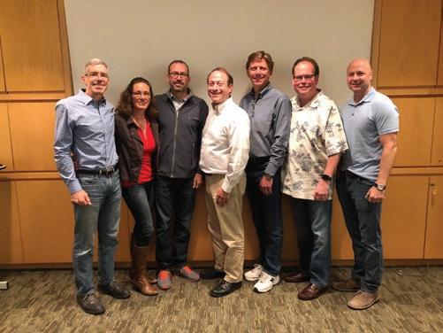
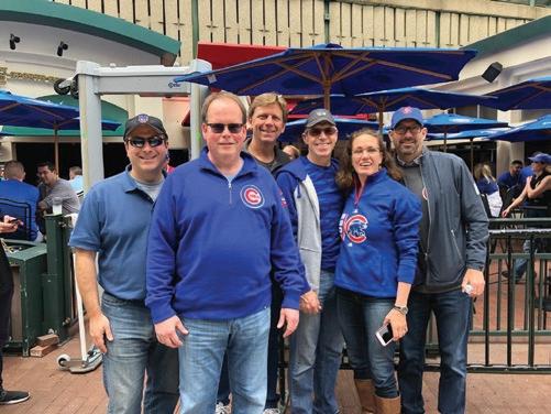
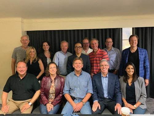
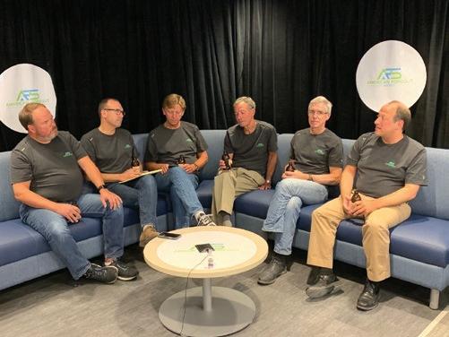
An interim board initially consisting of Jobe, Lipham, Buckley, Bell, Freeman, Lister, and Mike Smith put forth their own funds to hire a consulting frm to help guide them through the formation of the society – its goals, vision, mission, size, fnancial plan, and organizational structure. An unsociety society, starting from scratch without preconceptions of what a society should be. Over the summer of 2018, this interim board (which now included Ken Chang, David Katzka, John Pandolfno, Joel Richter, and Rena Yadlapati) met in person monthly and more often by phone or email (this was pre-Zoom), and some found time to relax together at a Cubbies game in Chicago (Figs. 1.1 and 1.2).
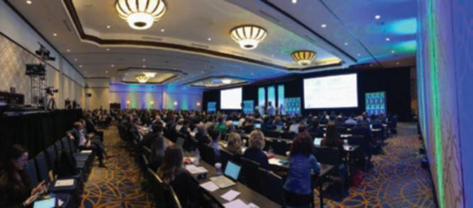
Bringing early reality to this dream of a society in the form of an inaugural annual meeting would deliver the message to a broader audience, initiate the conversations between gastroenterologists and foregut surgeons, and establish industry ties. John Lipham’s “go big or go home” encouraged needed contributions from board members to supplement the grant from Ethicon for the needed fnancial commitment to host the inaugural meeting in Las Vegas in March 2019. Dr. Felice Schnoll-Sussman, a gastroenterologist at Weill Cornell in NYC, and Reg Bell were course directors. Kate Freeman, who would become the executive director of AFS, along with the interim board and course directors hosted 550 attendees at a
3-day, state-of-the-art meeting with over 40 state-of-theart lectures from as many leaders in their respective felds (Fig. 1.3 ).
On the heels of an incredibly successful meeting, the inaugural AFS board was convened to deliver on the mission of improving education, patient care, and outcomes in the diseases of the foregut (Fig. 1.4).
With that our vision became clear: “To advocate personalized treatment strategies for patients with foregut disease through a collaborative partnership across disciplines.”… Our mission is “To help guide both the diagnosis and management of Foregut disease through collaboration between Gastroenterologists and Foregut Surgeons. To foster research that will culminate in the development of benchmarks for excellence while also establishing specialty specifc training
Fig. 1.1 The initial interim board, Chicago June 9, 2018. From left to right: Reginald Bell, Kate Freeman, Tripp Buckley, Mike Smith, John Lipham, Dan Lister, John Pandolfno (Blair Jobe in absentia)
Fig. 1.2 Enjoying a rare moment of relaxation
R. C. W. Bell and F. Schnoll-Sussman
programs that will ultimately translate into the improved care, safety and value for patients with Foregut diseases.”
Committees were formed, individualized sponsor relationships were developed, and a journal (later to be appropriately named Foregut with co-editors Philip Katz and Brian Louie) was founded. The 501(c)(3) AFS Foundation was established to provide long-term backing for the society’s goals of improving patient care including patient education and research.
Then, COVID altered everyone’s course in 2020; the planned in-person meeting for March was delayed until June, then September, and we all pivoted to virtual. Despite these barriers, over 400 people attended! Though Felice could not be physically present, her rousing “whoop whoop” cheered the exhausted in-person program committee! (Fig. 1.5).
A decision to keep the annual meeting in September led to an in-person 2021 meeting (fortuitously at a lull in COVID’s
1.6 The AFS board at the annual meeting in Opryland on September 23, 2021. From left to right: Lee Swanstrom, Ken Chang, Phil Katz, Joel Richter, Dan Lister, Mike Smith, John Lipham, Felice Schnoll-Sussman, Reg Bell, Prakash Gyawali, Peter Kahrilas, Christy Dunst, Kate Freeman (Executive Director), Kerry Dunbar
pandemonium) at the Gaylord Opryland with over 320 inperson and 100 virtual attending (Fig. 1.6).
Very early in this course, Ninh Nguyen and John Clarke had a vision for a textbook of foregut disease with chapters co-authored by gastroenterologists and surgeons, convinced Springer of the value of this endeavor; and it is through his vision and strong encouragement that this textbook has been brought to fruition.
The American Foregut Society is still in its defning stages with the desire to forge deep collaborative ties between foregut surgeons and gastroenterologists at the epicenter. The future is bright, and the time is now—“we are better together.”
Fig. 1.3 The inaugural AFS annual meeting, March 15, 2019
Fig. 1.4 The inaugural AFS board, 2019. From left to right: Mike Smith, Phil Katz, Felice Schnoll-Sussman, Christy Dunst, Kate Freeman, Santiago Horgan, John Lipham, Ken Chang, Dan Lister, Peter Kahrilas, Reg Bell, Tripp Buckley, Rena Yadlapati, Bob Ganz
Fig. 1.5 An exhausted crew, September 26, 2020
Fig.
GERD and Eosinoplhilic Esophagitis
GERD Pathophysiology: The Role of the Sphincter and Crural Diaphragm
Ravinder K. Mittal and John C. Lipham
A recent study that utilized National Gastrointestinal Survey using MyGiHealth determined the prevalence of GERD symptoms in the past, and persistence symptoms (heartburn or regurgitation, 2 or more days/past week) among participants taking proton pump inhibitors (PPIs). 44% of the subjects reported GERD symptoms in the past and 31% in the past week. Thirty-fve percent of those who experienced GERD symptoms were currently on therapy (55% on PPIs, 24% on histamine-2 receptor blockers, and 24% on antacids). Among 3229 participants taking daily PPIs, 54% had persistent GERD symptoms. On a global level, estimates of age-standardized prevalence of GERD for various locations in 2017 ranged from 4.4 to 14% cases. Age-standardized prevalence was highest (11%) in the USA, Italy, Greece, New Zealand, several countries in Latin America, Caribbean, north Africa, the Middle East, and eastern Europe. Global prevalence peaked at 19%, between ages of 75 and 79 years, and it increased by 18% between 1990 and 2017 because of the aging and population growth. One can argue whether these epidemiological studies that utilize standardized questionnaire which equate heartburn and regurgitation symptoms with GERD truly refect GERD, but at least they refect the scope of “GERD” in year 2021.
Gastric acid and pepsin are the major offenders and primarily responsible for the esophageal mucosal damage in refux diseases. Interestingly though, gastric acid secretion in majority, if not all, of patients with GERD is normal. Anatomic and functional abnormality of the sphincter mechanism at the esophagogastric junction (EGJ) and deranged esophageal peristalsis allow acid and possibly other noxious agents to reach and remain in the esophagus for extended
R. K. Mittal (*)
Division of Gastroenterology, Department of Medicine, University of California San Diego, San Diego, CA, USA
e-mail: rmittal@ucsd.edu
J. C. Lipham
The Division of Upper GI and General Surgery at the University of Southern California, Los Angeles, CA, USA
e-mail: John.Lipham@med.usc.edu
periods of time after refux events which induces esophageal mucosal damage and symptoms. What constitutes a normal sphincter mechanism at the EGJ and what goes wrong with it in patients with GERD is the focus of this chapter.
A person can stand upside down after eating a large hearty meal, yet no food backs up into the esophagus, and hence the presence of a sphincter/valvular mechanism at the EGJ is intuitively clear. Inglefnger wrote (1958) that the crural diaphragm, oblique entry of esophagus into the stomach which creates a fap valve, and intrinsic contraction at the gastroesophageal junction area (LES) constitute the antirefux barrier. However, he stated that the importance of each of these mechanisms can be supported or challenged on the basis of evidence, which is inconclusive. Sixty years later (2018), Pandolfno and Tach in a review article cited the same three structures, i.e., LES, crural diaphragm, and fap valve as the key components of the antirefux barrier. However, the difference between 1958 and 2018 is that the methodological improvements have provided better evidence for the anatomical and functional nature of the antirefux barrier.
Allison (1951) felt that a sliding hiatus hernia was the key player in the pathogenesis of GERD and reduction of hiatus hernia was the major element in his surgical treatment of refux disease. On the other hand, Nissen (1956) focused on improving the LES function by gastroplication which we now call fundoplication, as the major element in his antirefux surgery. Follow-up studies revealed that approximately half of the patients following Allison repair as well as following Nissen fundoplication repair had recurrence of GERD symptoms. In both repairs, recurrence of hiatus hernia is a major predictor of the recurrent symptoms. Based on the above observations, one may argue that the lack of attention to the LES in Allison repair and lack of crural diaphragm repair in the Nissen fundoplication may be the reasons for the failure of antirefux surgery. Current thinking is that hiatal dysfunction leads to the formation of hiatus hernia and repair of esophageal hiatus (by plication, mesh, or other methods) along with fundoplication should be the key elements of antirefux surgery. However, one must keep in mind
N. T. Nguyen et al. (eds.), The AFS Textbook of Foregut Disease, https://doi.org/10.1007/978-3-031-19671-3_2
though that the esophageal hiatus which is formed by the right crus of diaphragm is important for two reasons: (1) separating the thoracic and abdominal cavity, i.e., physically keeping the distal esophagus/LES in the abdomen, thus preventing migration of the stomach into the chest or hiatus hernia, and (2) a sphincter-like action at the EGJ that counteracts pressure gradients between the esophagus (located in the thorax) and stomach (located in the abdomen). Along with LES and crural diaphragm, phrenoesophageal ligament that anchors the two structures is the third key player at the EGJ in the maintenance of competent antirefux barrier, which will be reviewed in this chapter.
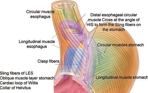
The sphincters, in general, separate adjacent organs by circumferential closure of the junction between those organs, i.e., esophagogastric junction (EGJ). Smooth muscle LES and crural diaphragm are the two sphincter mechanisms at the EGJ. Sphincters, as per English dictionary, are donutshaped structure. The latter implies that the LES should be a thick band of the circular muscle. However, no such distinct structure has been identifed when examining the EGJ visually, neither in the autopsy specimens nor at the time of surgery, which led to the belief that the LES is a functional rather than a distinct anatomical/morphological entity. However, high-frequency ultrasound imaging using catheter probes in the live humans reveal that the muscles of the LES region is approximately two times thicker than the adjacent muscles of the esophagus. Maybe, the loss of the LES muscle tone in the autopsy specimens and surgical specimen leads to the thinning of the tissue because thickness is related to muscle tone. Lieberman-Mefferet (1976) examined the distal esophagus and proximal stomach from the live donors using a dissecting microscope and concluded that the LES region has a unique anatomy; it consists of clasp (on the lesser curvature) and sling fbers (on the greater curvature of the stomach). In Lieberman-Mefferet study, some regions, especially along the greater curvature side, did have thicker muscle as compared to the other regions. Yassi et al., for the frst time, performed a computer 3D reconstruction of the microscopic images of the EGJ, 8.2 μm resolution, 652 sections, spaced 50 micron apart. Their intent was to visualize the arrangement of muscle fbers/fascicle inside the LES muscle at the microscopic level. However, the 3D data fles were larger than their computer memory to build the 3D anatomy of the LES, at the resolution of the captured images. Zifan et al. used images captured by Yassi et al. to create the 3D myoarchitecture of LES at the resolution of captured images. They found that the circular muscles at the lower end of the esophagus, from the right and left side, cross at the angle of His at the greater curvature of the stomach and continue onto the anterior and posterior wall of the stomach toward the lesser curvature of stomach, as the sling fbers of the LES (Fig. 2.1). These fbers appear to be same structures which in the literature have been called by several other
R. K. Mittal and J. C. Lipham
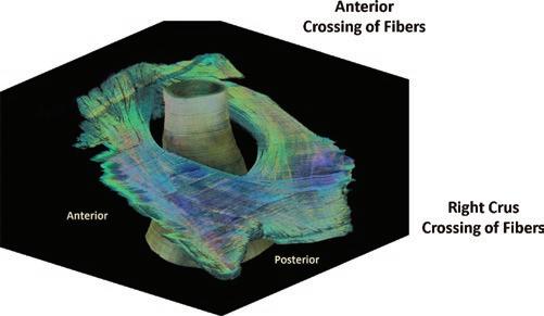
names, i.e., “collar of Helvetius,” cardiac loop of Willis, and oblique muscle layer of the stomach. The latter is also known as the innermost of the three muscle layers of the stomach. Circular muscle fbers from the lesser curvature of the stomach which are the same as the clasp fbers of the LES are inserted into the sling fbers. Finally, the longitudinal muscles of the esophagus also terminate into the sling fbers. Based on the above morphology, the LES is not a donutshaped muscle; instead it is like a “noose” at the EGJ. Similar to LES, the myoarchitecture of the right crus which forms the esophageal hiatus (crural diaphragm) also resembles a “noose.” The right crus muscle originates from the lumbar spine (L1, L2, and L3) and divides into two muscle bundles, fbers of which decussate in a “scissorlike fashion” before surrounding the esophagus to form the esophageal hiatus (Fig. 2.2). Interestingly, muscle fascicles of the right crus also decussate one more time, in the front of the esophagus
Fig. 2.1 Myoarchitecture of the lower esophageal Sphincter. Note the crossing of circular muscles of the esophagus at the angle of His and continuation as the sling fbers of the stomach. Clasp fbers of the stomach are inserted into the sling fbers of the stomach. (From Zifan A, Kumar D, Cheng LK, Mittal RK. Sci Rep. 2017 Oct 13;7 (1):13188
Fig. 2.2 Myoarchitecture of the esophageal hiatus is formed by the right and left crus muscles. Note that the two bundles of the right crus cross each other in a “scissorlike fashion” posterior, as well as anterior to the esophagus. From Zifan A, Kumar D, Cheng LK, Mittal RK. Sci Rep. 2017, 13;7 (1):13188
before merging into the central tendon of the diaphragm. Interestingly, the external anal sphincter also has “noose like” myoarchitecture, i.e., decussation of fbers at the anterior and posterior surfaces of anal canal. Might be, all sphincters are not donuts; instead they have “noose-like” myoarchitecture to cause circumferential closure. A longer length of muscle in the “noose-like myoarchitecture” provides mechanical advantage, i.e., it can create greater force upon contraction to cause circumferential closure.
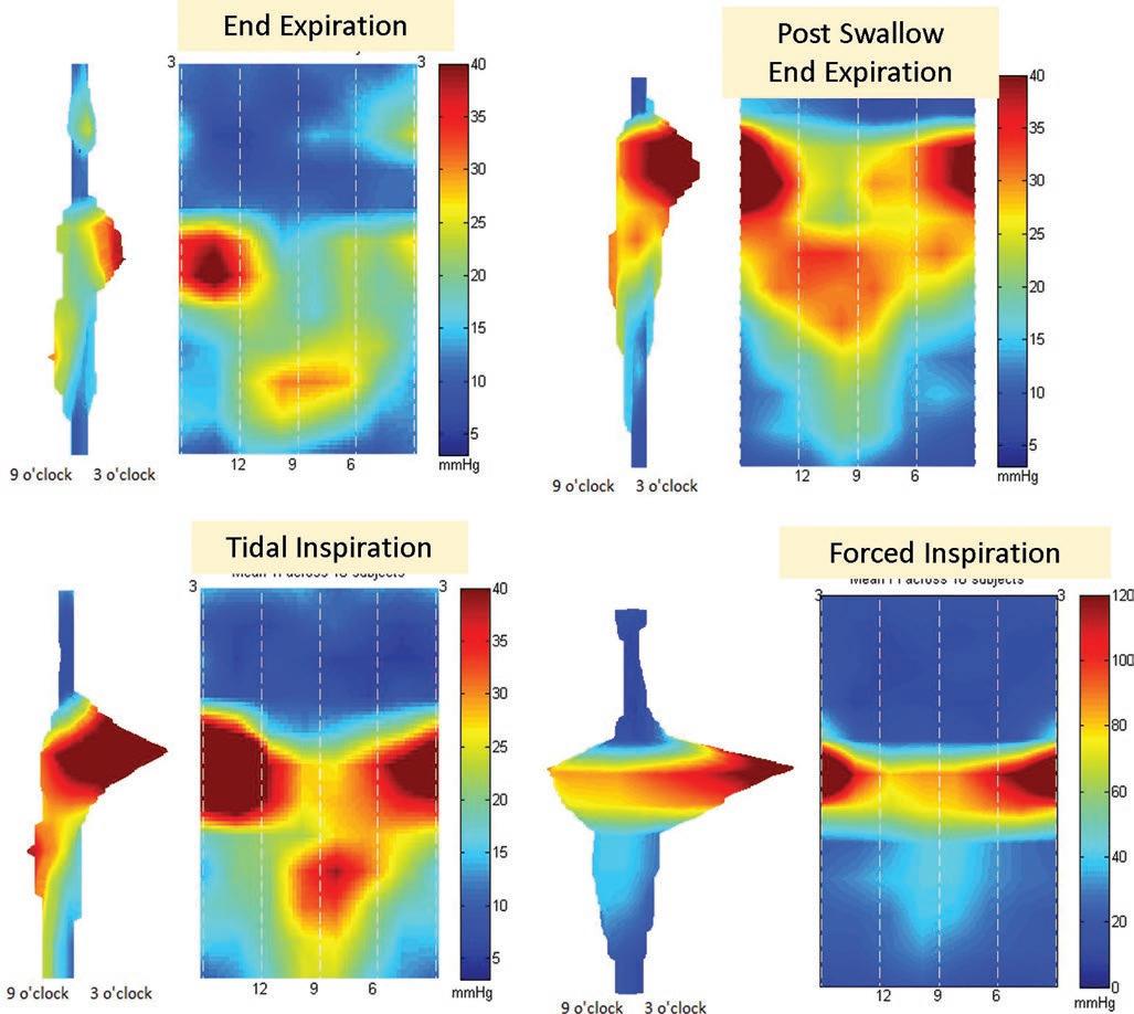
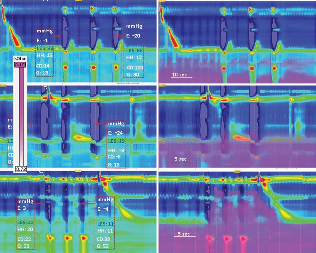
Manometry techniques to measure the antirefux barrier function have been improving approximately every 10 years since the 1950s when Code frst recorded the LES highpressure zone using water-flled catheters. Infusion manometry using side-hole catheters by Harris, Dent sleeve sensor, electrode sleeve sensor, solid-state pressure transducers, high-resolution manometry using color topography, and 3D high-resolution LES manometry have brought us to the current state of knowledge of the function of smooth muscle LES and skeletal muscle crural diaphragm. The 3D-LES manometry catheter, the newest of all the techniques, provides all information revealed by earlier methods and much more. The 3D catheter consists of 12 rings of pressure transducers, spaced 7.5 mm apart, with each ring containing 8 separate transducers spaced 45 degrees apart. Thus, 96 high fdelity pressure transducers can record the EGJ pressure continuously in real time. Color topography of the pressure plots allows one to visualize the 3D pressure profle of the EGJ high-pressure zone as a seamless signature over time. One can determine the radial and axial location of each pressure transducer in the subject by performing a CT scan while the catheter is in place in the subject, thus making it possible to align the LES function with the anatomy of the EGJ region. These studies in normal subjects reveal a unique EGJ pressure profle that has following features (Fig. 2.3):
1. The EGJ pressure profle is longer along the lesser curvature, almost double the length, as compared to the greater curvature of the stomach.
2. The highest pressure is located in the left-posterior direction, known in the past as circumferential LES pressure asymmetry. The reason for higher pressure on the left side is because high-pressure zone is shorter in length in that direction.
3. Crural diaphragm-related squeeze is superimposed on the cranial half of the LES pressure. In other words, the LES and crural diaphragm are anatomically superimposed on each other.
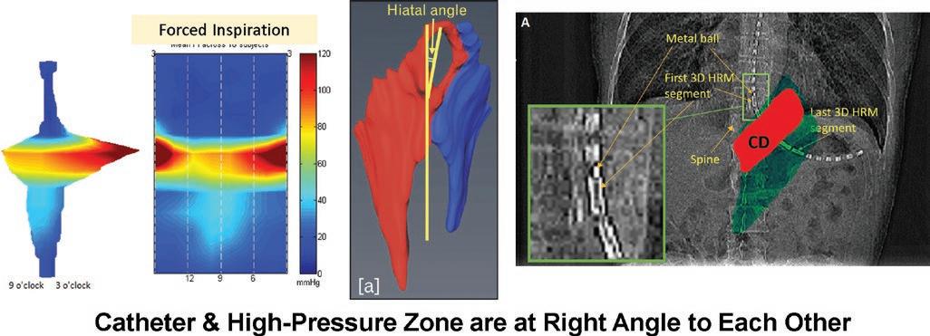
4. The cranial or the proximal edge of the EGJ pressure profle is horizontal because the LES turns to the left at the level of esophageal hiatus (crural diaphragm), as it enters into the abdomen, which brings the hiatus and catheter at right angle to each other (Fig. 2.4).
The EGJ pressure profle aligned with the anatomy of the region from the CT scan images makes it is clear that the right side of the high-pressure zone related to LES aligns to that part of the stomach where clasp fbers anchor into the anterior and posterior sling muscles. There are no reports of the 3D-LES manometry in patients with hiatus hernia, which will allow one to determine the 3D pressure profle of LES and crural diaphragm separately. However, high-resolution manometry studies clearly show two separate high-pressure zones, one related to the LES and the other to the crural diaphragm in patients with sliding hiatus hernia, each one with its own characteristics. The crural diaphragm is mostly active during the inspiratory phase of respiratory cycle. When the LES and crural diaphragm are superimposed on each other, as in normal subjects, the end expiratory EGJ pressure is generally due to the smooth muscle LES. Increase in gastric pressure related to migratory motor complex of the stomach results in LES contraction with increase in the EGJ pressure. On the other hand, increase in the EGJ pressure with inspiration is generally related to crural diaphragm; the amplitude of increase is directly related to the depth of inspiration or the force of diaphragmatic contraction. Crural diaphragm can contract continuously (tonically) during abdominal compression, straight-leg raises, coughing, Valsalva, and all those physical activities that increases intra-abdominal pressure. Change in the EGJ pressure related to LES, being a smooth muscle, occurs slowly (over seconds). On the other hand, the crural diaphragm being a skeletal muscle can contract much faster (over milliseconds). The two sphincters, LES and crural diaphragm, are critical in the prevention of GER because they must respond to counter pressure gradients between the esophagus and stomach which are infuenced by intrathoracic (pleural) and intra-abdominal pressures, respectively. The esophageal pressure is generally lower as compared to stomach during the entire respiratory cycle, and thus there is a pressure gradient in favor of GER; pressure gradient is 6–10 mmHg during end expiration, and it can be 50–100 mmHg during deep breathing and other physical maneuvers. Understanding of the two-sphincter concept, LES and crural diaphragm, is critical to understand the antirefux barrier function.
The esophagus enters into the stomach along its lesser curvature. It is suggested that fundus of the stomach can press on the distal most part of the esophagus or LES to make a gastric fap valve, which has a role in the prevention of GER. Based on the anatomy of the EGJ in normal subjects, as described in previous paragraphs, it is only the distal half of the LES (and not the esophagus) that is located in the abdomen, which can be infuenced by gastric pressure. The LES is a tonically contracted muscle and hence it is a stiff/ noncompliant muscle, which would make transmission of gastric pressure into the intraluminal LES pressure unlikely.
Fig. 2.3 3D-pressure topography of the esophagogastric junction (EGJ). Mean EGJ pressure profle from ten subjects. The EGJ pressure under three different end-expiration conditions: (a) end expiration (EE); (b) post-swallow end expiration (PSE) and with two different strengths of crural diaphragm contractions; (c) tidal inspiration (TI) and (d) forced inspiration (FI). The 9 and 3 o’clock positions are toward the lesser and greater curvature of the stomach, respectively, and 12 and 6 o’clock face toward the anterior and posterior direction, respectively.
Mittal RK, Zifan A, et al. Am J Physiol Gastrointest Liver Physiol. 2017 Sep 1;313 (3):G212-G219
R. K. Mittal and J. C. Lipham
Fig. 2.4 Relationship between 3D-EGJ pressure profle and anatomy of the EGJ seen on CT images. The EGJ pressure profle was obtained at the peak of inspiration. A CT scan was obtained with the 3D-LES manometry catheter in place in the subject. Crural diaphragm was segmented from the CT scan images of the region. Superimposition
The author’s opinion is that it is unlikely that the gastric fap valve by itself can contribute to the antirefux barrier. Gastric fap valve is disturbed in the setting of sliding hiatus hernia; it is more likely that the latter, rather than the gastric fap, contributes to the GERD (as described in the following paragraphs).
Gastroesophageal Refux Mechanisms
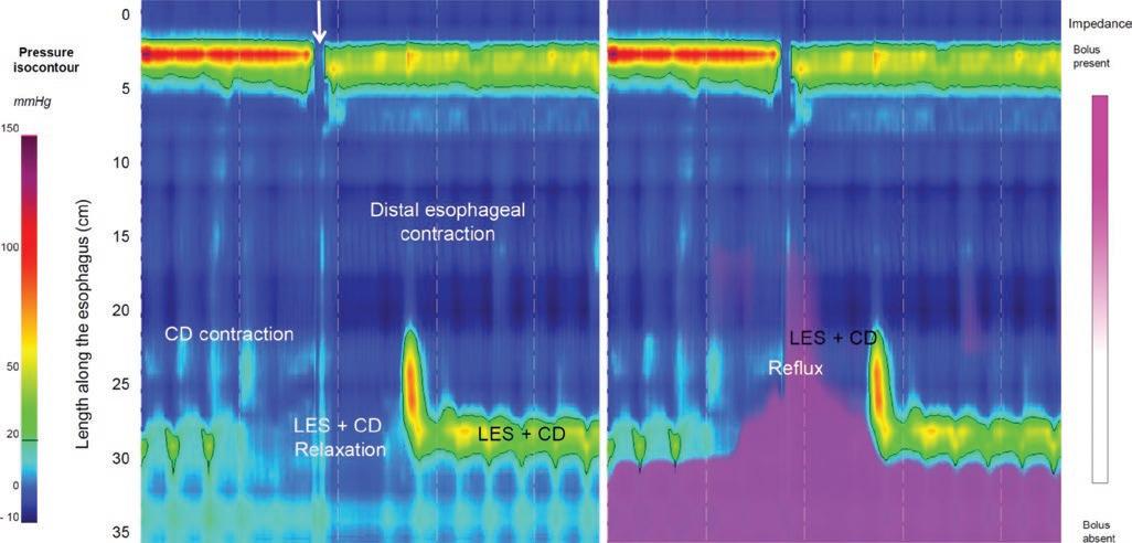
Refux of gastric contents into the esophagus occurs via several mechanisms, transient LES relaxation (TLESR), low LES pressure, crural diaphragm dysfunction, and sliding hiatus hernia. TLESR is the major mechanism of belching and GER in healthy subjects (Fig. 2.5). It is also the major mechanism of GER in patients with normal LES pressure, especially in patients with no evidence of sliding hiatus hernia. The TLESR is preceded by a unique pattern of longitudinal muscle contraction of the esophagus, which starts in the distal esophagus and traverses in a reverse peristaltic fashion toward the mouth. The longitudinal muscle contraction gets stronger as the LES and crural diaphragm relaxation becomes complete. Studies show that the contraction of longitudinal muscles activates inhibitory motor neurons of the myenteric plexus through a stretch-sensitive (mechanosensitive) mechanism to induce LES relaxation and concurrent inhibition of the crural diaphragm. The inhibition of crural diaphragm is essential for the occurrence of GER during TLESR in subjects with a normal EGJ anatomy, i.e., absence of sliding hiatus hernia. Gastric distension is the predominant stimulus for the induction of TLESR. Despite the importance of TLESR in the GERD pathogenesis, drugs targeting to inhibit
of the EGJ high-pressure zone over the fuoroscopic image of the EGJ. Note that the manometry catheter bends to the left as it enters the stomach. Even though the hiatus is oriented at an angle with the spine, it is at right angle to the hiatus because of bending of the catheter at the EGJ which gives a horizontal pressure profle at the proximal end
TLESRs have limited effcacy and signifcant adverse events. Crural myotomy in cats lead to an increase in the incidence of GER. Injection of botulinum toxin into the LES, used routinely in the treatment of achalasia and other spastic motor disorders, leads to partial/complete paralysis of the crural diaphragm contraction, which results in GER events during various physical maneuvers that increase gastroesophageal pressure gradients, including during the simple act of deep breath.
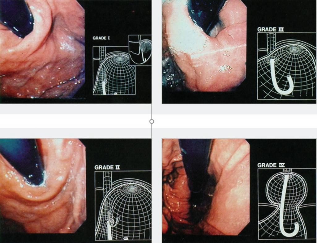
With each swallow-induced primary peristalsis, contraction of the longitudinal muscles of the esophagus pulls the LES in cranial direction (2–3 cm), into the thorax, resulting in what is generally known as a physiological hiatus hernia or phrenic ampulla. On the other hand, persistent herniation of the stomach into the chest resulting in anatomical separation of LES and CD is a pathologic entity. Clinicians have known for long time of strong association between sliding hiatus hernia and GERD. Separation of the LES and CD results in deformity at the EGJ, also known as the loss of fap valve of the stomach. One can grade the severity of deformity using Hill’s grade classifcation system (Fig. 2.6). Studies show that patients with Hill’s grade 3 and grade 4 fap valves have increased esophageal acid exposure, or in other words GERD. Prolonged HRM recordings show that the LES slide in and out of the hiatus (crural diaphragm) several times during the day. Interestingly, majority of GER events occurs in the setting when the LES and CD are anatomically separate, i.e., in the setting of a sliding hiatus hernia. The dynamic relationship (sliding) between LES and CD is possibly an important reason for the controversy surrounding the role of hiatus hernia in GERD. Studies show
R. K. Mittal and J. C. Lipham
Fig. 2.5 Transient lower esophageal sphincter relaxation (TLSER) on high-resolution manometry (on the left) and impedance (on the right). Lower esophageal sphincter (LES) relaxation is >10 seconds in duration, which occurs in the absence of swallow (no pharyngeal contraction). Upper
many mechanisms by which sliding hiatus hernia may cause GER events:
1. A small amount of acid is trapped in the hernia sac (between the LES and CD) that can refux repeatedly into the esophagus during swallow-induced LES relaxation.
2. Separation of LES and CD results in reduction of the LES pressure.
3. Widening of the esophagus hiatus by a herniated stomach impairs the sphincter function of crural diaphragm. A recent study describes respiration-induced changes in the pressure gradients between the stomach, hernia, and esophagus leads to the GER events (two-stage mechanical pump hypothesis) (Figs. 2.7 and 2.8).
In the frst stage, during the expiratory phase of respiratory cycle, gastric contents fow from the stomach (positive pressure environment of the abdomen) into the herniated stomach (negative pressure environment of thorax) because crural diaphragm contraction (barrier) is generally not active during expiratory phase of the respiratory cycle. In the second stage, with inspiration phase of respiratory cycle, contraction of the CD (hiatus) causes compartmentalization of the stomach (above and below the hiatus). Furthermore, with inspiration there is a greater decrease in the esophageal than the hernia pressure, resulting in a net pressure gradient directed toward the esophagus. The LES pressure response to inspiration varies among patients; in some patients there is
esophageal sphincter (UES) relaxation occurred during the TLESR. Also note crural diaphragm inhibition during TLESR. The TLESR is accompanied with gastroesophageal refux (observed on impedance). Roman, S, Holloway R, Keller J, et al. Neurogastroenterol Motil 2017;29
an increase in the LES pressure with inspiration and in others there is a decrease. In the latter setting, contents from the herniated stomach can refux across the LES into the esophagus. Acid refux fowing into the esophagus with each breath can be a major player in the pathogenesis of moderate to severe GERD, i.e., erosive esophagitis and Barrett’s esophagus. The GER events from hiatus hernia into the esophagus, usually small in amount, may travel only to the most distal part of the esophagus, which explains why refux-induced changes in the esophagus are greatest close to the squamocolumnar junction or the Z line.
Intrathoracic location of the LES is disadvantageous for its antirefux barrier function because a negative thoracic pressure can pull open a contracted LES muscle. It is not clear why in some hiatal hernia patients the LES pressure increases with inspiration which is advantageous for its antirefux barrier function. It is also interesting to note that all those animals that have an intra-abdominal length of the esophagus in the abdomen, such as mice, rat, rabbit, and opossum, can’t vomit. On the other hand, animals with a superimposed LES and crural diaphragm, such as cats, dogs, pigs, and humans, can vomit. Surgical literature suggests that an intra-abdominal location of the LES/esophagus is important in the competency of LES. A successful Nissen fundoplication reduces hiatus hernia, restores a small length of the esophagus back into the abdomen, wraps the distal 2 cm of the esophagus with gastric fundus, and reduces the size of esophageal hiatus by either plication or placement of a mesh.
In the setting of a large hiatal opening, a mesh is placed to reinforce the hiatal opening. Studies show several mechanisms by which Nissen fundoplication makes the refux barrier competent: (1) reduction of hiatus hernia, (2) increase in the basal LES pressure, (3) increase in the intra-abdominal length of LES length, and (4) reduction in the frequency of TLESRs and rendering TLESRs incomplete. Since there are several things done during a fundoplication, it is not clear which component of the repair leads to which component of the antirefux barrier more effective. It is likely that plication/ reinforcement of the hiatus prevents recurrence of sliding hiatus hernia; whether it leads to an improved sphincter function of crural diaphragm requires further study.
The role of phrenoesophageal ligament in the making of an effective antirefux barrier function can’t be overemphasized. As described earlier, in healthy subjects, the LES and CD are physically superimposed on each other. The two structures are anchored to each other by the upper and lower leaves of the phrenoesophageal ligament, which originate
from the thoracic and abdominal surfaces of diaphragm, respectively. The phrenoesophageal ligament penetrates deep into the connective tissue between the bundles of longitudinal and circular muscles of the esophagus, thus forming an extremely tight anchoring between the LES and crural diaphragm. With inspiration and expiration, the LES and crural diaphragm move in the caudal and cranial direction, respectively, without separating from each other. X-ray fuoroscopy studies show that metal clip placed endoscopically at the squamocolumnar junction (Z line) move 2–3 cm in the cranial/oral direction during primary and secondary peristalsis. They migrate from the intra-abdominal position before peristalsis into the thorax during peristalsis and return back to their baseline position at the completion of peristalsis. Z line is normally located in the middle of LES, which implies that the LES migrates into the thorax during peristalsis, or in other words, each swallow results in formation of a small sliding hiatus hernia, also referred to as physiologic sliding hiatus hernia, or phrenic ampulla. The pathological sliding
Fig. 2.7 High-resolution manometry impedance (HRMZ) recordings of three patients (A, B, and C) with sliding hiatus hernia (HH), without (left), and with impedance (right) recordings. Note pressure changes in the esophagus (E), lower esophageal sphincter (LES), hiatus hernia (HH), crural diaphragm (CD), and stomach (G) with three deep breaths. The boxes on each HRMZ recordings show pressure at the fve locations, before (left) and during deep inspirations (right). In patient A, deep breaths result in an increase in the LES pressure. On the other hand, in patients B and C, deep inspirations result in the decrease in
hiatus hernias are those in which LES and crural diaphragm remain separate, thus forming two high-pressure zone, which implies defective phrenoesophageal ligament. A pathological sliding hiatus hernia can be the result of either excessive pull on the esophagus (from the longitudinal muscle contraction), defective phrenoesophageal ligament, or excessive intra-abdominal pressure.
Several studies have examined for the structural defects in the crural diaphragm and phrenoesophageal ligament in GERD patients. Biopsy samples of the crura and phrenoesophageal ligament under transmission electron microscopy in GERD patients reveal abnormalities in 94% of the biopsied
LES pressure (more signifcantly in patient 3). The LES pressure during deep breath in patient 3 is same as esophageal pressure. Note the changes in impedance (pink color, as a marker of fuid movement, i.e., refux). In all patients, fuid moves from the stomach into HH in between deep inspiration (end expiration). There is no movement of fuid from HH into the esophagus in patient A, who has increase in LES pressure with deep breath. On the other hand, in patients B and C, reduction in the LES pressure with inspiration is associated with movement of fuid from HH into the esophagus
samples, with 75% of those having severe abnormalities. The latter were defned as extended disruption and degeneration of the crural muscle architecture. Other studies have found loss of myofbrils, infammatory infltrate, and neovascularization in the right and left crural diaphragm in hiatus hernia patients, but not in controls. Phrenoesophageal ligament samples obtained intraoperatively from patients with hiatal hernias found that the collagen content was 60% lower in the hiatal hernia patients (including type I and type III collagen), suggesting GERD as a genetic connective tissue disorder. Structural abnormalities of the CD have been explained on the basis of genetics studies linking altered collagen expression.
Fig. 2.8 Mean data of pressure in the esophagus (E), lower esophageal sphincter (LES), hiatus hernia (HH), crural diaphragm (CD), and stomach (G) in patients who show increase in LES pressure with inspiration (group 1) and who show decrease in the LES pressure with inspiration (group 2), before and during deep breaths. There was no difference in the G, HH, and esophageal pressure in the two groups. The LES pressure before deep breath was lower than during deep breath in group 2 (*). The LES pressure before deep breath was different between the two groups (**). The CD squeeze pressure was lower in group 2 compared to group 1 (#). The schematic shows movement of gastric fuid from the stomach into the HH during expiration
Children and adults, diagnosed with GERD and/or hiatal hernias, have been found to have higher prevalence of mutations within the collagen type III gene COL3A1. Above fndings have been confrmed in a case-control cohort study in which signifcant association with COL3A1 mutations was found in hiatal hernia patients compared to control patients, suggesting an altered expression of collagen III, which resulted in an imbalance between collagen I and collagen III levels.
In summary, the smooth muscle LES and skeletal muscle crural diaphragm, anchored to each other by the phrenoesophageal ligament, forms a normal antirefux barrier. Both the LES and crural diaphragm have “noose”-like myoarchitecture. The 3D-LES pressure recordings and CT scan imaging done simultaneously reveal the unique EGJ high-pressure zone profle, and for the frst time, it provides the relationship between EGJ pressure profle (function) and anatomy. Transient LES relaxation, a vagally mediated neurological event, which results in simultaneous relaxation of the LES and crural diaphragm is an important mechanism of GER in patients with the GERD. Disruption of phrenoesophageal ligament and crural diaphragm which results in formation of sliding hiatus hernia is also critical in the pathogenesis of GERD. Recurrence of sliding hiatus hernia following Nissen fundoplication leads to recurrence of symptoms, which suggests that repair of the crural diaphragm (hiatus) is a critical part of the antirefux surgery. Future studies need to focus on
advancing our knowledge at several fronts, i.e.: (1) genesis of GERD symptoms; (2) pathogenesis of transient LES relaxation, why, and when does a physiological event such as TLESR becomes a pathological one; and (3) all those factors that are important in the genesis of hiatus hernia, and recurrence of sliding hiatus hernia following Nissen fundoplication.
Questions
1. The key elements of a competent antirefux barrier are:
A. Smooth muscle lower esophageal sphincter
B. Crural diaphragm
C. Phrenoesophageal ligament
D. All of the above
Answer: D
2. Sliding hiatus hernia impairs antirefux barrier by the following mechanisms:
A. It reduces LES pressure.
B. It results in a defective gastric fap valve.
C. It acts as a two-stage mechanical pump that drives gastric contents into the hernia during expiratory phase of respiratory cycle and pumps hernia contents into the esophagus during inspiratory pump of the respiratory cycle.
D. All of the above.
Answer: D
Acknowledgement Financial Support: Dr. Mittal is supported by NIH Grant R01 DK109376
Confict of Interest None of the authors have any confict of interest.
Bibliography
1. Delshad SD, Almario CV, Chey WD, et al. Prevalence of gastroesophageal refux disease and proton pump inhibitor-refractory symptoms. Gastroenterology. 2020;158:1250–1261 e2.
2. The global, regional, and national burden of oesophageal cancer and its attributable risk factors in 195 countries and territories, 1990-2017: a systematic analysis for the global burden of disease study 2017. Lancet Gastroenterol Hepatol. 2020;5:582–97.
3. Allison PR. Refux esophagitis, sliding hiatal hernia, and the anatomy of repair. Surg Gynecol Obstet. 1951;92:419–31.
4. Nissen R. A simple operation for control of refux esophagitis. Schweiz Med Wochenschr. 1956;86:590–2.
5. Mittal R, Vaezi MF. Esophageal motility disorders and gastroesophageal refux disease. N Engl J Med. 2020;383:1961–72.
7. Zifan A, Kumar D, Cheng LK, et al. Three-dimensional Myoarchitecture of the lower EsophagealvvSphincter and Esophageal hiatus using optical sectioning microscopy. Sci Rep. 2017;7:13188.
8. Mittal RK, Zifan A, Kumar D, et al. Functional morphology of the lower esophageal sphincter and crural diaphragm determined by three-dimensional high-resolution esophago-gastric junction pressure profle and CT imaging. Am J Physiol Gastrointest Liver Physiol. 2017;313:G212–9.
9. Babei A, Korsapati H, Bhargava V, Zheng W, Mittal RK. A unique pattern of longitudinal muscle contraction during tran-
10. Mittal RK, Kumar D, Jiang Y. Sliding hiatus hernia: a two-step pressure pump of gastroesophageal refux. Gastroenterology. 2021;161:339–41.
11. Kumar D, Zifan A, Mittal RK. Botox injection into the lower esophageal sphincter induces hiatal paralysis and gastroesophageal refux. Am J Physiol Gastrointest Liver Physiol. 2020;318:G77–83.
12. Fei L, del Genio G, Rossetti G, et al. Hiatal hernia recurrence: surgical complication or disease? Electron microscope fndings of the diaphragmatic pillars. J Gastrointest Surg. 2009;13:459–64.
13. Fei L, Rossetti G, Allaria A, et al. Laparoscopic hiatal hernia repair. Is the mesh hiatoplasty justifed? Ann Ital Chir. 2014;85:38–44.
14. von Diemen V, Trindade EN, Trindade MR. Hiatal hernia and gastroesophageal refux: study of collagen in the phrenoesophageal ligament. Surg Endosc. 2016;30:5091–8.
15. Richter JE, Kumar A, Lipka S, et al. Effcacy of laparoscopic Nissen fundoplication vs transoral incisionless fundoplication or proton pump inhibitors in patients with gastroesophageal refux disease: a systematic review and network meta-analysis. Gastroenterology. 2018;154:1298–1308.e1297.
16. Yassi R, Cheng LK, Rajagopal V, et al. Modeling of the mechanical function of the human gastroesophageal junction using an anatomically realistic three-dimensional model. J Biomech. 2009;42:1604–9.
17. Bredenoord AJ, Weusten BL, Timmer R, et al. Intermittent spatial separation of diaphragm and lower esophageal sphincter favors acidic and weakly acidic refux. Gastroenterology. 2006;130:334–40.
18. Mittal RK. Regulation and dysregulation of esophageal peristalsis by the integrated function of circular and longitudinal muscle layers in health and disease. Am J Physiol Gastrointest Liver Physiol. 2016;311:G431–43.
19. Hill LD, Kozarek RA, Kraemer SJ, et al. The gastroesophageal fap valve: in vitro and in vivo observations. Gastrointest Endosc. 1996;44:541–7.
Empirical diagnosis of GERD based on symptomatology is common clinical practice. Based on the Montreal consensus statement, “GERD is a condition which develops when refux of stomach contents causes troublesome symptoms or complications” [1]. Typical symptoms include burning pain starting in the upper abdomen and moving upward, behind the chest and toward the throat as well as regurgitation although only 49% of patients with GERD report typical symptoms. Extra-esophageal syndromes (refux cough, laryngitis, asthma, dental erosion) are considered atypical symptoms. Full assessment by a physician leads to a sensitivity of approximately 65% for the diagnosis of GERD with a specifcity of 70% [2]. Furthermore, the detection rate of GERD by use of symptoms seems to decrease with age, with elderly patients often remaining asymptomatic despite high rate of mucosal injury and complications [3]. Unfortunately, lack of GERD symptoms also is more common in patients with Barrett’s esophagus, placing these patients at high risk for late detection of esophageal adenocarcinoma.
Clinical Questionnaires
The diffculty in ascertaining the presence of GERD accurately through physician assessment has led to the formation
S. Chandra (*)
Dignity Health Gastroenterology, Creighton University School of Medicine, Phoenix, AZ, USA
e-mail: subhash.chandra@creighton.edu
J. Gapp
Division of Gastroenterology, CHI Health Creighton University School of Medicine, Omaha, NE, USA
K. Wang
Division of Gastroenterology and Hepatology, Mayo Clinic School of Medicine, Rochester, MN, USA
e-mail: wang.kenneth@mayo.edu
of questionnaires. The goal being that if specifc patientreported information is elicited, then the sensitivity and specifcity for a GERD diagnosis might be improved. The Diamond study, performed in the UK, used the Refux Disease Questionnaire (RDQ) in an attempt to improve diagnosis. However, use of the RDQ did not appear to offer substantial diagnostic improvement above physician assessment when using endoscopy and pH monitoring as the standard [2]. While questionnaires may be useful in following disease progression and response to treatment, their use for diagnostic purposes is limited.
Empiric Acid Suppression Trial
Patients with empirical diagnosis of GERD often undergo a trial of acid suppression therapy with a maximal dose proton pump inhibitor (PPI) for 2–4 weeks prior to more invasive methods of evaluation. Based upon symptoms, approximately 50% of patients will have a response to PPI therapy [4]. In those with known GERD, 69% of patients will respond to PPI trial, while 51% of those without GERD will also have response to PPI therapy [5]. Previous studies have indicated that a trial of PPI therapy for diagnosis of GERD held a sensitivity of 54% and a specifcity of 65% when compared to combination of upper endoscopy and 24-hour pH monitoring. The sensitivity of a combination of empirical diagnosis with response to PPI therapy approach in 78% and specifcity of 54% [2]. Such a moderate performance in testing metrics along with the questionable management of patients with a partial response to high-dose acid suppression therapy often leads to the need for further evaluation. In cases where there is symptom improvement with acid suppression and recurrence of symptoms with cessation of therapy in the absence of red fag signs, it is still considered reasonable to make a diagnosis of GERD.
N. T. Nguyen et al. (eds.), The AFS Textbook of Foregut Disease, https://doi.org/10.1007/978-3-031-19671-3_3
3
Upper Endoscopy
Upper endoscopy for the evaluation of GERD is generally indicated in patients who do not respond to acid suppression therapy or have red fag signs consisting of iron defciency anemia, dysphagia, odynophagia, and weight loss or as part of screening for Barrett’s esophagus in high-risk populations. Upper endoscopy can establish the presence of pathologic GERD based on the presence of mucosal injury (Los Angeles grade C or D esophagitis) or its complications (peptic stricture or Barrett’s esophagus) occurring in 2.2%, 11%, and 3.3%, respectively, in those not already on PPI therapy [6]. However, the aforementioned fndings often are not present in patients with symptomatic GERD, particularly since patients are often placed on PPI therapy prior to endoscopy. While the criteria for a defnitive GERD diagnosis often are not met with upper endoscopy alone, a thorough endoscopic exam of the gastroesophageal junction and biopsies of the esophagus provide information to be used in conjunction with supplementary studies in forming a diagnosis of GERD as well as for ruling out other disease entities with a shared symptomatology. A high-quality endoscopic exam is also a prerequisite in the case that antirefux surgery is to be considered in the future.
Box
3.1 Defnitive Evidence for GERD on Upper Endoscopy
LA class C or D esophagitis. Peptic stricture. Barrett’s esophagus.
The endoscopic exam performed for an indication of GERD should include four principal components. First, thorough examination of the esophagus on forward view should be performed to determine the presence of esophagitis, Barrett’s esophagus, peptic strictures, or other abnormalities that suggest tumor or achalasia. Esophagitis should be graded based upon the Los Angeles (LA) esophagitis system and if high grade (grade C or D) would require 8 weeks of therapy with follow-up endoscopy to assess healing and exclude Barrett’s esophagus and long-term treatment (Fig. 3.1). Peptic strictures are most common at the squamocolumnar junction, and careful observation in this area with a high degree of distention increases detection. If salmon-colored mucosa >1 cm from the top of the gastric folds or mucosal irregularities are observed, biopsies should be performed. Examination of the esophagus for dilatation, presence of fuid in the esophagus upon initial esophageal intubation, and diffculty traversing the gastroesophageal junction with
the gastroscope can indicate the presence of achalasia or pseudo-achalasia which often shares common symptomatology to patients with severe refux disease.
Second, mucosal biopsies are not routinely performed though histologic fndings of basal cell hyperplasia and papillary elongation support a diagnosis of refux disease and are sensitive but have poor specifcity for GERD. Findings of dilated intercellular spaces due to infammatory changes are also present; however, such studies are not normally performed outside of a research setting. The one circumstance that biopsies should routinely be obtained is if the patient’s symptom profle includes dysphagia in which case eosinophilic esophagitis needs to be considered. Normal histology in the distal esophagus does not exclude GERD as patients are often on PPIs at the time of endoscopy.
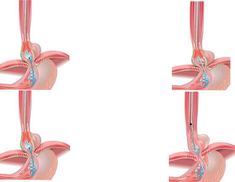
Third, endoscopic evaluation of the GEJ on both forward and retrofexed views can signifcantly alter suspicion for refux disease when correctly performed. Upon examination of the lower esophagus, the presence of a hiatal hernia should be recorded and is considered to be part of a high-quality upper endoscopy. The presence of a hiatal hernia indicates mechanical failure of the antirefux barrier wherein the diaphragmatic crura and the intrinsic circular smooth muscle LES no longer function in tandem to form an effective barrier to refuxate. During a retrofexed exam of gastroesophageal junction, a Hill grade (I–IV) should be assigned as an assessment of the gastroesophageal fap valve (GEFV). Proper assignment of a Hill grade to the GEFV requires adequate insuffation and close inspection and is as follows. In Hill grade I, the tissue ridge is snug to the endoscope and intra-abdominal esophagus extends 3–4 cm along the lesser curvature. In Hill grade II, the tissue ridge is present and GEJ opens with respiration yet closes promptly. In Hill grade III, the tissue ridge is not present (i.e., loss of gastroesophageal fap valve). In grade IV, there is wide-open diaphragmatic hiatus (i.e., hiatal hernia) (Fig. 3.2). The loss of mechanical function of a Hill grade IV gastroesophageal fap valve is fairly evident on the retrofexed exam. However, while the changes consistent with Hill grade III GEFV are more subtle endoscopically, they also represent the presence of a failed valve, and are a precursor to formation of a hiatal hernia and correlate with the presence of esophagitis [7]. Therefore, Hill grades III and IV should increase suspicion for the presence of refux disease. Documentation of the Hill grade for endoscopy performed for GERD as an indication can be useful in communicating likelihood of a compromised refux barrier as cause for refux disease.
Finally, location of the Z line should be documented to guide placement of pH sensor for refux monitoring in the future, should it be required.