Basic Components: Structure and Function
1.1 Introduction
The immune system evolved as a defence against infectious diseases. Individuals with markedly deficient immune responses, if untreated, succumb to infections in early life. There is, therefore, a selective evolutionary pressure for a really efficient immune system. Although innate systems are fast in response to pathogens, the evolution to adaptive responses provided greater efficiency. However, a parallel evolution in pathogens means that all species, plants, insects, fish, birds and mammals have continued to improve their defence mechanisms over millions of years, giving rise to some redundancies as well as resulting in apparent complexity. The aim of this chapter is to provide an initial description of the molecules involved, moving on to the role of each in the immune processes rather than the more traditional sequence of anatomical structure, cellular composition and then molecular components. It is hoped that this gives a sense of their relationship in terms of immediacy and dependency as well as the parallel evolution of the two immune systems. An immune response consists of five parts:
• Recognition of material as foreign and dangerous.
• An early innate (non-specific) response to this recognition.
• A slower specific response to a particular antigen, known as an adaptive response.
• Non-specific augmentation of this response.
• A memory of specific immune responses, providing a quicker and larger response when that particular antigen is encountered the second time.
Innate immunity, though phylogenetically older and important in terms of speed of response, is less efficient. Humoral components (soluble molecules in the plasma) and cells in blood and tissues are involved. Such responses are normally accompanied by inflammation and occur within a few hours of stimulation (Table 1.1).
Adaptive immune responses are also divided into humoral and cellular responses. Adaptive humoral responses result in the generation of antibodies reactive with a particular antigen. Antibodies are proteins with similar structures, known collectively as immunoglobulins (Ig). They can be transferred passively to another individual by injection of serum. In contrast, only cells can transfer cellular immunity. Good examples of cellular immune responses are the rejection of a graft by lymphoid cells as well as graft-versus-host disease, where viable transferred cells attack an immunologically compromised recipient that is unable to fight back.
Antibody-producing lymphocytes, which are dependent on the bone marrow, are known as B cells. In response to antigen stimulation, B cells will mature to antibody-secreting plasma cells. Cellular immune responses are dependent on an intact thymus, so the lymphocytes responsible are known as thymus-dependent (T) cells. The developmental pathways of both cell types are fairly well established (Fig. 1.1).
The recognition phase is common to both adaptive and innate immunity. It involves professional cells, known as classical dendritic cells (DCs), that recognize general pathogen features or specific antigenic molecules, process the antigens and present antigen fragments to the other cells of the immune system, as well as initiating non-specific inflammation to the pathogen. In the effector phase, neutrophils and macrophages (innate immunity) and antibodies and effector T lymphocytes (adaptive immunity) eliminate the antigen.
In terms of disease, like other organs, the immune system may fail (immunodeficiency), may become malignant (lymphoid malignancies) or may produce aberrant responses (such as in autoimmunity or allergy). This chapter describes the normal immune system in order to lay the basis for discussing these ways in which it can go wrong and so cause disease.
1.2 Key molecules
Many types of molecules play vital roles in both phases of immune responses; some are shared by both the innate and the adaptive systems. Antigens are substances that are recognized by immune components. Detection molecules on innate cells recognize general patterns of ‘foreignness’ on non-mammalian cells, whereas those on adaptive cells are specific for a wide range of very particular molecules or fragments of molecules. Antibodies are not only the surface receptors of B cells (BCRs) that recognize specific antigens, but, once the appropriate B
cells are activated and differentiate into plasma cells, antibodies are also secreted into blood and body fluids in large quantities to prevent that antigen from causing damage. T cells have structurally similar receptors for recognizing antigens, known as T-cell receptors (TCRs). Major histocompatibility complex (MHC) molecules provide a means of self-recognition and also play a fundamental role in T lymphocyte effector functions. Effector mechanisms are often dependent on messages from initiating or regulating cells; soluble mediators, which carry messages between cells, are known as interleukins, cytokines and chemokines.
Table 1.1 Components of innate and adaptive immunity
FeaturesInnateAdaptive
Foreign molecules recognizedStructures shared by microbes, recognized as patterns (e.g. repeated glycoproteins), PAMPs
Wide range of very particular molecules or fragments of molecules on all types of extrinsic and modified self-structures
Nature of recognition receptorsGermline encoded – limited PRRsSomatic mutation results in a wide range of specificities and affinities
Speed of responseImmediateTime for cell movement and interaction between cell types
MemoryNoneEfficient
Humoral componentsComplement componentsAntibodies
Cellular componentsDendritic cells, neutrophils, macrophages, NK cells, NKT cells, B1 cells, epithelial cells, mast cells
iNKT cells, γδ T cells
Lymphocytes – T (Th1, Th2, Th17, Tregs), B
NK, natural killer; PAMPs, pathogen-associated molecular patterns; pattern-recognition receptors (PRRs); Tregs, regulatory T cells.
Peripheral effector cells
Fig. 1.1 Development of different types of blood cells from a pluripotential stem cell in the bone marrow. The developmental pathway for natural killer (NK) cells is shown separately because it is thought that NK cells may develop in both the thymus and the bone marrow.
1.2.1 Molecules recognized by immune systems
Foreign substances are recognized by both the innate and adaptive systems, but in different ways, using different receptors (see section 1.2.2). The innate system is activated by ‘danger
signals’, due to pattern-recognition receptors on DCs recognizing conserved microbial structures directly, often repeated polysaccharide molecules, known as pathogen-associated molecular patterns. Toll-like receptors (receptors that serve a
similar function to toll receptors in drosophila) make up a large family of non-antigen-specific receptors for a variety of individual bacterial, viral and fungal components such as DNA, lipoproteins and lipopolysaccharides. Activation of DCs by binding to either of these detection receptors leads to inflammation and subsequently activation of the adaptive system
Phagocytic cells also recognize particular patterns associated with potentially damaging materials, such as lipoproteins and other charged molecules or peptides.
Traditionally, antigens have been defined as molecules that interact with components of the adaptive system, i.e. T- and B-cell recognition receptors and antibody. An antigenic molecule may have several antigenic determinants (epitopes); each epitope can bind with an individual antibody, and a single antigenic molecule can therefore provoke many antibody molecules with different binding sites. Some low-molecular-weight molecules, called haptens, are unable to provoke an immune response themselves, although they can react with existing antibodies. Such substances need to be coupled to a carrier molecule in order to have sufficient epitopes to be antigenic. For some chemicals, such as drugs, the carrier may be a host (auto) protein. The tertiary structure, as well as the amino acid sequence, is important in determining antigenicity. Pure lipids and nucleic acids are poor antigens, although they do activate the innate system and can be inflammatory.
Antigens are conventionally divided into thymus-dependent and thymus-independent antigens. Thymus-dependent antigens require T-cell participation to provoke the production of antibodies; most proteins are examples. Thymus-independent antigens require no T-cell cooperation for antibody production; they directly stimulate specific B lymphocytes by virtue of their ability to cross-link antigen receptors on the B-cell surface, produce predominantly IgM and IgG2 antibodies and provoke poor immunological memory. Such antigens include bacterial polysaccharides, found in bacterial cell walls. Endotoxin, another thymus-independent antigen, not only causes specific B-cell activation and antibody production, but also acts as a stimulant for all B cells regardless of specificity.
Factors other than the intrinsic properties of the antigen can also influence the quality of the immune response (Table 1.2). Substances that improve an immune response to a separate, often rather weak, antigen are known as adjuvants . The use of adjuvants in humans, important in vaccines against infective agents and tumours, is discussed in Chapter 7, section 7.3.2.
Superantigen is the name given to those foreign proteins that are not specifically recognized by the adaptive system but do activate large numbers of T cells regardless of specificity, via direct action with an invariant part of the TCR (see Chapter 2, section 2.4.2).
Self-antigens are not recognized by DCs, so inflammation and co-stimulation of T cells (see section 1.4.1) is not induced. There are mechanisms to control any aberrant adaptive responses to self-antigens, by prevention of the production of specific receptors and regulation of the response if the immune system is fooled into responding (see Chapter 5).
Table 1.2 Factors influencing the immune response to an antigen, i.e. its immunogenicity
1Nature of molecule:
Protein content
Size
Solubility
2Dose:
Low doses provoke small amounts of antibody with high affinity and restricted specificity
Moderate doses provoke large amounts of antibody but mixed affinity and broad specificity
High doses provoke tolerance
3Route of entry:
ID, IM, SC→regional lymph nodes
IV→spleen
Oral→Peyer’s patches
Inhalation→bronchial lymphoid tissue
4Addition of substances with synergistic effects, e.g. adjuvants
5Genetic factors of recipient animal:
Species differences
Individual differences
ID, intradermal injection; IM, intramuscular injection; IV, intravenous injection; SC, subcutaneous injection.
1.2.2 Recognition molecules
There are several sets of detection molecules on DCs (Table 1.3): pattern-recognition receptors (PRRs), such as Tolllike receptors, as well as chemotactic receptors and phagocytic receptors. PRRs may be soluble or attached to cell membranes. Mannan-binding lectin is a protein that binds sugars on microbial surfaces; if attached to a macrophage, it acts as a trigger for phagocytosis and, if soluble, it activates the complement cascade, resulting in opsonization. Others belonging to this family are less well defined.
Toll-like receptors (TLRs) are part of this family too and are expressed either on the cell surface or intracellularly on endosomal membranes (Table 1.4). These are evolutionarily conserved proteins found on macrophages, DCs and neutrophils. At least ten different TLRs are found in humans, each TLR recognizing a range of particular motifs on pathogens, such as double-stranded RNA of viruses (TLR3), lipopolysaccharides of Gram-negative bacterial cell walls (TLR4), flagellin (TLR5) and bacterial DNA (TLR9), all highly conserved motifs unique to microorganisms. Upon binding to their ligands, TLRs induce signal transduction, via a complex cascade of intracellular adaptor molecules and kinases, culminating in the induction of nuclear factor kappa B transcription factor (NFκB)-dependent gene expression and the induction of proinflammatory cytokines (Fig. 1.2). The clinical consequences of a defective TLR pathway are discussed in Chapter 3, section 3.4.1 (see Box 1.1 in this chapter also).
Inflammasomes are a complex of intracellular proteins that are assembled in response to sensing pathogen-associated
Table 1.3
Markers on dendritic cells
Immature dendritic cellsMature myeloid dendritic cells
FunctionAntigen captureAntigen presentation to immature T cells for specific differentiation
Co-stimulatory molecule expression, e.g. CD80, CD86
Absent or low++
Adhesion molecules, e.g. ICAM-1Absent or low++
Cytokine receptors, e.g. IL-12RAbsent or low++
Pattern-recognition receptors, e.g. mannose receptor
MHC class II: TurnoverVery rapidPersist >100 h
DensityReduced (approx. 1 × 10⁶)Very high (approx. 7 × 10⁶)
Location and ligands for toll-like receptors (TLRs)
TLR1, TLR2Cell surfaceBacterial lipopeptides, peptidoglycan
TLR3Endosomal membraneds viral RNA
TLR4Cell surfaceLipopolysaccharide
TLR6Cell surfaceBacterial lipopeptides
TLR7Endosomal membranessRNA
TLR8Endosomal membranessRNA
TLR9EndosomeCpG DNA
Fig. 1.2 Sequential cellular events induced by engagement of Toll-like receptors on dendritic cells, neutrophils and macrophages by microbial ligands. IKB, inhibitor kappa B; IRAK, interleukin-1 receptorassociated kinase; MAPK, mitogen-activated protein kinase; TRAF, TNF receptor-associated factor.
ICAM-1, Intercellular adhesion molecule-1; IL, interleukin; MHC, major histocompatibility complex.
Table 1.4
Box 1.1 Clinical consequences of a defective Toll-like receptor pathway
In humans, deficiency of IRAK-4 (interleukin-1 receptor-associated kinase) or MyDD88, key intracellular molecules responsible for TLR signal transduction (Fig. 1.2), is associated with recurrent pyogenic bacterial infections accompanied by failure to mount an appropriate acute-phase response (see Chapter 3, Case 3.6).
Mice lacking TLR4 are exceptionally susceptible to infection with Gram-negative bacteria.
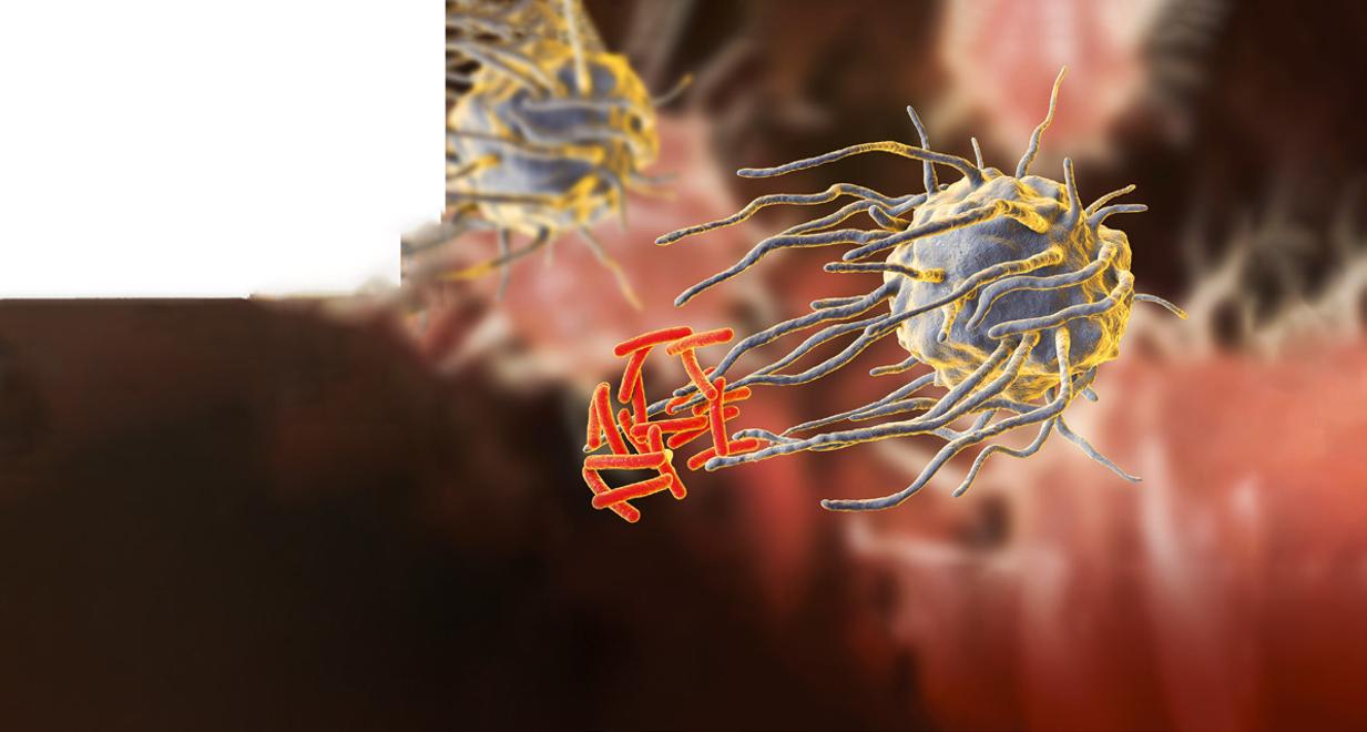
Recruitment of Caspase proteaser
Assembly of NLRP3 in ammassre
Actiotion of 3 enes for NLR 3, IL-1
Schematic representation of NLRP3 inflammasome activation.
molecular patterns (PAMPs) and danger-associated molecular patterns (DAMPs), derived from external pathogens or damaged host cells (Fig. 1.3). A typical inflammasome is built around a scaffold containing NOD-like receptors (NLRs). An example of a clinically relevant, well-characterized inflammasome is NLRP3, also known as cryopyrin. NLRP3 plays a key role in innate immunity by orchestrating the release of proinflammatory cytokines, in particular interleukin (IL)-1 and IL18. Mutations in the gene encoding cryopyrin are associated with a number of hereditary periodic fever syndromes (see Chapter 10), which are effectively treated by inhibitors of the IL-1 pathway (e.g. Anakinra).
CD1 molecules are invariant proteins (MHC-like and associated with β2-microglobulin – see later) that are present on DCs and epithelial cells. CD1 combine with lipids, which are poor antigens and not usually well presented to the adaptive immune system, and so act as recognition molecules for the
ZAP70p56lckp59fyn
Fig. 1.4 Diagram of the structure of the T-cell receptor (TCR). The variable regions of the alpha (ɑ) and beta (β) chains make up the T idiotype, i.e. antigen/peptide-binding region. The TCR is closely associated on the cell surface with the CD3 protein that is essential for activation.
intestine and other microbial-rich surfaces. CD1 present lipids to the immune cells of the gut in particular, namely nonMHC-restricted natural killer (NK) T cells and γδ T cells in the epithelium.
Antigenic epitopes, having been processed by DCs, are recognized by cells of the adaptive system by means of specific receptors. Each T cell, like B cells, is pre-committed to a given epitope. It recognizes this by one of two types of TCRs, depending on the cell’s lineage and thus its effector function. T cells have either αβTCR – a heterodimer of alpha (α) and beta β) chains – or γδTCR – a heterodimer of gamma γ and delta (δ) chains. αPTCR cells predominate in adults, although 10% of T cells in epithelial structures are of the γδTCR type. In either case, TCRs are associated with several transmembrane proteins that make up the cluster differentiation 3 (CD3) molecule (Fig. 1.4), to make the CD3–TCR complex responsible for taking the antigen recognition signal inside the cell (signal transduction). Signal transduction requires a group of intracellular tyrosine kinases (designated p56 lck, p59 fyn, ZAP 70) to join with the cytosolic tails of the CD3–TCR complex and become phosphorylated. Nearby accessory molecules, CD2, LFA-1, CD4 and CD8, are responsible for increased adhesion (see section 1.2.6), but are not actually involved in recognizing presented antigenic fragments.
The genes for TCR chains are on different chromosomes: β and γ on chromosome α7 and α and δ on chromosome 14. Each of the four chains is made up of a variable and a constant domain. The variable regions are numerous (although less so than immunoglobulin-variable genes); they are joined by D and J region genes to the invariant (constant) gene by recombinases, RAG1 and RAG2, the same enzymes used for making antigen receptors on B cells and antibodies (section 1.4.1). The diversity of T-cell antige0.n receptors is achieved in a similar way for immunoglobulin, although TCRs are less diverse since somatic mutation is not involved; perhaps the risk of ‘selfrecognition’ would be too great. The diversity of antigen binding is dependent on the large number of V genes and the way
Fig. 1.3
in which these may be combined with different D and J genes to provide different V domain genes. The similarities between TCRs and BCRs led to the suggestion that the genes evolved from the same parent gene and both are members of a ‘supergene’ family. Unlike immunoglobulin, TCRs are not secreted and are not independent effector molecules.
A particular TCR complex recognizes a processed antigenic peptide in the context of MHC class I or II antigens (section 1.4.1), depending on the type of T cell; helper T cells recognize class II with antigen, and this process is enhanced by the surface accessory protein CD4 (see later) and intracellular signals. Cytotoxic T cells (CTL/Tc) recognize antigens with class I (see section 1.3.1) and use CD8 accessory molecules for increased binding and signalling. Since the number of variable genes available to TCRs is more limited, reactions with antigen might not be sufficient if it were not for the increased binding resulting from these accessory mechanisms. Recognition of processed antigen alone is not enough to activate T cells. Additional signals, through soluble cytokines (interleukins), are needed; some of these are generated during ‘antigen processing’ (see section 1.4.1).
MHC molecules were originally known as ‘histocompatibility antigens’ because of the vigorous reactions they provoked during mismatched organ transplantation. However, these molecules are known to play a fundamental role in immunity by presenting antigenic peptides to T cells. Histocompatibility antigens in humans (known as human leucocyte antigens, HLAs) are synonymous with the MHC molecules. MHC molecules are cell-surface glycoproteins of two basic types: class I and class II (Fig. 1.5). They exhibit extensive genetic polymorphism with multiple alleles at each locus. As a result, genetic variability between individuals is very great and most unrelated individuals possess different MHC (HLA) molecules. This means that it is very difficult to obtain perfect HLA matches between unrelated persons for transplantation (see Chapter 8). The extensive polymorphism in MHC molecules is best explained by the need of the immune system to cope with an
ever-increasing range of pathogens adept at evading immune responses (see Chapter 2).
The TCR of an individual T cell will only recognize antigen as a complex of antigenic peptide and self-MHC (Fig. 1.6). This process of dual recognition of peptide and MHC molecule is known as MHC restriction, since the MHC molecule restricts the ability of the T cell to recognize antigen (Fig. 1.6). The importance of MHC restriction in the immune response was recognized by the award of the Nobel Prize in Medicine to Peter Doherty and Rolf Zinkernagel, who found that virusspecific CTLs would only kill cells of the same particular allelic form of MHC molecule.
MHC class I antigens are subdivided into three groups: A, B and C. Each group is controlled by a different gene locus within the MHC region on chromosome 6 (Fig. 1.7) in humans (different in mice). The products of the genes at all three loci are chemically similar. All MHC class I antigens (see Fig. 1.5) are made up of an a heavy chain, controlled by a gene in the relevant MHC locus, associated with a smaller chain called β2-microglobulin, controlled by a gene on chromosome 12. The differences between individual MHC class I antigens are due to variations in the a chains; the β2-microglobulin component is constant. The detailed structure of class I antigens was determined by X-ray crystallography. This shows that small antigenic peptides (approx. nine amino acids long) can be tightly bound to a groove produced by the pairing of the two extracellular domains (a1 and a2) of the a chain. The affinity (tightness of binding) of individual peptide binding depends on the nature and shape of the groove, and accounts for the MHC restriction mentioned earlier.
MHC class II antigens have two heavy chains, α and β, both coded for by genes in the MHC region of chromosome 6. The detailed structure of MHC class II antigens was also
Fig. 1.5 Diagrammatic representation of major histocompatibility complex (MHC) class I and class II antigens. CHO, carbohydrate side chain; β2m, β2-microglobulin.
Fig. 1.6 Major histocompatibility complex (MHC) restriction of antigen recognition by T cells. T cells specific for a particular peptide and a particular MHC allele (i) will not respond if the same peptide were to be presented by a different MHC molecule as in (ii) or as in (iii) if the T cell were to encounter a different peptide. APC, antigen-presenting cell; TCR, T-cell receptor.
Chromosome 6
Fig. 1.7 Major histocompatibility complex on chromosome 6; class III antigens are complement components. TNF, tumour necrosis factor.
determined by X-ray crystallography. It has a folded structure similar to class I antigens, with the peptide-binding groove found between the α and β chains (see Fig. 1.5). Whereas most nucleated cells express class I molecules, expression of class II molecules is restricted to a few cell types: DCs, B lymphocytes, activated T cells, macrophages, inflamed vascular endothelium and some epithelial cells. However, other cells (e.g. thyroid, pancreas, gut epithelium) can be induced to express class II molecules under the influence of interferon (IFN)-γ released during inflammation. In humans, there are three groups of variable class II antigens: the loci are known as HLA-DP HLA-DQ and HLA-DR.
In practical terms, there are different mechanisms by which antigens in different intracellular compartments can be captured and presented to CD4+ or CD8+ T cells (Fig. 1.8). Endogenous antigens (including viral antigens that have infected host cells) are processed by the endoplasmic reticulum and presented by MHC class I-bearing cells exclusively to CD8+ T cells. Prior to presentation on the cell surface, endogenous antigens are broken down into short peptides, which are then actively transported from the cytoplasm to endoplasmic reticulum by proteins. These proteins act as a shuttle and are so named ‘transporters associated with antigen processing’ (TAP-1 and TAP-2). TAP proteins (also coded in the MHC class II region) deliver peptides to MHC class I molecules in the endoplasmic reticulum, from whence the complex of MHC and peptide is delivered to the cell surface. Mutations that affect function in either TAP gene prevent surface expression of MHC class I molecules.
In contrast, exogenous antigens are processed by the lysosomal route and presented by MHC class II antigens to CD4+ T cells (Fig. 1.8). As with MHC class I molecules, newly synthesized MHC class II molecules are held in the endoplasmic reticulum until they are ready to be transported to the cell surface. While in the endoplasmic reticulum, class II molecules are prevented from binding to peptides in the lumen by a protein known as MHC class II-associated invariant chain. The invariant
Presentation of endogenous/viral antigens by MHC class I molecules
Presentation of exogenous antigens by MHC class II molecules
Vesicle
Golgi
Complex with MHC I
Endoplasmic reticulum
Viral antigen complexed with TAP
Viral antigenic peptide
Class I mRNA
Viral mRNA
Viral DNA
Nucleus
Class II mRNA endoplasmic reticulum
Vesicle
Endosome
Invariant chain is cleaved on fusion to enable class II molecules to bind antigen in the groove Viral DNA
Viral antigen/autoantigen MHC class I molecule
TAP (transporters associated with antigen processing)
Processed exogenous antigen MHC class II molecule
Invariant chain protects antigen binding groove
Fig. 1.8 Different routes of antigen presentation, depending on the nature of the antigen. MHC, major histocompatibility complex.
chain also directs delivery of class II molecules to the endosomal compartment, where exogenous antigens are processed and made available for binding to class II molecules.
The MHC class III region (see Fig. 1.7) contains genes encoding proteins that are involved in the complement system (see section 1.4.1): the early components C4 and C2 of the classical pathway and factor B of the alternative pathway. Some inflammatory proteins, e.g. tumour necrosis factor (TNF), are also encoded in adjacent areas. Invariant MHC-like proteins, such as CD1 lipid-recognition receptors (see earlier), are not coded for on chromosome 6, despite being associated with β2-microglobulin.
In contrast to TCRs, the antigen BCRs are surface-bound immunoglobulin molecules that can be secreted as soluble molecules. As with TCRs, they have predetermined specificity for epitopes and are therefore extremely diverse. The immune system has to be capable of recognizing all pathogens, past and future. Such diversity is provided by the way in which all three types of molecules, TCR, BCR and antibody, are produced.
The basic structure of the immunoglobulin molecule is shown in Fig. 1.9. It has a four-chain structure: two identical
Fig. 1.9 Basic structure of an immunoglobulin molecule. Domains are held in shape by disulfide bonds, though only one is shown. C1-3, constant domain of a heavy chain; CL, constant domain of a light chain; =S=, disulfide bond; VH, variable domain of a heavy chain; VL, variable domain of a light chain.
The amino (N) terminal domains of the heavy and light chains include the antigen-binding site. The amino acid sequences of these N-terminal domains vary between different antibody molecules and are known as variable (V) regions. Most of these differences reside in three hypervariable areas of the molecule, each only 6–10 amino acid residues long. In the folded molecule, these hypervariable regions in each heavy and light chain come together to form, with their counterparts on the other pair of heavy and light chains, the antigen-binding site (Fig. 1.9). The structure of this part of the antibody molecule is unique to that molecule and is known as the idiotypic determinant. In any individual, approximately 106–107 different antibody molecules could be made up by 103 different heavy chain variable regions associating with 103 different light chain variable regions, though there are even more epitopes due to further variation during the later processing (see section 1.4.1).
Fig. 1.10 Schematic representation of immunoglobulin (Ig)M pentamer (MW 800 kDA).
The part of the immunoglobulin chain next to the V region in either heavy or light chains is the constant (C) region; this is made up of one domain in a light chain (CL) and three or four in a heavy chain (CH) (Fig. 1.9). There are two alternative types of CL chain, known as kappa (κ) and lambda (λ); an antibody molecule has either two κ or two λ light chains, never one of each. Of all the antibodies in a human individual, roughly 60% contain κ and 40% contain λ light chains. There are no known differences in the functional properties between κ and λ light chains. In contrast, there are several possible different types of CH domain, each with important functional differences (Table 1.5). The heavy chains determine the isotype of the antibody and the ultimate physiological function of the particular antibody molecule. Once the antigen-binding site has reacted with its antigen, the molecule undergoes a change in the conformation of its heavy chains in order to take part in effector reactions, depending on the isotype of the molecule.
Fig. 1.11 Schematic representation of dimeric secretory immunoglobulin (Ig)A (MW 385 kDA).
heavy (H) chains (mol. wt. 50 kDa) and two identical light (L) chains (mol. wt. 25 kDa). Each chain is made up of domains of about 110 amino acids held together in a loop by a disulphide bond between two cysteine residues in the chain. The domains have the same basic structure and many areas of similarity in their amino acid sequences. The heavy chains determine the isotype of the immunoglobulin, resulting in pentameric IgM (Fig. 1.10), dimeric IgA (Fig. 1.11) or monomeric IgG.
The processes by which the components of this supergene family are produced are identical for TCR and BCR and are known as recombination. Immunoglobulin production, whether for BCR or antibody production, is the same initially. As for the TCR, the genes for the different chains in a BCR are carried on different chromosomes (Fig. 1.12). Like those coding for other macromolecules, the genes are broken up into coding segments (exons) with intervening silent segments (introns). The heavy chain gene set on chromosome 14 is made up of small groups of exons representing the constant regions of the heavy chains – e.g. mu (μ) chain – and a very large number of V region genes, perhaps as many as 103. Between the V and C genes are two small sets of exons, D and J (Fig. 1.12). In a single B cell, one V region gene is selected, joined to one D and J on the same chromosome; the VDJ product is then joined at the level of RNA processing to Cμ when the B cell is making IgM. The cell can make IgG by omitting the Cμ and joining VDJ to a Cγ. Thus, the cell can make IgM, IgD and IgG/A/E in sequence, while still using the same variable region. The same enzymes are used for the TCRs, and coded for by two recombination-activating genes controlling VDJ gene recombination: RAG1 and RAG2. Disruption of the RAG1 or RAG2
Table 1.5
Immunoglobulin classes and their functions
IgM μ 0.5–2.0Neutralization and opsonization +++–L
IgG1 γ15.0–12.0Opsonization+++++M, N, P, L, E
IgG2 γ22.0–6.0+±P, L
IgG3 γ30.5–1.0Opsonization+++++M, N, P, L, E
IgG4 γ40.1–1.0–+N, L, P
IgA1 α10.5–3.0Neutralization at mucosal surfaces ––M, N
IgA2 α20.0–0.2–––
IgD δ TraceLymphocyte membrane receptor
IgE εΣ TraceMast cell attachment––B, E, L
* Normal adult range in g/L.
† Classical pathway.
‡ Fc receptors on: basophils/mast cells, B; eosinophils, E; lymphocytes, L; macrophages, M; neutrophils, N; platelets, P.
function in infants who have mutations in these genes causes profound immune deficiency, characterized by absent mature B and T cells, as neither TCRs or BCRs can be produced. On a different chromosome (either chromosome 22 for λ chains or chromosome 2 for κ chains) in the same cell, a V gene is joined to a J gene (there is no D on the light chain) and then the VJ product is joined at the RNA level to the Cκ or Cλ (Fig. 1.12).
The wide diversity of antigen binding is dependent on the large number of V genes and the way in which these may be combined with different D and J genes to provide different rearranged VDJ gene segments. Once V, D and J rearrangement has taken place to produce a functional immunoglobulin molecule, further V region variations are introduced only at a much later
Fig. 1.12 Immunoglobulin genes (see text for explanation).
stage, when antibodies rather than BCRs are produced by the process of somatic mutation in germinal centres.
Natural killer cells also have recognition molecules. These cells are important in killing virally infected cells and tumour cells. They have to be able to recognize these targets and distinguish them from normal cells. They recognize and kill cells that have reduced or absent MHC class I, using two kinds of receptors – inhibitory (KIR) and activating (KAR) –to estimate the extent of MHC expression. They also have one type of Fc IgG (Fcγ) receptor, that for low-affinity binding of IgG antibodies, and so NK cells are able to kill some cells with large amounts of antibody on their surfaces. Further subsets of NK-like cells that contribute to innate immunity
The product is VHC, i.e. an IgM heavy chain with a particular variable regionFinal product IgMκ or IgMλ
include NKT cells and invariant NKT cells (section 1.3.6); these are thought to be particularly important in tumour immunology (section 1.5.1).
The major purpose of the complement pathways is to provide a means of removing or destroying antigens, regardless of whether or not these are coated with antibody. This requires that complement components recognize damaging material such as immune complexes (antigen combined with antibodies) or foreign antigens. The complement pathways are discussed in more detail in section 1.3.5.
1.2.3 Accessory molecules
The binding of a processed antigen–MHC class II complex on an antigen-presenting cell (APC) to the corresponding TCR provides an insufficient signal for T-cell activation; the binding of accessory molecules on the two cell surfaces provides additional stimuli. Accessory molecules are lymphocyte surface proteins, distinct from the antigen-binding complexes, which are necessary for efficient binding, signalling and homing Accessory molecules are invariant, non-polymorphic proteins. Each accessory molecule has a particular ligand – a corresponding protein to which it binds. These ligands are present on all cells that require close adhesion for functioning; for example, there are those on T cells for each of the many cell types that can activate or respond to T cells (APCs, endothelial cells, etc.); similar ligands are present on B cells for efficiency of T-cell help as well as stimulation by follicular DCs.
There are several families of accessory molecules, but the most important appear to be the immunoglobulin supergene family of adhesion molecules, which derives its name from the fact that its members contain a common immunoglobulin-like structure. Members of the family strengthen the interaction between APCs and T cells (Fig. 1.13); those on T cells include CD4, CD8, CD28, CTLA-4, CD45R, CD2 and lymphocyte function antigen 1 (LFA-1). For interaction with B cells, CD40 ligand and ICOS are important for class switching
(see section 1.4.3). Adhesion molecules, for binding leucocytes (both lymphocytes and polymorphonuclear leucocytes) to endothelial cells and tissue matrix cells, are considered in section 1.2.6. On B cells, such molecules include CD40 (ligand for CD40L, now named CD154; see Chapter 3, Case 3.2), B-7-1 and B7-2 (ligands for CD28).
1.2.4 Effector molecules for immunity
There are humoral and cellular effector molecules in both the innate and the adaptive immune systems (Table 1.6). Several of the same mechanisms are used in both types of immune responses, especially in killing of target cells, suggesting that the evolution of immune responses has been conservative in terms of genes, though with much redundancy to ensure the life- preserving nature of the immune systems in the face of rapid evolution of pathogenic microbes.
Antibodies
Antibodies are the best-described effector mechanisms in adaptive immunity. They are the effector arm of B cells and are secreted as soluble molecules by plasma cells in large
Table 1.6 Effector molecules in immunity
HumoralComplement components for opsonization or lysis
CellularPerforin in NK cells creates pores in target cell membranes
Specific antibodies for opsonization and phagocytosis or lysis with complement
Perforin in cytolytic (CD8) T cells creates pores in specific target cell membranes, allowing entry of granzymes to cause apoptosis
NKT cells induce apoptosis by perforin production
Granzymes in NK cells induce apoptosis in target cells
Lysosomes in phagocytic vacuoles result in death of ingested microbes
Fig. 1.13 Diagrammatic representation of adhesion molecules on T cells and their ligands on antigen-presenting cells (APCs)/ virus-infected target cells. MHC, major histocompatibility complex; TCR, T-cell receptor.
Preformed histamine and related vasoactive substances as well as leukotrienes in mast cells
NK, natural killer
quantities, to be carried in the blood and lymph to distant sites. As shown in Table 1.5, there are five major isotypes of antibodies, each with different functions (see also Box 1.2).
IgM is a large molecule whose major physiological role is intravascular neutralization of organisms (especially viruses). IgM has five complement-binding sites, resulting in excellent complement activation and subsequent removal of the antigen–antibody–complement complexes by complement receptors on phagocytic cells or complement-mediated lysis of the organism (see section 1.4).
IgG is a smaller immunoglobulin that penetrates tissues easily. Placental transfer is an active process involving specific placental receptors for the Fc portion of the IgG molecule, termed FcRn (Fc receptor of the neonate). The FcRn receptor is also present on epithelial and endothelial cells and is an important regulator of IgG metabolism (see Chapter 7, section 7.4 and Fig. 7.8). Of the four subclasses, IgG1 and IgG3 activate complement most efficiently and are responsible for clearing most protein antigens, including the removal of microorganisms by phagocytic cells (see section 1.5). IgG2 and IgG4 react predominantly with carbohydrate antigens (in adults) and are relatively poor opsonins (promoters of phagocytosis).
IgA is the major mucosal immunoglobulin. Attachment of the ‘secretory piece’ prevents digestion of this immunoglobulin
Box 1.2 Immunoglobulin isotypes and their significance
IgM is phylogenetically the oldest class of immunoglobulin. It is a large molecule (Fig. 1.10) and penetrates poorly into tissues. IgM has five complement-binding sites, which results in excellent activation of the classical complement pathway.
IgG is smaller and penetrates tissues easily. It is the only immunoglobulin to provide immune protection to the neonate (Table 1.5) as IgG is actively transported across the placenta. There are four subclasses of IgG, with slightly different functions.
IgA is the major mucosal immunoglobulin – sometimes referred to as ‘mucosal antiseptic paint’. IgA in mucosal secretions consists of two basic units joined by a J chain (Fig. 1.11); the addition of a ‘secretory piece’ prevents digestion of this immunoglobulin in the intestinal and bronchial secretions.
IgD is synthesized by antigen-sensitive B lymphocytes and is not secreted, acting as a cell-surface receptor for activation of these cells by the specific antigen relating to the B-cell receptor; it is essential for activation of antigen-responsive B cells.
IgE is produced by plasma cells, but is taken up by specific IgE receptors on mast cells and basophils. IgE then provides an antigen-sensitive way of expelling intestinal parasites by increasing vascular permeability and inducing chemotactic factors via mast cell degranulation (see section 1.7).
in the intestinal and bronchial secretions. IgA2 is the predominant subclass in secretions and neutralizes antigens that enter via these mucosal routes. IgA, the monomeric IgA in serum, is capable of neutralizing antigens that enter the circulation, but IgA1 is sensitive to bacterial proteases and therefore less useful for host defence at mucosal surfaces. IgA has additional functions via its receptor (FcaR or CD89), present on mononuclear cells and neutrophils, for activation of phagocytosis, inflammatory mediator release and antibody-dependent cell-mediated cytotoxicity (ADCC) (see section 1.5).
There is little free IgD or IgE in serum or normal body fluids, since these act as surface receptors on mature B cells or mast cells, respectively.
As mentioned previously, mechanisms of recombination in immunoglobulin production, whether for BCR or antibody production, are the same initially (Fig. 1.12). Once V, D and J region rearrangement has taken place, further variation is introduced when antibodies are made, by the introduction of point mutations in the V region genes. This process, known as somatic hypermutation, occurs in the lymphoid germinal centres and is critically dependent on activation-induced cytidine deaminase (AID), an enzyme responsible for deamination of DNA. Somatic hypermutation helps to increase the possible number of combinations and accounts for the enormous diversity of antibody specificities (1014), which by far exceeds the number of different B cells in the body (1010).
Cytokines and chemokines
Cytokines are soluble mediators secreted by macrophages or monocytes (monokines) or lymphocytes (lymphokines). These mediators act as stimulatory or inhibitory signals between cells; those between cells of the immune system were known as interleukins (a phrase that has fallen out of general usage since the range of soluble molecules has widened so tremendously, though the individual names persist to avoid confusion). As a group, cytokines share several common features (see Box 1.3). Among the array of cytokines produced by macrophages and T cells, IL-1 and IL-2 are of particular interest due to their pivotal role in amplifying immune responses. IL- 1 acts on a wide range of targets (Table 1.7), including T and B cells. In contrast, the effects of IL-2 are largely restricted to lymphocytes. Although IL-2 was originally identified on account of its ability to promote the growth of T cells, it has similar trophic effects on IL-2 receptor-bearing B and NK cells. The considerable overlap between actions of individual cytokines and interleukins is summarized in Table 1.8.
Cytokines that induce chemotaxis of leucocytes are referred to as chemokines, a name derived from chemo + kine, i.e. something chemical to help movement. Some cytokines and interleukins have been redefined as chemokines as their function becomes clearer, e.g. IL-8 = CXCL8. Chemokines are structurally similar proteins of small molecule size (8–10 kDa), which are able to diffuse from the site of production to form a local concentration gradient along which granulocytes and
Box 1.3 Common features of cytokines
• Their half-lives are short, so any potential harm due to persistent action is controlled.
• They are rapidly degraded as another method of regulation and thus difficult to measure in the circulation.
• Most act locally within the cell’s microenvironment, which confines their action to a particular site.
• Some act on the cell of production itself, promoting self-activation and differentiation through highaffinity cell-surface receptors.
• Many cytokines are pleiotropic in their biological effects, i.e. affecting multiple organs in the body.
• Most exhibit biologically overlapping functions, illustrating the redundancy of the group. For this reason, therapeutic targeting of individual cytokines in disease has had limited success so far (effects of deletion of individual cytokine genes are listed in Table 1.8).
chemokines (CXC), coded for by genes on chromosome 17 and attractants for granulocytes, and the homeostatic chemokines, acting as attractants for lymphocytes (CC) and coded by genes on chromosome 4. The corresponding receptors on inflammatory cells are designated CXCR on neutrophils and CCR on lymphocytes; there are exceptions!
Molecules for lysis and killing
The other major sets of effector molecules are the cytolytic molecules, though less is known about their diversity or mechanisms of action. They include perforin , a C9-like molecule present in secretory lysosomes in CD8 T cells and in NK cells that polymerizes to form pores to enable large proteins to enter the cell. These cell types also secrete granzymes , enzymes that induce apoptosis in target cells (Table 1.6). Macrophages and polymorphonuclear leucocytes also contain many substances for the destruction of ingested microbes, some of which have multiple actions, such as TNF. The duplication of the functions of this essential phylogenetically ancient protein during evolution underlines the continued development of mammalian immunity to keep up with microbial invaders.
Target cellEffect
T lymphocytesProliferation
Differentiation
Lymphokine production
Induction of IL-2 receptors
B lymphocytesProliferation
Differentiation
NeutrophilsRelease from bone marrow
Chemoattraction
Macrophages
FibroblastsProliferation/activation
Osteoblasts
Epithelial cells
OsteoclastsReabsorption of bone
HepatocytesAcute-phase protein synthesis
HypothalamusProstaglandin-induced fever
MuscleProstaglandin-induced proteolysis
IL, interleukin
lymphocytes can migrate towards the stimulus. There are two types of movement: migration of leucocytes to sites of inflammation and that of differentiating cells moving to a specific activation site (see section 1.2.5); chemokines are involved in both. There are therefore two main types: the inflammatory
1.2.5 Receptors for effector functions
Without specific cytokine receptors on the surface of the target cells, cytokines are ineffective; this has been demonstrated in those primary immune deficiencies in which gene mutations result in absence or non-functional receptors, such as the commonest X-linked form of severe combined immune deficiency (see Chapter 3, Case 3.5), IL-12 receptor or IFN-y receptor deficiencies (see Chapter 3). Some cytokines may have unique receptors, but many others share a common structural chain, such as the γ-chain in the receptors for IL-2, IL-4, IL-7, IL-9, IL-15 and IL-23, suggesting that these arose from a common gene originally. There are other structurally similar cytokine receptors, leading to the classification of these receptors into five families of similar types of receptors, many of which have similar or identical functions, providing a safety net (redundancy) for their functions, which are crucial for both the innate and adaptive immune systems.
Chemokine receptors from a family of G protein-coupled receptors – meaning that they are transmembrane and able to activate internal signalling pathways. These receptors also function as differentiation ‘markers’, as they become expressed as an immune reaction progresses and cells move in inflammatory responses.
Receptors for the Fc portions of immunoglobulin molecules (FcR) are important for effector functions of phagocytic cells and NK cells. There are at least three types of Fcγ receptors: FcRγl are high-affinity receptors on macrophages and neutrophils that bind monomeric IgG for phagocytosis; FcRγII are low-affinity receptors for phagocytosis on macrophages and neutrophils and for feedback inhibition on B cells; and FcRγIII on NK cells as mentioned earlier. There are also FcRn involved in the transfer of IgG across the placenta and these receptors
Table 1.7 Actions of interleukin-1
Table 1.8 Clinically important cytokines grouped by effect on immune or inflammatory responses, to show source and site of action
CytokinesAction
(a) Promotion of non-specific immunity and inflammation
Interleukin-17 (IL-17)Increases chemokine production for inflammatory cells
Interleukin-1 (IL-1)(See Table 1.7)
Interleukin-6 (IL-6)Growth and differentiation of T, B and haematopoietic cells
Production of acute-phase proteins by liver cells
Interleukin-8 (now CXCL8)Chemotaxis and activation of neutrophils and other leucocytes
Interferon-α (IFN-α)Antiviral action by activation of NK cells, upregulation of MHC class I antigens on virally infected cells, inhibition of viral replication
Interleukin-5 (IL-5)Activation of B cells, especially for IgE production
Activation of eosinophils
Tumour necrosis factor (TNF)Promotion of inflammation by: activation of neutrophils, endothelial cells, lymphocytes, liver cells (to produce acute-phase proteins)
Interferes with catabolism in muscle and fat (resulting in cachexia)
Interferon-γ (IFN-γ)Activation of macrophages, endothelial cells and NK cells. Increased expression of MHC class I and class II molecules in many tissues; inhibits allergic reactions (↓IgE production)
(b) Lymphocyte activation, growth and differentiation, i.e. specific immunity
Interleukin-2 (IL-2)Proliferation and maturation of T cells, induction of IL-2 receptors and activation of NK cells
Interleukin-4 (IL-4) and interleukin-5 (IL-5)
Induction of MHC class II, Fc receptors and IL-2 receptors on B and T cells
Induction of isotype switch in B cells
Facilitation of IgE production (mainly IL-4)
Activation of macrophages
Proliferation of bone marrow precursors
Interleukin-12 (IL-12)† Synergism with IL-2; regulates IFN-y production Activation of NK cells
Interleukin-13 (IL-13)Actions overlap with IL-4, including induction of IgE production IL-13 receptor acts as a functional receptor for IL-4
Interleukin-15 (IL-15)Similar to IL-12
Interleukin-16 (IL-16)Chemotaxis and activation of CD4 T cells
(c) Colony stimulation of bone marrow precursors
GM-CSFStimulates growth of polymorph and mononuclear progenitors
G-CSFStimulates growth of neutrophil progenitors
M-CSFStimulates growth of mononuclear progenitors
(d) Regulatory cytokines
Interleukin-10 (IL-10); also called cytokine synthesis inhibitory factor‡
Transforming growth factor-ß (TGF-ß)
Inhibition of cytokine production
Growth of mast cells
Antiinflammatory Inhibits cell growth
Table 1.8 (Continued ) CytokinesAction
(e) Chemokines
Interleukin-8 (IL-8)See under section (a)
RANTES (regulated on activation, normal T cell expressed and secreted)
Monocyte chemotactic protein (MCP 1, 2, 3)
Chemoattractant for eosinophils, monocytes
Chemoattractant for monocytes
ExotaxinChemoattractant for eosinophils; synergistic with IL-5
Evidence from murine models. See appendix for web address for update on knockout mice.
† IL-12 family of cytokines includes IL-23 and IL-27.
‡ IL-10 family includes IL-19, IL-20 and IL-22.
G-CSF, granulocyte colony-stimulating factor; GM-CSF, granulocyte-macrophage colony-stimulating factor; Ig, immunoglobulin; M-CSF, macrophage colony-stimulating factor; MHC, major histocompatibility complex; NK, natural killer.
are involved in IgG recirculation and catabolism. IgE receptors are found on mast cells, basophils and eosinophils for triggering degranulation of these cells. IgA receptors ensure the transport of polymeric IgA across the mucosal cells and other, possibly important functions are slowly being defined.
Complement receptors for fragments of C3 produced during complement activation also provide a mechanism for phagocytosis and are found on macrophages and neutrophils. However, there are several types of complement receptors : those on red blood cells for transport of immune complexes for clearance (CR1), those on B cells and follicular DCs in lymph nodes to trap antigen to stimulate a secondary immune response (CR2) (see section 1.4.3), and those on macrophages, neutrophils and NK cells to provide adhesion of these blood cells to endothelium, prior to movement into tissues (CR3).
1.2.6 Adhesion molecules
Adhesion molecules comprise another set of cell surface glycoproteins with a pivotal role, not only in immune responses by mediating cell-to-cell adhesion, but also for adhesion between cells and extracellular matrix proteins. Adhesion molecules are grouped into two major families: (i) integrins and (ii) selectins (Table 1.9). The migration of leucocytes to sites of inflammation is dependent on three key sequential steps mediated by adhesion molecules (Fig. 1.14): rolling of leucocytes along activated endothelium is selectin dependent; tight adhesion of leucocytes to endothelium is integrin dependent; and transendothelial migration occurs under the influence of chemokines. Cytokines also influence the selectinand integrin-dependent phases.
Integrins are heterodimers composed of non-covalently associated α and β subunits. Depending on the structure of the
β subunit, integrins are subdivided into five families (β1 to β5 integrins). β1 and β2 integrins play a key role in leucocyte–endothelial interaction. β1 integrins mediate lymphocyte and monocyte binding to the endothelial adhesion receptor called vascular cell adhesion molecule (VCAM-1). β2 integrins share a common β chain (CD18) that pairs with a different α chain (CD11a, b, c) to form three separate molecules (CD11a CD18, CD11b CD18, CD11c CD18); they also mediate strong binding of leucocytes to the endothelium. Examples in other systems include β3 to β5 integrins mediating cell adhesion to extracellular matrix proteins such as fibronectin and vitronectin in the skin and laminin receptor in muscle.
The selectin family is composed of three glycoproteins designated by the prefixes E (endothelial), L (leucocyte) and P (platelet) to denote the cells on which they were first described. Selectins bind avidly to carbohydrate molecules on leucocytes and endothelial cells and regulate the homing of the cells to sites of inflammation (see section 1.6.1, Chapter 11, section 11.1 and Table 1.10).
1.3 Functional basis of innate responses
The aim of an immune response is to destroy foreign antigens, whether these are inert molecules or invading organisms. To reach the site of invasion and destroy the pathogens, the components of the immune systems have to know where to go and to how to breach the normal barriers, such as the endothelial cells of the vascular system. Humoral factors (such as antibodies and complement) are carried in the blood and enter tissues following an increase in permeability associated with inflammation . Immune cells (innate and antigen specific) are actively attracted to a site of inflammation and enter the tissues via specific sites using active processes of adhesion.
Table 1.9 Examples of clinically important adhesion molecules
Adhesion moleculeLigand
β1 integrin family
VLA-4 (CD49d-CD29) expressed on lymphocytes, monocytes
β2 integrin family
CD18/CD11 expressed on leucocytes
VCAM-1 on activated endothelium
Clinical relevance of interactionConsequences of defective expression
Mediates tight adhesion between lymphocytes, monocytes and endothelium
β3 integrin family
Expressed on platelets
Selectin family
E-selectin (CD62E) expressed on activated endothelial cells
L-selectin (CD62L) expressed on all leucocytes
ICAM-1 on endothelium
Mediates tight adhesion between all leucocytes and endothelium
Impaired migration of lymphocytes and monocytes into tissue. Defective expression of either ß1integrins or VCAM-1 has not yet been described in humans. However, blockade of ß1integrins by natalizumab, a therapeutic monoclonal antibody, is associated with a high risk of progressive multifocal leucoencephalopathy, a severe viral infection of the brain
Defective expression of CD18/CD11 is associated with severe immunodeficiency, characterized by marked neutrophil leucocytosis, recurrent bacterial and fungal infection, and poor neutrophil migration into sites of infection
FibrinogenInteracts during clottingClotting disorder Glanzmann’s disease
Sialyl Lewis X (CD15) on neutrophils, eosinophils
CD34, Gly CAM on high endothelial venules
Mediates transient adhesion and rolling of leucocytes on monocytes
L-selectin mediates transient adhesion and rolling of leucocytes in lymph nodes, and also acts as a homing molecule directing lymphocytes into lymph nodes
Defective expression of CD15 is associated with severe immunodeficiency – clinical features similar to CD18 deficiency. Mice deficient in both E- and P-selectin exhibit a similar clinical phenotype
L-selectin-deficient mice exhibit reduced leucocyte rolling and impaired lymphocyte homing
Fig. 1.14 Adhesion molecules and leucocyte–endothelial interactions.
ICAM, intercellular adhesion molecule; VCAM, vascular cell adhesion molecule; VLA, very late activation antigen.
Table 1.10 Proteins controlling classical and alternative complement pathways*
ProteinFunctionClinical consequences of deficiency
Circulating inhibitors
C1 esterase inhibitorBinds to activated C1r, C1s, uncoupling it from C1q
Factor HBinds C3b displacing Bb; co-factor for factor I
Factor ISerine protease that cleaves C3b; acts synergistically with factor H
Membrane inhibitors
Complement receptor 1 (CR1; CD35)
Decay accelerating factor (DAF; CD55)
Uncontrolled activation of classical pathway leading to hereditary angioneurotic oedema
Total deficiency causes recurrent bacterial infection, glomerulonephritis and renal failure; partial deficiency with familial (atypical) haemolytic uraemic syndrome; a particular allele with adult macular degeneration
As for factor H
Receptor for C3bProtect mammalian cells. Low CR1 numbers on red cells in SLE is a consequence of fast turnover
Accelerates decay of C3b Bb by displacing Bb
Protectin (CD59)Inhibits formation of lytic pathway complex on homologous cells; widely expressed on cell membranes
* This is not an exhaustive list. SLE, systemic lupus erythematosus.
Non- specific immunity is older, in evolutionary terms , than antibody production and antigen- specific T cells. The major cells involved in the innate system are phagocytic cells (macrophages and polymorphonuclear leucocytes), which remove antigens including bacteria, and DCs, which are the first cells to react to invaders. The major humoral components of the four complement pathways can either directly destroy an organism or initiate/facilitate its phagocytosis. DCs recognize pathogens in order to provide a rapid initial cytokine response (such as interferon- a in a viral infection by plasmacytoid DCs) and to process antigen for presentation to specific TCRs alongside MHC for activation (classical DCs) (section 1.4.1).
1.3.1 Endothelial cells
The endothelium forms a highly active cell layer lining the inside of blood vessels and thus is present in all tissues. In addition to its critical role in maintaining vasomotor tone, the endothelium is closely involved in inflammation, wound healing and the formation of new blood vessels (angiogenesis). Immunologically, endothelial cells are intimately involved in interactions with leucocytes prior to leaving the circulation to enter sites of tissue damage (Fig. 1.14). The endothelium also plays an important role in regulating the turnover of IgG, through the presence of FcRn, a receptor that prevents IgG from undergoing lysosomal degradation (see section 1.2.4 and Chapter 7, section 7.4). The immunological importance of the endothelium is summarized in Box 1.4.
DAF deficiency alone does not cause disease
In combination with DAF deficiency leads to paroxysmal nocturnal haemoglobinuria (see Chapter 16, section 16.2.4)
Box 1.4 Immunological importance of the endothelium
• Expresses a wide range of molecules on the cell surface (E-selectin, intercellular adhesion molecule-1 [ICAM-1], vascular cell adhesion molecule [VCAM-1], complement receptors) and thus plays a critical role in leucocyte–endothelial interactions (Fig. 1.14).
• Major site of immunoglobulin (Ig)G turnover due to FcRn (Fc receptor of the neonate).
• Forms an important part of the innate immune response by expressing Toll-like receptors to recognize foreign pathogens.
• Capable of antigen presentation.
1.3.2 Neutrophil polymorphonuclear leucocytes
Neutrophils are short-lived cells that play a major role in the body’s defence against acute infection. They synthesize and express adhesion receptors so that they can stick to, and migrate out of, blood vessels into the tissues. Neutrophils move in response to chemotactic agents produced at the site of inflammation; substances include CXCL8, complementderived factors (such as C3a and C5a), kallikrein, cytokines released by THh1 cells and chemotactic factors produced by mast cells.
Neutrophils are phagocytic cells. They are at their most efficient when activated after entering the tissues. Morphologically,
the process of phagocytosis is similar in both neutrophils and macrophages. Neutrophils are able to kill and degrade the substances that they ingest. This requires a considerable amount of energy and is associated with a ‘respiratory burst’ of oxygen consumption, increased hexose monophosphate shunt activity and superoxide production. Genetically defective respiratory burst activity is associated with chronic granulomatous disease (see Chapter 3).
1.3.3 Macrophages
Macrophages and monocytes represent the mononuclear phagocytic system, which, along with DCs, forms the cells of the innate system. Lymphoid and myeloid cells are derived from closely related stem cells in the bone marrow (Fig. 1.1); each cell lineage has a different colonystimulating factor and, once differentiated, they have entirely different functions. Polymorphonuclear leucocytes develop in the bone marrow and emerge only when mature. Monocytes circulate for only a few hours before entering the tissues, where they may live for weeks or months as mature macrophages or DCs . Macrophages differentiate in the tissues, principally in subepithelial interstitial and lymphatic sinuses in liver, spleen and lymph nodes, sites where antigens gain entry. Tissue macrophages are heterogeneous in appearance, in metabolism and also in function; they include freely mobile alveolar and peritoneal macrophages, fixed Kupffer cells in the liver and those lining the sinusoids of the spleen. When found in other tissues, they are called histiocytes.
A major function of the mononuclear phagocyte system is to phagocytose- invading organisms and other antigens. Macrophages have prominent lysosomal granules containing acid hydrolases and other degradative enzymes with
Receptors and functions of mononuclear phagocytic cells.
Microbial components
which to destroy phagocytosed material. That material may be an engulfed viable organism, a dead cell, debris, an antigen or an immune complex. In order to carry out their functions effectively, macrophages must be ‘activated’; in this state, they show increased phagocytic and killing activity. Stimuli include cytokines (see section 1.2.5), substances that bind to other surface receptors (such as IgG: Fc receptors, TLRs for endotoxin and other microbial components, receptors for bacterial polysaccharides and for soluble inflammatory mediators such as C5a; see Fig. 1.15). Activation may result in release of cytokines from monocytes or DCs such as TNF or IL-1, which may cause further damage in already inflamed tissues.
1.3.4 Dendritic cells
Classical or myeloid dendritic cells are mononuclear cells derived from bone marrow precursors and closely related to monocytes. There are many subsets, but there are differences between these subsets in mice compared with humans and other primates, particularly in their surface markers. So only those relating to humans are described here, though clearly their corresponding functions have been described in all mammalian species studied so far.
Immature dendritic cells are ubiquitous, particularly in epithelia that serve as a portal of entry for microbes, where they capture antigens as well as reacting to pathogen components quickly, within a few hours of invasion. Subsequently, the activated DCs migrate to draining lymph nodes and mature to present antigen to cells of the adaptive system (Fig. 1.16).
DCs have a range of functions: as well as processing antigens (Fig. 1.8), they are able to recognize and respond to pathogens by secreting IFN-α, produce IL-12 and chemokines as well as causing the differentiation of immature T cells to a
e.g. lipids, chemokines, peptides, etc.
e.g. lipids, viral RNA e.g. carbohydrates in outer bacterial walls
Transmembrane receptors
Effect
α helical receptors Toll-like receptors – intra and extracellular
Increase in integrin activity and cytoskeletal changes for migration into tissues
Mannose receptor
Cytokine production and acute phase responses
Phagocytosis via complement activation and killing of microbes
Fig. 1.15
Present to:MobilitySiteAppearanceCell
Interdigitating dendritic cells
Paracortex of lymph node
Langerhans' cellsSkin
Veiled cellsLymph
Follicular dendritic cells
Macrophages
B cell (especially if activated)
Fig. 1.16 Antigen-presenting cells and their associated sites.
MobileT cells
MobileT cells
MobileT cells
Lymph node folliclesStaticB cells
Lymph node medulla
Liver (Kupffer cells)
Brain (astrocytes)
Lymphoid tissue
T cell receptor (TCR)
MHC class I or II
(B7.1) CD80CTLA-4
(B7.2) CD86CD28
Cell membrane of APC
Cell membrane of T cell
Mobile Static Static T and B cells
MobileT cells
T cell receptor (TCR)
MHC class I or II
(B7.1) CD80CTLA-4
(B7.2) CD86CD28
Cell membrane of APC
(a)(b) Activation
Interrupted costimulatory pathway
Cell membrane of T cell
Unresponsiveness (anergy) or apoptosis
Fig. 1.17 (a, b) Role of co-stimulatory pathway in T-cell activation. APC, antigen-presenting cell; MHC, major histocompatibility complex.
variety of effector T cells. Depending on the environment of the cell, which is not entirely understood, mature DCs can activate CD4+ cells to become Th1, Th2, Th17, CTLs and regulatory T cells (Tregs) or to induce apoptosis and so induce tolerance (see section 1.4). Immature and mature DCs have different sets of surface proteins (which act as distinct markers), in keeping with their different functions (see Table 1.3), depending on whether they are immature (for antigen capture/sensing pathogens via PRRs) or mature (for presentation of antigen to T cells).
The interaction between DCs and T cells is strongly influenced by a group of cell surface molecules that function as costimulators: CD80 (also known as B7-1) and CD86 (B7-2) on the activated DC, each of which engages with counter receptors on the T-cell surface referred to as CD28 and CTLA4. A functional co-stimulatory pathway is essential for T-cell activation. In the absence of a co-stimulatory signal, interaction
between DCs and T cells leads to T-cell unresponsiveness (Fig. 1.17). The importance of the co-stimulatory pathway is underlined by the ability of antagonists to co-stimulatory molecules to interrupt immune responses both in vitro and in vivo. This observation has been exploited therapeutically in mice with advanced lupus, in which treatment with a CTLA-4 conjugated protein to block CD28 leads to significant improvement in disease activity. Translation to human therapeutic monoclonal antibodies has succeeded despite a rocky start (see Chapter 7, Case 7.3). Abatacept, a CTLA-4 fusion protein, is licensed for the treatment of rheumatoid arthritis. T-cell activation by DCs also depends on cytokines secreted by activated DCs such as IL-12.
Processed antigen is presented to T cells complexed with the MHC class II antigens on the APC surface, since T cells do not recognize processed antigen alone. The most efficient APCs are the interdigitating dendritic cells found in the T-cell regions
of a lymph node (Figs 1.15 and 1.17). These dendritic cells have high concentrations of MHC class I and II molecules, co-stimulatory molecules (CD80, CD86) as well as adhesion molecules on their surfaces (Table 1.3) and limited enzymatic powers, which enable antigen processing but not complete digestion. Being mobile, they are able to capture antigen in the periphery and migrate to secondary lymphoid organs, where they differentiate into mature dendritic cells and interact with naive T cells. These cells are known as Langerhans cells when present in the skin.
These cells differ from the follicular dendritic cells in the follicular germinal centre (B-cell area) of a lymph node (see Figs 1.15 and 1.17). Follicular dendritic cells have receptors for complement and immunoglobulin components and their function is to trap immune complexes and to feed them to B cells in the germinal centre. This is part of the secondary immune response, since preexisting antibodies are used, accounting for B-cell memory.
Plasmacytoid DCs are found in blood and mucosalassociated lymphoid tissues and can secrete large quantities of type I IFNs in response to viral infections. However, their precise role and repertoire and therefore clinical significance remain unclear.
The few DCs in the blood are typically identified and enumerated in flow cytometry. Three types of DCs have been defined in human blood and these are the CD11 c + myeloid DCs, the CD141+ myeloid DCs and the CD303+ plasmacy-
toid DCs (as per the IUIS Nomenclature committee). Dendritic cells in blood are less mature and have no projections from their surface (dendrites). Still, they can perform complex functions, including chemokine production in CD11c+ myeloid DCs, cross-presentation of antigen in CD 141+ myeloid DCs and IFN-a production in CD303+ plasmacytoid DCs.
Monocyte-derived DCs, also known as myeloid DCs, are activated (mature) DCs that are found in inflammatory sites, from whence they travel to draining lymph nodes. As with other mature DCs, they express co-stimulatory molecules and so can activate T cells. Under certain circumstances they can even secrete TNF-α and nitric oxide. The ability to culture these cells from human blood monocytes has led to the concept of DC vaccines for cancers.
Activated B cells themselves are also able to present antigen (Fig. 1.16).
1.3.5 Complement
The complement system consists of a series of heat- labile serum proteins that are activated in turn. The components normally exist as soluble inactive precursors; once activated, a complement component may then act as an enzyme (Fig. 1.19), which cleaves several molecules of the next component in the sequence (rather like the clotting cascade). Each precursor is cleaved into two or more fragments. The major fragment has two biologically active sites: one for
Afferent lymphatics
Capillaries
Extravascular spaces
Germinal centre (composed of B cells proliferating in response to antigen) 2 follicle (B cells, follicular dendritic cells, T cells - 5%)
Mantle of mature but unstimulated B cells
Lymph node
Efferent lymphatics containing effector T cells
Blood 1 follicle (B cells)
Lymphatics
Naive T cells
Mature B cells
Tissues
Post-capillary venule in paracortex
Medulla (plasma cells) Cortex paracortex +follicles
Fig. 1.18 Organization of spleen.
Small fragment (a)
Inactive precursor
Large fragment (b)
Enzyme
Attachment
Mediator of in ammation
Fig. 1.19 Basic principle underlying the cleavage of complement components.
binding to cell membranes or the triggering complex and the other for enzymatic cleavage of the next complement component (Fig. 1.20). Control of the sequence involves spontaneous decay of any exposed attachment sites and specific inactivation by complement inhibitors. Minor fragments (usually prefixed ‘a’) generated by cleavage of components have important biological properties in the fluid phase, such as the chemotactic activity of C5a.
The history of the discovery of the complement pathways has made the terminology confusing. Several of the components have numbers, but they are not necessarily activated in
numerical order; the numbering coincides with the order of their discovery and not with their position in the sequence. Activated components are shown with a bar over the number of the component (e.g. C1 is activated to C1 ‒) and fragments of activated components by letters after the number (e.g. C3 is split initially into two fragments, C3a and C3b).
The major purpose of the complement pathways is to provide a means of removing or destroying antigen, regardless of whether or not it has become coated with antibody (Fig. 1.20). The lysis of whole invading microorganisms is a dramatic example of the activity of the complete sequence of complement activation, but that is not necessarily its most important role. The key function of complement is probably the opsonization of microorganisms and immune complexes; microorganisms coated (i.e. opsonized) with immunoglobulin and/or complement are more easily recognized by macrophages and more readily bound and phagocytosed through IgG: Fc and C3b receptors.
Similarly, immune complexes are opsonized by their activation of the classical complement pathway (see later); individuals who lack one of the classical pathway components suffer from immune complex diseases (see section 1.6). Soluble complexes are transported in the circulation from the inflammatory site by erythrocytes bearing CR1, which bind to the activated C3 (C3b) in the immune complex. Once in the spleen or liver, these complexes are removed from the red cells, which are then recycled (Fig. 1.21).
Minor complement fragments are generated at almost every step in the cascade and contribute to the inflammatory response. Some increase vascular permeability (C3a), while others attract neutrophils and macrophages for subsequent
Fig. 1.20 Functions of complement pathways. MASP, MBL-associated serine protease; MBL, mannan-binding lectin.
Opsonization
Key enzymes
C3 Convertases
C5 Convertases C3b4b2b