Instant ebooks textbook Textbook of pediatric gastroenterology, hepatology and nutrition: a comprehe
Visit to download the full and correct content document: https://ebookmass.com/product/textbook-of-pediatric-gastroenterology-hepatology-an d-nutrition-a-comprehensive-guide-to-practice-1st-ed-2016-edition-ebook-pdf-version/
More products digital (pdf, epub, mobi) instant download maybe you interests ...
GO! with Microsoft Excel 2016 Comprehensive (GO! for Office 2016 Series) 1st Edition – Ebook PDF Version
60 Chronic Viral Hepatitis B and C 681 Stefan Wirth
61 Bacterial, Fungal and Parasitic Infections of the Liver ...................................... 693 Anita Verma
62 Liver Disease in Primary Immunodeficiencies 701 Nedim Hadzic
63 Autoimmune Liver Disease 705 Giorgina Mieli-Vergani and Diego Vergani
64 Inherited Metabolic Disorders and the Liver ...................................................... 721
Hugh Lemonde and Mike Champion
Contributors
Marina Aloi Pediatric Gastroenterology and Liver Unit, Sapienza University of Rome, Rome, Italy
Ruba K. Azzam Section of Gastroenterology, Hepatology and Nutrition, Department of Pediatrics, Comer Children’s Hospital, University of Chicago, Chicago, IL, USA
R. Mark Beattie Southampton Children’s Hospital, University Hospital Southampton, Southampton, UK
Marc A. Benninga Department of Pediatric Gastroenterology and Nutrition, Emma Children’s Hospital, Academic Medical Centre, Amsterdam, The Netherlands
Roberto Berni Canani Department of Translational Medical Science—Section of Pediatrics, University of Naples “Federico II”, Naples, Italy
European Laboratory for the Investigation of Food Induced Diseases and CEINGE Advanced Biotechnologies, University of Naples “Federico II”, Naples, Italy
European Laboratory for the Investigation of Food Induced Diseases, University of Naples “Federico II”, Naples, Italy
Zulfiqar A. Bhutta Division of Women and Child Health, Department of Pediatrics, Aga Khan University, Karachi, Pakistan
SickKids Center for Global Child Health, Hospital for Sick Children, Toronto, ON, Canada
Departments of Pediatrics, Nutritional Sciences and Public Health, SickKids Peter Gilgan Centre for Research and Learning, University of Toronto, Toronto, ON, Canada
Osvaldo Borrelli Division of Neurogastroenterology and Motility, Department of Pediatric Gastroenterology, Great Ormond Street Hospital for Children, NHS Foundation Trust, London, UK
Athos Bousvaros GI Division—Inflammatory Bowel Disease Center, Children’s Hospital, Boston, MA, USA
Eugenia Bruzzese Department of Translation Medical Science, Section of Pediatrics, University of Naples “Federico II”, Naples, Italy
Vittoria Buccigrossi Department of Translational Medical Science, Section of Pediatrics, University of Naples “Federico II”, Naples, Italy
Mike Champion Department of Pediatric Inherited Metabolic Disease, Evelina London Children’s Hospital, St Thomas’ Hospital, London, UK
Virginie Colomb Department of Pediatric Gastroenterology and Nutrition, Hôpital NeckerEnfants Malades, Paris Cedex 15, France
Corina Gabriela Cotoi Institute of Liver Studies, Kings College Hospital, London, UK
Tommaso Cozzolino Department of Translational Medical Science—Section of Pediatrics, University of Naples “Federico II”, Naples, Italy
Salvatore Cucchiara Department of Pediatrics, Pediatric Gastroenterology and Liver Unit, Sapienza University of Rome, Rome, Italy
Lorenzo D’Antiga Department of Pediatric Hepatology, Gastroenterology and Transplantation, Hospital Papa Giovanni XXIII—Bergamo, Bergamo, Italy
Jai K. Das Division of Women and Child Health, Department of Pediatrics, Aga Khan University, Karachi, Pakistan
Georges Daube Département des Sciences des Denrées alimentaires, Faculté de Médecine vétérinaire, Université de Liège, Liège, Belgium
Mark Davenport Department of Pediatric Surgery, King’s College Hospital, London, UK
Nicholas O. Davidson Department of Medicine, Washington University School of Medicine, St. Louis, MO, USA
Pauline De Bruyne Department of Pediatrics, Ghent University Hospital, Ghent, Belgium
Ruth De Bruyne Department of Pediatric Gastroenterology, Hepatology and Nutrition, Ghent University Hospital, Ghent, Belgium
Mario De Curtis Department of Pediatrics, Neonatology Unit, Policlinico Umberto I, Sapienza University of Rome, Rome, Italy
Annamaria Deganello Department of Radiology, King’s College Hospital, London, UK
Maesha Deheragoda Institute of Liver Studies, King’s College Hospital, London, UK
Ashish P. Desai Department of Pediatric Surgery, King’s College Hospital, London, UK
Niranga Manjuri Devanarayana Department of Physiology, Faculty of Medicine, University of Kelaniya, Ragama, Sri Lanka
Anil Dhawan Professor and Director, Pediatric Liver, GI and Nutrition Center, Clinical Director, Child Health, King’s College Hospital, London, UK
Margherita Di Costanzo Department of Translational Medical Science—Section of Pediatrics, University of Naples “Federico II”, Naples, Italy
Angelo Di Giorgio Department of Pediatric Hepatology, Gastroenterology and Transplantation, Hospital Papa Giovanni XXIII—Bergamo, Bergamo, Italy
Giovanni Di Nardo Department of Pediatrics, Pediatric Gastroenterology and Liver Unit, Sapienza University of Rome, Rome, Italy
Valentina Discepolo Department of Translational Medical Science—Section of Pediatrics, University of Naples “Federico II”, Napoli, Italy
Department of Medicine, University of Chicago, Chicago, IL, USA
Christopher Duggan Division of Gastroenterology, Hepatology and Nutrition, Boston Children’s Hospital, Boston, MA, USA
Christophe Dupont Department of Pediatric Gastroenterology and Nutrition, Necker— Enfants malades Hospital, Paris Descartes University, AP-HP, Paris, France
Evangelia Farmaki Pediatric Immunology and Rheumatology Referral Centre, First Dept of Pediatrics, Aristotle University of Thessaloniki, Thessaloniki, Greece
Emer Fitzpatrick Professor and Director, Pediatric Liver, GI and Nutrition Center, Clinical Director, Child Health, King’s College Hospital, London, UK
Antonietta Giannattasio Section of Pediatrics, Department of Translational Medical Science, University of Naples “Federico II”, Naples, Italy
Francesca Paola Giugliano Section of Pediatrics, Department of Transitional Medical Science, University of Naples “Federico II”, Naples, Italy
Praveen S. Goday Division of Gastroenterology, Department of Pediatrics, Medical College of Wisconsin and Children’s Hospital of Wisconsin, Milwaukee, WI, USA
Olivier Goulet Department of Pediatric Gastroenterology and Nutrition, Hôpital NeckerEnfants Malades, Paris Cedex, France
Tassos Grammatikopoulos Pediatric Liver, GI and Nutrition Centre, NHS Foundation Trust, King’s College Hospital, London, UK
Stefano Guandalini Department of Pediatrics, Section of Gastroenterology, Hepatology and Nutrition, Comer Children’s Hospital, University of Chicago, Chicago, IL, USA
Alfredo Guarino Department of Translation Medical Science, Section of Pediatrics, University of Naples “Federico II”, Naples, Italy
Section of Pediatrics, Department of Translational Medical Science, University of Naples “Federico II”, Naples, Italy
Nedim Hadzic Pediatric Centre for Hepatology, Gastroenterology and Nutrition, King’s College Hospital, London, UK
Nigel J. Hall Southampton Children’s Hospital, University of Southampton, Southampton, UK
Anna Hames Institute of Liver Studies, King’s College Hospital, London, UK
Nigel Heaton King’s Health Partners, Institute of Liver Studies, Kings College Hospital FT NHS Trust, London, UK
Margot L. Herman Division of Gastroenterology, Department of Medicine, University of Washington School of Medicine, Seattle, WA, USA
Leslie M Higuchi GI Division—Inflammatory Bowel Disease Center, Children’s Hospital, Boston, MA, USA
Susan Hill Department of Gastroenterology, Division of Intestinal Rehabilitation and Nutrition, Great Ormond Street Hospital for Children NHS Foundation Trust, London, UK
Iva Hojsak Referral Center for Pediatric Gastroenterology and Nutrition, Children’s Hospital Zagreb, University of Zagreb School of Medicine, Zagreb, Croatia
Geert Huys Laboratory of Microbiology & BCCM/LMG Bacteria Collection, Faculty of Sciences, Ghent University, Ghent, Belgium
Warren Hyer The Polyposis Registry, St Mark’s Hospital, Middx, UK
Stacy A. Kahn Section of Gastroenterology, Hepatology and Nutrition, Department of Pediatrics, University of Chicago, Comer Children’s Hospital, Chicago, IL, USA
Nicolas Kalach Department of Pediatric Gastroenterology and Nutrition, Necker—Enfants malades Hospital, Paris Descartes University, AP-HP, Paris, France
Department of Pediatrics, Saint Antoine Pediatric Clinic, Saint Vincent de Paul Hospital, Groupement des Hôpitaux de l’Institut Catholique de Lille (GH-ICL), Catholic University, Lille, Nord, France
Deepa Kamat Nutrition and Dietetics Department, King’s College Hospital NHS Foundation Trust, London, UK
Binita Maya Kamath Division of Gastroenterology, Hepatology and Nutrition, The Hospital for Sick Children, University of Toronto, Toronto, ON, Canada
Ino Kanavaki Department of Pediatrics, Swiss Center for Liver Disease in Children, University Hospitals Geneva, Geneva, Switzerland
Jess L. Kaplan Division of Pediatric Gastroenterology and Nutrition, MassGeneral Hospital for Children, Boston, MA, USA
Tuomo J. Karttunen Department of Pathology, Oulu University Hospital, Medical Research Center Oulu, University of Oulu, Oulu, Finland
Chayarani Kelgeri Department of Pediatric Gastroenterology, Hepatology and Nutrition, Global Hospital and Health City, Chennai, India
Professor and Director, Pediatric Liver, GI and Nutrition Center, Clinical Director, Child Health, King’s College Hospital, London, UK
Barbara S. Kirschner Section of Gastroenterology, Hepatology and Nutrition, Department of Pediatrics, University of Chicago, Comer Children’s Hospital, Chicago, IL, USA
Jutta Köglmeier Division of Intestinal Rehabilitation and Nutrition, Department of Gastroenterology, Great Ormond Street Hospital for Children NHS Foundation Trust, London, UK
Hugh Lemonde Department of Pediatric Inherited Metabolic Disease, Evelina London Children’s Hospital, St Thomas’ Hospital, London, UK
Emile Levy Research Centre, CHU Ste-Justine and Department of Nutrition, Université de Montréal, Montreal, QC, Canada
B U. K. Li Division of Gastroenterology, Hepatology and Nutrition, Medical College of Wisconsin, Milwaukee, WI, USA
Keith J. Lindley Division of Neurogastroenterology and Motility, Department of Gastroenterology, Great Ormond Street Hospital for Children NHS Foundation Trust, London, UK
Department of Pediatric Gastroenterology, Division of Neurogastroenterology and Motility, Great Ormond Street Hospital for Children NHS Foundation Trust, London, UK
Andrea Lo Vecchio Section of Pediatrics, Department of Translational Medical Science, University of Naples “Federico II”, Naples, Italy
Alfredo J. Lucendo Department of Gastroenterology, Hospital General de Tomelloso, Tomelloso, Ciudad Real, Spain
Ylenia Maddalena Department of Translational Medical Science—Section of Pediatrics, University of Naples “Federico II”, Naples, Italy
Monica Malamisura Department of Translational Medical Science—Section of Pediatrics, University of Naples “Federico II”, Naples, Italy
Sara Mancell Nutrition and Dietetics Department, King’s College Hospital NHS Foundation Trust, London, UK
Massimo Martinelli Department of Translational Medical Sciences, Section of Pediatrics, University of Naples “Federico II”, Naples, Italy
Alisha Mavis Division of Gastroenterology, Department of Pediatrics, Children’s Hospital of Wisconsin, Medical College of Wisconsin, Milwaukee, WI, USA
Valérie McLin Department of Pediatrics, Swiss Center for Liver Disease in Children, University Hospitals Geneva, Geneva, Switzerland
Erasmo Miele Section of Pediatrics, Department of Transitional Medical Science, University of Naples “Federico II”, Naples, Italy
Giorgina Mieli-Vergani Pediatric Liver, GI and Nutrition Centre, King’s College Hospital, London, UK
Marinita Morelli Department of Translational Medical Science—Section of Pediatrics, University of Naples “Federico II”, Naples, Italy
Natalia Nedelkopoulou Pediatric Liver, GI and Nutrition Centre, King’s College Hospital, London, UK
Agostino Nocerino Department of Pediatrics, Azienda Ospedaliero-Universitaria “S. Maria della Misericordia” University of Udine, Udine, Italy
Rita Nocerino Department of Translational Medical Science—Section of Pediatrics, University of Naples “Federico II”, Naples, Italy
Lydia O’Sullivan Department of Pediatric Surgery, King’s College Hospital, London, UK
Salvatore Oliva Department of Pediatrics, Pediatric Gastroenterology and Liver Unit, Sapienza University of Rome, Rome, Italy
Rosemary Pauley-Hunter Department of Pediatric Gastroenterology, Boys Town National Research Hospital, Boys Town, NE, USA
Vincenza Pezzella Department of Translational Medical Science—Section of Pediatrics, University of Naples “Federico II”, Naples, Italy
Maria Giovanna Puoti Department of Translational Medical Science—Section of Pediatrics, University of Naples “Federico II”, Naples, Italy
Alberto Quaglia Clinical Lead Liver Pathology, Institute of Liver Studies, King’s College Hospital, London, UK
Paolo Quitadamo Department of Translational Medical Science, Section of Pediatrics, University of Naples “Federico II”, Naples, Italy
Shaman Rajindrajith University Pediatric Unit, Teaching Hospital, Ragama, Sri Lanka Department of Pediatrics, Faculty of Medicine, University of Kelaniya, Ragama, Sri Lanka
Whitney M. Rassbach Division of Allergy and Immunology, Department of Pediatrics, The Elliot and Roslyn Jaffe Food Allergy Institute, Kravis Children’s Hospital, Icahn School of Medicine at Mount Sinai, New York, NY, USA
Mettu Srinivas Reddy Institute of Liver Disease and Transplantation, National Foundation for Liver Research, Global Health City, Chennai, India
Brian P Regan GI Division—Inflammatory Bowel Disease Center, Children’s Hospital, Boston, MA, USA
Mohamed Rela Institute of Liver Disease and Transplantation, National Foundation for Liver Research, Global Health City, Chennai, India
Institute of Liver Studies, King’s College Hospital, London, UK
Jacques Rigo CHU de Liege, CHR de la Citadelle, University of Liege, Liege, Belgium
Service Universitaire de Néonatologie, CHU de Liège, University of Liège, CHR de la Citadelle, Liège, Belgium
Nathalie Rock Department of Pediatrics, Swiss Center for Liver Disease in Children, University Hospitals Geneva, Geneva, Switzerland
Véronique Rousseau Department of Pediatric Surgery, Necker—Enfants malades Hospital, Paris Descartes University, AP-HP, Paris, France
Rehana A. Salam Division of Women and Child Health, Department of Pediatrics, Aga Khan University, Karachi, Pakistan
Efstratios Saliakellis Division of Neurogastroenterology and Motility, Department of Pediatric Gastroenterology, Great Ormond Street Hospital for Children, NHS Foundation Trust; UCL Institute of Child Health, London, UK
Marianne Samyn Pediatric Liver, GI and Nutrition Centre, King’s College Hospital, London, UK
Maria E. K. Sellars Department of Radiology, King’s College Hospital, London, UK
Thibault Senterre CHU de Liege, CHR de la Citadelle, University of Liege, Liege, Belgium
Department of Neonatology, CHU de Liège, Université de Liège, Liège, Belgium
Service Universitaire de Néonatologie, CHR de la Citadelle, Liège, Belgium
Timothy A. Sentongo Department of Pediatrics, Section of Gastroenterology, Hepatology and Nutrition, Comer Children’s Hospital, University of Chicago, Chicago, IL, USA
Naresh P. Shanmugam Department of Pediatric Gastroenterology, Hepatology and Nutrition, Global Hospital and Health City, Chennai, India
Pediatric Liver, GI & Nutrition Center, King’s College Hospital, London, UK
Department of Pediatric Hepatology, Gastroenterology and Nutrition, Global Hospitals and Health City, Chennai, India
Shailee Sheth Department of Pediatric Surgery, King’s College Hospital, London, UK
Scott H. Sicherer Division of Allergy and Immunology, Department of Pediatrics, The Elliot and Roslyn Jaffe Food Allergy Institute, Kravis Children’s Hospital, Icahn School of Medicine at Mount Sinai, New York, NY, USA
Piotr Socha Department of Gastroenterology, Hepatology and Nutrition Disorders, The Children’s Memorial Health Institute, Warsaw, Poland
Maria Immacolata Spagnuolo Department of Translational Medical Science, Section of Pediatrics, University of Naples “Federico II”, Naples, Italy
Annamaria Staiano Section of Pediatrics, Department of Transitional Medical Science, University of Naples “Federico II”, Naples, Italy
Department of Translational Medical Sciences, Section of Pediatrics, University of Naples “Federico II”, Naples, Italy
Bhanu Sunku Department of Pediatrics, Mount Kisco Medical Group, Mount Kisco, NY, USA
Christina M. Surawicz Division of Gastroenterology, Department of Medicine, University of Washington School of Medicine, Seattle, WA, USA
Stuart Tanner Academic Unit of Child Health, Sheffield Children’s Hospital, University of Sheffield, Sheffield, UK
Marta Tavares Porto Children’s Hospital, Porto, Portugal
Gianluca Terrin Department of Perinatal Medicine, Neonatology Unit, Sapienza University of Rome, Rome, Italy
Department of Pediatrics, Neonatology Unit, Policlinico Umberto I, Sapienza University of Rome, Rome, Italy
Department of Gynaecology—Obstetrics and Perinatal Medicine, University of Rome “La Sapienza”, Rome, Italy
Rakesh Kumar Thakur Department of Pediatric Surgery, King’s College Hospital, London, UK
Nikhil Thapar Division of Neurogastroenterology and Motility, Department of Pediatric Gastroenterology, Great Ormond Street Hospital for Children, NHS Foundation Trust; UCL Institute of Child Health, London, UK
Richard J. Thompson Institute of Liver Studies, King’s College Hospital, London, UK
Mike Thomson Department of Gastroenterology, Sheffield Children’s Hospital, Sheffield, South Yorkshire, UK
Mark P. Tighe Department of Pediatrics, Poole Hospital, NHS Foundation Trust, Poole, UK
Sami Turunen Department of Pediatrics, Oulu University Hospital, Medical Research Center Oulu, University of Oulu, Oulu, Finland
Aliye Uc Stead Family Department of Pediatrics, University of Iowa Children’s Hospital, Iowa City, IA, USA
Babu Vadamalayan Pediatric Liver, GI and Nutrition Centre, King’s College Hospital, London, UK
Yvan Vandenplas Department of Pediatrics, Universitair Ziekenhuis Brussel, Vrije Universiteit Brussel, Brussels, Belgium
Department of Pediatrics, UZ Brussel, Brussels, Belgium
Jon A. Vanderhoof Department of Pediatrics, Boston Children’s Hospital, Harvard Medical School, Boston, MA, USA
Boys Town National Research Hospital, Boys Town, NE, USA
Diego Vergani Institute of Liver Studies, King’s College Hospital, London, UK
Anita Verma Institute of Liver Studies, King’s College Hospital, NHS, Foundation Trust, London, UK
Steven L. Werlin Division of Gastroenterology, Department of Pediatrics, Medical College of Wisconsin and Children’s Hospital of Wisconsin, Milwaukee, WI, USA
Michael Wilschanski Pediatric Gastroenterology Unit, Hadassah Hebrew University Medical Center, Jerusalem, Israel
Stefan Wirth HELIOS Medical Centre, Department of Pediatrics, Witten/Herdecke University, Wuppertal, Germany
Sona Young Section of Gastroenterology, Hepatology and Nutrition, Department of Pediatrics, Comer Children’s Hospital, University of Chicago, Chicago, IL, USA
Elke Zani-Ruttenstock Department of Pediatric Surgery, King’s College Hospital, London, UK
Indre Zaparackaite Department of Pediatric Surgery, Great Ormond Street Hospital, Brighton, UK
Part I
Gastroenterology and Nutrition
Microvillus Inclusion Disease and Tufting Enteropathy
Agostino Nocerino and Stefano Guandalini
Introduction
The Larger Group of “Intractable Diarrheas of Infancy”
Before focusing on microvillus inclusion disease and tufting enteropathy, we briefly review similarly presenting entities. In 1968 Avery, Villavicencio and Lilly were the first to describe a severe chronic diarrhea in 20 infants and they named it “infantile intractable diarrhea”; according to their description “(it) was prolonged and intractable despite extensive hospital therapy” [1].
This syndrome was defined on the basis of some clinic characteristics, namely: (1) diarrhea of more than 2 weeks duration, (2) age, less than 3 months, (3) three or more stool cultures negative for bacterial pathogens, (4) necessity of intravenous rehydration, and (5) prolonged and intractable diarrhea despite hospital therapy.
The death rate was very high: 9 out of the 20 babies (45 %) in Avery et al.’s record had died; it was even higher in Hyman et al.’s (70 %) record [2].
Heterogeneity and lack of specificity are evident in Avery’s original report: different pathologies were grouped in it, some of which with a diagnosis were well defined even at that time. Only autoptic material was available for the first cases, and only after the introduction of total parenteral nutrition (TPN) at the beginning of the 1970s [3] it was possible to study the matter more in depth, thanks to proximal small-intestinal biopsy [4] and later on to the development of
A. Nocerino ()
Department of Pediatrics, Azienda Ospedaliero-Universitaria “S. Maria della Misericordia” University of Udine, P.le S. Maria della Misercordia 15, Udine 33100, Italy
e-mail: agostino.nocerino@uniud.it
S. Guandalini
Department of Pediatrics, Section of Gastroenterology, Hepatology and Nutrition, Comer Children’s Hospital, University of Chicago, Chicago, 60637, IL, USA
endoscopic techniques, which were safe and adequate for the infant as well. It became consequently possible to discriminate different causes for the so-called intractable diarrhea of infancy [5] but its definition superimposes on the definition of “protracted diarrhea of infancy”: The latter has duration in common with it, but a failure to gain weight is enough to define the picture [6].
Many cases of “protracted diarrhea of infancy” are diet associated, as a consequence of cow milk or lactose intolerance or malnutrition. Malnutrition causes intestinal atrophy and consequently a malabsorption syndrome and diarrhea, which apparently gets better with fasting. These features have almost disappeared in the developed countries.
The main causes of “intractable diarrhea of infancy,” including more severe and longer forms, can thus be summed up (see also Table 1.1):
Autoimmune Enteropathy
This rare disorder mostly occurring in young infants and children (6–18 months old), is characterized by severe diarrhea and small-intestinal mucosal atrophy resulting from immune-mediated injury. It remains a challenging diagnosis because of its clinicopathologic variability. This entity is dealt with in Chap. 2.
Small-Intestinal Enteropathy of Unknown Origin
This entity could be a variation of autoimmune enteropathy, as the increase in inflammatory cells in the lamina propria shows. It appears in less than 12-month-old infants, with a lower death rate compared to those with autoimmune enteropathy, but it can be very severe. Infants can be dependent from TPN [5].
S. Guandalini et al. (eds.), Textbook of Pediatric Gastroenterology, Hepatology and Nutrition, DOI 10.1007/978-3-319-17169-2_1
Table 1.1 Main causes of protracted diarrhea in infancy
Small-intestinal enteropathy of unknown origin
Intractable ulcerating enterocolitis of infancy
Congenital enterocyte heparan sulfate deficiency
Congenital intestinal integrin deficiency
Congenital secretory diarrheas
Congenital chloridorrhea
Congenital Na-losing diarrhea
Autoimmune enteropathy
Diseases of the intestinal epithelium
Microvillus inclusion disease
Tufting enteropathy
Intractable Ulcerating Enterocolitis of Infancy
A rare disease initially described in 1991 in five children presenting in the first year of life with intractable diarrhea, ulcerating stomatitis, and large ulcers with overhanging edges throughout the colon within the first year of life [7]. The affected infants can show such a severe colitis that a subtotal colectomy is necessary, even if long-term prognosis is good. It has been suggested that the affected children have a genetically determined primary immune dysregulation [8].
Congenital Enterocyte Heparan Sulfate Deficiency
Described in 1995 in three infants who, within the first weeks of life, presented secretory diarrhea and massive enteric protein loss [9]. The small-intestinal mucosa is normal on light microscopy, but histochemical examinations show a complete absence of enterocyte heparan sulfate. The sulfated glycosaminoglycans of the basocellular membrane are mostly deficient, particularly heparan sulfate, while distribution of vascular and lamina propria glycosaminoglycans is normal [9]. Diarrhea is so severe to make TPN necessary, associated to repeated albumin infusions because of severe protein-losing enteropathy. Studies in men and mice show that heparan sulfate is essential in maintaining intestinal epithelial barrier function [10], and that the specific loss of heparan sulfate proteoglycans from the basolateral surface of intestinal epithelial cells is common to many forms of protein-losing enteropathy [11].
Congenital Intestinal Integrin Deficiency
In 1999, Lachaux et al. described an intractable diarrhea starting from 9 days after birth, associated to pyloric atresia and total epithelial detachment of gastric and intestinal mucosa. Immunofluorescence analysis showed α6β4 integrin deficiency at the intestinal epithelium—lamina propria junction [12].
Mutations in α6 or β4 integrins cause junctional epidermolysis bullosa with pyloric atresia. In 2008, two Kuwaitian brothers with pyloric atresia were described, respectively affected by intractable diarrhea and episodes of protein-losing enteropathy, with a novel mutation in β4 integrin not associated to its reduced expression in tissues [13].
Congenital Secretory Diarrheas
Includes congenital chloridorrhea and congenital sodium diarrhea, dealt with in Chap. 36.
Diseases of the Intestinal Epithelium
Microvillus inclusion disease and tuft enteropathy are the best-known diseases of the intestinal epithelium causing intractable diarrhea of infancy.
In 1994, Girault et al. described eight infants with earlyonset severe watery diarrhea associated to facial deformities and unusual tufts of woolly hair with trichorrhexis nodosa. Duodenal biopsies showed moderate to severe villous atrophy, with normal or hypoplastic crypts; colon biopsies were grossly normal. As a consequence, severe malabsorption was present. All patients had no antibody response to immunization antigens; the immunological response to vaccinations was poor. Five children died despite TPN [14]. Two children from the series of Girault et al. had hepatic cirrhosis; six additional patients had signs and symptoms compatible with this new “syndromic diarrhea”, associated to hepatic involvement (Tricho-Hepato-Enteric Syndrome, THES) characterized by fibrotic livers with marked hemosiderosis [15–17].
Nine different mutations in TTC37 gene (5q14.3–5q21.2) were found in 12 children from 11 families with classical features of THES. TTC37 codes for a protein that has been named thespin (THES ProteIN) [18].
Enlarged platelets with abnormal α-granule secretion can be observed in some patients. The estimated incidence of the syndrome is 1 in 400,000 to 1 in 500,000 live births.
Microvillus Inclusion Disease
In 1978, Davidson et al. described five infants presenting an intractable diarrhea of infancy characterized by secretive diarrhea and malabsorption, starting in the first hours after birth with hypoplastic villous atrophy in the small-intestinal biopsy. Four of these infants had a deceased brother who had presented similar features.
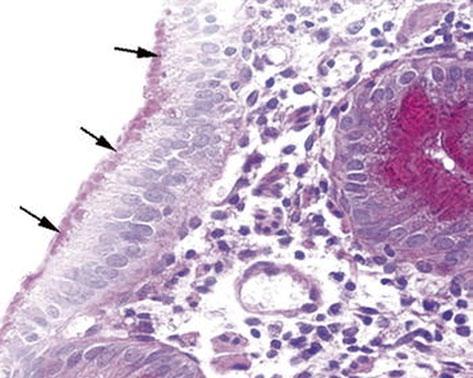
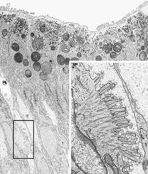
In one of these infants, the electron microscopy identified the presence of a peculiar abnormality of the microvilli of the enterocytes [19] (Fig. 1.1).
Three new cases with the same clinic and histological characteristics of the latter were described in France in 1982,
Fig. 1.1 Microvillous in the original label inclusion disease. PAS staining highlights abundant PAS-positive material (arrows) in the apical part of the enterocyte cytoplasm. PAS × 260. PAS peroidic acid-Schiff (Reprinted from Ref. [20], Fig. 1, with kind permission from Springer Science and Business Media)
and the four of them were grouped in a new disease called congenital microvillus atrophy [21, 22]. Two new cases were described in Great Britain in 1985 [23] and one in Italy in 1986; a subsequently born brother of the latter resulted affected too [24]. A survey completed in 1987 among centers known for their involvement in pediatric gastroenterology identified more than 30 cases worldwide. Additional cases were later published.
In 1989, Cutz et al. proposed the use of the term “microvillus inclusion disease” to highlight the characteristic ultrastructural lesions of the disease [25].
Clinical presentation: case report
First child of parents with no blood relation, A.G. was born after 37 weeks of gestation, the pregnancy having been complicated by a risk of miscarriage in the 5th month. His weight was 3500 g.
The infant was hospitalized when he was 40 days old because of an abundant diarrhea (15–20 evacuations a day of liquid stools), starting on the 6th day of its life and resistant to numerous dietary and pharmacological therapies.
Entering the hospital, the patient weighed 2800 g, it was in severe general conditions with dystrophia and dehydration; a TPN was therefore immediately started. The acid–basic balance showed hyponatremic acidosis (pH 7.2; EB −8,3; Na 128 mEq/l). The secretive nature of diarrhea was confirmed by its entity (about 100 ml/kg/die) with a total absence of oral nutrition and with the persistence of TPN in progress.
Moreover, the typical absence of ionic gap in the stools was present: osmolality 226 mOsm/l, Na 86 mEq/l, K 23.5 mEq/l (gap 7 mOsm/l).
Loperamide and chlorpromazine increased intestinal absorption, but did not change the clinical picture.
Microbiological examinations included an electronic microscope examination of the feces for the identification of viruses and the search for enterotoxigenic bacteria and parasites with specific methods were repeatedly negative.
The abdominal ultrasound showed adrenal hyperplasia associated to hyperaldosteronism (1160 ng/ml, v.n. < 125 ng/ ml).
Jejunal biopsy showed a picture of villus atrophy with no hyperplastic crypts and periodic acid-Schiff (PAS)-positive material stored in the apical cytoplasm of enterocytes. Electron microscopy was diagnostic for microvillus inclusion disease.
Microvillus Inclusion Disease is a Congenital Secretory Diarrhea Starting in Neonatal Age
Severe diarrhea typically appears in the first days of life, usually within the first 72 h, and it is immediately life threatening. The stools are watery, and the stool output is 100–500 ml/kg/day when the infant is fed, a volume comparable to or higher than that observed in cholera. The diarrhea is of secretory type; therefore, it persists at a stable rate of 50–300 ml/kg/day despite fasting, and the electrolyte content of the stools is increased, without an osmotic gap. However, the mucosal atrophy causes osmotic diarrhea. For this reason, feeding increases the fecal output and oral feeding in nutritionally significant amounts is impossible. Due to the high output, patients can lose up to 30 % of their body weight within 24 h, resulting in profound metabolic acidosis and severe dehydration, unless vigorous intravenous rehydration is started.
In a small percentage of cases (which was in the past considered to be around 20 %, and presently around 5 % of the cases [26]), diarrhea starts later in life, between 1 and 3 months, usually at 6–8 weeks of age. This less severe form has been denominated late-onset microvillus inclusion disease, while the classical form beginning at neonatal age has been denominated early-onset microvillus inclusion disease[27].
The hallmark of the disease is the electron microscopic finding of disrupted enterocytic microvilli (i.e., digitations of the apical membrane of the intestinal epithelial cell protruding into the lumen) and the appearance of characteristic inclusion vacuoles, whose inner surfaces are lined by typical microvilli. Both lesions are seen only with the electronic microscopy.
A few cases have been termed atypical microvillus inclusion disease, in which the onset can be early or late, but the histologic picture is different, particularly for the absence of detectable microvillus inclusions [28].
Therefore, three variants of the disease have been identified: early-onset microvillus inclusion disease, late-onset microvillus inclusion disease, and atypical microvillus inclusion disease. However, because of the sparse distribution of microvillus inclusions, it is not certain that their absence could be limited to the sample.
Microvillus inclusion disease is usually characterized by growth retardation and some developmental delay later in infancy. No other specific findings can be detected. However, the disease can be associated with other abnormalities, indicated in Table 1.2
Histologic Findings
Findings from duodenal biopsy must not be considered diagnostic. Histologic results of duodenal biopsy samples can range from essentially normal to mildly abnormal, showing the following:
• Thin mucosa caused by hypoplastic villus atrophy
• Diffuse villus atrophy (loss of villus height)
• Crypt hypoplasia
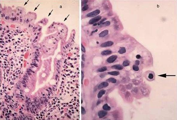
PAS staining of the intestinal biopsy sample does not show the usual linear staining along the brush border, but reveals PAS-positive material in the apical cytoplasm. The PAS staining material corresponds to the increased number of electron dense secretory granules in the epithelium. The abnormal pattern of staining appears in the upper crypt region and continues over the villus [29] (Fig. 1.2).
PAS accumulates in low crypts in atypical microvillus atrophy, in upper crypts in congenital microvillus atrophy, and in low villi in late-onset microvillus atrophy.
Fig. 1.2 Microvillous in the original label inclusion disease. Villous enterocytes: the boxed area shows microvilli on the lateral membrane. Inset: Enlargement of the boxed area × 6200, inset × 22,500. (Reprinted from [20, Fig. 5], with kind permission from Springer Science and Business Media)
Similar results were obtained with anti-CD10 immunohistochemistry: In affected children, the normal linear staining in surface enterocytes is absent, while prominent cytoplasmic reactivity is seen [30]. CD10 is a neutral membraneassociated peptidase; thus, abnormal stain findings with PAS or anti-CD10 immunohistochemistry are expressions of the abnormalities in microvillar structure.
Table 1.2 Anomalies described in association to microvillus inclusion disease
Rectal biopsy findings demonstrate microvillus involutions and an increased number of secretory granules. This test has been proposed as a relatively easy method for making an early diagnosis. Anti-CD10 immunohistochemistry can aid in the diagnosis, because abnormal cytoplasmic CD10 staining of absorptive colonocytes has been observed in microvillus inclusion disease [31].
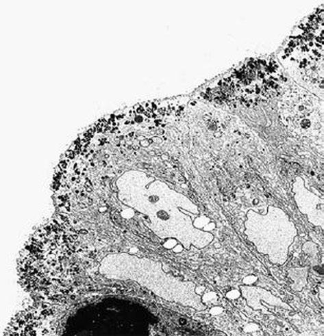
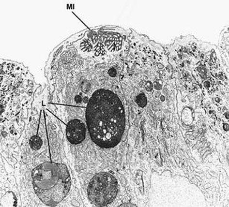
The diagnosis rests on findings demonstrated by electron microscopy (see Figs. 1.3 and 1.4.). Electron microscopy shows well-preserved crypt epithelium with abundant microvilli. Villus enterocytes are severely abnormal, particularly toward the apices of the short villi. The microvilli are depleted in number, short, and irregularly arranged. Some of the enterocytes contain the typical microvillus involutions, which are intracellular vacuoles where microvilli are observed lining the inner surface. A striking feature is a number of small, membrane-bound vesicles containing electrondense material (see Figs. 1.3 and 1.4). A few cases have been
Fig. 1.3 Microvillous in the original label inclusion disease. The apical cytoplasm of villous epithelium shows an increased number of secretory granules associated with microvillus alterations × 2400. (Reprinted from [20, Fig. 4], with kind permission from Springer Science and Business Media)
Fig. 1.4 Microvillous in the original label in the original label inclusion disease. The villous enterocytes lack brush-border microvilli, whereas their apical cytoplasm contains a microvillus inclusion (MI) and numerous lysosomes (L) × 5.500. (Reprinted from [20, Fig. 2], with kind permission from Springer Science and Business Media)
described in which the classic microvillus inclusions are shadowed by other features, such as large aggregates of electron lucent, vermiform membranous vesicles in enterocyte cytoplasm, corresponding to the PAS-positive material [32].
Epidemiology
The cases published or gathered in an online registry were 137 in 2014 [26].
A female preponderance had been observed among the published cases, with a female-to-male ratio of 2:1, but in the total 137 cases there is a 1.54 male to female ratio. A blood relation is present in 41 % of the assessable cases with a genre preference for males. A cluster of cases from the Navajo reservation in northern Arizona suggests an incidence as high as 1 case per 12,000 live births.
Pathophysiology
Due to their alterations, mature enterocytes inefficiently absorb ions and nutrients, causing a malabsorption syndrome; however, the diarrhea is caused mainly by active secretion of water and electrolytes in the intestinal lumen (secretory diarrhea). The pathogenesis of the secretory diarrhea is unknown; it is assumed to result from an imbalance between decreased absorption and unaltered secretion.
Measurement of stool electrolytes and osmolality enables rapid and accurate assessment of the pathogenesis of this chronic diarrhea (osmolar versus secretory) and greatly narrows the differential diagnosis.
Fecal electrolytes demonstrate a typical pattern of secretory diarrhea. Fecal sodium levels are high (approximately 60–120 mEq/l), and no osmotic gap is found. In patients with secretory diarrhea, the following formula applies: 2(Na concentration + K concentration) = stool osmolarity ± 50. In osmotic diarrhea, stool osmolarity exceeds 2(Na concentration + K concentration) by 100 or more.
Secretory diarrhea occurs in the fasting state and is associated with large output losses that cause dehydration and metabolic acidosis.
In osmotic diarrhea, findings on stool microscopy are negative for white blood cells (WBCs), blood (exudative diarrhea), and fat (steatorrhea).
Even if there are data about the anomalies in water and electrolytes transportation in the small intestine, it is not known whether and how the colon mucosa participates to the absorption alterations in the disease.
In one of the Italian cases, we used the technique of rectal perfusion that showed a decrease in sodium absorption, only partially corrected by chlorpromazine administration [33].
Pathogenesis
Severe perturbation of the microvillar cytoskeleton may disrupt the transport of brush border components that have
to be assembled at the apical membrane. The postulated abnormality in the cytoskeleton causes a block in exocytosis, mainly of PAS-positive material (e.g., polysaccharides, glycoproteins, glycolipids, neutral mucopolysaccharides). As a consequence, small secretory granules that contain a PASpositive material accumulate in the apical cytoplasm of epithelial cells.
In 2008, the presence of mutations in the MYO5B gene was described in seven patients (out of ten tested), predominantly of Turkish origin [34]. Homozygous mutations in the same gene were subsequently found in seven cases of Navajo origin; five parents were heterozygote [35]. A total of 41 unique MYO5B mutations in 40 patients have been identified so far: in more detail 16 different homozygous mutations in 25 patients, and 25 heterozygous mutations in 15 patients [26].
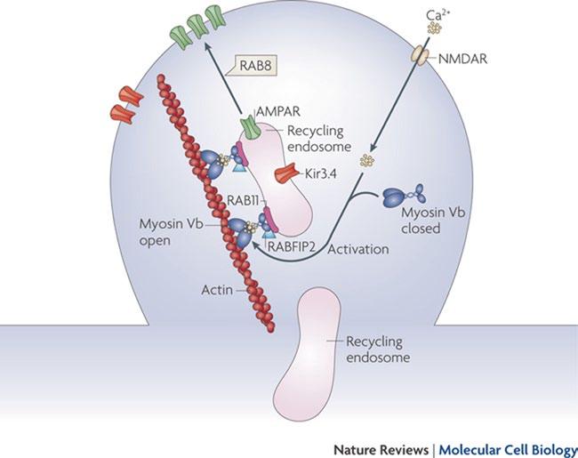
The MYO5B gene codifies myosine Vb, an actin-based motor protein which carries the recycling endosomes to the apical plasma membrane along the actin filaments of the microtubules. The functional deficiency of this protein alters the intracellular trafficking of resident apical plasma membrane proteins to the cell surface, and this could be the cause for the impaired apical brush border membrane development [34]. Actually, in microvillus inclusion disease the MYO5B mutations associate to a defective myosin Vb expression in enterocytes.
Myosine Vb carries on its action after having bound to a specific small guanosine-5′-triphosphatase (GTPase) rab proteins, such as Rab 11, located on the surface of recycling endosomes [36]. Thanks to this link the recycling endosomes move along the actine filaments (see Fig. 1.5) [37].
Fig. 1.5 Endocytic recycling. Myosin Vb is a conformation-dependent binding partner of Rab11-FIP2. Activation of myosin Vb induces translocation of recycling endosomes and their cargo. Final transport from the recycling endosome to the cell surface is mediated by Rab8. (Reprinted by permission from Macmillan Publishers Ltd and Nature Publishing Group [37])
A. Nocerino and S. Guandalini
When myosine Vb has an altered function, the recycling endosomes are not carried in a normal way: in the enterocytes of the subjects with microvillus inclusion disease, no regular accumulation of myosine Vb and of the recycling endosomeassociated proteins (one of these is Rab 11) can be observed close to the apical membrane, and no specific staining pattern is present [38]. Therefore, the Rab 11 distribution in the enterocytes can be a helpful diagnostic tool [39].
Other biochemical mechanisms depending on myosine Vb which can produce alterations in the structure of the microvilli are presently being studied [40].
Myosine Vb is expressed in all the epithelial tissues and, actually, microvillus inclusions in the stomach and colon, in addition to less well-defined inclusions in gallbladder epithelium and in renal tubular epithelial cells, have been reported in some patients with microvillous inclusion disease (MVID). Nevertheless, no extraintestinal symptoms are generally reported. Two children with renal Fanconi syndrome who carried mutation MYO5B did not show alterations in the apical brush border morphology and the PAS staining pattern in renal tubular epithelial cells, which makes it unlikely for it to be the cause of proximal tubular renal dysfunction [41].
Recently, Dutch investigators have found [42] that the mild variant of MVID appears to be caused by loss of function of syntaxin 3 (STX3), an apical receptor involved in membrane fusion of apical vesicles in enterocytes. In fact, whole-exome sequencing of DNA from patients with variant MVID revealed homozygous truncating mutations in STX3; and in addition, patient-derived organoid cultures and overexpression of truncated STX3 in CaCo2 cells recapitulated most characteristics of variant MVID.
Prenatal Diagnosis
Pregnancy and birth are usually normal in individuals with microvillus atrophy, and polyhydramnios is usually absent, in contrast to the clinical picture of patients with other causes of congenital secretory diarrhea [43]. Nevertheless, in some cases, polyhydramnios and bowel dilation in the third trimester have been described [44]. In one case, a high fetal alpha-fetoprotein in the second trimester was observed [45]. Authors have speculated that the fetal alpha-fetoprotein elevation might possibly be caused by in utero body fluid leakage into the amniotic fluid through fetal enteropathy.
Identification of the gene responsible for the disease allows its prenatal diagnosis [46].
Treatment
The prognosis of early-onset microvillus inclusion disease is poor. If patients are untreated, the disease is rapidly fatal because of dehydration and malnutrition.
In late-onset microvillus inclusion disease diarrhea tends to be less severe, and some alimentation is possible.
Medical Care
Agents tentatively given to induce a better growth of the intestinal mucosa (e.g., epithelial growth factor, colostrum) are ineffective. Several drugs (e.g., somatostatin, octreotide, loperamide, chlorpromazine) have been tried to counteract the massive secretory diarrhea in patients with microvillus atrophy; however, none has proven effective.
At present, the only available therapy is TPN. Children with late-onset microvillus inclusion disease usually have less severe diarrhea; with age they can reduce the requirements of TPN to 1–2 per week.
If patients are treated with TPN, their prognosis entirely depends on the complications of this approach. These complications include cholestasis with subsequent liver damage leading to cirrhosis, catheter-related sepsis due to infection with bacterial or fungal agents, and progressive lack of vascular access.
In the observed cases, cholestasis appears worsened by transplant.
The study of eight patients who developed cholestatic liver disease suggests that cholestasis is enhanced by the impairment of the MYO5B/RAB11A apical recycling endosome pathway in hepatocytes [47].
Surgical Care
Successful outcomes of small-intestinal transplantation have been reported, and evidence suggests that an early transplant might be beneficial. The limited experience accumulated in a few centers worldwide reflects an overall survival rate of
approximately 50 % at 5 years after small-bowel transplantation; this is a much better outcome than is seen with other indications for intestinal transplantation [48]. Patients who did not receive colonic transplant weaned later from parenteral nutrition.
The analysis of 16 patients who underwent a small-bowel transplantation shows a lower death rate compared to those who did not (23 versus 37 %) after an average observation period of 3.5 years (but variable between 3 months and 14 years). In all of the cases, apart from the first two, colon had been transplanted too [49].
Although only small series have been reported, evidence suggests that early small-bowel transplantation should be performed, at least in children with early-onset microvillus inclusion disease. Patients with late-onset microvillus atrophy appear to have an improved prognosis.
Transplantation appears to be the only option for patients who do not fare well with long-term TPN (e.g., because of sepsis, liver damage, lack of vascular access). For patients in whom transplantation is successful, a gradual return to a normal diet is considered possible.
In the observed cases, TPN-related cholestasis appears worsened by transplant. Therefore, in children with cholestasis, the worsening of this picture after the transplant points to a combined liver-intestinal transplantation.
In 1994, Reifen et al. described two infants less than a month old with protracted diarrhea. The diarrhea was so profuse to make TPN necessary but it improved when enteral nutrition was interrupted. The jejunal biopsies showed a peculiar picture characterized by the presence of focal aggregations of packed enterocytes whose shape resembled of a teardrop, as a consequence of an apical rounding of the plasma membrane. These focal areas resembled a tuft and that is why the term “tufting enteropathy” was coined [50]. Curiously, a case with the same characteristics was identified among those presented by Davidson et al. in the same paper where the first case of microvillus inclusion disease had been described [19].
Clinical Expression
The incidence of the disease has been estimated to be 1:100,000 live births in Western Europe [51], but it seems higher in people of Arabic origin [52].
The picture is a severe secretory diarrhea starting in the first weeks of life. During pregnancy, there is no polyhydramnios, as in the microvillus inclusion disease and
differently from congenital sodium diarrhea and congenital chloridorrhea.
The alterations in the enterocytes, in any case, cause an accentuation of the diarrhea with nutrition, including total enteral nutrition, as it had already been observed since the first described cases.
There are two different clinical forms: one is isolated and the other is syndromic, associated to different anomalies, particularly to facial dysmorphism with choanal atresia and superficial punctuated keratitis [53, 54].
Pathophysiology
In 2008, a mutation of the gene for Epithelial Cell Adhesion Molecule (EpCAM) was identified in two ill children in the same family, and in three children from unrelated kindreds [55]. EpCAM is a transmembrane protein involved in cell proliferation, differentiation, and adhesion.
In 2010, a mutation in SPINT2 gene was found in a case affected by a syndromic form of tufted enteropathy. SPINT2 is a transmembrane protein that seems to be involved in epithelial regeneration [56].
It is interesting to note how mutations in SPINT2 gene are also present in the syndromic congenital sodium diarrhea, where choanal atresia, hypertelorism, and corneal erosions are particularly frequent and anal atresia can be found in certain cases [57].
Analyzing 57 patients, mutations in the gene for EpCAM were found in 73 % of the cases, all of them presenting an isolated intestinal disease.
But in 21 % of the cases, all showing a syndromic form of the disease, there are mutations the SPINT2 gene.
According to this study, tufting enteropathy could be separated into at least three genetic classes, each with specific phenotypes [58].
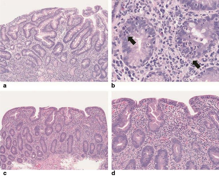
Fig. 1.6 a Numerous tufts of enterocytes on the mucosal surface of the duodenum. b A characteristic tear-drop-shaped structure (arrow) in an epithelial tuft (H&E stain; original magnification: a–x 80; b–x 400). (Reprinted from Ref. [59, Fig. 1], with kind permission from Springer Science and Business Media)
However, it seems impossible at present to discriminate “tufting enteropathy” isolated from the syndromic one, even from a genetic point of view.
Histologic Features
Jejunal biopsy shows a picture of partial villous atrophy associated to crypt hyperplasia. The most characteristic feature, the one which gave the name to the disease, is the presence of “tufts,” small focal aggregates of teardrop-shaped enterocytes with an apical rounding (See Fig. 1.6a, b).
The “tufts” are not a characteristic exclusive to intestinal epithelial dysplasia, because they have been observed in other mucosal enteropathies and in normal jejunum. In the latter cases, anyway they were present in < 10 % of the epithelial surface, while in “tufting enteropathy” they are present in more than 80 % of the jejunal surface. But the picture is not always so evident in the earliest period of the disease. Attempts at immunohistochemical analysis (including beta-catenin, E-cadherin, desmoglein, laminins) have not been easily applicable [60]. On the contrary, the staining with EpCAM/MOC31 antibody, an EpCAM antibody clone, showed a sensitivity and specificity of 100 % for loss of staining in 15 studied patients [61].
Electronic microscopy shows relatively normal microvilli, and it is not particularly useful for diagnosis, if only to exclude a microvillus inclusion disease.
A mild inflammation of the lamina propria is also present. An infiltration of T lymphocytes within the lamina propria had been observed since the original description, even if inferior to celiac disease, but it sometimes arises suspicion of autoimmune enteropathy [50].
Treatment
Tuft enteropathy is associated to a severe secretory diarrhea, which worsens with nutrition. That is why affected children have to be treated with TPN.
Some cases seem to have a less severe course and they can be given a partial parenteral nutrition [62].
Cases totally dependent on TPN are candidates for intestinal transplantation.
References
1. Avery GB, Villavicencio O, Lilly JR, Randolph JG. Intractable diarrhea in early infancy. Pediatrics 1968;41:712–22
2. Hyman CJ, Reiter J, Rodnan J, Drash AL. Parenteral and oral alimentation in the treatment of the nonspecific protracted diarrheal syndrome of infancy. J Pediatr. 1971;78:17–29.
3. Shwachman H, Filler RM, Khaw KT. A new method of treating malnourished infants with severe chronic diarrhea. Acta Pediatr Scand. 1970;59:446–7.
4. Shwachman H, Lloyd-Still JD, Khaw KT, Antonowicz I. Protracted diarrhea of infancy treated by intravenous alimentation. II: studies of small intestinal biopsy results. Am J Dis Child. 1973;125:365–8.
5. Walker-Smith J A. Intractable diarrhea of infancy. Saudi J Gastroenterol. 1995;1:152–6.
6. Larcher VF, Shepherd R, Francis DE, Harries JT. Protracted diarrhoea in infancy. Analysis of 82 cases with particular reference to diagnosis and management. Arch Dis Child. 1977;52:597–605.
7. Sanderson IR, Risdon RA, Walker-Smith JA. Intractable ulcerating enterocolitis of infancy. Arch Dis Child. 1991;66:295–9.
8. Thapar N, Shah N, Ramsay AD, Lindley KJ, Milla PJ. Long-term outcome of intractable ulcerating enterocolitis of infancy. J Pediatr Gastroenterol Nutr. 2005;40:582–8.
9. Murch SH, Winyard PJ, Koletzko S, Wehner B, Cheema HA, Risdon RA, et al. Congenital enterocyte heparan sulphate deficiency with massive albumin loss, secretory diarrhoea, and malnutrition. Lancet 1996;347:1299–301.
10. Bode L, Salvestrini C, Park PW, Li JP, Esko JD, Yamaguchi Y, et al. Heparan sulfate and syndecan-1 are essential in maintaining murine and human intestinal epithelial barrier function. J Clin Invest. 2008;118:229–38.
11. Bode L, Freeze HH. Applied glycoproteomics–approaches to study genetic-environmental collisions causing protein-losing enteropathy. Biochim Biophys Acta. 2006;1760:547–59.
12. Lachaux A, Bouvier R, Loras-Duclaux I, Chappuis JP, Meneguzzi G, Ortonne JP. Isolated deficient alpha6beta4 integrin expression in the gut associated with intractable diarrhea. J Pediatr Gastroenterol Nutr. 1999;29:395–401.
13. Salvestrini C, McGrath JA, Ozoemena L, Husain K, Buhamrah E, Sabery N, et al. Desquamative enteropathy and pyloric atresia without skin disease caused by a novel intracellular beta4 integrin mutation. J Pediatr Gastroenterol Nutr. 2008;47:585–91.
14. Girault D, Goulet O, Le Deist F, Brousse N, Colomb V, Césarini JP, et al. Intractable infant diarrhea associated with phenotypic abnormalities and immunodeficiency. J Pediatr. 1994;125:36–42.
15. Stankler L, Lloyd D, Pollitt RJ, Gray ES, Thom H, Russell G. Unexplained diarrhoea and failure to thrive in two siblings with unusual facies and abnormal scalp hair shafts: a new syndrome. Arch Dis Child. 1982;57:212–6.
16. Verloes A, Lombet J, Lambert Y, Hubert AF, Deprez M, Fridman V, et al. Tricho-hepato-enteric syndrome: further delineation
of a distinct syndrome with neonatal hemochromatosis phenotype, intractable diarrhea, and hair anomalies. Am J Med Genet. 1997;68:391–5.
17. Fabre A, André N, Breton A, Broué P, Badens C, Roquelaure B. Intractable diarrhea with “phenotypic anomalies” and tricho-hepato-enteric syndrome: two names for the same disorder. Am J Med Genet A. 2007;143A:584–8
18. Hartley JL, Zachos NC, Dawood B, Donowitz M, Forman J, Pollitt RJ, et al. Mutations in TTC37 cause trichohepatoenteric syndrome (phenotypic diarrhea of infancy). Gastroenterology 2010;138:2388–98,.e1–2.
19. Davidson GP, Cutz E, Hamilton JR, Gall DG. Familial enteropathy: a syndrome of protracted diarrhea from birth, failure to thrive, and hypoplastic villus atrophy. Gastroenterology 1978;75:783–90.
20. Morroni M, Cangiotti AM, Guarino A, Cinti S. Unusual ultrastructural features in microvillous inclusion disease: a report of two cases. Virchows Arch. 2006;448:805–10.
21. Schmitz J, Ginies JL, Arnaud-Battandier F, et al. Congenital microvillous atrophy, a rare cause of neonatal intractable diarrhoea. Pediatr Res. 1982;16:1014.
22. Goutet JM, Boccon-Gibod L, Chatelet F, Ploussard JP, Navarro J, Polonovski CI. Familial protracted diarrhoea with hypoplastic villous atrophy: report of two cases. Pediatr Res. 1982;16:1045.
24. Guarino A, Nocerino A, Cinti S, Berni Canani R, Terracciano L, Raimondi F, Guandalini S. Atrofia congenita dei microvilli intestinali. Riv Ital Ped. 1992;18:150–3.
25. Cutz E, Rhoads JM, Drumm B, Sherman PM, Durie PR, Forstner GG. Microvillus inclusion disease: an inherited defect of brush-border assembly and differentiation. N Engl J Med. 1989;320:646–51.
26. van der Velde KJ, Dhekne HS, Swertz MA, Sirigu S, Ropars V, Vinke PC, et al. An overview and online registry of microvillus inclusion disease patients and their MYO5B mutations. Hum Mutat. 2013;34:1597–605.
27. Phillips AD, Schmitz J. Familial microvillous atrophy: a clinicopathological survey of 23 cases. J Pediatr Gastroenterol Nutr. 1992;14:380–96.
28. Mierau GW, Wills EJ, Wyatt-Ashmead J, Hoffenberg EJ, Cutz E. Microvillous inclusion disease: report of a case with atypical features. Ultrastruct Pathol. 2001;25:517–21.
29. Phillips AD, Szafranski M, Man LY, Wall WJ. Periodic acid-Schiff staining abnormality in microvillous atrophy: photometric and ultrastructural studies. J Pediatr Gastroenterol Nutr. 2000;30:34–42.
30. Groisman GM, Amar M, Livne E. CD10: a valuable tool for the light microscopic diagnosis of microvillous inclusion disease (familial microvillous atrophy). Am J Surg Pathol. 2002;26:902–7.
31. Koepsell SA, Talmon G. Light microscopic diagnosis of microvillus inclusion disease on colorectal specimens using CD10. Am J Surg Pathol. 2010;34:970–2.
32. Weeks DA, Zuppan CW, Malott RL, Mierau GW. Microvillous inclusion disease with abundant vermiform, electron-lucent vesicles. Ultrastruct Pathol. 2003;27:337–40.
33. Guandalini S, Nocerino A, Saitta F, Fasano A, Ascione G, De Curtis M, et al. Valutazione dell’assorbimento di elettroliti ed acqua nel colon di un lattante affetto da atrofia congenita dei microvilli. Riv Ital Pediatr. 1987;13:76
34. Müller T, Hess MW, Schiefermeier N, Pfaller K, Ebner HL, Heinz-Erian P, et al. MYO5B mutations cause microvillus inclusion disease and disrupt epithelial cell polarity. Nat Genet. 2008;40:1163–5.
35. Erickson RP, Larson-Thomé K, Valenzuela RK, Whitaker SE, Shub MD. Navajo microvillous inclusion disease is due to a mutation in MYO5B. Am J Med Genet A. 2008;146A:3117–9.
36. Schafer JC, Baetz NW, Lapierre LA, McRae RE, Roland JT, Goldenring JR. Rab11-FIP2 interaction with MYO5B regulates movement of Rab11a-containing recycling vesicles. Traffic 2014;15:292–308.
37. Grant BD, Donaldson JG. Pathways and mechanisms of endocytic recycling. Nat Rev Mol Cell Biol. 2009;10:597–608.
38. Szperl AM, Golachowska MR, Bruinenberg M, Prekeris R, Thunnissen AM, Karrenbeld A, et al. Functional characterization of mutations in the myosin Vb gene associated with microvillus inclusion disease. J Pediatr Gastroenterol Nutr. 2011;52:307–13.
39. Talmon G, Holzapfel M, DiMaio DJ, Muirhead D. Rab11 is a useful tool for the diagnosis of microvillous inclusion disease. Int J Surg Pathol. 2012;20:252–6.
40. Dhekne HS, Hsiao NH, Roelofs P, Kumari M, Slim CL, Rings EH, van Ijzendoorn SC. Myosin Vb and Rab11a regulate phosphorylation of ezrin in enterocytes. J Cell Sci. 2014; 127(Pt 5):1007–17.
41. Golachowska MR, van Dael CM, Keuning H, Karrenbeld A, Hoekstra D, Gijsbers CF, et al. MYO5B mutations in patients with microvillus inclusion disease presenting with transient renal Fanconi syndrome. J Pediatr Gastroenterol Nutr. 2012;54:491–8.
42. Wiegerinck CL, Janecke AR, Schneeberger K, Vogel GF, van Haaften-Visser DY, Escher JC, et al. Loss of syntaxin 3 causes variant microvillus inclusion disease. Gastroenterology 2014;147:65–8.
44. Kennea N, Norbury R, Anderson G, Tekay A. Congenital microvillous inclusion disease presenting as antenatal bowel obstruction. Ultrasound Obstet Gynecol. 2001;17:172–4.
45. Chen CP, Su YN, Chern SR, Wu PC, Wang W. Prenatal diagnosis of microvillus inclusion disease. Taiwan J Obstet Gynecol. 2011;50:399–400.
46. Chen CP, Chiang MC, Wang TH, Hsueh C, Chang SD, Tsai FJ, et al. Microvillus inclusion disease: prenatal ultrasound findings, molecular diagnosis and genetic counseling of congenital diarrhea. Taiwan J Obstet Gynecol. 2010;49:487–94.
47. Girard M, Lacaille F, Verkarre V, Mategot R, Feldmann G, Grodet A, et al. MYO5B and BSEP contribute to cholestatic liver disorder in microvillous inclusion disease. Hepatology 2013;60:301–16.
48. Ruemmele FM, Jan D, Lacaille F, Cézard JP, Canioni D, Phillips AD, et al. New perspectives for children with microvillous inclusion disease: early small bowel transplantation. Transplantation 2004;77:1024–8.
49. Halac U, Lacaille F, Joly F, Hugot JP, Talbotec C, Colomb V, et al. Microvillous inclusion disease: how to improve the prognosis of a severe congenital enterocyte disorder. J Pediatr Gastroenterol Nutr. 2011;52:460–5.
50. Reifen RM, Cutz E, Griffiths AM, Ngan BY, Sherman PM. Tufting enteropathy: a newly recognized clinicopathological entity associated with refractory diarrhea in infants. J Pediatr Gastroenterol Nutr. 1994;18:379–85.
51. Goulet O. Intestinal epithelial dysplasia: a new entity. Arch Pediatr. 1996;3(suppl 1):324s–5s.
52. Goulet O, Salomon J, Ruemmele F, de Serres NP, Brousse N. Intestinal epithelial dysplasia (tufting enteropathy). Orphanet J Rare Dis. 2007;2:20.
53. Bird LM, Sivagnanam M, Taylor S, Newbury RO. A new syndrome of tufting enteropathy and choanal atresia, with ophthalmologic, hematologic and hair abnormalities. Clin Dysmorphol. 2007;16:211–21.
54. Roche O, Putterman M, Salomon J, Lacaille F, Brousse N, Goulet O, Dufier JL. Superficial punctate keratitis and conjunctival erosions associated with congenital tufting enteropathy. Am J Ophthalmol. 2010;150:116-21.e1.
55. Sivagnanam M, Mueller JL, Lee H, Chen Z, Nelson SF, Turner D, et al. Identification of EpCAM as the gene for congenital tufting enteropathy. Gastroenterology. 2008;135:429–37.
56. Sivagnanam M, Janecke AR, Müller T, Heinz-Erian P, Taylor S, Bird LM. Case of syndromic tufting enteropathy harbors SPINT2 mutation seen in congenital sodium diarrhea. Clin Dysmorphol. 2010;19:48.
57. Heinz-Erian P, Müller T, Krabichler B, Schranz M, Becker C, Rüschendorf F, et al. Mutations in SPINT2 cause a syndromic form of congenital sodium diarrhea. Am J Hum Genet. 2009;84:188–96.
58. Salomon J, Goulet O, Canioni D, Brousse N, Lemale J, Tounian P, et al. Genetic characterization of congenital tufting enteropathy: EpCAM associated phenotype and involvement of SPINT2 in the syndromic form. Hum Genet. 2014;133:299–310.
59. El-Matary W, Dalzell AM, Kokai G, Davidson JE. Tufting enteropathy and skeletal dysplasia: is there a link? Eur J Pediatr. 2007;166:265–8.
60. Patey N, Scoazec JY, Cuenod-Jabri B, Canioni D, Kedinger M, Goulet O, Brousse N. Distribution of cell adhesion molecules in infants with intestinal epithelial dysplasia (tufting enteropathy). Gastroenterology 1997;113:833–43.
61. Ranganathan S, Schmitt LA, Sindhi R. Tufting enteropathy revisited: the utility of MOC31 (EpCAM) immunohistochemistry in diagnosis. Am J Surg Pathol. 2014;38:265–72.
62. Lemale J, Coulomb A, Dubern B, Boudjemaa S, Viola S, Josset P, et al. Intractable diarrhea with tufting enteropathy: a favorable outcome is possible. J Pediatr Gastroenterol Nutr. 2011;52:734–9.
The Spectrum of Autoimmune Enteropathy
Natalia Nedelkopoulou, Evangelia Farmaki, Maesha Deheragoda and Babu Vadamalayan
Introduction
Chronic, unexplained diarrhea in children younger than 3 months old was first characterized as “intractable diarrhea” [1]. The term “protracted diarrhea” was used later to describe infants with frequent and loose stools severe enough to often require parenteral alimentation as nutritional support [2]. The differential diagnosis of enteropathies in infancy and childhood includes inherited epithelial and congenital transport defects, enzymatic deficiencies and allergic enteropathy (Table 2.1).
The most frequent diagnosis in children with protracted diarrhea is autoimmune enteropathy (AIE) [3, 4]. It is a rare, immune-mediated disorder starting usually within the first months of life. The age of onset is between 1 month and 5 years (median age 17 months) [5], but late-onset adult forms have been also reported [6–9]. The disease was first described by Walker-Smith et al. in 1982 in a male child with clinical features of coeliac disease and villous blunting unresponsive to gluten-free diet [10] and represents a heterogeneous group of disorders rather than a discrete entity. The incidence is estimated at less than 1 in 100,000 infants. The diagnostic criteria are debatable but the presence of circulating anti-enterocyte antibodies and the lack of immunodefi-
B. Vadamalayan ()
Pediatric Liver, GI and Nutrition Centre, King’s College Hospital, Denmark Hill, London SE59RS, UK e-mail: babu.vadamalayan@nhs.net
N. Nedelkopoulou
Pediatric Liver, GI and Nutrition Centre, King’s College Hospital, London SE59RS, UK
E. Farmaki
Pediatric Immunology and Rheumatology Referral Centre, First Dept of Pediatrics, Aristotle University of Thessaloniki, Thessaloniki, Greece
M. Deheragoda
Institute of Liver Studies, King’s College Hospital, London SE59RS, UK
ciency have been proposed as the hallmark features of AIE [5, 11]. The latter criterion has been challenged by clinical experience and better understanding of the immunology of autoimmunity and self-tolerance [12].
AIE is characterized by variable clinical expression, ranging from isolated gastrointestinal involvement to severe systemic disease [13, 14]. Patients diagnosed with the disease often exhibit extra-intestinal manifestations of autoimmunity, in contrast to those with tufting enteropathy and microvillus inclusion disease [15]. Based on a genetic approach combined with immunological evaluation, three different forms of AIE have been proposed:
1. A predominately or isolated gastrointestinal form of AIE with typical anti-enterocyte antibodies in both sexes
2. A systemic X-linked form of AIE associated with different endocrinopathies, haematological symptoms and severe eczematous skin disease, known as immune dysregulation, polyendocrinopathy, AIE X-linked syndrome (IPEX) occurring only in males
3. An IPEX-like form, a priori FOXP3 independent occurring in both sexes
IPEX and autoimmune polyendocrinopathy-candidiasisectodermal dystrophy (APECED) syndrome (APR-1/autoimmune phenomena, polyendocrinopathy, candidiasis and ectodermal dystrophy) are systemic forms of AIE [16].
Clinical Presentation
Chronic, secretory diarrhea refractory to bowel rest that leads to dehydration, malabsorption and severe weight loss is the typical clinical presentation of AIE. Diarrhea usually begins between 2 and 4 weeks of age and the secretory component can be delayed for a few months [3, 11, 17]. The symptoms are often debilitating and the disease is potentially life threatening. The establishment of the diagnosis is crucial in order to ensure optimal treatment. Patients typically require immunosuppressive therapies and total parenteral
S. Guandalini et al. (eds.), Textbook of Pediatric Gastroenterology, Hepatology and Nutrition, DOI 10.1007/978-3-319-17169-2_2
Table 2.1 Differential diagnosis of diarrhea in infancy and childhood
nutrition (TPN) for hydroelectrolytic balance and nutritional support [18, 19].
Despite the fact that the mucosal abnormality is primarily confined to the small intestine, the term “generalized autoimmune gut disorder” has been used to describe the association between AIE and autoimmune colitis [8]. Emerging evidence suggests that AIE can be a manifestation of a more diffuse autoimmune disorder of the gastrointestinal system, which comprises gastritis, colitis, hepatitis and pancreatitis with positivity of a variety of autoantibodies, including antiparietal, anti-goblet cell and anti-smooth muscle antibodies [6, 20–22].
Furthermore, the involvement of extra-intestinal organs can be present during the course of the disease. Multisystem extra-intestinal manifestations include endocrine, renal, pulmonary, hematologic and musculoskeletal. Hypothyroidism with interstitial fibrosis and lymphocytic infiltration of the thyroid gland, nephrotic, nephritic syndrome and membranous glomerulonephritis, interstitial pneumonopathy, periportal fibrosis and bronchitis, haemolytic anaemia, rheumatoid arthritis and dermatitis/atopic eczema have all been reported [5, 11, 22, 23].
Thymus plays a key role in the deletion of potentially self-reactive clones of T cells. The association between AIE and thymoma has been described in both pediatric and adult patients and provides further evidence about the role of thymoma and the development of autoimmunity [24, 25].
Pathogenesis
The underlying immunologic and molecular mechanisms in AIE have not yet been fully elucidated and are widely debatable. However, it has been established that an autoimmune response is involved in the pathogenesis of the disease. Thymus orchestrates a healthy immune system. The intrathymic maturation of T lymphocytes is crucial for the deletion of potentially self-reactive clones of T cells. The dysfunction of the thymus results in the non-deletion and presence of selfreactive T cells that can induce the expansion of anti-self B cells [11, 26, 27].
In AIE, the gut is the site where the autoimmune reaction takes place and is mediated by the activation of self-reactive T cells locally, resulting in the typical histological lesions. In normal states, the expression of human leukocyte antigen (HLA) class II molecules on the enterocyte surface is crucial in establishing and maintaining the oral tolerance as the epithelial cells present exogenous peptides to the clonotypic T cell receptors. The overexpression of HLA-DR antigens in enterocytes and the inappropriate expression of HLA class II molecules in the crypt epithelium of the proximal small intestine in children with AIE have been reported [15, 28].
An increase in the levels of cluster of differentiation CD4 and CD8 lymphocytes in the lamina propria in subjects affected by AIE provides further evidence that the T cells are involved in the pathogenesis of the disease [29, 30]. The intestinal T lymphocytes cause damage to the enterocytes by exerting direct cytotoxicity, via the production of lymphokines or through an antibody-dependent cytotoxicity resulting in cellular apoptosis [31–33]. The loss of the regulatory function of T lymphocytes and the activation of the immune system are implicated in the pathogenesis of IPEX syndrome, whereas AIE is partly attributed to a humoral immune response with the presence of anti-enterocyte antibodies [34].
A variety of circulating autoantibodies, such as antibodies against gastric parietal cells, pancreatic islets, glutamic acid decarboxylase, insulin, smooth muscle, endoplasmic reticulum, reticulin, gliadin, adrenal cells, nuclear antigens, deoxyribose nucleic acid (DNA), thyroglobulin and thyroid microsomes, has been detected in patients with AIE [7, 18]. The presence of antibodies against goblet cells, enterocytes and colonocytes is supportive of the diagnosis. These antibodies are directed against components of the intestinal brush border membrane, with an increasing intensity from the crypts towards villous tip [5, 13]. However, they are neither diagnostic nor specific for the disease and have been also identified in other disorders such as the cow’s milk allergy, inflammatory bowel disease and in adults with human immunodeficiency virus (HIV) infection. Moreover, the appearance of the autoantibodies after the onset of the mucosal damage, the lack of correlation between the titer and the
histological severity and their disappearance after treatment, but before the complete mucosal restoration support the hypothesis that these antibodies are most likely a secondary event in the pathogenesis of the disease in response to bowel injury [10, 35–37].
The nature of the gut antigen that elicits the immune response and results in the alteration of the intestinal permeability has been extensively investigated. A 55-kD protein located in both the gut and renal epithelial cells that reacted with serum autoantibodies was first identified by Colletti et al. in 1991 in a patient with complicated presentation of AIE with small bowel and glomerular involvement [38]. A few years later, a 75-kD auto-antigen that is distributed through the whole intestine and the kidney was recognized in patients with X-linked AIE associated with nephropathy [39]. The intestinal auto-antigen in autoimmune polyendoendocrine syndrome type 1 (APECED) is tryptophan hydroxylase (TPH), which is mainly present in the enterochromaffin cells of the mucosa [40].
Emerging evidence has pointed towards an uncontrolled inflammatory reaction caused by the disturbance of the effector–regulatory T-cell interaction and leading to the production of autoantibodies, such as anti-enterocyte antibodies [41]. The understanding of the underlying molecular mechanism and the identification of the genetic defect in AIE was achieved due to the clinical similarities between scurfy mice and boys with the disease. Scurfy mice are a naturally occurring X-linked mutant that presents with massive lymphoproliferation, diarrhea, intestinal bleeding, scaly skin, anaemia, thrombocytopenia and hypogonadism [42]. Based on the observation that the disease-causing mutation in scurfy mice was on the X chromosome, the human IPEX locus was identified on chromosome Xp11.23-q13.3 and the gene was named FOXP3. It comprises 11 exons which encode the FOXP3 protein or scurfin, a 48-kDa protein of the forkhead (FKH)/winged-helix transcription factor family that is predominantly expressed in CD4+CD25+ T cell with regulatory function, at significantly lower levels in CD4+CD25-T cells and not at all in CD8+ or B220+ cells [43–46].
Increasing experimental evidence has shown that scurfin is implicated in the thymic maturation of T cells that are designated to acquire regulatory function. CD4+CD25+ Treg represent a small subset (5–10 %) of CD4+ T helper cells in humans and mice. Studies on CD4+CD25+ T cells from IPEX patients with the use of anti-CD127 have shown that FOXP3 plays a crucial role in the generation of functional T regulatory cells (Treg) and intact FOXP3 is indispensable for the development of fully functional Tregs, whereas FOXP3 with amino acid substitutions in the FKH domain is sufficient for the generation of functionally immature Tregs [47].
FOXP3 has DNA-binding activity and due to its structure may serve as nuclear transcription factor and act as a repressor of transcription and regulator of T cell activation
[48, 49]. The transcription of a reporter containing a multimeric-FKH-binding site is repressed by intact FOXP3. Such FKH-binding sites are located adjacent to nuclear factor of activated T cells (NFAT), regulatory sites in various cytokine promoters such as interleukin (IL)-2, or granulocyte–macrophage colony-stimulating factor enhancer. Therefore, intact scurfin protein appears to be capable to directly repress NFAT-mediated transcription of the IL-2 gene in CD4+ cells upon activation [50].
Despite the evidence that FOXP3 plays a key role in the development and function of Treg cells, the underlying mechanisms have not been fully understood. Data from animal models with transgenic induction of FOXP3 have shown that the overexpression of scurfin in normal mice leads to a tremendous suppression of immune functions, whereas the depletion of Tregs in healthy mice results rapidly in the development of different T-cell-mediated autoimmune disorders, similar to scurfy in mice or IPEX in humans that go in complete remission upon reconstitution with Treg cells [51, 52].
The three domains that are crucial for the function of FOXP3 are the C-terminal region, which contains the forkhead domain that directly binds DNA regions, the central domain with a zinc finger and leukine zipper that promotes the oligomerization of the FOXP3 molecule and the repressor domain located in the N-terminal region that binds the NAFT [53, 54]. Genetic screening on X chromosome in patients with AIE revealed that the majority of mutations cluster primarily within the FKH domain and the leukine zipper within the coding region of the FOXP3 gene causing potentially absent FOXP3 protein expression or a protein product with loss of function [13, 53].
Histopathology
Histologic evaluation of the small bowel in typical AIE reveals partial or total villous blunting/atrophy and crypt hyperplasia. In addition, there is a marked infiltration of the lamina propria by mixed inflammatory cells with a prominence of mononuclear cells, including T lymphocytes [15]. Apoptotic bodies and intraepithelial lymphocytes are present in the crypt epithelia. Most cases show a relative paucity of surface lymphocytosis in contrast to coeliac disease. The lymphocytic infiltration of the intestinal mucosa is constituted by CD4–CD8 T lymphocytes and macrophages. Goblet, Paneth and/or enterochromaffin cells may be reduced in number or absent. Cryptitis and crypt abscesses have been reported in severe AIE. Crypt enterocytes commonly show an increased expression of HLA-A, -B, -C molecules [8, 55, 56]. (See Table 2.2; Fig. 2.1) [12].
AIE primarily involves the small bowel with the histologic lesions being most prominent in the proximal small
Table 2.2 Histological findings in autoimmune enteropathy
Histological findings in autoimmune enteropathy
Partial or total villous blunting/atrophy and crypt hyperplasia
Marked infiltration of mononuclear cells, including activated T lymphocytes in the lamina propria
Apoptotic bodies and intraepithelial lymphocytes present in the crypt/gland epithelia, but relative paucity of surface lymphocytosis
Crypt abscesses in severe autoimmune enteropathy
Increased expression of HLA-A, -B, -C molecules in crypt enterocytes
intestine. However, changes have been also described in the oesophagus, stomach and colon in both pediatric and adult patients supporting the hypothesis for a diffuse disease process involving the entire gastrointestinal tract.
Recent reports describe the infiltration of the squamous epithelium by lymphocytes or eosinophils in the oesophagus. Gastric biopsies can show features of chronic nonspecific gastritis with or without reactivity. Atrophic gastritis, intestinal metaplasia and glandular destruction have been also described. There may be increased apoptosis of glandular epithelium [6, 58]. The colonic morphological lesions vary from diffuse mild active colitis with inflammatory cell infiltration to severe active chronic colitis with goblet cell depletion, Paneth cell metaplasia, distortion of crypt architecture and crypt abscess formation. An increase in intraepithelial lymphocytes has also been described [21, 58].
Diagnosis
The diagnostic criteria for AIE were originally proposed by Unsworth and Walker-Smith et al. and included (a) protracted diarrhea and severe enteropathy with small intestinal villous atrophy, (b) no response to exclusion diets, (c) evidence of predisposition to autoimmune disease (presence of circulating enterocyte antibodies or associated autoimmune disease) and (d) no severe immunodeficiency [18]. A more recent adult study proposed the updated criteria and now the diagnosis is established when all of the criteria are present (Table 2.3). The disease should be considered in the differential diagnosis in all patients presenting with severe, unexplained diarrhea requiring parenteral nutritional support particularly in infants, since AIE is the most common cause of protracted diarrhea in infancy [3]. The endoscopic examination with small bowel biopsy is the cornerstone of investigations.
The diagnostic work up should also include information regarding the birth and family history and the time of onset of diarrhea. The disease is characterized by secretory diarrhea, nonresponsive to bowel rest. Most of the affected infants have no history of gluten ingestion at the time of presentation. Furthermore, the lack of response to a gluten-free diet points towards AIE [5, 11, 59].
Serum immunoglobulin assays show normal immunoglobulin (Ig) M and decreased IgG attributed to proteinlosing enteropathy. IgA is often within normal range, but
Fig. 2.1 a, b Low and high magnifications, respectively. In some cases of pediatric autoimmune enteropathy, the small intestinal biopsies show cryptitis and crypt abscesses that may obscure the salient finding of autoimmune enteropathy, crypt apoptosis (arrows). There is also an absence of Paneth cells. c, d As described in adult patients, small intestinal biopsies can demonstrate a combination of both autoimmune enteropathy and sprue-like histologic findings, characterized by severe villous blunting, marked intraepithelial lymphocytosis, diffuse mononuclear inflammatory infiltrate and prominent crypt apoptosis. Of note, goblet cells are lacking within this specimen [12]. (Reprinted by permission from Macmillan Publishers Ltd, Nature Publishing Group: Ref. [57])
Table 2.3 Diagnostic criteria for AIE [14, 18]
Diagnostic criteria for AIE by Unsworth and Walker-Smith [18]
Protracted diarrhea and severe enteropathy with small intestinal villous atrophy
Updated diagnostic criteria for AIE by Akram et al. [14]
Chronic diarrhea (> 6 weeks)
No response to exclusion dietsMalabsorption
Evidence of predisposition to autoimmune disease (presence of circulating enterocyte antibodies or associated autoimmune disease)
Small bowel histology showing partial or complete villous blunting, deep crypt lymphocytosis, increased apoptotic bodies, minimal intraepithelial lymphocytosis
No severe immunodeficiencyExclusion of other causes of villous atrophy, including coeliac disease, refractory sprue and intestinal lymphoma
Presence of anti-enterocyte and/or anti-goblet cell antibody supports the diagnosis and sometimes correlates with disease improvement, but is not required to make the diagnosis
AIE autoimmune enteropathy
IgA deficiency associated with villous atrophy has been also reported in AIE. T and B cell function tests, the lymphocytic subsets and polymorphonuclear cell counts are generally normal. Anti-smooth muscle, anti-nuclear and anti-thyroid microsomal autoantibodies have been identified in the course of the disease [5, 8, 22, 60].
Determination of faecal inflammatory markers, like faecal calprotectin, is a simple method that is helpful in distinguishing constitutive intestinal epithelial disorders, such as microvillus atrophy and epithelial dysplasia from immune-inflammatory etiologies such as AIE and inflammatory colitis. It has been proposed that the dramatically increased levels of faecal calprotectin in neonates and infants with immuneinflammatory disorder can distinguish these disorders from constitutive epithelial disorders with 100 % specificity [61].
Treatment
Early recognition and accurate diagnosis of AIE are mandatory to ensure the optimal treatment. The disease is characterized by life-threatening diarrhea often nonresponsive to bowel rest. TPN represents an important step in the management of AIE for nutritional support, adequate rehydration and optimal growth [11, 14, 62]. However, the pediatric patients are not always TPN dependent during the course of the disease [19]. When the gastrointestinal involvement is less severe, elemental or low-carbohydrate-containing formula is recommended to promote enteral delivery of nutrients and calories. The potential tolerance to enteral feeds and the
concomitant inflammatory changes affecting the colon make small bowel transplantation not an ideal treatment option for AIE [17, 63].
Long-term immunosuppression is the mainstay of treatment for the disease. Standard immunosuppressive therapies include corticosteroids, cyclosporine, azathioprine and 6-mercaptopurine. Steroids in the form of prednisolone or budesonide are often needed to induce remission. However, the disease can be refractory to steroids or diarrhea recurs when they are tapered [64].
Since the early 1990s, pediatric patients with AIE have been successfully treated with oral cyclosporine A. Studies have shown that a relatively low drug level (50 ng/mL) led to improvement in growth, intestinal carbohydrate absorption and small bowel histology. However, a number of patients do not respond to the medication and a possible reason is the inefficient absorption of the oral compound due to the underlying chronic enteropathy [65, 66].
Tacrolimus has been used as a therapeutic treatment option with beneficial effects in a variety of autoimmune diseases, including autoimmune hepatitis, primary sclerosing cholangitis and steroid-refractory nephrotic syndrome. Its mechanism of action is similar to cyclosporine. Both drugs block the gene activation for cytokine production by inhibiting the antigenic response of helper T lymphocytes, suppressing IL and interferon-γ [17]. Bousvaros et al. first used tacrolimus as an alternative therapy for AIE and concluded that it can be efficacious if other immunosuppressive regimens fail. Clinical improvement occurred between 1 and 4 months once therapeutic levels were achieved, and serum drug levels were obtained between 5 and 15 ng/mL. Tacrolimus’ absorption is less dependent on mucosal integrity compared to cyclosporine; however, mucosal healing improves its absorption, and the dosage should be adjusted to achieve the desired blood levels of the drug. The need for long term or lifelong treatment with tacrolimus in AIE necessitates baseline and frequent monitoring in order to prevent its potential complications, including nephrotoxicity, neurotoxicity, increased predisposition to infections and lymphoproliferative disease [67].
The combination of tacrolimus and infliximab has been proven successful in controlling the inflammation in severe AIE in both pediatric and adult patients. The synergistic effect of these agents is based on the different aspects of the immune system on which they act. Emerging evidence supports that infliximab itself is a highly effective tool for achieving clinical remission and restoring small bowel villous architecture in AIE. The drug has been introduced because of its tumour necrosis factor (TNF)-α antagonistic effect since high levels of this cytokine are being produced by intestinal intraepithelial T lymphocytes of patients with AIE. The response to infliximab is usually rapid and the quality of life of the patients improves dramatically. However, it has