Download full Fifty years of evolution in biological research: progress and decline jacques balthaza
Visit to download the full and correct content document: https://ebookmass.com/product/fifty-years-of-evolution-in-biological-research-progres s-and-decline-jacques-balthazart/
More products digital (pdf, epub, mobi) instant download maybe you interests ...
Fifty Years of Bangladesh, 1971-2021: Crises of Culture, Development, Governance, and Identity Taj Hashmi
First published 2023 in Great Britain and the United States by ISTE Ltd and John Wiley & Sons, Inc.
Apart from any fair dealing for the purposes of research or private study, or criticism or review, as permitted under the Copyright, Designs and Patents Act 1988, this publication may only be reproduced, stored or transmitted, in any form or by any means, with the prior permission in writing of the publishers, or in the case of reprographic reproduction in accordance with the terms and licenses issued by the CLA. Enquiries concerning reproduction outside these terms should be sent to the publishers at the undermentioned address:
ISTE Ltd
John Wiley & Sons, Inc. 27-37 St George’s Road 111 River Street London SW19 4EU Hoboken, NJ 07030 UK USA
The rights of Jacques Balthazart to be identified as the author of this work have been asserted by him in accordance with the Copyright, Designs and Patents Act 1988.
Any opinions, findings, and conclusions or recommendations expressed in this material are those of the author(s), contributor(s) or editor(s) and do not necessarily reflect the views of ISTE Group.
Library of Congress Control Number: 2023936094
British Library Cataloguing-in-Publication Data
A CIP record for this book is available from the British Library
ISBN 978-1-78630-878-8
4.1.
4.2.
4.3.
6.1.
6.2.
7.1.4.
7.1.5.
7.1.6.
7.1.7.
7.2.
7.2.1.
Preface
As I finish writing this book, in the summer of 2022, I am now 73 years old. I am a biology researcher who has been officially retired for eight years, but I continue my scientific activity from a place of passion. As I often told my friends and colleagues, I have been paid my entire working life for an activity that was also a hobby, and I see no reason to stop this activity just because my salary has been turned into a pension. This being said, this “job”, if it is a job at all, has changed profoundly over the course of my 50-year career, sometimes for the better, but often for the worse. This is what I would like to explain in this book while evoking what the life of a researcher in the fundamental sciences interested in knowledge can be, independently of the profit that can be made from it.
I initially experienced the profound changes in research conditions during my career with enthusiasm. This was probably related to my naivety and inexperience, but retrospectively, I really think that most of the changes that occurred in the research world during the last quarter of the 20th century have had largely positive effects. First of all, there have been significant changes in analytical techniques, particularly at the biochemical and then at the genetic level, which have made it possible to tackle questions that were previously impossible to study. In addition, research tools have become considerably more sophisticated. First, calculation tools and also all the equipment used in the laboratory have become miniaturized and automated. These changes have rapidly made it possible to carry out a much larger amount of work in one day.
x Fifty Years of Evolution
However, at the same time, new constraints and obligations have gradually developed, while the expectations of what the researcher should produce have grown exponentially. This initially led to a limitation of the researcher’s ability to make progress, but then we saw a gradual decrease in the quality of research, for some in any case, as a result of the increasing pressure to publish that gradually developed.
It is my gradual discovery of this state of affairs that has prompted me to pick up my pen and try, first of all, to take stock of the current situation, to identify as precisely as possible the causes of the problem and to propose the solutions that seem possible at this stage. I hope that this short book will serve as a catalyst for the scientific community and the organizations that fund it to correct this state of affairs as much as possible. That said, research has by now become a commercial enterprise not quite like any other, but is nevertheless subject to significant constraints. Even so, I hope that this creative activity at the highest level and which is eminently satisfying can survive in a disinterested form by promoting above all curiosity and the thirst for learning.
May 2023
Acknowledgments
In retrospect, I think I was truly blessed to have lived a life as a research scientist, both for all the happy moments I experienced and for what was accomplished scientifically. This of course would have been impossible without the help, mentoring and collaboration of so many people that it is clearly impossible to list them here. This list includes:
– the exceptional teachers I had the honor of having during my primary and secondary education – I think especially of my French, physics and biology teachers who gave me the basics to learn science effectively;
– my professor of zoology at university, Michel Chardon, who pushed me to switch from veterinary studies, which I had started by mistake, to zoology, a step I have never regretted;
– my PhD supervisor, Ernest Schoffeniels who gave me total freedom to develop the research project I had in mind even though this project was quite far from the interests of his laboratory. Nevertheless, he was always completely supportive of my research and a source of endless enthusiasm.
All the work I have published has been carried out with a large number of collaborators, including more than 30 students doing their undergraduate thesis (now renamed graduate work), 21 PhD students, 17 postdoctoral students, 15 international collaborators who stayed in the laboratory for long periods of time and finally my long-time collaborators with whom I have worked for 10, 20 and sometimes 30 years with great pleasure and success.
It is impossible to mention all these people here and I will only mention the collaborators who have played a major role in this story:
– Michael Schumacher, who developed the first measurement of aromatase activity in the laboratory.
– Michelle Baillien, who developed a faster test for the activity of this enzyme, based on the measurement of tritiated water production. She was also instrumental in launching our research program on rapid regulation of brain aromatase activity.
– Gian Carlo Panzica and his wife Carla Viglietti, from the University of Turin, who taught me neuroanatomy and with whom I have published many articles since 1986.
– Annemie Van der Linden (University of Antwerp), who introduced me to the world of in vivo nuclear magnetic resonance imaging. We have published regularly together since 1998.
– And of course Gregory (Greg) Ball, my scientific twin. He developed an interest in ethology, endocrinology and neurosciences through paths very similar to mine. Having received very similar training, we understood each other from the beginning implicitly. In fact, one word evoked a whole stream of ideas for us where others would have needed whole sentences to understand. This collaboration which began in 1985 can be considered a model, I think, of creativity, productivity and, above all, fun!
I would also like to offer special thanks to Charlotte Cornil who has worked in collusion with me since her dissertation in 1999 and took over the leadership of the behavioral neuroendocrinology research group when I became emeritus in 2014.
And finally, I would like to publicly thank my wife Claire. She has not only given me 50 years of love and two beautiful boys who now have children of their own, that is our private life, but more relevantly for this discussion concerning science, she has also been my friend and companion during all these years, attentively encouraging me when I was demoralized, giving me advice when I was in doubt and trying to calm me down when I was irritated. And, above all, she created for me an incredibly favorable environment for me to develop my work and my passion. For many, many
years, she took on most, I should probably say almost all, of the burden of daily life: all the tasks that go into raising a family and maintaining a functional and very pleasant home. I will be forever grateful to her for what she did for all those years when I was developing my scientific career. Thank you, Claire, I love you!
Introduction
This book speaks of the profound changes which have affected the conditions in which fundamental scientific research is pursued, i.e. research motivated essentially, if not exclusively, by curiosity and the desire to discover the laws that govern the functioning of the universe. It concerns very directly research in biology and more particularly the field of behavioral endocrinology which was the subject of the author’s studies, but most of the observations and conclusions apply mutatis mutandis to all fields of fundamental research.
Fundamental research changed drastically during the second half of the 20th century and the beginning of the 21st century. In 1950–1960, researchers were still working largely independently and collaborations were still a rarity. Funding for laboratories was plentiful and not closely linked to their performance. The university where I worked from 1971 to the present had large budgets directly from the government and redistributed these funds to the various laboratories that could easily conduct their research activity without spending considerable time writing research proposals to obtain their funding. This situation changed in the 1970s when laws regulating the funding of universities in Belgium drastically reduced it. A similar change also took place in other Western countries. This progressively initiated a race for grants and in parallel a race for publications, which became the only criterion for the quality of research. At the same time, from 1975 onwards, a quantitative method of evaluating the so-called quality of scientific journals was developed, the impact factor, which measures in a comparative way the quantity of citations of articles published by a given journal within the past
two years. Quite quickly, the impact factor was more or less unconsciously transformed into a measure of the quality of the articles themselves and thus of the work of the researchers. This has resulted in a race to publish in journals with a high impact factor, which in my opinion has been very damaging for the overall quality of research. I will come back to this in more detail.
At the same time, and whether or not this is related, we can discuss it, we have seen a significant decrease in the confidence that the general public has in the scientific process over the last 50 years. In 1960–1970, the general public thought that science was able to solve all our problems and to lead us to the Moon, which it did. Paradoxically, while scientific progress has never been so important, we have witnessed in the last few decades a resurgence of various irrational beliefs and a questioning of the value of the scientific consensus. I will try to explain, at least in part, this surprising shift in public opinion. The “profession” of researcher in biology has thus changed considerably over the last 50 years, for the better (technical progress) and also in a less positive way (various constraints, relative disaffection of the general public). It is this change that I would like to present here.
This description is largely based on the author’s personal experience, who for more than 50 years has been actively involved in various research activities in the field of biology and more specifically in the field of behavioral endocrinology. This activity has led to two fundamental observations. On the one hand, it has led to a clear awareness of the fact that changes in research methods and conditions have a profound effect on the career of a particular individual. On the other hand, it has become increasingly clear that fundamental research cannot be planned. Chance, more elegantly called serendipity, plays a very important role in its evolution. We will come back to this when we discuss research funding methods.
The author’s scientific activity has indeed shifted over time from one issue to another. Even though all these questions were intellectually linked, it is clear that serendipity has played an important role in this evolution. These changes in research themes were made through discoveries, meetings at conferences, contacts with other researchers first by mail and then by e-mail, and finally through the reading of particularly impressive articles. This
accumulated experience shows that it is particularly vain to try to plan fundamental research. Discoveries are made by chance meetings between different ideas, between different researchers or following the appearance of a technique that allows a new question or an old question to be approached from a new angle.
Over the past few decades, there has been an increasing tendency to allocate research funds to respond to specific questions defined by planning bodies. Thus, the majority of research credits distributed by the European Union finance projects submitted in response to specific requests in a defined field. This is what is known as the “top-down” approach: researchers are asked to solve a specific problem. This approach works, of course, in applied research, but not in fundamental research. For a long time, fundamental research was based on a “bottom-up” approach, where researchers propose a work program based on their interests and skills. The federal research support agencies in the United States, the NIH (National Institutes of Health) and the NSF (National Science Foundation) have made extensive use of this funding method. This funding mode was also widespread during the second half of the 20th century among the national funds that finance research in the countries of Western Europe (Belgium, France, Italy, etc.). Many countries, however, have gradually transferred their resources to the European Community, which has become a major source of research funding in Europe and has operated for 20–30 years almost exclusively using the “top-down” model. This attitude has been problematic for fundamental research and has led to the virtual disappearance of entire fields of research in Europe. The creation of the ERC (European Research Council) in 2007 has fortunately partially reversed this situation, and this organization now funds research programs initiated by the researchers themselves. It is only regrettable that the amount of money distributed by the ERC remains far less (less than 20% of the total) than that made available to researchers via the interventionist approach. One of the goals of this book will therefore be to try to convince as wide an audience as possible that the planning of fundamental science leads largely to its sterilization.
The Evolution of Techniques
In the last few decades, the techniques for investigating biological phenomena have developed in a literally extraordinary way. This was probably the “golden period” of biology, following the rapid development of chemistry and physics in the previous two centuries. It may be useful to briefly summarize these technical developments here insofar as they are relevant to the author’s field of research, namely behavioral neuroendocrinology. They have also conditioned the evolution of research in general which is the subject of this book. This evolution has recently been presented with more technical detail in a special volume of the journal Hormones and Behavior published on the occasion of its 50th anniversary (Balthazart 2020). Eight main types of technical developments should be mentioned.
1.1. Hormone assays
Increasingly sensitive techniques have been progressively developed, enabling hormones present in small biological samples to be determined. In the 1960s, circulating hormones could only be measured by biological assays which, although specific, were not very sensitive. Thus, prolactin, a pituitary hormone, was measured in samples by injecting them into pigeons or ring doves and measuring a few days later the growth of the crop (a bulge in the digestive tract that secretes a caseous liquid, the crop milk used to feed the young), which is directly controlled by this hormone (Nicoll 1967). In the same way, a pregnancy test in females was achieved by injecting urine extract into rabbits and observing whether this induced ovulation (Cabrera 1969). Studies including hormone assays were therefore very rare. The first
volume of the journal Hormones and Behavior published in 1969 contained only two articles including such assays performed in large volumes of urine, whereas such assays are now the rule in most publications.
Chemical methods (fluorimetric assays, paper chromatography, spectrophotometry, gas chromatography) were gradually developed (Jallageas and Attal 1968), but these time-consuming and laborious methods were only slightly more sensitive and only allowed the determination of hormones in large volumes of blood from large animals. In 1959, Solomon Aaron Berson and Rosalyn Yalow developed a first radioimmunoassay for measuring insulin in small volumes of blood (Yalow and Berson 1959). This discovery, which earned the authors the Nobel Prize in 1977, marked the beginning of a revolution in biological and biomedical research, even though this new technique was initially greeted with skepticism. The technique was quickly applied to the determination of protein hormones, but it took another 10 years before this type of determination could be adapted for steroid hormones such as estradiol and testosterone (Abraham 1969; Furuyama et al. 1970).
The exquisite sensitivity of the radioimmunoassay which goes down in the best cases to a few picograms (10-12 g or one thousandth of a billionth of a gram) has made it possible to measure these hormones in a few microliters of blood or in microbrain samples. This technique is also relatively fast and simple, allowing hundreds of samples to be measured quickly. This made it possible for the first time to undertake a long-term study of the evolution of the hormonal environment of small species such as mice or songbirds (see, for example, Romero and Wingfield (2016) and Wingfield (2017)).
More recently, a new approach to steroid assays has been developed. It is based on a fine separation of the different steroids present in a sample by chromatography or high performance liquid chromatography (HPLC) followed by their identification and quantification by mass spectrometry (Liere et al. 2000; Jalabert et al. 2021). This approach can determine dozens of different steroids in the same very small sample size (sensitivity equal to or better than radioimmunoassay) with an unequalled degree of specificity. The only disadvantage is that only one sample can be assayed at a time, even though the analysis can be automated in sequence for multiple samples. It also requires the use of sophisticated and expensive equipment. The combined use of these two techniques, however, provides unparalleled
opportunities for the analysis of relationships between hormones and physiological processes including behavior.
1.2. Techniques for the identification of steroid hormone action sites in the brain
In the early 1970s, the sites in the brain where sex steroids (testosterone, estradiol, progesterone) act to activate sexual behavior and other physiological mechanisms were essentially unknown. The appearance at that time of radioactive steroids made it possible for the first time to address this issue effectively. Two laboratories led by Donald Pfaff at the Rockefeller University in New York and Walter Stumpf at the University of North Carolina in Chapel Hill had independently developed the in vivo autoradiography technique for visualizing these binding sites (Pfaff 1968a, 1968b; Stumpf 1970; Sar and Stumpf 1972). These sites are mainly concentrated in the preoptic area, the hypothalamus and some nuclei of the telencephalon such as the amygdala and the bed nucleus of the stria terminalis. They represent in fact the places where nerve cells express receptors for these steroids, namely, androgen, estrogen and progesterone receptors (Kelley and Pfaff 1978; Morrell and Pfaff 1978). The mapping of these sites could, over the next two decades, be confirmed by visualizing corresponding receptor proteins or RNA messengers respectively by immunocytochemistry or in situ hybridization. Over time, the sensitivity and specificity of all these methods have progressively improved, and they have thus made it possible to localize with cellular resolution all the sites in the brain where sex steroids act.
For the researcher interested in the control mechanisms of behavior, however, the identification of these binding sites was only a first step in the investigations. It remained to establish which of these sites are directly involved in the activation of behavior. This goal was progressively achieved from 1960 onwards thanks to the development of stereotactic surgery techniques which make it possible to modulate in an anatomically specific way the activity of precise nerve sites (Lisk 1960; Michael 1962; Barfield 1969). To do this, the anesthetized animal is fixed in a standardized way in a metal frame which carries gallows calibrated to a tenth of a millimeter. These then make it possible to guide either electrodes in the brain to produce microlesions in sexually active animals or cannulas which will make it possible to inject or implant steroids in animals deprived of their gonads and
therefore sexually inactive. It has thus been possible to establish in a variety of animal models (mammals, birds, reptiles, etc.) that the sexual behavior of the male is largely controlled by the action of androgens or their estrogenic metabolites which are derived by aromatization mainly in the preoptic area. The sexual behavior of the female, on the contrary, depends on the action of estrogens, possibly associated with progesterone, at the level of the hypothalamus, especially the ventromedial hypothalamus. This being said, it is clear that many other brain sites are involved in the control of these behaviors, but their description is clearly beyond the scope of this book. The interested reader can find a synthesis of this research in more specialized journal articles (Hull and Dominguez 2015; Pfaus et al. 2015).
1.3. Molecular biology and sequencing techniques
The last 50 years have also seen the rapid development of incredibly sophisticated and increasingly efficient techniques for isolating and characterizing nucleic acids, DNA (deoxyribonucleic acid) and RNA (ribonucleic acid) which are the basis of the genetic code and its use in all living organisms.
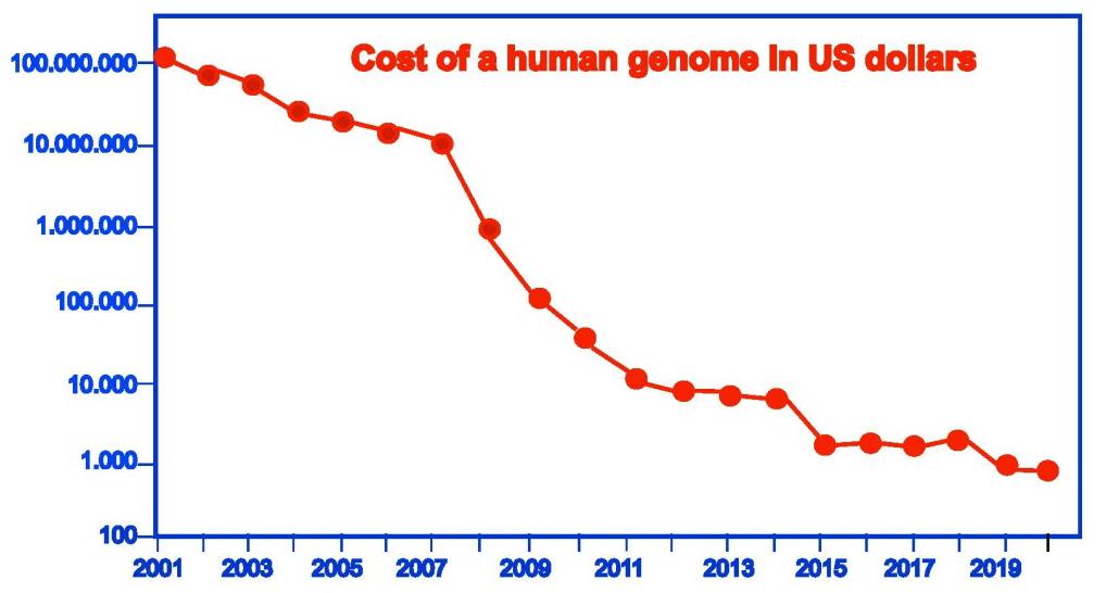
The chemical structure of DNA was formally identified in 1953 by James Watson and Francis Crick (Nobel Prize in Physiology or Medicine in 1962) as essentially consisting of a double chain formed in a specific order of four different components, the nucleotides adenine, thymine, cytosine and guanine (abbreviated as A, T, C, G) (Watson and Crick 1953). However, it was not until 1977 that a reasonably easy method for determining the sequence of this DNA appeared, the so-called Sanger method, which also earned its discoverer the Nobel Prize in 1980 (Sanger et al. 1977). However, this method could only determine the sequence of short portions of DNA (a few hundred nucleotides) and the sequencing of a long DNA chain therefore required the analysis of multiple partially overlapping segments, which was time-consuming and laborious. More sophisticated and rapid methods have developed over the years, however, and these advances still continue today (Heather and Chain 2016; Shendure et al. 2017). As a result, the speed of DNA sequencing has improved dramatically and its cost has decreased proportionately. As an example, the first complete sequencing of the human genome published in 2000 cost between $300 million and $1,000 million and involved hundreds of researchers over a period of around 13 years
(Lander et al. 2001; Venter et al. 2001). The same work can now be done in a day for about $1,000! And there is reportedly a new sequencing technique that puts the human genome at about $100 (Pennisi 2022).
Figure 1.1. The phenomenal decrease in the cost of DNA sequencing illustrated by the cost of sequencing a human genome1. For a color version of this figure, see www.iste.co.uk/balthazart/fiftyyears.zip
We have thus moved from the first-generation Sanger sequencing technique, to the second-generation techniques known as Illumina methods, and then to the third-generation methods or the Nanopore method2. Illumina sequencing techniques use reversible chain-terminating dyes to identify DNA sequences. The sample is first cleaved into short sequences that are then bound to generic adapters, and each fragment is amplified into many copies by the polymerase chain reaction or PCR technique (see end of this section). Each fragment is separated into single strands and sequenced in a medium that contains the different fluorescently labeled nucleotides, DNA polymerase, and a terminator that interrupts the PCR cycle. As a result of the terminator, only one base is added per strand to each cycle. The cycle terminator is then removed allowing the addition of the next fluorescent base. Based on the fluorescent signals emitted, a computer detects the base
1 Redrawn from data in: www.genome.gov/about-genomics/fact-sheets/Sequencing-HumanGenome-cost.
2 See www.differencebetween.com/difference-between-nanopore-and-illumina-sequencing.
that has been added in each cycle. This approach makes it possible to identify a DNA sequence in 4–56 hours, depending on the length of the chain in question. However, this approach only allows the sequencing of short strands of 100–150 base pairs, which must then be reassembled in a rather tedious process.
The Nanopore technique is based on an entirely different principle in which the passage of DNA through a very small pore (nanopore) sequentially induces a signal characteristic of each nucleotide which can therefore be recorded. In general, a flow cell has a number of nanopores that are integrated into an electro-resistive cell. Each nanopore is associated with an electrode and a sensor chip that measures in real time the current flowing through the nanopore. This current is characteristic for each nucleotide, which makes it possible to determine the sequence of the nucleic acid in real time without the need to use modified nucleotides or to split the DNA into short sequences. This process is therefore much less laborious, less expensive and much faster.
This technical progress has opened the door to scientific studies that, until recently, would have been considered science fiction. The genome of a rapidly growing number of species is now known and provides valuable information on the evolution and its mechanisms. One example is a series of studies that have sequenced the genomes of 48 species of birds representing the different major existing groups. These data have led to a better understanding of the evolution and adaptation of these birds to different lifestyles (Zhang et al. 2014), a better understanding of the evolution of their beaks from a tooth-bearing jaw (Meredith et al. 2014) or the acquisition of learned vocalizations (Pfenning et al. 2014). Subtle genetic mutations and differences between individuals are being identified and can be linked to various pathologies which allows for considerable medical progress and will lead in the near future to the development of personalized medicine. Ancient DNA can be detected and sequenced, which sheds new light on the evolution of the human species (Paabo 2015). And these are just a few of the countless examples to which these new techniques are being applied.
Multiple techniques have also been developed to analyze and quantify the expression of genes contained in this DNA. The RNA resulting from the expression of DNA (transcription) can be easily identified, amplified and
quantified in a relatively simple manner, thanks in particular to the polymerase chain reaction or PCR, technique (Mullis et al. 1986) whose name has become familiar to the general public since the Covid-19 pandemic. The set of genes expressed at a given time in an organism, an organ or even a single cell can thus be determined quantitatively and linked to the physiology of the structure. This technical approach enables an analysis of the physiology and behavior of an individual, whether healthy or suffering from a pathology, with an unprecedented precision. This leads to considerable progress in both fundamental biology and medical research.
1.4. Controlled modification of gene expression
Advances in molecular biology have naturally made it possible to specifically study the physiological role of genes identified by sequencing whose transcription, and therefore, the concentration of the corresponding RNA) is correlated with the expression of a specific behavior. This approach has, of course, also been developed in all other fields of biology and medicine. A technique was first developed that allowed the expression of a gene and thus of the corresponding protein to be completely inactivated in mice (Capecchi 1989). This gave rise to numerous knock-out (KO) mouse lines which made it possible to specify the physiological role of a given protein, for example, the estradiol receptor (Lubahn et al. 1993; Krege et al. 1998) or aromatase, the enzyme that controls the conversion of testosterone to estradiol (Fisher et al. 1998; Honda et al. 1998).
By 1980, this technique fundamentally transformed the ability of researchers to analyze the function of individual genes in mice. The National Institutes of Health (NIH) in the United States actually allocated hundreds of millions of dollars over the next two decades to generate KO mice for numerous genes resulting in the production of more than 4,000 different lines. They then realized that it was necessary to coordinate all of these efforts which led to the Knockout Mouse Project (KMOP) Repository (the archive of KO mice) which stores and distributes live mice that are KO for more than 8,500 different genes and also the corresponding engineered stem cells and frozen embryos (Lloyd 2011).
However, this method suffered from a number of practical and conceptual problems. The comparison of KO mice and control mice allows us to define functional deficits linked to the absence of expression of a specific protein, but it is often impossible to confirm the conclusions obtained by the reverse experiment, which would consist of restoring the affected function.
An exception to this problem is when the KO affects an enzyme such as aromatase. It is then possible to restore the function affected by suppressing the transformation of testosterone into estradiol by administering exogenous estradiol to the affected animal (Bakker et al. 2004).
Moreover, for practical reasons that are only partially understood, the KO technique has so far only been applied extensively to mice of a particular strain (Sv129), which limits the possibility of generalizing the results obtained, especially because these mice have a large number of peculiarities such as the absence of the corpus callosum, the main communication between the two cerebral hemispheres (Wahlsten 1982). The recent development of the technique known as CRISPR-Cas9, commonly known as molecular scissors, which allows for easy modification of a specific point in an individual’s genome (by Doudna and Charpentier (2014), Nobel Prize in Chemistry 2020) has removed this limitation by potentially creating KOs in all animal species (Doudna and Charpentier 2014).
Finally, and more embarrassingly, suppression of gene expression by KO is present from early development and throughout the life of the individual. These early deficits often induce compensatory mechanisms that mask functional deficits (see, for example, Apostolakis et al. (2002)). Moreover, it is impossible to separate effects related to developmental changes from those that specifically affect function in adults. The solution to the latter problem, however, has been found by developing conditional KOs in which the DNA of a gene has been altered so as to suppress the expression of that gene, but the suppression becomes effective only if the animal is exposed to a chemical trigger (e.g. injection of an antibiotic or tamoxifen, an anti-estrogenic compound not normally present in animals). This leads to having normally developing subjects in whom suppression of the protein of choice is induced at the desired time (Sauer and Henderson 1988; Zhang et al. 2012). More sophisticated variations of this method have also been developed that allow, for example, the expression of a protein to be
suppressed uniquely in a particular cell type, such as only those cells that express a given receptor or a given neurotransmitter (Yeh et al. 2002; Gibson and Muse 2009; Raskin et al. 2009; Naule et al. 2016). However, these approaches are not without methodological problems and require the performance of controlled manipulations that are unfortunately not always implemented (Harno et al. 2013).
1.5. Techniques for temporarily modifying the activity of neurons
In all neurosciences, and in particular in behavioral neuroendocrinology, technical progress has made it possible to manipulate the activity of neurons or groups of neurons in an increasingly precise manner in order to determine their specific role. The stereotaxic surgery techniques already mentioned make it possible to damage or administer a hormone in an anatomically precise manner to a limited group of neurons. This approach was initially used to delineate groups of cells or nuclei involved in the activation of specific behaviors. It was then rapidly combined with the use of pharmacological tools that, for example, stimulate or block specific steroid receptors or inhibit the transformation of one steroid into another, for example the aromatization of testosterone into estradiol.
Molecular biology has also come to the rescue in this endeavor by making it possible to temporarily block the synthesis of proteins suspected of being involved in the control of a physiological function or behavior. A variety of technologies based on blocking the translation of a specific RNA into the corresponding protein have been developed (McCarthy et al. 1993a). Any protein results from the transcription of a gene present in the DNA into an RNA that will itself be translated into a protein. It is possible to block or at least greatly reduce this translation by injecting an RNA fragment whose sequence is complementary to that of the targeted RNA. This injected RNA is called antisense and, by binding with the target RNA, it will prevent its translation and promote its degradation, so that the production of the corresponding protein will be temporarily inhibited until a new stock of this RNA is transcribed from the DNA. It will thus be possible to evaluate the function of the protein concerned following this blockage obtained by the injection of antisense or similar compounds (morpholinos, small inhibitory RNA or siRNA, etc.). This technique has namely been used to assess the role
of the estrogen receptor in the masculinization of rodent behavior (McCarthy et al. 1993b) or of the steroid receptor coactivator SRC1 in the activation of sexual behavior in female rats (Molenda et al. 2002) or male quail (Charlier et al. 2005).
Several other techniques have also been developed in recent years to modulate the activity of specific neurons in an even more specific way. One of them involves inducing in these cells, by genetic manipulation (e.g. via CRISPR-Cas9) or by injecting a virus containing the gene of interest, the expression of a receptor that is only activated in the presence of a compound that does not exist physiologically in the body. This type of receptor is called DREADD (Designer Receptor Exclusively Activated by Designer Drugs), a receptor created to be activated exclusively by a synthetic drug. This receptor, which belongs to a very specific class of membrane receptors, the GPCRs (or G protein coupled receptors), is then incorporated into the membrane of the target cells (the entire brain or a specific class of neurons that have been targeted by the virus), and in the presence of the synthetic drug (very often CNO or clozapine N-oxide) it will depolarize and induce the electrical activity of that cell. This DREADD technique therefore makes it possible to test the role of a specific group of neurons by rapidly inducing (a few seconds after the injection) for a relatively short time (a few minutes to an hour) their activity via injection of CNO.
An even more timely activation of a few seconds of specific groups of neurons can finally be obtained by a technique known as optogenetics, which combines light stimulation with a genetic manipulation of the content of these neurons (Boyden et al. 2005; Deisseroth et al. 2006). In this technique, the expression of a light-sensitive membrane ion channel is induced in the target neurons, by genetic manipulation or viral injection. When exposed to a specific wavelength of light, this channel can, depending on its nature, either depolarize (activate) or hyper polarize (inhibit) the cell that contains it with a temporal resolution of a few milliseconds. To study the role of these neurons in the control of a behavior, it is then possible to implant an optical fiber precisely in the area of the brain located dorsally to the site that expresses the light-sensitive channel. A point illumination with light of a precise wavelength emitted at high frequency (2–20 Hertz) by a light source connected to a computer will then make it possible to instantly activate or block the activity of the neurons expressing the light-sensitive