Visit to download the full and correct content document: https://ebookmass.com/product/endovascular-interventions-a-step-by-step-approachnov-6-2023_1119467780_wiley-blackwell-jose-wiley/
More products digital (pdf, epub, mobi) instant download maybe you interests ...
Elementary Statistics: A Step By Step Approach 10th Edition, (Ebook PDF)
All rights reserved No part of this publication may be reproduced, stored in a retrieval system, or transmitted, in any form or by any means, electronic, mechanical, photocopying, recording or otherwise, except as permitted by law. Advice on how to obtain permission to reuse material from this title is available at http://www wileycom/go/permissions
The right of Jose M Wiley, Cristina Sanina, George D Dangas, and Prakash Krishnan to be identified as the authors of the editorial material in this work has been asserted in accordance with law.
Registered Offices
John Wiley & Sons, Inc., 111 River Street, Hoboken, NJ 07030, USA
John Wiley & Sons Ltd, The Atrium, Southern Gate, Chichester, West Sussex, PO19 8SQ, UK
For details of our global editorial offices, customer services, and more information about Wiley products visit us at www.wiley.com.
Wiley also publishes its books in a variety of electronic formats and by printondemand. Some content that appears in standard print versions of this book may not be available in other formats
Trademarks: Wiley and the Wiley logo are trademarks or registered trademarks of John Wiley & Sons, Inc. and/or its affiliates in the United States and other countries and may not be used without written permission All other trademarks are the property of their respective owners John Wiley & Sons, Inc. is not associated with any product or vendor mentioned in this book.
Limit of Liability/Disclaimer of Warranty
The contents of this work are intended to further general scientific research, understanding, and discussion only and are not intended and should not be relied upon as recommending or promoting scientific method, diagnosis, or treatment by physicians for any particular patient. In view of ongoing research, equipment modifications, changes in governmental regulations, and the constant flow of information relating to the use of medicines, equipment, and devices, the reader is urged to review and evaluate the information provided in the package insert or instructions for each medicine, equipment, or device for, among other things, any changes in the instructions or indication of usage and for added warnings and precautions While the publisher and authors have used their best efforts in preparing this work, they make no representations or warranties with respect to the accuracy or completeness of the contents of this work and specifically disclaim all warranties, including without limitation any implied warranties of merchantability or fitness for a particular purpose No warranty may be created or extended by sales representatives, written sales materials, or promotional statements for this work. The fact that an organization, website, or product is referred to in this work as a citation and/or potential source of further information does not mean that the publisher and authors endorse the information or services the organization, website, or product may provide or recommendations it may make. This work is sold with the understanding that the publisher is not engaged in rendering professional services The advice and strategies contained herein may not be suitable for your situation. You should consult with a specialist where appropriate. Further, readers should be aware that websites listed in this work may have changed or disappeared between when this work was written and when it is read Neither the publisher nor authors shall be liable for any loss of profit or any other commercial damages, including but not limited to special, incidental, consequential, or other damages.
Library of Congress CataloginginPublication Data
Names: Wiley, Jose M , author | Sanina, Cristina, author | Dangas, George D , author | Krishnan, Prakash, author
Title: Endovascular interventions: a stepbystep approach / Jose M. Wiley, Cristina Sanina, George D. Dangas, Prakash Krishnan.
Description: Hoboken, NJ : WileyBlackwell, 2023 | Includes bibliographical references and index
Identifiers: LCCN 2023012153 (print) | LCCN 2023012154 (ebook) | ISBN 9781119467786 (paperback) | ISBN 9781119467847 (adobe pdf) | ISBN 9781119467861 (epub)
Department of Cardiovascular Diseases, John Ochsner Heart & Vascular Institute, The Ochsner Clinical School, University of Queensland School of Medicine New Orleans, LA, USA
Saadat Shariff, MD
Department of Cardiothoracic & Vascular Surgery (Vascular Surgery) Albert Einstein College of MedicineMontefiore Medical Center Bronx, NY, USA
Isabella Alviz, MD
Department of Medicine, Albert Einstein College of MedicineMontefiore Medical Center Bronx, NY, USA
Cornelia Rivera, MD
Department of Medicine, Albert Einstein College of MedicineMontefiore Medical Center Bronx, NY, USA
Michelle Cortorreal, MD
Department of Medicine, Albert Einstein College of MedicineMontefiore Medical Center Bronx, NY, USA
James S. Jenkins, MD
Department of Cardiovascular Diseases, John Ochsner Heart & Vascular Institute, The Ochsner Clinical School, University of Queensland School of Medicine New Orleans, LA, USA
Tamunoinemi BobManuel, MD
Department of Cardiovascular Diseases, John Ochsner Heart & Vascular Institute, The Ochsner Clinical School, University of Queensland School of Medicine New Orleans, LA, USA
Aksim G. Rivera, MD
Department of Surgery (Vascular Surgery), Albert Einstein College of MedicineJacobi Medical Center Bronx, NY, USA
Patricia Yau, MD
Department of Surgery (Vascular Surgery), Albert Einstein College of MedicineJacobi Medical Center Bronx, NY, USA
John Denesopolis, MD
Department of Surgery (Vascular Surgery), Albert Einstein College of MedicineJacobi Medical Center Bronx, NY, USA
John Futchko, MD
Department of Surgery (Vascular Surgery), Albert Einstein College of MedicineJacobi Medical Center Bronx, NY, USA
Katie MacCallum, MD
Department of Surgery (Vascular Surgery), Albert Einstein College of MedicineJacobi Medical Center Bronx, NY, USA
Mohammad Hashim Mustehsan, MD
Division of Cardiology, Albert Einstein College of MedicineMontefiore Medical Center, Bronx, NY,
Jose D. Tafur, MD
Department of Cardiovascular Diseases, John Ochsner Heart & Vascular Institute, The Ochsner Clinical School, University of Queensland School of Medicine New Orleans, LA, USA
Cristina Sanina, MD
Division of CardiologyDepartment of MedicineBeth Israel Deaconess Medical Center Harvard Medical School Boston, MA, USA
David A. Hirschl, MD
Department of Radiology, Albert Einstein College of MedicineMontefiore Medical Center Bronx, NY, USA
Michael S. Segal, DO
Department of General Surgery Wyckoff Heights Medical Center Brooklyn, NY, USA
Sameh Elrabie, DO
Department of General Surgery Wyckoff Heights Medical Center Brooklyn, NY, USA
Rajesh K. Malik, MD
Division of Vascular Surgery Wyckoff Heights Medical Center Brooklyn, NY, USA
Sahil A. Parikh, MD
Division of Cardiovascular Diseases Columbia University Irving Medical Center, New York, NY, USA
Joseph J. Ingrassia, MD
Division of Cardiovascular Diseases Columbia University Irving Medical Center, New York, NY, USA
Matthew T. Finn, MD
Division of Cardiovascular Diseases Columbia University Irving Medical Center, New York, NY, USA
Raman Sharma, MD
Division of Cardiology, The Zena and Michael A. Weiner Cardiovascular Institute, Icahn School of Medicine at Mount Sinai, New York, NY, USA
Prakash Krishnan, MD
Division of Cardiology, The Zena and Michael A. Weiner Cardiovascular Institute, Icahn School of Medicine at Mount Sinai, New York, NY, USA
Roberto CerrudRodriguez, MD
Division of Cardiology, Albert Einstein College of MedicineMontefiore Medical Center, Bronx, NY, USA
Shunsuke Aoi, MD
Division of Cardiology, Albert Einstein College of MedicineMontefiore Medical Center, Bronx, NY, USA
Amit M. Kakkar, MD
Division of Cardiology, Albert Einstein College of MedicineJacobi Medical Center, Bronx, NY, USA
Ehrin J. Armstrong, MD
University of Colorado School of MedicineRocky Mountain Regional VAMedical Center, CO, USA
Rory Brinker, MD
University of Colorado School of MedicineRocky Mountain Regional VAMedical Center, CO, USA
Manaf Assafin, MD
Division of Cardiology, Albert Einstein College of MedicineMontefiore Medical Center, Bronx, NY, USA
Miguel AlvarezVillela, MD
Division of Cardiology, Albert Einstein College of MedicineMontefiore Medical Center, Bronx, NY, USA
Robert Pyo, MD
Division of Cardiology, Renaissance School of Medicine at Stony Brook University, NY, USA
Pedro CoxAlomar, MD
Division of Cardiology, Louisiana State University School of Medicine New Orleans, LA, USA
Vishal Kapur, MD
Division of Cardiology, The Zena and Michael A. Weiner Cardiovascular Institute, Icahn School of Medicine at Mount Sinai, New York, NY, USA
Sagar Goyal, MD
Division of Cardiology, The Zena and Michael A. Weiner Cardiovascular Institute, Icahn School of Medicine at Mount Sinai, New York, NY, USA
Asma Khaliq, MD
Department of Cardiology, Lenox Hill Heart & Vascular Institute Donald and Barbara Zucker School of Medicine at Hofstra/Northwell Health New York, NY, USA
Sandrine Labrune, MD
Department of Cardiology, Lenox Hill Heart & Vascular Institute Donald and Barbara Zucker School of Medicine at Hofstra/Northwell Health New York, NY, USA
Seth I. Sokol, MD
Division of Cardiovascular Diseases, Albert Einstein College of MedicineJacobi Medical Center Bronx, NY, USA
Wissam A. Jaber, MD
Division of Cardiology, Emory University Hospital, Atlanta, GA, USA
Yosef Golowa, MD
Department of Radiology, Albert Einstein College of MedicineMontefiore Medical Center Bronx, NY, USA
Juan Terre, MD
Division of Cardiology, Albert Einstein College of MedicineMontefiore Medical Center, Bronx, NY, USA
Nelson Chavarria, MD, MSc
Division of Cardiology, Albert Einstein College of MedicineMontefiore Medical Center, Bronx, NY, USA
Jose M. Wiley, MD, MPH
Section of Cardiology John W. Deming Department of Medicine Tulane University School of Medicine New Orleans, LA, USA
George D. Dangas, MD, PhD
Division of Cardiology, The Zena and Michael A. Weiner Cardiovascular Institute, Icahn School of
Innominate & Carotid Artery Intervention in HighRisk Patients
Tyrone J. Collins
Department of Cardiovascular Diseases, John Ochsner Heart & Vascular Institute, The Ochsner Clinical School, University of Queensland School of Medicine, New Orleans, LA, USA
Introduction
Revascularization of supraaortic arterial disease (complicated peripheral artery disease) is usually elective and prophylactic to prevent initial or recurrent ischemic events. Surgical revascularization was once considered the treatment of choice [1]. Successful reports of percutaneous transluminal angioplasty (PTA) and stenting introduced endovascular treatment as an equal or possibly better than surgery option [2]. Each patient is unique, and the risk is multifactorial with both demographic and anatomic risk factors. Several “highrisk” features are generally considered when treating carotid artery disease in these patients [3] (Table 1.1). Some of these features are also risk factors for innominate intervention. The level of stenosis and/or occlusion, vessel tortuosity, amount of calcification, presence or absence of thrombus, concomitant vascular abnormalities, and comorbid conditions will also affect the risk with revascularization of the other supraaortic vessels.
Although some authors may consider endovascular therapy the treatment of choice for innominate atherosclerotic disease, surgical therapy has been shown to be safe and effective [4]. During a period of almost 20 years from1974 to 1993, Kieffer et al. revascularized surgically 148 patients with acceptable rates of complications, late mortality, longtermpatency, freedomfromneurologic events, and reoperation [4].
Table 1.1 Highrisk features reported in the literature.
1. CAS in females
2. CAS in octogenarians
3. CAS with type II, type III, or bovine arch
4. Tortuous common carotid artery, angulated ICA, and/or distal ICA
5. Long lesions ≥15 mm
6. Ostialcentered lesions
7. Calcified arch and/or heavily calcified lesion
8. Highgrade stenosis
9. Contralateral carotid occlusion
10. Presence of vertebral artery occlusion and/or stenosis
11. Patient with CKD
Innominate Interventions in HighRisk Patients
Catheterbased Therapyfor An Innominate (Brachiocephalic) Stenosis
Step 1. Identification of the level of stenosis is the initial step. Computed tomography angiography (CTA) can be useful prior to an invasive procedure. This can allow for planning the interventional strategy and considering alternative forms of treatment. Additionally, CTAcan be used to size the reference vessels.
When considering the choice of arterial access remember that catheter size is limited with radial access and the need to cross the stenosis is usually necessary fromthe radial or brachial approach. If intervention is planned, injections are against the direction of blood flow when working fromthe armapproach. I prefer the femoral approach to innominate stenoses.
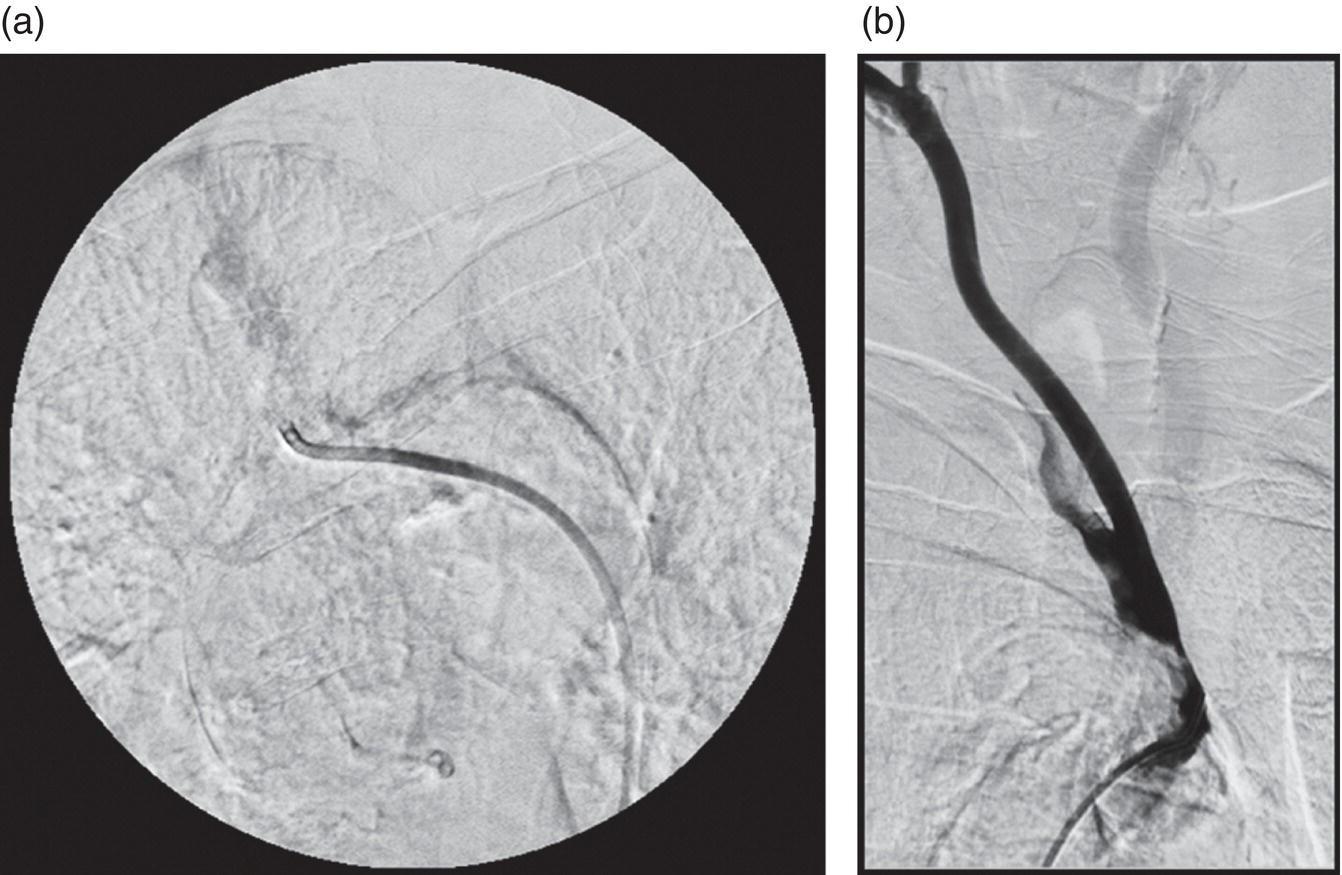
Invasive angiography can be done with digital and/or subtraction angiography. Apigtail catheter is positioned in the ascending aorta proximal to the origin of the innominate artery. The angiography is performed in the 30° left anterior oblique (LAO) projection. Selective angiography is done with a Judkins right diagnostic catheter or guiding catheter (Figure 1.1a,b). Other diagnostic catheters can be used for selective angiography. The “working view” is the angulation that allows for delineation of the stenosis, any adjacent branches, and the ostiumof the innominate. Road mapping may be useful but also take advantage of any vascular calcification as a point of reference.
Step 2. After the decision to intervene and baseline angiography has been performed, the innominate is engaged with an 8 Fr guide catheter. Adifferent approach is to use a diagnostic catheter to engage the innominate artery, cross the stenosis with the appropriate wire, and introduce a 6 Fr sheath over the wire to the ostiumof the innominate. Anticoagulation to achieve an activated clotting time (ACT) > 250 s is administered. Depending on the available balloons and stents, the appropriate wire (0.014–0.035 in.) is steered across the stenosis. The tip of the wire is passed into the subclavian artery. Wire tip can also be placed in the common or external carotid artery. Innominate artery PTAand stenting is usually performed without utilizing a distal embolic protection device (EPD). If you choose to use EPD, the necessary wire or filter can be positioned in the internal carotid artery. Horesh reported a case of innominate stenting with a covered stent and distal protection [5]. He emphasized the need to individualize patients and consider using embolic protection in highrisk patients. Hybrid procedures have been performed using balloon occlusion to trap embolic debris.
Step 3. Predilatation with a balloon is performed. The initial balloon is usually undersized but gives an idea of the ability to distend the lesion (Figure 1.2). The Shockwave Lithoplasty System(Medical Inc.) has been used to successfully treat severely calcified innominate stenosis prior to stenting [6]. This systemhas also been used in a hybrid operation [7]. Use the balloon inflation to help decide on stent sizing (diameter and length).
Step 4. Stent implantation is done after ensuring the correct position of the delivery system(Figure 1.3). If necessary, magnify the image to demonstrate the stent is appropriately placed. Remember, an undersized stent can be implanted so that the delivery sheath or catheter does not have to be “upsized.” Alarger balloon (Figure 1.4) can subsequently be employed to adequately expand the stent without changing the sheath or catheter.
1.2 Predilatation with undersized balloon.
Figure
Figure 1.3 Stent in position at ostiumof innominate.
Step 5. Assessment of the poststent result is performed to determine stent apposition and size (Figure 1.5). If necessary, the stent can be postdilated with a larger balloon.
Step 6. After hemostasis the patient is usually monitored overnight and discharged the following day. Dual antiplatelet therapy is maintained for at least one month if there are no contraindications.
Figure 1.4 Larger balloon inflation.
Figure 1.5 Final angiogram.
Carotid Artery Intervention in HighRisk Patients
Endovascular Treatment of A Carotid Stenosis
Left common carotid artery stenoses are treated endovascularly similarly to innominate artery stenoses. Distal embolic protection is not used routinely. There are endovascular, hybrid, and surgical alternatives.
Transcarotid artery revascularization (TCAR) offers alternative to both carotid endarterectomy (CEA) and carotid artery stenting (CAS) which are done via a transfemoral approach.
CAS can be performed with distal embolic protection and/or flow reversal. Distal embolic protection is the most commonly used choice. It is readily available and technically easier to deploy. However, it is not the best choice for tortuous common and/or internal carotid arteries, heavily calcified vessels, and “string signs.” Distal protection devices require crossing the diseased segment without protection compared to proximal protection where this is not necessary. Additionally, if anatomy warrants, CEAcan be the treatment of choice.
Catheterbased Therapyfor Carotid Stenosis
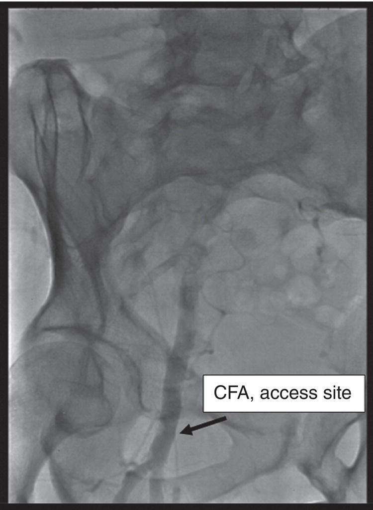
Step 1. Arterial access is obtained for distal embolic protection and flow reversal cases. Distal EPD can be done via femoral, radial, or brachial access. Flow reversal, because of the larger diameter sheath required, is performed via the femoral artery route. Access is obtained with ultrasound guidance or using anatomic landmarks. Femoral angiography is usually performed at the initiation of the case to document the appropriateness of the access and to plan for use of a closure device (Figure 1.6).
Figure 1.6 Femoral artery access.
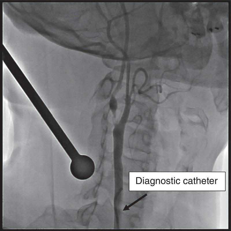
Figure 1.7 Baseline selective angiography with reference object.
Step 2. Selective carotid angiography (Figure 1.7) of the culprit vessel is performed with a diagnostic catheter. The best angle to visualize the lesion is chosen. Quantitative angiography is done. This can be with a reference object placed at the level of the lesion or with online software. Of note, angiography of all the arch and intracranial vessels is performed prior to intervention. This can be done during the CAS procedure or at an earlier date. Other imaging modalities (CTAor MRI) can be done prior to the CAS. Imaging of the intracranial circulation is necessary in the event that the rare occurrence of neurorescue is needed and it is necessary to document the baseline anatomy.
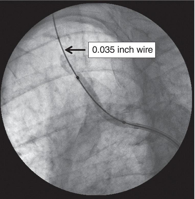
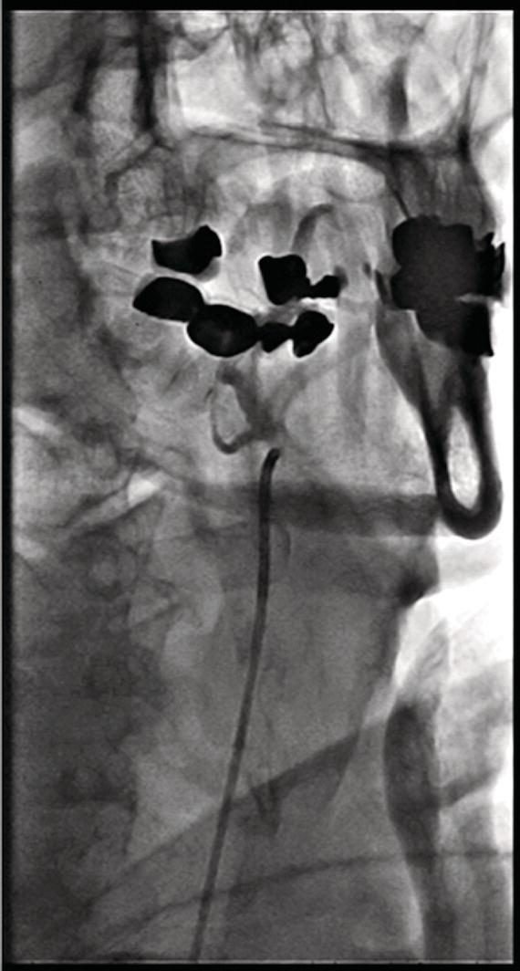
Step 3. Heparin is administered to achieve an ACT greater than 250 s. Asheath, guide catheter, or neuroprotection device is exchanged over a stiff wire (exchange length 0.035 in. Amplatz wire). The tip is
placed in the common carotid artery. During the exchange procedure, the tip of the wire is positioned in the common carotid or external carotid artery. For the flow reversal case, it is necessary to place the stiff wire in the external carotid artery (ECA) through the diagnostic catheter (Figure 1.8) before the exchange procedure.
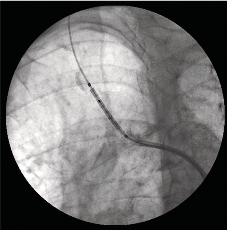
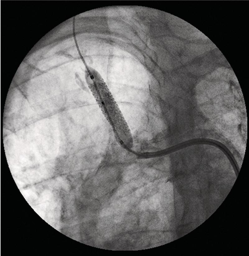
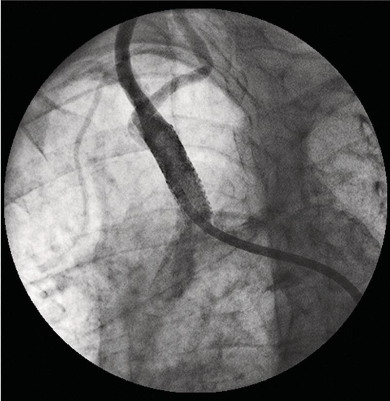
Step 4. This is where the proximal and distal protection significantly differ. With distal protection, a device is passed through the lesion into the internal carotid artery (Figure 1.9). The EPD is deployed. Balloon predilatation (Figures 1.10) is usually done with a smaller than the reference vessel sized balloon (generally 2.5–3 mm). Astent is deployed (Figure 1.11) and usually postdilatation is skipped. Completion angiography is done (Figure 1.12) along with the intracranial images. Hemostasis can be obtained with a closure device of choice. Generally, these patients are observed in the hospital overnight.