Visit to download the full and correct content document: https://ebookmass.com/product/lippincott-illustrated-reviews-flash-cards-biochemistrylippincott-illustrated-reviews-series-1st-edition-ebook-pdf/
More products digital (pdf, epub, mobi) instant download maybe you interests ...
The authors wish to thank John Swaney, PhD, our colleague at Drexel University College of Medicine, for his careful reading of the manuscript and constructive comments. Any errors are ours alone.
We thank the publishing team assembled by Wolters Kluwer. Stephanie Roulias, product development editor, and Kelly Horvath, freelance development editor, along with Doug Smock, Teresa Exley, and David Orzechowski, gave invaluable assistance in the development and production of the finished product. We also thank Robin R. Preston, PhD, for his design of the flash card format.
Dedication
The authors dedicate this work to the medical, biomedical graduate, and professional studies students of Drexel University. You have challenged and inspired us, and have made us better teachers.
Card 3.6 Question and Answer: Modified photo courtesy of Photodyne Incorporated, Hartland, WI.
Card 4.2 Answer: Kronauer and Buhler, Images in Clinical Medicine, The New England Journal of Medicine, June 15, 1995, Vol. 332, No. 24, p. 1611.
Card 4.5 Question and Answer: 1. Modified photo from Web site Derma.de. 2. Modified
Figure Credits
from Jorde LB, Carey JC, Bamshad MJ, et al. Medical Genetics. 2nd ed. St. Louis, MO: Mosby; 2000. http://medgen.genetics. utah.edu/index.htm
Card 13.6 Answer: From the Crookston Collection, University of Toronto.
Card 21.4 Question and Answer: From Custom Medical School Stock Photo, Inc.
Card 22 Case Card Question: Modified from WebMD Inc. http://www.samed.com/sam/ forms/index.htm.
Card 23.6 Question and Answer: Modified from Cryer PE, Fisher JN, Shamoon H. Hypoglycemia. Diabetes Care. 1994;17: 734–753.
1.1 Question Amino Acid Structure
What effect will raising pH from an acidic value to the physiologic value of 7.4 have on the structural features shown in red at right?
Free amino acid
At physiologic pH, what will be the charge on the side chain (R group) of free Asp? Of Lys?
These are common to all `-amino acids.
Which amino acid(s) contains a side-chain hydroxyl group that can be glycosylated?
A secondary amino group?
Is Val ionized when incorporated into a protein?
H ` Amino group
Carboxyl group
Side chain is distinctive for each amino acid. `-Carbon is linked to the carboxyl, amino, and R groups.
1.1 Answer Amino Acid Structure
Raising the pH from an acidic value to the physiologic value of 7.4 will result in deprotonation (ionization) of the -carboxyl group (pK 2) to COO . The -amino group (pK 9) will remain protonated.
At physiologic pH, the charge on the side chain (R group) of free Asp is negative. Lys is positive.
Ser and Thr each contain a hydroxyl group that can be O-glycosylated. [Note: The hydroxyl group can also be phosphorylated.] Pro contains a secondary amino group. Its -amino N and R group form a rigid ring.
Val is not ionized when incorporated into a protein because (1) the -amino and -carboxyl groups are involved in peptide bonds and, consequently, are unavailable for ionization, and (2) the side chain is nonpolar.
These are common to all `-amino acids.
Side chain is distinctive for each amino acid. `-Carbon is linked to the carboxyl, amino, and R groups.
1.2 Question Amino Acid Structure
Based on the figure, where would Leu likely be located in a protein that spans the membrane? In a soluble protein? What term refers to the tendency of nonpolar molecules (or regions of molecules such as amino acid side chains) to cluster together in a polar environment such as an aqueous solution?
Nonpolar amino acids ( ) cluster in the interior of soluble proteins.
In sickle cell anemia (SCA), why does the replacement of a Glu by a Val on the surface of the deoxyHb molecule result in the association of these molecules?
Polar amino acids ( ) cluster on the surface of soluble proteins.
Nonpolar amino acids ( ) cluster on the surface of membrane proteins.
membrane
Cell C l
Soluble proteinMembrane protein
Amino Acid Structure 1.2
Leu, a nonpolar amino acid, would likely be located within the hydrophobic membrane-spanning domain of the protein. It would likely be located in the interior of a soluble protein.
The term hydrophobic effect refers to the tendency of nonpolar molecules (or regions of molecules such as amino acid side chains) to cluster together in a polar environment such as an aqueous solution.
The replacement of polar Glu by nonpolar Val creates a hydrophobic region on the surface of the deoxyHb molecule that will interact with a hydrophobic region on other deoxyHb molecules. This interaction creates rigid polymers of deoxyHb that deform RBCs. Thus, it is the hydrophobic effect that drives the association of deoxyHb molecules in SCA.
Nonpolar amino acids ( ) cluster in the interior of soluble proteins.
Nonpolar amino acids ( ) cluster on the surface of membrane proteins.
Polar amino acids ( ) cluster on the surface of soluble proteins.
Which structure shown (A or B) represents L-Ala?
1.3 Question Amino Acid Structure
Which amino acid does not possess a chiral (asymmetric) carbon?
Which peptide is less soluble in an aqueous (polar) environment, Ala-Gly-Asn-Ser-Tyr or Gly-Met-Phe-Leu-Ala?
1.3 Answer Amino Acid Structure
Structure A represents L-Ala. The L isomer of an amino acid has the -amino group on the left. The D isomer has the -amino group on the right. D and L isomers are mirror images of each other (enantiomers).
Gly, with its two H substituents, does not possess a chiral (asymmetric) carbon.
Because the Gly-Met-Phe-Leu-Ala peptide contains no charged or polar uncharged amino acids, it is less soluble than Ala-Gly-Asn-Ser-Tyr in an aqueous (polar) environment.
Acidic and Basic Properties of Amino Acids
What relationship is described by the Henderson–Hasselbalch equation shown?
Is an acid with a large pKa stronger or weaker than one with a small pKa?
The pKa of acetic acid (CH3COOH) is 4.8. What is the pH of a solution containing acetic acid and its conjugate base (CH3COO ) in a ratio of 10 to 1?
Physiologic buffers are important in resisting blood pH changes. Maximal buffering occurs when the pH is equal to the , while effective buffering can occur within .
1.4 Answer Acidic and Basic Properties of Amino Acids
The Henderson–Hasselbalch equation describes the relationship between the pH of a solution and the concentration of a weak acid [HA] and its conjugate base [A ].
An acid with a large pKa is weaker than one with a small pKa because the large pKa reflects less ionization (fewer H released). This is because pKa log Ka.
Because pH pKa log [A ]/[HA], when pKa is 4.8 and the ratio of the acid to its conjugate base is 10 to 1, the pH is equal to 4.8 log of 0.1. Therefore, pH 4.8 ( 1) 3.8.
Physiologic buffers are important in resisting blood pH changes. Maximal buffering occurs when the pH is equal to the pKa, while effective buffering can occur within 1 pH unit of the pKa.
Acidic and Basic Properties of Amino Acids
Which FORM (I, II, or III) shown represents the isoelectric form of Ala?
Calculate the pI for Arg, which has three pKs: pK1 2.2, pK2 9.2, and pK3 12.5.
What will happen to the charge on His residues in a protein that moves from the cytoplasm (pH 7.4) to a lysosome (pH 5.0)?
1.5 Answer Acidic and Basic Properties of Amino Acids
= 2.3
The isoelectric form has no net charge. It is the zwitterionic (“two ion”) form. Therefore, FORM II is the isoelectric form of Ala.
The pI corresponds to the pH at which an amino acid is electrically neutral, that is, the average of the pKs on either side of the isoelectric form. For Arg, a dibasic amino acid with pK1 (most acidic group) 2.2, pK2 9.2, and pK3 (least acidic group) 12.5, the pI is 10.8 (the average of 9.2 and 12.5).
In a protein, the imidazole R group of His can be charged or uncharged depending on the local environment. It will be uncharged (deprotonated) at pH 7.4 and charged (protonated) at pH 5.0. [Note: In free His the pK of the R group is 6.0.]
Question Acidic and Basic Properties of Amino Acids
Based on the bicarbonate buffer system shown, what will happen to the availability of HCO3 when H is lost, such as with emesis (vomiting)?
Use the Henderson–Hasselbalch equation to determine what will happen to pH when HCO3 is lost (e.g., with diarrhea) and when CO2 is increased (e.g., with pulmonary obstruction).
Aspirin (pKa 3.5) is largely protonated and uncharged in the stomach (pH 1.5). What percentage of the aspirin will be in this lipid-soluble form at pH 1.5?
1.6 Answer Acidic and Basic Properties of Amino Acids
With emesis (vomiting), the loss of H (rise in pH) results in increased availability of HCO3 as the result of a compensatory rightward shift in the bicarbonate buffer system.
The Henderson–Hasselbalch equation is used to calculate how the pH of a system changes in response to changes in the concentration of an acid or its conjugate base. For the bicarbonate buffer system, pH pK log [HCO3 ]/[CO2]. Therefore, both the loss of HCO3 (base) with diarrhea and the increase in CO2 (acid) because of decreased elimination with pulmonary obstruction result in decreased pH.
pH pK log [Drug ]/[Drug-H]. Therefore, for aspirin in the stomach, 1.5 3.5 ( 2). Because the antilog of 2 is 0.01, the ratio of [Drug ]/[Drug-H] is 1/100. This means that 1 out of 100 (1%) of the aspirin molecules will be the Drug form and 99 out of 100 (99%) will be the uncharged, lipid-soluble, Drug-H form.
At the pH of the stomach (1.5), a drug like aspirin (weak acid, pK = 3.5) will be largely protonated (COOH) and, thus, uncharged.
Uncharged drugs generally cross membranes more rapidly than do charged molecules.
2.1 Question Protein Structure
Which level of protein structure depicted can be correctly described as the “three-dimensional shape of a folded polypeptide chain”?
Mutations that insert, delete, or replace amino acids change this level of protein structure.
How many different isoforms of the tetrameric enzyme PK can be made from M and/or L subunits?
How many different tetrapeptides could be generated from three different amino acids?
2.1 Answer Protein Structure
The “three-dimensional shape of a folded polypeptide chain” describes a protein’s tertiary structure (No. 3 shown).
At a minimum, the primary structure (amino acid sequence) will change with mutations that insert, delete, or replace amino acids. [Note: Changes in the primary structure can also affect the higher levels of protein structure (No. 2 to 4 shown). Such changes frequently result in protein misfolding and can lead to loss of function, aggregation, or degradation.]
Five different forms of tetrameric PK can be made from M and/or L subunits: M4, M3L, M2L2, ML3, and L4. Because PK is composed of more than one subunit, it has a quaternary structure.
There are 34 or 81 (where 3 the number of amino acids and 4 the chain length) different tetrapeptides that could be generated from three different amino acids.
Primary Structure of Proteins
What is the name given to the bond outlined by the black box shown?
What are the characteristics of this bond?
With fever, why might proteins begin to unfold but not be hydrolyzed to peptides and free amino acids?
amino end of peptide
2.2 Question
Free carboxyl end of peptide
2.2 Answer Primary Structure of Proteins
A peptide bond, a type of amide bond, is outlined by the black box. Peptide bonds link the amino acid residues in a peptide or protein by joining the -amino group of one amino acid to the -carboxyl group of the next as water is released.
The peptide bond has partial double-bond character, is rigid and planar, uncharged but polar, and almost always in the trans configuration that reduces steric interference by the R groups.
Peptide bonds are resistant to conditions (such as the heat from a fever) that can denature proteins and cause them to unfold. However, they are susceptible to cleavage by enzymes known as proteases or peptidases. [Note: Strong acids or bases at high temperatures can nonenzymatically cleave peptide bonds.]
2.3 Question Primary Structure of Proteins
Sequencing large polypeptides involves cleavage reactions, as shown. Which sites in a peptide are susceptible to cleavage by the endopeptidase trypsin? By cyanogen bromide?
Peptide of unknown sequence
What is the Edman degradation method?
What is the amino acid sequence of a nonapeptide if trypsin digestion yields three products (Asn, Met-Gln-Lys, and Ala-Gly-Met-Leu-Arg) and cyanogen bromide cleavage yields three products (Leu-Arg-Met, Gln-Lys-Asn, and Ala-Gly-Met)?
2. Determine sequence of peptides using the Edman method
What is the correct order?
Peptide of unknown sequence
1. Cleave with cyanogen bromide
2. Determine sequence of peptides using the Edman method
1. Cleave with trypsin
Peptide B
Peptide A
Peptide XPeptide Y
Peptide C
2.3 Answer Primary Structure of Proteins
Trypsin, an endopeptidase, cleaves at the carboxyl side of Lys and Arg residues within a peptide.
[Note: Exopeptidases remove the terminal amino acid.] Cyanogen bromide cleaves at the carboxyl side of Met residues.
The Edman degradation method chemically determines the sequence of amino acids through the sequential removal and identification of the N-terminal amino acids in the small peptides generated from a polypeptide by cleavage reactions.
Based on the overlapping amino acids in the products of the trypsin (Asn, Met-Gln-Lys, and Ala-Gly-Met-Leu-Arg) and the cyanogen bromide (Leu-Arg-Met, Gln-Lys-Asn, and Ala-Gly-Met) cleavage reactions, the amino acid sequence of the nonapeptide is Ala-Gly-Met-Leu-Arg-Met-Gln-Lys-Asn.
[Note: The sequence of amino acids in a protein is always written from the N-terminal to the C-terminal amino acid.]
Peptide of unknown sequence
1. Cleave with trypsin at lysine and arginine
2. Determine sequence of peptides using the Edman method
What is the correct order?
Peptide of unknown sequence
1. Cleave with cyanogen bromide at methionine
2. Determine sequence of peptides using the Edman method
Original sequence of
Peptide B Peptide A
Peptide XPeptide Y
Peptide C
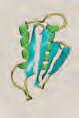
2.4 Question Secondary Structure of Proteins
Which type of secondary structure is illustrated at right?
How does the orientation of the hydrogen bonds differ between the -helix and the -sheet structures?
In proteins (e.g., the GPCRs for glucagon and the catecholamines) that contain several -helical membrane-spanning domains, why would Pro not be one of the amino acids found in these domains?
2.4 Answer Secondary Structure of Proteins
The figure illustrates an -helix, a right-handed, helical, secondary structural element commonly encountered in both fibrous and globular proteins.
The hydrogen bonds in a coiled -helix are intrachain bonds that are parallel to the polypeptide backbone, whereas those in a -sheet (an extended structure) can be intra- or interchain bonds (depending on whether they form between sections of one polypeptide or between two polypeptides) that are perpendicular to the backbone. [Note: -Helices and -sheets may be components of supersecondary structures (motifs), such as a -barrel.]
Pro contains a secondary amino group that is not compatible with the right-handed spiral of the -helix because (1) it cannot participate in the hydrogen bonding and (2) it causes a kink in the protein. Consequently, Pro is not found in the membrane-spanning domains of proteins such as GPCRs. [Note: Amino acids with bulky or charged R groups can also disrupt formation of an -helix.]
2.5 Question Tertiary Structure of Proteins
What type of molecular interaction involved in stabilizing the tertiary structure of a protein is shown?
What type of interaction would likely occur between Asp and Lys?
The tertiary structures of proteins (such as albumin) that function in the extracellular environment are stabilized by the formation of covalent links between the oxidized side chains of which sulfur-containing amino acid(s)?
2.5 Answer Tertiary Structure of Proteins
Shown are hydrophobic interactions between Ile and Leu, two amino acids with nonpolar R groups.
Ionic interactions (salt bridges) would likely occur between Asp (acidic R group) and Lys (basic R group).
Two sulfur-containing Cys residues, brought into close proximity by the folding of the peptide(s), are covalently linked through oxidation of their thiol side chains. The disulfide bonds formed stabilize the tertiary structure of the folded peptide, preventing it from becoming denatured in the oxidizing extracellular environment. [Note: Cys-containing albumin transports hydrophobic molecules (e.g., fatty acids and bilirubin) in the blood. Its levels are used as an indicator of nutritional status.]
2.6 Question Protein Misfolding
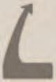
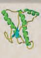
As illustrated, what secondary structural feature is enriched in the infectious form of a prion protein (PrP) as compared to the noninfectious form?
Why do most large denatured proteins not revert to their native conformations even under favorable environmental conditions?
What misfolded peptide formed by abnormal proteolytic cleavage is the dominant component of the plaque that accumulates in the brains of individuals with Alzheimer disease?
Interaction of the infectious PrP molecule with a normal PrP causes the normal form to fold into the infectious form.
Noninfectious PrPC
2.6 Answer Protein Misfolding
The -sheet secondary structure is enriched in the infectious PrPSc form of a PrP, which causes the transmissible spongiform encephalopathies, as compared to the noninfectious PrPC form that is -helical rich.
The folding of most large proteins is a facilitated process that requires the assistance of proteins known as chaperones and ATP hydrolysis.
A is the misfolded peptide produced by abnormal proteolytic cleavage of amyloid precursor protein by secretases. A forms an extended -sheet and spontaneously aggregates to form fibrils that are the dominant component of the amyloid plaque that accumulates in the brains of individuals with Alzheimer disease. [Note: The -sheets in A have exposed hydrophobic amino acid residues. The hydrophobic effect drives the aggregation and precipitation of A .]
Interaction of the infectious PrP molecule with a normal PrP causes the normal form to fold into the infectious form.
Infectious PrPSc (contains a-sheets)
Infectious PrPSc (contains a-sheets)
Noninfectious PrPC (contains `-helix)
Cell membrane
Amyloid
Spontaneous aggregation to form insoluble fibrils of a-pleated sheets