For more information about Scrivener publications please visit www.scrivenerpublishing.com.
All rights reserved. No part of this publication may be reproduced, stored in a retrieval system, or transmitted, in any form or by any means, electronic, mechanical, photocopying, recording, or otherwise, except as permitted by law. Advice on how to obtain permission to reuse material from this title is available at http://www.wiley.com/go/permissions.
Wiley Global Headquarters
111 River Street, Hoboken, NJ 07030, USA
For details of our global editorial offices, customer services, and more information about Wiley products visit us at www.wiley.com.
Limit of Liability/Disclaimer of Warranty
While the publisher and authors have used their best efforts in preparing this work, they make no representations or warranties with respect to the accuracy or completeness of the contents of this work and specifically disclaim all warranties, including without limitation any implied warranties of merchantability or fitness for a particular purpose. No warranty may be created or extended by sales representatives, written sales materials, or promotional statements for this work. The fact that an organization, website, or product is referred to in this work as a citation and/or potential source of further information does not mean that the publisher and authors endorse the information or services the organization, website, or product may provide or recommendations it may make. This work is sold with the understanding that the publisher is not engaged in rendering professional services. The advice and strategies contained herein may not be suitable for your situation. You should consult with a specialist where appropriate. Neither the publisher nor authors shall be liable for any loss of profit or any other commercial damages, including but not limited to special, incidental, consequential, or other damages. Further, readers should be aware that websites listed in this work may have changed or disappeared between when this work was written and when it is read.
Library of Congress Cataloging-in-Publication Data
ISBN 978-1-119-85720-4
Cover image: Pixabay.Com
Cover design by Russell Richardson
Set in size of 11pt and Minion Pro by Manila Typesetting Company, Makati, Philippines
5
T. Graceshalini, S. Rathnamala and M. Prabhanantha
5.1
5.2
5.2.1
5.3
5.3.1.2
5.3.1.3
5.4
5.5
M. Menagadevi , S. Mangai, S. Sudha and
6.1.1
6.1.3
6.2
6.4
6.5
6.5.1
6.9
6.7.1
6.7.2
8
7.4
9
S. Vairaprakash and S. Rajagopal
8.3.3
8.3.4
8.4
9.1
Rajdeep Ghosh, Nidul Sinha and Souvik
B. Paulchamy, R. Uma Maheshwari, D. Sudarvizhi AP(Sr. G), R. Anandkumar AP(Sr. G) and Ravi G.
Preface
The Brain-Computer Interface (BCI) is an emerging technology that is developing to be more functional in practice. The aim is to establish, through experiences with electronic devices, a communication channel bridging the human neural networks within the brain to the external world. For example, creating communication or control applications for locked-in patients who have no control over their bodies will be one such use. Recently, from communication to marketing, recovery, care, mental state monitoring, and entertainment, the possible application areas have been expanding. Machine learning algorithms have advanced BCI technology in the last few decades, and in the sense of classification accuracy, performance standards have been greatly improved. For BCI to be effective in the real-world, however some problems remain to be solved.
The book provides the reader with the fundamental theories, concepts, and methods in neuroscience, brain recording and stimulation technologies, signal processing, and machine learning. Readers have the chance to review their knowledge and assess their comprehension of the subjects presented in each chapter with exercises and questions at the end of each chapter. Some assignments provide the student the option to explore topics outside of those covered in the textbook by looking for new information online and following leads in research articles. Highlighting most of the research directions in the digital world, this book is more suitable for researchers from biomedical background, data analysts, AI researchers, machine and deep learning engineers, students and academicians.
The book is organized as follows: In chapters 1 and 2 provides an introduction to Brain–Computer Interface: Applications and Challenges. Chapter 3 discusses the statistical learning of brain compute interface was discussed. Chapter 4 begins with the impact of brain computer interface on the lifestyle of elderly people. Chapter 5 reviews the innovation to human augmentation in brain computer interface and its potential limitations in artificial intelligence. Chapter 6 details the Resting-State fMRI: large data analysis in neuro imaging. Chapter 7 describes early detection of
Preface
epileptic seizure using deep learning algorithms. Chapter 8 describes the application of brain computer interface based on the real time upper limb protheses to improve the quality of the elderly. Chapter 9 describes another application of brain computer interface to assisted automated wheelchair control management. Chapter 10 shows the application of convolutional neural network to identify Bengali vowels from EEG signal using activation map. Chapter 11 discusses the optimized feature selection techniques for classifying electrocorticography signals. Chapter 12 reviews some of the challenges, application and advancements in brain computer interface. The editors thank all contributors for their time and effort and have collectively delivered high quality work.
The Editors December 2022
1 Introduction to Brain–Computer Interface: Applications and Challenges
Jyoti R. Munavalli1*, Priya R. Sankpal1, Sumathi A.1 and Jayashree M. Oli2
1ECE, BNM Institute of Technology, Bangalore, India
2Amrita School of Engineering, ECE, Bengaluru, Amrita Vishwa Vidyapeetham, India
Abstract
Brain–Computer Interface (BCI) is a technology that facilitates the communication between the brain and the machine. It is a promising field that has lot of potential to be tapped for various applications. To begin with, this chapter explains the basics of the brain and its function. It describes the BCI technology and the steps: from signal acquisition to applications. The signal capturing is done through invasive and non-invasive methods. The features from the brain signals are extracted and classified using various advanced machine learning classification algorithms. BCI is extensively helpful for health-related problems but it also has applications in education, smart homes, security and many more. BCI has its own share of challenges that it has to overcome so that it could be beneficial in the future use. We discuss about all the issues like ethical, technical and legal. This chapter provides an overview on BCI through basics, applications, and challenges.
“A man sitting in a garden enjoying his regular walk. There are three devices that are in use in the garden; a drone, a wheelchair, and a laptop. Each of them is controlled by the man without using any remote
controller. Yes, he is controlling them with his mind. This is one of the examples of brain-machine interface and we will be having numerous of them in the near future.”
In the past 20 years, the world has seen tremendous changes in the technology. Many technologies were invented that really affected the society for/in their well-being. We are witnessing new arenas like Artificial Intelligence, Virtual Reality, electronic health records, robotics, Data Science, and many more. All these have revolutionized the healthcare delivery system. Artificial Intelligence has paved its way in diagnosis, prediction of diseases through its advanced algorithms like machine learning and deep learning [1]. Virtual reality assists in treatment plans like phobias and neurological disorders [2]. EMR-based real time optimization has improved the efficiency of hospital systems and aid in decision making, again through technological intervention [3–7]. It has been observed that robotic assisted surgeries and the extent to which data science was utilized during pandemic are the big marking of technology in healthcare (Healthcare 4.0). With these technological interventions, Brain Computing Interface (BCI) is one among them.
In 1920, the first record to measure brain activity of human was by means of EEG but the device was very elementary. Later in 1970, research on BCI that was particularly for neuro-prosthetic, began at the University of California, Los Angeles, but it was in 1990s that these devices were actually implemented in humans.
A Brain–Computer Interface is also referred as Brain Machine Interface or Mind-Machine Interface. BCI is a computer-based system that acquires the signals based on the activities in the brain and analyzes and translates the neuronal information into commands that can control external environment (either hardware or software). It is an Artificial Intelligence system that identifies the patterns from the collected brain signals. The electrical signals that are generated during brain activities are used in interaction or change with the surroundings. It allows individuals that are not capable to talk and/or make use of their limbs for operating the assistive devices that help them in walking and handling and controlling the objects [8]. BCI is extensively used in Medicine and Healthcare [9].
This chapter presents the overview of BCI: its history and basics, the process details with hardware components, its applications and then finally the challenges faced while dealing with BCI. We begin with the description of functional areas of brain.
1.2 The Brain – Its Functions
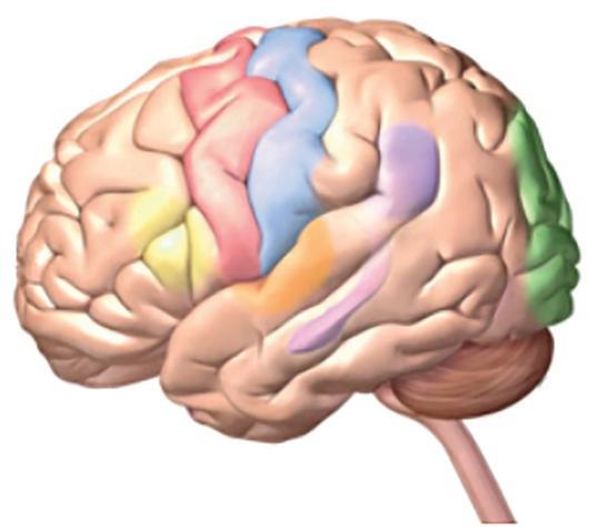
The brain is a soft mass made up of the nerves and tissues that are connected to the spinal cord. The main parts of the brain are Cerebrum, Cerebellum and Brain stem (see Figure 1.1). Frontal lobe, temporal lobe, parietal lobe and occipital lobe, are the four lobes of cerebrum. They are responsible for reading, learning, thinking, emotions, walking, vision, and hearing (regarding senses). Cerebellum is responsible for balancing and coordination. Brain stem is responsible for heartbeat, breathing, blood pressure, swallowing, and eye movements [10, 11].
Brain generates many signals and the electrical signals generated are used in BCI system. These signals are measured using invasive or noninvasive techniques.
1.3 BCI Technology
BCI as mentioned earlier is a communication channel between the brain and the external processing device. The goal of BCI technology is to give a communication model to those people who are severely paralyzed and do not have control over their muscles [12]. It takes the bio-signals measured from a person and predicts some abstract facet of cognitive state.
Frontal lobe
Occipital lobe
Parietal lobe
Temporal lobe
Cerebellum
Brainstem
Figure 1.1 Brain parts.
4 Brain-Computer Interface
Most commonly, the BCI focuses on patients that have problems with motor state and cognitive state. In normal humans, there is an intersection of brain activity, eye movement, and body movements. If any one of them is missed, it results in constrained state. Figure 1.2 shows this intersection. It is observed that BCI is applicable to the areas where patients have normal to major cognition levels working along with no motor state response to minor motor state response. So under this umbrella, we get patients that experience completely locked-in syndrome (CLIS) or Locked-in Syndrome (LiS) [13].
Motor state
Normal
Minor problem
Major problem
Figure 1.2 BCI domain.
1.3 Block diagram of BCI.
BCI Application
Figure
Locked-in syndrome is a neurological disorder also known as pseudo coma where patient is completely paralyzed that is losing control of voluntary muscles, except the eye movements. Therefore, such people can think and analyze but not speak and move. In recent past, it is seen that chronic LIS can be unlocked with the aid of BCI [14].
The block diagram of BCI is as in Figure 1.3. It begins with recording of signals from brain, then processing of these recorded signals. Here various features from the signals are extracted and classified as per their properties or characteristics. Based on these signals’ commands are generated and the BCI device works accordingly.
1.3.1 Signal Acquisition
In BCI, signal acquisitor plays an important role. There are different recording techniques in BCI and are broadly classified as invasive and noninvasive methods as shown in the Figure 1.4. These methods aid to bring out/pull out electric and magnetic signals of brain activity.
Figure 1.4 Types of BCI signal acquisitor.
1.3.1.1
Invasive Methods
Electrodes are implanted in the scalp to extract the required parameters and in non-invasive method, external sensors are used to measure the parameters.
a. Intra-Cortical Recording:
A single electrode or sometimes array of electrodes are in the cortex of the brain. These interfaces are been used for the past 70 years and some of the popular kinds of hardware for intracortical recording are as follows:
i. Wire-based arrays
ii. Micro-machined micro-electrodes
iii. Polymer microelectrodes
i. Wire-Based Arrays
They are also called Microwire arrays, Wire arrays are made up of insulated metal wires with an uninsulated tip that is used to observe the bipotential form of neurons in a bipolar environment [15]. The diameter of those wires is in the range of 10–200 micrometers the limitations of microwire-based arrays are as follows:
• They are limited because of recording failures and FBR effects.
• Microwire arrays are highly prone to variation, disappearance, or disappearance of recorded signal in the timeframe spanning from weeks to months post-implantation [16].
• The wires are tedious to place and route to microelectronic packages.
• Isolation cracks, corrosion – analysis of tungsten microwaves extracted from rats after 9 months of use, revealed material deterioration in the form of isolation fractures and defamation.
• Extensive use of electrodes leads to electric leakages which result in errors when recording.
ii. Micro-Machined Micro-Electrodes
The introduction of photolithography and subsequent advancements in micromachining technology prompted the development of a new generation of silicon-based brain probes. (micromachined microelectrodes) [17]. Ex: Michigan Planar electrode arrays, Utah Electrode arrays [18].
The limitations are as follows:
• They degrade with time.
• Recording loss due to vascular mutilation.
• Failures in interconnection.
• Size and rigidity of the probes.
• Expensive (GoldPlatinum, Iridium are widely used in planar recording areas).
• They are prone to fracture.
• Failure in persistent recordings is mainly because of the 2D. geometry of MMEA-based reading electrodes.
• The size and mechanical mismatch of silicon-based and wire microelectrode arrays with the brain are two of the most important problems limiting the quality of neural recordings.
iii. Polymer Microelectrodes
The disadvantages of stiff materials can be potentially deviated using Polymers [19]. But they have certain limitations:
• The accuracy and depth of implantation of soft and flexible implants into the brain is hampered, making them difficult to implant [20].
• Complicated structural design.
• Expensive methods of fabrication.
• As these are internally placed, they pose challenge for using in long term cases [21]. Fabrication methods and the characteristics of the materials used also impact on its durability.
b. Electrocorticography (ECoG)
Intracranial electroencephalography is a technique for recording brain signals by putting electrode grids on the cortex’s surface. ECoG is an invasive BCI recording method that records with electrodes put directly on the brain’s exposed surface [22]. These are used when performing an internal brain surgery.
• They are expensive.
• They are bulky.
• They are prone to the formation of scar tissue, which obstructs the signal when the body reacts to the foreign item.
Non invasive Invasive
Intracor tical
• Limited sampling time – Seizures may not be recorded with EcoG [23, 24].
• The region of the exposed cortex and operation duration restrict the number of electrodes that may be placed. Errors in sampling are possible.
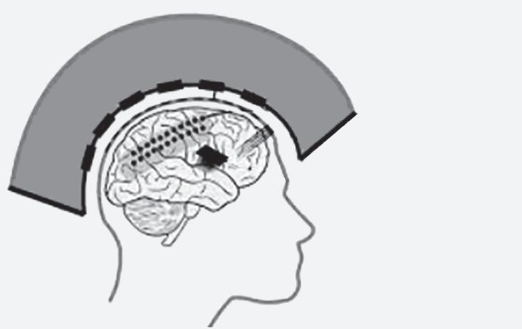
ECoG is a minimal invasive method. Stereotactic electroencephalography (sEEG) also used electrodes to measure brain activity. sEEG provides measurements from much deeper brain structures than ECoG, yet it has received very less attention in BCI applications [25, 26]. All, the intracortical, ECoG and sEEG are invasive methods and their placing in cortex is as shown in the Figure 1.5. ECoG is sometimes referred as semi-invasive method.
1.3.1.2
Non-Invasive Methods
a. EEG (Electroencephalogram)
EEG is a method of signal acquisition that records the electrical signals of the brain by the help of metal discs(electrodes) that are attached to the scalp [27]. There are four types of electrodes:
1) Traditional wet electrodes
2) Dry electordes
3) Active electrodes
4) Passive electrodes
Figure 1.5 Recording places in/on brain.
Hardware concerns with EEG-based BCI equipment with wet electrodes:
• Maintenance and use of wet electrodes is cumbersome.
• Electrical impedance of the skin has its impact the signal acquisition, that is the quality of recording the brain signals. The water content in association with the electrodes reduces electrical impedance whereas the air in association with the electrodes increases the electrical impedance.
• The interface betweeen the skin and electrode causes noises which considerably affect the signal.
• The setup of wet electrode is not well tolerated by subjects over longer periods of recording a) because of the electrolytic gel used which causes irritation to the subjects b) because of the discomfort caused by the elastic straps to hold the eeg cap in place [28].
• The electrical impedance also depends on various factors like surface area of the electrode, room temperature, and the interface layer.The people who were taking the EEG readings must keep all these factors in check.
Hardware concerns with EEG-based BCI equipment with dry electrodes:
• The Quality of the signal obtained using dry electrodes is low when compared to wet electrodes [29].
• For dry electrodes the electrical impedance deteriorate rapidly with use and generally should be replaced after 30 days of usage.
• The electrode caps are prone to movement since there will be no gel to hold the caps in place.
• Highquality electrode caps are generally made of gold and titanium, for prolonged hours of usage change of electrode caps is recommended so it will be costly to buy and maintain the electrodes [30].
• Elastic straps are used to maintain the electric caps in place which causes discomfort to the subject over longer periods of time.
b. MEG (Magnetoencephalography)
It is an imaging test which reflects the activity of the brain by recording the magnetic fields produced by electric currents occuring naturally in the brain [31].
10 Brain-Computer Interface
Hardware concerns with MEG-based BCI equipment:
• The MEG equipment is very expensive.
• MEG equipment requires liquid helium to maintain its superconducting equipment.
• The equipment must be used in a magnetic shielded room and the food used by the subjects and the examiner must also be administered ad managed.
• The patients need to remain relatively still during a MEG examination and the patients with a vagus nerve simulator, pacemaker, or similar device may not be able to undergo an MEG study [32].
c. Functional Magnetic Resonance Imaging (fMRI)
This method acquires the brain activity parameters based on blood flow changes. So, this method depends on cerebral blood flow coupled with neuronal activation. When brain is in use, the blood flow varies depending to the task being performed. Hence, the parameters also vary. fMRI is used to detect and evaluate the brain abnormalities that could not be captured in other imaging techniques like x-ray or MRI.
d. Near-Infrared Spectroscopy (NIRS)
This method measures brain activity in frontal cortex. Here light absorption is used to calculate oxygen and hemoglobin levels.
Before extracting the features from the measured parameters, preprocessing is required. Pre-processing generally consists of Referencing, temporal filtering, and signal enhancement. Referencing is comparing the measured brain signal to a standard or reference signal in the form of voltage. It can be common reference, average reference, or current source density. Temporal filtering removes unwanted noise signals that are present in the measured brain signals. Signal enhancement techniques like Principal component Analysis (PCA), Surface Laplacian, automatic enhancement methods are used to enhance the parameters measured.
1.3.2 Feature Extraction
Based on the signal processing, the commands are generated. So, identification of correct features is an essential step. Feature extraction in BCI is recognizing the events or useful properties that are captured by various neuroimage methods. This will reduce the complexity and help in