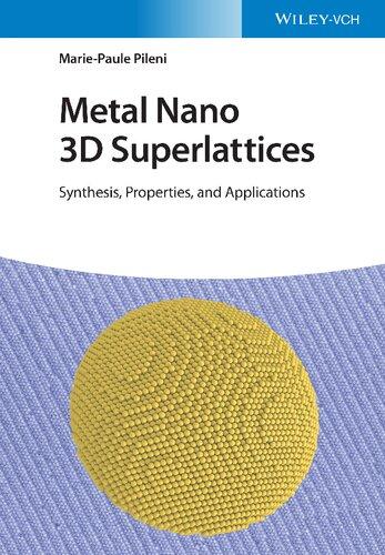
Metal Nano 3D Superlattices
Synthesis, Properties, and Applications
Marie-Paule Pileni
Author
Prof. Marie-Paule Pileni
Sorbonne Université
Department of Chemistry
4 Place Jussieu
75005 Paris Cedex 05
France
Cover Image: © Marie-Paule Pileni, Sorbonne Université
All books published by WILEY-VCH are carefully produced. Nevertheless, authors, editors, and publisher do not warrant the information contained in these books, including this book, to be free of errors. Readers are advised to keep in mind that statements, data, illustrations, procedural details or other items may inadvertently be inaccurate.
Library of Congress Card No.: applied for
British Library Cataloguing-in-Publication Data
A catalogue record for this book is available from the British Library.
Bibliographic information published by the Deutsche Nationalbibliothek
The Deutsche Nationalbibliothek lists this publication in the Deutsche Nationalbibliografie; detailed bibliographic data are available on the Internet at <http://dnb.d-nb.de>.
© 2023 Wiley‐VCH GmbH, Boschstr. 12, 69469 Weinheim, Germany
All rights reserved (including those of translation into other languages). No part of this book may be reproduced in any form – by photoprinting, microfilm, or any other means –nor transmitted or translated into a machine language without written permission from the publishers. Registered names, trademarks, etc. used in this book, even when not specifically marked as such, are not to be considered unprotected by law.
Typesetting Straive, Chennai, India
Print ISBN 978‐3‐527‐34477‐2
ePDF ISBN 978‐3‐527‐34478‐9
ePub ISBN 978‐3‐527‐34480‐2
oBook ISBN 978‐3‐527‐34479‐6
To Vannina, Christophe, Alix, and Paul
Contents
Acknowledgments xii
Introduction xiii
1 Syntheses of Metal Nanocrystals 1
1.1 Nanocrystal Growth Processes and Control of Size and Distribution 1
1.2 Crystalline Structure of Metal Nanocrystals 3
1.2.1 Co Nanocrystals 3
1.2.2 Au, Ag, and Cu Nanocrystals 4
1.3 Various Techniques Used to Produce Metal Nanocrystals and Control Their Sizes and Distribution 5
1.3.1 Reverse Micelles 5
1.3.2 Inorganic Chemical Reaction to Produce Au and Ag Nanocrystals 8
1.3.2.1 Synthesis of Au Nanocrystals Differing by Their Diameters 8
1.3.2.2 Synthesis of 5-nm Polycrystalline Silver Nanocrystals 11
1.3.3 Thermal Decomposition 11
1.3.4 Hot Injection 13
1.4 An Example to Show the Importance of the Reactant Sequence to Produce Nanocrystals 15
1.5 N-Heterocyclic Carbene Ligands for Au Nanocrystals
Stabilization 17
1.6 Conclusion 20
References 20
2 Influence of the Nanoparticle Crystalline Structures Called Nanocrystallinities on Various Properties 27
2.1 Nano-Kirkendall 27
2.1.1 Influence of the Atom Diffusion Processes on Rather Large Nanoparticles (7 nm/8 nm) Differing by Their Nanocrystallinities 30
2.1.1.1 Amorphous Nanoparticles 30
2.1.1.2 hcp Nanocrystals 31
2.1.1.3 fcc Nanocrystals 33
2.1.1.4 Epsilon-Phase Nanocrystals 33
2.1.2 Influence of the Nanocrystal Size Related to the Various Crystalline Structures 34
2.1.3 Nanoparticle Environment Effect 36
2.1.3.1 Isolated Nanocrystals 36
2.1.3.2 Influence of the Electron Beam Irradiation on the Final Nanocrystals 37
2.1.4 Conclusions 39
2.2 Local Surface Plasmon Resonance (LSPR) of Au Nanocrystals Differing by Their Nanocrystallinity 39
2.3 Acoustic Vibrational Modes 43
2.3.1 Breathing Mode 43
2.3.2 Quadrupolar Mode 50
2.4 Crystal Growth Process 58
2.5 Mechanical Properties 58
2.6 Conclusion 59 References 59
3 Au 3D Superlattices Produced by Solvent Evaporation Process 65
3.1 Au 3D Superlattice Morphology of Au Nanocrystals Coated with Thiol Derivatives 65
3.1.1 Au 3D Superlattice Morphology Produced at Zero Solvent Vapor Pressure 66
3.1.1 Au 3D Superlattice Morphologies Produced Under Various Solvent Vapor Pressures 73
3.1.2 Influence of Temperature During the Evaporation Process on Au 3D Superlattice Morphologies 78
3.1.3 Hierarchy in the 3D Superlattice Crystallinity 80
3.2 Interparticle Distance Between Nanocrystals Coated with Thiol Derivatives 80
3.3 Au 3D Superlattices Coated with N-Heterocyclic Carbene 88
3.4 Conclusions 92 References 92
4 3D Superlattice Growth in a Thermodynamic Equilibrium 97
4.1 Simultaneous 3D Superlattices Heterogeneous and Homogeneous Growth Processes 97
4.1.1 General Behavior of the Two Simultaneous Supracrystal Growth Processes of Au Nanocrystals 97
4.1.2 Analogy with Nature: Air–Solvent Interface Acts as Perfect Substrate 102
4.1.3 Case of 5-nm Au Nanocrystals in Toluene Saturation 103
4.1.4 Nanocrystals of 4-nm Dispersed in Toluene 108
4.2 Submillimeter-Size Single 3D Superlattices of 5-nm Au Nanocrystals 109
4.3 Conclusions 111 References 112
5 Ag 3D Superlattices 115
5.1 Control of the Crystalline Structure of Ag 3D Superlattices 115
5.2 Optical Properties 122
5.2.1 Thin Film 123
5.2.2 Thick Films 131
5.3 Stability 134
5.4 Conclusions 134
References 135
6 Mesostructures of Magnetic Nanocrystals Subjected to Applied Magnetic Field 139
6.1 Maghemite Nanocrystals 140
6.1.1 Stripes Formed by Applying a Magnetic Field: van der Waals Versus Dipolar Forces Controlling Mesoscopic Organizations of Magnetic Nanocrystals 140
6.1.2 Applied Magnetic Field Perpendicular to the Substrate: Liquid–Gas Phase Transition 149
6.2 Cobalt Nanocrystals 151
6.2.1 Alignments Induced by Dipolar Interactions Between Co nanoparticles Subjected to a Magnetic Field Parallel to the Substrate 151
6.2.2 Columns and Labyrinths of Co Nanoparticles: Nanocrystal Size Distribution as a Key Parameter on the Mesostructures 154
6.3 Conclusion 163
References 163
7 Binary 3D Superlattices 167
7.1 Structure of 3D Superlattices Predicted by the Hard-Sphere Model 169
7.1.1 Co/Ag Binary 3D Superlattices of Co and Ag Nanoparticles 169
7.1.2 Co/Co Binary 3D Superlattices of Amorphous Co Nanoparticles 171
7.1.3 Ag/Ag Binary 3D Superlattices of Polycrystalline Ag Nanoparticles 172
7.2 Limitation of the Hard-Sphere Model 175
7.2.1 Ligand Exchange 175
7.2.2 Relative Concentration of the Small and Large Nanoparticles on the Binary 3D Superlattices 182
7.2.3 Temperature Effect 183
7.2.3.1 Influence on the Binary Phase Diagram 183
7.2.3.2 Structural Transformation of CoAAu13 Subjected to High Temperature 184
7.2.3.3 Influence of the Magnetic Properties on Co/Ag Binary Systems 186
7.2.4 Unexpected Behavior Induced by Mixing Small and Large Nanoparticles 190
7.3 Solvent-Mediated Crystallization of Nanocrystal 3D Assemblies of Silver Nanocrystals: Unexpected Superlattice Ripening 191
7.4 Collective Properties Involved in Self-Assemblies of Binary 3D Superlattices 191
7.5 Conclusion 193 References 194
8 Analogy Between 3D Lattices and Atomic Crystals: Crystalline Structure 199
8.1 Atomic Crystals, Shaped 3D Lattices, and Minerals 199
8.2 Negative 3D Lattices 204
8.2.1 How Negative 3D Lattices Are Produced? 204
8.2.2 Analogy Between Negative 3D Lattices, Atomic Crystals, and Minerals 211
8.3 Vicinal Surface of 3D Lattices 211
8.4 Quasi-3D Lattices 220
8.5 Conclusions 221 References 222
9 Analogy Between 3D Superlattices and Atomic Crystals: Physical Properties 225
9.1 Magnetic Properties 225
9.2 Coherent Longitudinal Acoustic Phonons in Small 3D Superlattices 227
9.3 Breathing Modes 230
9.4 Conclusions 235 References 235
10 3D Superlattice Stability 237
10.1 Influence of Temperature 237
10.2 Edging Process 238
10.2.1 Ag Nanocrystals 238
10.2.2 Au Nanocrystals 246
10.2.3 Influence of the Coating Agent 247
10.3 Solvent-Mediated Crystallization of Nanocrystal 3D Assemblies 251
10.4 Conclusions 260 References 261
11 Intrinsic Properties Related Due to the Self-Assemblies of Nanocrystals 265
11.1 Epitaxial Crystal Growth as a Result of the Nanocrystal Ordering 265
11.2 Unexpected Electronic Properties of Micrometer-Thick 3D Superlattices of Au Nanocrystals 274
11.3 Collective Magnetic Properties of Co Nanocrystals Self-Assembled in 3D Superlattices 278
11.3.1 Influence of Nanocrystal Ordering on the Magnetic Properties 279
11.3.2 Influence of Co Nanocrystallinity on fcc 3D Superlattices 280
11.3.3 Magnetic Properties of Single-Domain ε-Phase Co Nanocrystals at Various Interactions Scales 282
11.3.3.1 Magnetic Properties of Co (ε-Phase) Nanocrystals Dispersed in PMMA 282
11.3.3.2 Magnetic Properties of Co (ε-Phase) Nanocrystals Deposited on Substrate 283
11.4 Super-Spin Glass Behavior of fcc Supracrystals 286
11.5 Alignment of Magnetic Nanocrystals 288
11.5.1 Do the Mesoscopic Structures Play the Major Role on the Magnetic Properties? 288
11.5.2 Comparison of the Influence or the Easy Axis’ Orientation and Dipolar Interactions 292
11.6 Co 3D Superlattice Collective Properties of Amorphous Nanoparticles 297
11.7 Conclusion 297 References 298
12 Mechanical Properties of 3D Superlattices 303
12.1 Measurements of Mechanical Properties Using Atomic Force Microscope (AFM) 303
12.1.1 Oliver and Pharr 303
12.1.2 Plate Model 304
12.1.3 Validity of Mechanical Properties Deduced from AFM 305
12.2 3D Superlattices Produced Under Thermodynamic Processes 306
12.2.1 Influence of the 3D Superlattice Growth Mechanism 306
12.2.2 Tuning of the Stiffness of Au 3D Superlattices 309
12.3 3D Superlattice Produced Through Heterogeneous 3D Superlattice Growth Process 312
12.3.1 Au 3D Superlattices 312
12.3.1.1 Size and Coating Agent Effect on the Mechanical Properties 312
12.3.1.2 Nanocrystallinity 315
12.3.2 Co 3D Superlattices 318
12.3.2.1 Epsilon Phase Co 3D Superlattice: Influence of Nanocrystal Size and 3D Superlattice Morphology on the Mechanical Properties 318
12.3.2.2 Hierarchical Mechanical Behavior of Co 3D Superlattices Related to Nanocrystallinity 321
12.3.3 Ag 3D Superlattices: Highly Weak Young Moduli 325
12.4 Do the Apparent Discrepancies of the Young Moduli Produced with a Large Variety of Metallic Nanocrystals Self-Assembled in fcc Structures Remain Valid or Not? 329
Contents x
12.5 Mesoscopic Assemblies of Co Nanocrystals Differing by Their Size
Distribution: Mechanical Intrinsic Properties 333
12.6 Conclusions 335 References 335
13 Cracks in Nanocrystal Film 339
13.1 Cracks of Nanocrystal Films 339
13.1.1 Isotropic Cracks 340
13.1.2 Orientational Cracks 345
13.1.3 Universal Feature 349
13.2 Cracks in Nature and Current Life 352
13.3 Conclusions 354
References 354
14 Water-Dispersive Hydrophobic Suprastructures: Specific Properties 357
14.1 Au and Co “Clustered” Structures 358
14.1.1 Fabrication and Characterization 358
14.1.2 Specific Properties 365
14.1.2.1 Magnetic Properties 365
14.1.2.2 Optical Properties 367
14.2 Colloidosomes and Supraballs 369
14.2.1 Colloidosome: Fabrication and Characterization 369
14.2.2 Supraballs: Fabrication and Characterization 372
14.3 Nanoheaters 375
14.4 Conclusion 378
References 379
15 Nanocrystal Self-Assembly in Cells 383
15.1 Ferrite Colloidosomes and Supraballs 383
15.2 Intracellular Fate of Hydrophobic Nanocrystal Self-Assemblies in Tumor Cells 383
15.2.1 Ability of Colloidosomes and Supraballs to be Uptaken into Tumor Cells 383
15.2.2 Internal Distribution of Colloidosomes and Supraballs in Tumor Cells 385
15.2.2.1 Nanocrystal Dispersions 385
15.2.2.2 Colloidosomes 385
15.2.2.3 Supraballs 387
15.2.3 Structural Organization of Nanocrystals in Tumor Cells 389
15.2.4 Interactions with Lysosomes 390
15.2.4.1 Lysosome Size 391
15.2.4.2 Lysosome Shape 392
15.2.4.3 Spatial Distribution and Density of Nanocrystals in Lysosomes 393
15.2.4.4 Proximity of Nanocrystals to the Lysosome Membrane 394
15.2.5 Magnetic Response of Internalized Nanocrystals 396
15.3 Conclusion 399
References 400
16 Photothermal Effects in the Tumor Microenvironment 403
16.1 Colloidosomes and Supraballs 403
16.2 Photothermal Properties: Apparent Contradiction Between the Global Heating and Cell Death 404
16.2.1 Pellet of Nanocrystal-Loaded Cells 406
16.2.2 Monolayers of Cells Having Internalized the Fe3O4 Nanocrystals 406
16.3 Suprastructures: Photothermal Properties in the in vivo Tumor Microenvironment 410
16.4 Suprastructures Modulate the Distribution of Fe3O4 Nanocrystals in the Tumor Microenvironment 413
16.5 Suprastructures: Photothermal Effects on the Tumor Extracellular Matrix 416
16.6 Conclusion 421 References 423
Index 427
Acknowledgments
The experiments for the data presented in this book have been performed by MPP team. The experiments were confirmed by several researchers from the group. This reduces the potential artifacts. Furthermore, long-term collaborations with limited group number are another strength.
MPP would like to thank
* Long-term collaborations: Dr. P.A. Albouy from Orsay, France; Dr. P. Bonville of CEA-Saclay, France; Professors N. Cerullo, D. Polli, and G. De la Valle of Politecnico di Milano, Italy; Professor M. El Sayed of Georgia Tech, USA; Professor K. Holmberg of Chalmers University, Professor B. W. Ninham of Canberra University, Australia; Sweden; Professor F. Willig of Fritz-Haber Institute der MPG, Germany; and Professor Z. Yang of Key Laboratory of Colloid and Interface Chemistry of MOE, School of Chemistry and Chemical Engineering, Shandong, University, China
* Short-term collaborations: Professor S. Ball of University of Antwerp, Belgium; Professor F. Carn of Université de Paris, Dr. F. Charra of CEA-Saclay, France; Professor P. M. Claesson of KTH, Sweden; Dr. I. Dobryden of KTH, Sweden, Professors J. Freund and J. Urban of Fritz-Haber Institut der MPG, Germany; Dr. F. Gazeau of Université de Paris, France; Professor J. Israelashivili of Santa Barbara, USA; Professor P. Moriarty of University of Nottingham, UK; Dr. A. Nicolas- Boluda of Université de Paris, and Professor D. M. Smilgies of Cornell University, USA and Dr. N. Winckelmans of University of Antwerp, Belgium.
* Nothing would be possible without my collaborators. Nice collaborative atmosphere has been the key parameter in the success of this book. When that was not possible, the collaboration was stopped. Most of them were PhD students or postdoctorates. Some of them remain in the group after the thesis.
People involved in this book are: E. Alphandéry, I. Arfaoui, A. Çolak, A. Courty, M. Gauvin, V. Germain, A. I. Henry, Kh. Khazen, E. KLecha, X. Ling, Y. Lalatonne, I. Lisiecki, S. Mourdikoudis, T. Ngo, D. Parker, H. Portales, J. Richardi, S. Roland, J. Romann, C. Salzeman, N. Schaeffer, Y. Wan, L. Wang, J. Wei, C. Yan, J. Yang, N. Yang, P. Yang, and Z. Yang
Other collaborators not involved in this book are: P. André, F. Billoudet+, A. Brioude, Q. Darugar, D. Ingert, N. Feltin, A. Filamkenbo, T. Jian, X. C. Jiang, J. Legrand, L. Levy, W. Qian, M. Maillard, F. Michel, L. Motte, K. Naoe, C. Petit, N. Pinna, F. Pitré, J. Tanori, and H. Taleb.
Introduction
Self-organization of entities into two-dimensional (2D) or three-dimensional (3D) materials, forming highly ordered structures such as atoms in bulk materials, cations and anions in salts, and organic molecules in biological materials, is rather basic. For instance, the gem opal assembled from silica spheres with a closely packed locally periodic structure under moderate compression in geology; the crystal of the tobacco mosaic virus in biological systems. When referring to science and technology, self-organization with a multitude of tunable parameters, such as the types of building blocks, the assembly driving force as well as potential applications of these assembled architectures, has attracted a broad interest among countless chemists, biologists, and physicists. Hence, self-assembly of objects into ordered functional superstructures is a universal process and a prevalent topic in science.
In this book, we concentrate our efforts on self-assemblies of metal nanocrystals. The nanocrystal size is in the range from 2 to 13 nm. To keep their integrities, these metal nanocrystals are coated with various organic molecules. The size distribution of the nanocrystals used here is rather low. Nanocrystals are dispersed in solution. The produced colloidal solution is optically clear and colored. The difference between nanoparticles and nanocrystals is related to the crystallinity of the core. Hence, the core of nanoparticles is amorphous, whereas that of nanocrystals is crystalline. This latter term does not take into account the ordering mode of atoms in the core (single domain or polycrystal). The crystalline structures of nanocrystals called nanocrystallinity permit to differentiate between single domain and polycrystal but not the crystalline phase. As matter of fact, single domain is a generic name and corresponds to various crystalline phases such as face-centered-cubic (fcc), hexagonal-close-packed (hcp), and body-centered-cubic (bcc) structures. For Co nanocrystals, the ε-phase exists at the nanoscale. Either by slow solvent evaporation of the colloidal solution or by keeping it under saturated solvent atmosphere (the solvent needs to be a bad solvent for the alkyl chains), nanocrystals tend to self-assemble either as films or as aggregates. A careful characterization shows that both films and aggregates are usually assembled in 3D superlattices called colloidal crystals, supercrystals, or supracrystals When the distribution of nanocrystals is random without regular order, the films and aggregates are considered as amorphous. All over these chapters, the building blocks of 3D superlattices are in the size range
between 5 and 10 nm. Similar crystalline structures are obtained in nature. However, the particle sizes (most of the cases ferrite) are in the range of micrometers. In Chapter 1, syntheses of nanocrystals with low size distribution are described. Chapter 2 shows some properties of nanocrystals differing by their nanocrystallinity. It has not been possible to control the nanocrystallinity of all the metal nanocrystals studied. This is due to the techniques used (soft chemistry) to produce nanocrystals. Here, we concentrate on the final structures of Co nanocrystals subjected to oxygen and differing by size and nanocrystallinity. Such diffusion processes in metal have been largely studied with bulk metal and nowadays at the nanoscale. This process is called nano-Kinkerdall effect. Hence, with ε-phase, whatever be the nanocrystal size, CoO hollow nanocrystals are produced. With amorphous, fcc, and hcp Co nanocrystals, various final structures are produced such as Co oxide (CoO), shell or yolk/shell structures with CoO shell, and CoO hollow structures, whereas the Co cores keep the crystalline structure they had before oxygen treatment. Some physical properties of metal nanocrystals differing by their nanocrystallinities are described: the localized surface plasmon resonance of Au nanocrystals and the acoustic breathing mode of Au and Co nanocrystals do not depend on the nanocrystallinity. Conversely, the fundamental quadrupolar mode of single-domain Au nanocrystals split, whereas the polycrystalline counterpart does not. Such splitting will be used all over the book chapters as a tool to identify a large collection of nanocrystals differing by their nanocrystallinities. In Chapter 3, the various assemblies obtained by solvent evaporation of Au colloidal solution are described. It is shown that the coating agents, the nanocrystal size, solvent vapor pressure, and temperature play key roles in the final crystalline structures and in the interparticle distances of the 3D superlattices. Chapter 4 shows that by keeping the colloidal solution under solvent saturation during several days, simultaneous crystal growth process takes place: a 3D superlattice film at the solvent–air interface and shaped 3D superlattices precipitated are produced. Such simultaneous 3D superlatticesgrowth processes are produced if the solvent of the colloidal solution has to have a large surface tension as the air–solvent interface and be a bad solvent for the alkyl chain used as coating agent. Very surprisingly, in Chapter 5, it is shown that crystalline structure of colloidal crystals keeps the memory, after evaporation, of the solvent used to disperse Ag nanocrystals. Hence, by tuning the solvent used to disperse nanocrystals, coating agents of nanocrystals, and temperature of solvent evaporation, i.e. solvent volatility, various crystalline structures such as fcc, bcc, and hcp can also be tuned. Finally, keeping the colloidal solution under solvent saturation during several days, simultaneous crystal growth process takes place: a 3D superlattice film at the solvent–air interface and shaped 3D superlattices precipitated are produced, which will be discussed in Chapter 4. To produce such segregation, the solvent of the colloidal solution has to have a large surface tension as the air–solvent interface and be a bad solvent for the alkyl chain used as coating agent. Chapter 6 shows that, by slow solvent evaporation of a magnetic colloidal solutions under magnetic field, long superimposed cylinders with a very regular structure are
Introduction xv
produced when the applied field is parallel to the substrate. Such mesoscopic structures are obtained when the distance between nanocrystals is very short. The control of interparticle distance between nanocrystals through the coating agent permits the control of the mesoscopic structure from superimposed cylinders to undulated films. When the applied field is perpendicular to the substrate, dots and labyrinths are produced. The difference between these two last structures is related to the size distribution of nanocrystals, i.e. with nanocrystal ordering: With highly ordered structures, dots are produced, whereas labyrinths are obtained with building blocks having a large size distribution. This is attributed to the fact that the stiffnesses of 3D superlattices are larger than amorphous mesoscopic structure. This was confirmed by the mechanical properties described in Chapter 12. Chapter 7 shows that by using nanocrystals having two well-defined sizes, their assemblies, governed by hard sphere interactions, favor the formation of a very large variety of 3D superlattice crystalline structures such as NaCl, AlB2, NaZn13, and Laves phase (MgZn2, MgCu2, MgNi2) structures. However, either through ligand exchange of the coating agent used to coat the nanocrystals, by using ferromagnetic nanocrystals, or by adding surfactant molecules, quasi-3D superlattices are produced. The driving force involved in the formation of quasi-3D superlattices remains an open question. Furthermore, the nanocrystallinity of nanocrystals involved in binary systems and the relative amount of small and large nanocrystals also play a role in the final structure. In some experimental conditions, 3D superlattices characterized by very stable vicinal surfaces are produced. Very surprisingly, Chapters 8 and 9 show analogies between atomic crystals and 3D superlattices. The atoms are replaced by (uncompressible) nanocrystals and atomic bonds by coating agents (carbon chains) that act like mechanical springs holding together the nanocrystals. These analogies are observed in the crystal growth processes, magnetic responses as well as vibrational processes. Hence, similar crystalline structures are obtained with 3D superlattices and atomic crystals. Furthermore, nanocrystals breath coherently in a 3D superlattice as atoms in a nanocrystal. Longitudinal propagation waves are observed in thin 3D superlattice film deposition on a substrate as well as thin atomic crystals grown on a wafer. Chapter 10 points out the importance of the coating agents on the nanocrystal ability to self-assemble in 3D superlattices and on their stabilities. Chapters 11 and 12 point out many of intrinsic properties due to the nanocrystal ordering in 3D superlattices. Hence, mild annealing of an assembly of nanocrystals induces epitaxial growth of triangular single atomic crystals. Their sizes are directly correlated to the coherence length of the nanocrystal assemblies. Furthermore, electronic transport properties through very thick 3D superlattices are observed by scanning tunneling microscopy and spectroscopy at 5 K. The mechanism of such unexpected process remains an open question. The magnetic properties governed by dipolar interactions are pointed out. The mechanical properties of 3D superlattices depend on their growth processes, the crystalline structure of nanocrystals used as building blocks, and the material used (Co, Au, Ag nanocrystals). In Chapter 13, we describe cracks of nanocrystal films as observed in nature, paintings,
etc. It is shown that, in the range of few nanometers to centimeters, the surface of the cracks linearly depends on the thickness of the film. This is probably similar in nature and paintings but difficult to search. Finally, from Chapters 14 to 16, it is demonstrated that assemblies of hydrophobic nanocrystals dispersed in aqueous solution are characterized by specific properties. The optical properties of Au 3D superlattices show both collective modes related to the 3D superlattice size and the fingerprint of nanocrystals used as building blocks. Such latter properties are also described in Chapter 12 where Coulomb blockage of isolated nanocrystals is observed in a 3D superlattice. These new suprastructures of either Au or ferrite nanocrystals behave as universal nanoheaters. Such behavior opens a new research area. In the presence of cancer cells, these suprastructures are internalized keeping the nanocrystal assemblies. This also opens a new research area. As a matter of fact, we can expect collective properties in cancer cells. The first example is shown by studying the photothermal properties of suprastructures internalized in cancer cells. Such property is not limited to cells. Veal tendon can be burnt by laser irradiation of 3D superlattices deposited on it.
Obviously, these metallic 3D superlattices are a new generation of materials in their infancy and many new intrinsic properties will be discovered in the future. The analogy between 3D superlattices and atomic crystals (Chapters 8 and 9) allows us to claim that other specific properties will be discovered in a large number of research are as involving energy release, catalysis, properties of transport, biomedicine, etc. As already observed in some cases, the scaling laws in 3D superlattices with respect to atomic crystals will have to be revisited. This claim is strongly supported by some results presented here such as (i) The possibility of identifying, as in Nature, a bipyramidal morphology as an intermediate in the growth process of truncated to triangular tetrahedral assemblies (Chapter 8). (ii) The speed of sound through 3D superlattices is very low compared to that measured in atomic crystals (Chapter 9). (iii) The frequency of the respiration process of nanocrystals in 3D superlattices is greater than that of atoms in nanocrystals. (iv) Very thick 3D superlattices (up to 5 µm) can be surprisingly imaged by STM and their electronic properties studied (Chapter 11). (v) Large single crystals of triangular atoms are produced by soft annealing of 3D superlattices obtained under ultra high vacuum (UHV) (Chapter 11), etc.
1
Syntheses of Metal Nanocrystals
Metal nanocrystals have been extensively developed over this last two decades. Here, we will focus on spherical metal nanocrystals. We study the most important factors [1] involved to control size, size distribution, composition, and crystalline structure called nanocrystallinity. However, other parameters could emerge in the future.
1.1 Nanocrystal Growth Processes and Control of Size and Distribution
The nanocrystal growth processes with control of size and size distribution have been largely studied over these past decade [1–18]. LaMer developed a theoretical model of hydrosols nucleation and growth processes [19, 20]. Ostwald observed that the atomic mobility by transfer matter from small to larger particles reduces the free energy associated with the particle/matrix interfacial area. This is called Ostwald ripening. These models have been experimented for nanoparticles/nanocrystals. The nucleation process is where nuclei (seeds) acts as templates for crystal growth. Homogeneous nucleation occurs when nuclei form uniformly throughout the parent phase, whereas heterogeneous nucleation forms at structural inhomogeneities (container surfaces, impurities, grain boundaries, dislocations). In liquid phase, heterogeneities occur much easier since a stable nucleating surface is already present. The growth of nanoparticles depends on the surface reaction and the monomer’s diffusion to the surface. In many cases, the same synthesis does not produce the same final size and shape (see below). This phenomenon may be attributed to either the presence of some impurities, additives present in a very low concentration in the reactants, or sequence of reactants inducing heterogeneous nucleation. The latter is due to the changes on the growth mechanism that markedly differs with seed size and/or internal crystallinity. Consequently, various chemical methods have been developed to control the size and shape of these nanocrystals by changing the preparative conditions such as the kind and/or the amount of protecting agent.
Metal Nano 3D Superlattices: Synthesis, Properties, and Applications, First Edition. Marie-Paule Pileni. © 2023 WILEY-VCH GmbH. Published 2023 by WILEY-VCH GmbH.
1 Syntheses of Metal Nanocrystals 2
Coating agents are needed to keep the integrity of nanocrystals. However, in reality as shown below, ligands have a vital role not only in influencing the size and size distribution of the nanocrystals but also, more importantly, in defining their interaction/interface with the environment. The ligand-mediated nanocrystal synthesis is still an open question. Actually, the role of nanocrystal/ligand differs with the metal ion and the organic ligand used. Hence, the ligand and metal nature are key parameters. At this point, we can mention the strength of ligand–nanocrystal surface interaction and the labile nature of the ligands either in the free or complex forms with the formation of clusters that it is produced with the nanocrystal constituents. The most used coating agents are surfactants. Here, we will concentrate to surfactant molecules. For most of the studies, the head polar groups are either thiol (SH), amine (NH2), or carboxylic (COOH) groups, whereas the alkyl chains are either alkane or oleic groups. In most of the cases, size selection processes are needed to produce nanocrystals with low size distribution. Carboxyl acid is covalently bound with Co and Fe atoms [21, 22]; thiol derivatives are “quasi” covalently bound to Au atoms, whereas it is lesser than Ag ones [23]. Conversely, amine groups [24] are weakly bound to most of the metallic atoms (Figure 1.1).
To control the size distribution, the digestive ripening permits to convert polydispersed to uniform nanocrystals: the first step is to break the nanocrystal large size distribution into ligand/metal complexes by ligand addition, then isolate the ligandstabilized nanocrystals, and finally heat, at high boiling point, the solvent of the dispersed nanocrystals in the presence of ligand [25–27]. Then, the control in size and size distribution, based on van der Waals or dipolar forces, can be reached by size selection procedure. By mixing bad and good solvents for the coating agents, as obtained with polymers [28], the bad solvent for the alkyl chains induces precipitate of the largest nanocrystals, leaving the smaller nanocrystals in solution. After centrifugation, the precipitate is dissolved with a good solvent for the alkyl chains,
Figure 1.1 Various coating agents used to coat nanocrystals: (a) oley group (C18H36X) and (b) alkyl chains (C12H25X). X is the head polar group: X = SH thiol derivatives, X = COOH acid derivatives, and X = NH2 amine derivatives.
(a) x x (b)
1.2 rystalllneStrrctrreof MetalNanocrystals 3
leading to a homogeneous clear solution, and again a bad solvent is added inducing a new precipitate. Similarly, the supernatant solvent is evaporated, and the same procedure as the precipitate is proceeded. Such process is repeated several times before reaching a sufficient low size distribution (≤10%) and consequently to be able to self-assemble in 2D or 3D superlattices. A rather large number of mixed solvents is used to size select nanocrystals: the solvent/nonsolvent pairs for the coating agent, e.g. hexane/ethanol, heptane/ethanol, chloroform/ethanol, chloroform/ methanol, and pyridine/hexane, are usually used. Ligands, considered as capping/ passivating agents, play a major role in the control of nanocrystal size and is far from only providing stability to the nanocrystal against aggregation.
With the most used syntheses to produce well-defined metal nanocrystals with a very low size distribution, several procedures are used: reverse micelles, inorganic chemical reaction, hot injection processes, and thermal decomposition. They are based on chemical reduction of the metal ion by various reducing agents such as hydrazine, sodium borohydride (NaBH4), citrate, and glycol. The control of nanocrystal size depends on (i) the relative concentrations of the reactants, (ii) the coating agent concentration, (iii) the structure of the coating agent, (iv) the relative amount of reactant and that of the coating agent (the higher the stabilizer agent compared with reactants, the smaller nuclei formed and consequently smaller nanocrystal size), and (v) the mixture of two coating agents (one binds tightly to the nanocrystal surface hindering growth, and the other less tightly bound favors the rapid growth).
1.2 Crystalline Structure of Metal Nanocrystals
Generally, the nanocrystal size is determined by the number of atoms produced and consequently by the number of the nanoparticles formed depending on the kinetics of nucleation and growth processes. The nanocrystal crystalline structure called nanocrystallinity is determined by the competition between the kinetics of structural transformation of the nanoparticles and that of delivering separation between nucleation/ growth processes (e.g. a burst of nucleation) and diffusion-controlled growth [29–34].
1.2.1 Co Nanocrystals
Cobalt has three nearly isoenergetic crystal structures (Figure 1.2): hexagonal closepacked (hcp), face-centered cubic (fcc), and epsilon (ε). Bulk cobalt displays two stable phases of hcp and fcc at ambient pressure. At the nanoscale, it can possess the epsilon phase with a complex cubic primitive structure, which is similar to that of β-manganese, with a unit cell parameter of 6.097 Å [35]. This metastable crystal structure (ε phase) can be transformed to hcp or fcc structure by annealing the sample at a suitable temperature [36]. Due to its relatively smaller magnetocrystalline anisotropy constant (Kc ≈ 1.5 × 10⁵ J m−3), ε-structure is widely known as a softer magnetic material, compared with the hcp and fcc phases.
1.2.2 Au, Ag, and Cu Nanocrystals
At the atomic level, the densest possible packing of four spheres is a tetrahedron [29, 37–39]. At the earliest stages of growth of a solid, the atoms reorganized into a completely new structure each time an atom is added. For fcc materials, the nanocrystals considered as compact packing of spheres are cuboctahedra, decahedra, and icosahedra (Figure 1.3). The surface of a cuboctahedral nanocrystals consisted of eight (111) planes and six (100) planes with closed atomic shells and represents the most stable form. The defined number of atoms resulted exclusively from the geometric arrangements and cannot be explained by the electronic shell model. The decahedra and icosahedra are structures with fivefold symmetry and are called multiple-twinned particles (MTPs). In fact, decahedral nanocrystals consisted of five deformed tetrahedral subunits twinned by their {111} planes, whereas icosahedral clusters originated from the twinning of 20, (111) faces, deformed tetrahedral subunits.

Various shapes of fcc nanocrystal arrangements.
Icosahedra
Decahedra
Figure 1.3
(c)
Figure 1.2 Crystalline structures of Co nanocrystals (a) hcp, (b) fcc, and (c) ε-phase.
1.3 Various Techniques Used to Produce Metal Nanocrystals and Control Their Sizes and Distribution
A very large number of techniques have been used to produce nanocrystals, with very low size distribution. Significant advantages of methodologies using highboiling-point solvents and organometallic precursors in the presence or absence of surfactants favor the control of nanoparticles size and improve the crystalline structure of the nanocrystals [11, 33, 40].
1.3.1 Reverse Micelles
Reverse micelles [2, 17, 41–48] are well known water-in-oil droplets stabilized by surfactants [49]. The best surfactant providing uniform droplets is sodium bis(2ethylhexyl)sulfosuccinate, Na(AOT), called Aerosol OT. The amount of water controls the droplet size (Figure 1.4a). The average droplet diameter increases linearly with the water content, w, defined as w = [H2O]/[AOT]. By collisions these droplets exchange their water contents and form again two independent droplets (Figure 1.4b). This process is used to produce nanosized material by either chemical reduction of metal ions or coprecipitation reactions. At low water content, the number of water molecules per surfactant is too small to hydrate the counterions and the head polar groups. This induces strong interactions between water molecules and the head polar groups. The water molecules are then considered as “bound water.”
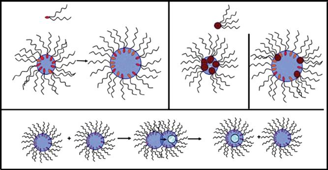
Figure 1.4 (a) Control the size of the droplet with the amount of water with Na bis(2-ethylhexyl)sulfosuccinate, Na(AOT). Similar behavior is observed with Ag(AOT). (b) Exchange process by collision between two droplets to favor chemical reaction. (c) Reverse micelles from functionalized surfactant Co and Cu (tetra (2-ethylhexyl) sulfosuccinate), X(AOT)2. (d) Mixed micelles.
(a)
An increase of the water content induces a progressive appearance of “free” water molecules in the center of the droplets, whereas some of water molecules remain bound to the interface. With Na(AOT), the bulk water phase is reached around w = 10. To produce metal nanocrystals without any oxide formation, the metal ions have to be in environment of bound water molecules and not into the bulk phase, i.e. they have to be located at the oil–water interface of the droplets. This explains why we synthesized functionalized surfactants Xn(AOT)n with n = 1 or 2, i.e. the sodium ions of AOT are replaced by metallic ones (Figure 1.4c). With Xn(AOT)n, reverse micelles are formed to produce reverse micelles, but the amount of water inside reverse micelles is limited (w ≠ 5) [42]. To provide a control of the droplet size by the water content, we used mixed micelles, Na(AOT), Xn(AOT)n. In such condition, the size of the droplet remains controlled by the amount of water molecules involved in the formation of reverse micelles that can then play the role of template (Figure 1.4d).
To synthesize amorphous Co nanoparticles, mixed micelles [Co(AOT)2/Na(AOT)] are used. Sodium borohydride (NaBH4) added to the micellar solution reduces the cobalt ions. Immediately after NaBH4 addition, the color of the micellar solution changes from pink to black, indicating the formation of colloidal Co nanoparticles. Coating agents with carboxylic as head group dodecanoic acid molecules added to the colloidal solution induce a chemical bond between the oxygen of carboxyl group and the Co atoms located at the interface. The coated Co nanoparticles are then washed and centrifuged several times with ethanol to remove all the AOT surfactant. Moreover, the black powder obtained is dispersed in hexane, and the colloidal solution is isotropic. Size selection process as described above takes place to produce Co nanoparticles with a ~10% size distribution. The entire synthesis is carried out in an N2 glove box using de-oxygenated solvents to prevent nanoparticle oxidation. The ratios of [NaBH4]/[Co(AOT)2] and [Na(AOT)]/[Co(AOT)2] control the nanoparticle size. In contrary to others, the nanoparticles are amorphous. The average diameter and size distribution evolve from 4 to 9 nm and 9% to 13%, respectively (Figure 1.5a). The relative ratio between Na(AOT) and Co(AOT)2 controls the nanoparticle size. By annealing process, under an inert atmosphere, the crystalline structure of Co nanoparticles is improved, and a transition from amorphous to single-domain hcp structure takes place [47, 48]. The annealing process does not significantly affect the nanocrystal diameter and its size distribution.
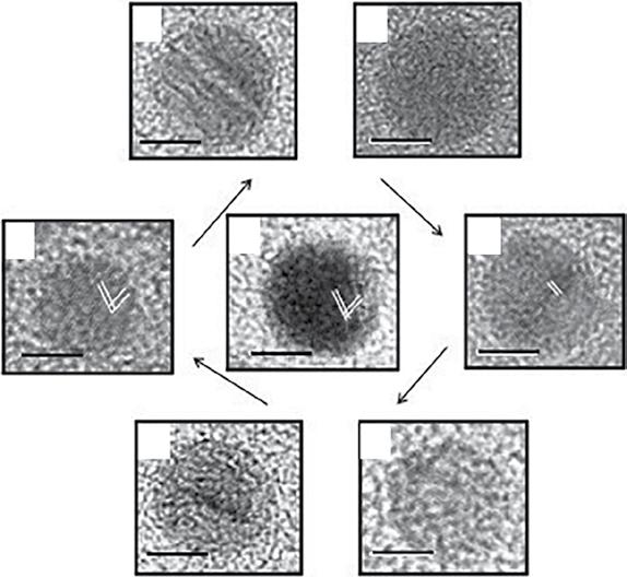
Amorphous Co nanoparticles deposited on the TEM grid are annealed under inert gas, 60 minutes at 250 °C. During such process, as shown in Figure 1.6, a transition of the crystalline Co nanoparticles from amorphous to single-domain hcp phase takes place. Note the hcp nanocrystal size increases compared with the corresponding amorphous counterparts. This is due to hcp lattice fringes. Hence, the crystallinity of Co nanoparticles is controlled by keeping the other parameters constant as size and surface coating. The control of amorphous Co nanoparticle size is obtained by mixing Na(AOT) with Co(AOT)2 [50].
To produce Ag nanocrystals by using reverse micelles, a colloidal solution of Na(AOT) and Ag(AOT) is mixed with Na(AOT) reverse micelles containing hydrazine [51]. The nanocrystals are extracted from the micellar solution and then are
1.3 arlors echnlires seed to roedrceMetalNanocrystalsanedontrol helrSliesaned lstrlirtlon 7
Figure 1.5 (a) Increase in the size of Co nanoparticles with controlling R, w, and the relative amount of Co(AOT)2. (b) Reduction of mixed reverse micelles of Ag(AOT)/ Na(AOT) (6/4) by hydrazine with w = 2. By controlling the ratio R = [Ag(AOT)]/[hydrazine] from 4 to 0.7, the Ag nanocrystal sizes are (a) 4, (b) 5, (c) 6, and (d) 7 nm, respectively.
Figure 1.6 Schematic illustration with high-resolution TEM (HRTEM) images of the evolution process of 7.2-nm Co nanocrystals: (a) amorphous Co nanoparticles, (b) hcp Co with large domains, (c) and (d) very large domains, (e) small hcp single crystals, (f) hcp single crystals with stacking faults, and (g) hcp single crystals. The scale bar is 3 nm.Source: Yang et al. [48]/American Chemical Society.
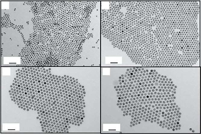
1 Syntheses of Metal Nanocrystals 8 subjected to the ethanol/hexane wash cycles. Changing the water content and the ratios of [Ag(AOT)]/[hydrazine] and [Ag(AOT)]/[Na(AOT)] controls the Ag nanocrystal from 4 to 7.3 nm (Figure 1.5b).
1.3.2 Inorganic Chemical Reaction to Produce Au and Ag Nanocrystals
1.3.2.1 Synthesis of Au Nanocrystals Differing by Their Diameters
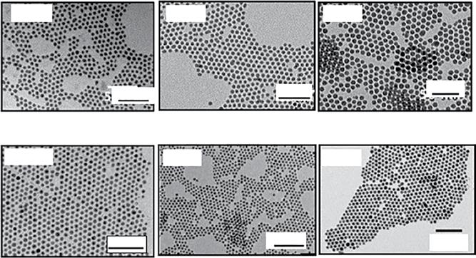
Au nanocrystals are synthesized by revisiting the Stucky method [51–54]: two solutions (A) and (B) are mixed under nitrogen protection. (A) is a preheated solution of chlorotriphenylphosphine Au(I) dissolved in toluene containing dodecanethiol, and (B) is amine–borane complex dissolved in toluene. The ratio of the volume of A/B controls the size of the nanocrystals from 7.8 to 5.8 nm. To decrease the size from 5.1 to 4 nm, the solution B is replaced by tert-butylamine and ammonia borane complexes, respectively (Figure 1.7). For simplicity, details on the synthesis to control the nanocrystal size are given in Table 1.1.
Let us consider 5-nm Au nanocrystals: at the end of an organometallic synthesis, a drop of Au nanocrystals dispersed in toluene is deposited on TEM grid. The darkfield TEM image (Figure 1.8) shows homogeneous or inhomogeneous contrasts corresponding to single and polycrystalline nanocrystals, respectively. After analyzing
Figure 1.7 TEM image and corresponding histograms of 4 (a), 5 (b), 7 (c), and 8 nm (d) obtained by deposition of a drop of colloidal solution of Au nanocrystals coated with dodecanethiol and dispersed in toluene .Source: Goubet et al. [55]/JOHN WILEY & SONS, INC.