For more information about Scrivener publications please visit www.scrivenerpublishing.com.
1st edition (2014), 2nd edition (2023)
All rights reserved. No part of this publication may be reproduced, stored in a retrieval system, or transmitted, in any form or by any means, electronic, mechanical, photocopying, recording, or otherwise, except as permitted by law. Advice on how to obtain permission to reuse material from this title is available at http://www.wiley.com/go/permissions.
Wiley Global Headquarters
111 River Street, Hoboken, NJ 07030, USA
For details of our global editorial offices, customer services, and more information about Wiley products visit us at www. wiley.com.
Limit of Liability/Disclaimer of Warranty
While the publisher and authors have used their best efforts in preparing this work, they make no representations or warranties with respect to the accuracy or completeness of the contents of this work and specifically disclaim all warranties, including without limitation any implied warranties of merchant-ability or fitness for a particular purpose. No warranty may be created or extended by sales representatives, written sales materials, or promotional statements for this work. The fact that an organization, website, or product is referred to in this work as a citation and/or potential source of further information does not mean that the publisher and authors endorse the information or services the organization, website, or product may provide or recommendations it may make. This work is sold with the understanding that the publisher is not engaged in rendering professional services. The advice and strategies contained herein may not be suitable for your situation. You should consult with a specialist where appropriate. Neither the publisher nor authors shall be liable for any loss of profit or any other commercial damages, including but not limited to special, incidental, consequential, or other damages. Further, readers should be aware that websites listed in this work may have changed or disappeared between when this work was written and when it is read.
Library of Congress Cataloging-in-Publication Data
ISBN 978-1-394-16624-4
Cover images: Pixabay.Com
Cover design by Russell Richardson
Set in size of 11pt and Minion Pro by Manila Typesetting Company, Makati, Philippines
2.7.4
2.5.5.1
3.3.5
3.3.6
3.3.7
3.3.8
3.3.9
3.3.10
3.4
3.4.2.1
3.4.3
5.2.2
5.2.3
5.3
5.2.1.1
5.2.2.1
5.2.2.2
6.1.1
6.1.2
6.2 Development of Enzyme-Based Glucose Biosensors
6.2.1 First Generation of Enzyme-Based Electrochemical Glucose Biosensors
6.2.2 Second-Generation Enzyme-Based GBs
6.2.3 Third-Generation Enzyme-Based
6.3 Fabrication of Enzymatic Glucose Biosensors
6.3.1
6.3.2 Immobilization of Enzyme for the Development of Glucose Biosensors
6.3.2.1 Adsorption Technique
6.3.2.2 Covalent Immobilization of GOx
6.3.2.3 Entrapment of GOx Into a Polymer Matrix
6.3.3 Application of Nanomaterials for the Development of a Transducer for Glucose Biosensors
6.3.3.1 Using Nanoparticles as an Artificial Peroxidase for the Fabrication of
6.3.3.2 Using Nanoparticles as Bifunctional Tools for Developing Label-Free Glucose
Nelson Malini, Sepperumal Murugesan and Ayyanar Siva
13 Application of Optical
Riyanka Das, Rajeshwari
13.2.3
13.2.3.4
13.2.4
Vipul Prajapati and Princy Shrivastav
16.1 Introduction
16.2 Antibodies: A Brief Overview
16.2.1 What Are Antibodies?
16.2.2 Types of Antibodies
16.2.3 Production and Purification of Antibodies
16.2.3.1 Polyclonal Antibodies
Antibodies as Bioreceptors
16.3 Antigen–Antibody Reactions
16.3.3 Complement
16.3.4 Radiomunoassay
16.3.5
16.3.6 Western Blotting
16.4 Antibody-Based Biosensors (Immunosensors)
16.4.1 What are Immunosensors?
16.4.2 Selection of Antibodies
16.4.3
16.4.3.4
16.4.4
16.4.5
16.4.6
16.4.7
Vipul Prajapati, Jenifer Ferreir, Riya Patel, Shivani Patel and Pragati Joshi
18.3.3
18.4.3.4
18.4.4
18.5.3 Measurement of the Light Output in the Tissues of the Living Animals From the Bioluminescence-Based Biosensor
Preface
The biosensor industry began as a small, niche activity in the 1980s and has since developed into a large, global industry. Nanomaterials have substantially improved not only nonpharmaceutical and healthcare uses, but also telecommunications, paper, and textile manufacture. Biological sensing aids in the understanding of living systems and may be used in a variety of sectors, including medicine, drug discovery, process control, environmental monitoring, food safety, military, and personal protection. It brings up new opportunities in bionics, power generation, and computing, all of which will benefit from a greater understanding of the bio-electronic relationship, as advances in communications and computational modeling are forcing us to reconsider how we offer healthcare and perform R&D and manufacturing to the modern world.
As a result of the customization of everything from health to environmental control, new payment structures and commercial models will arise. Wearable, mobile, and integrated sensors are being used in an increasing variety of products, but the majority of these devices still rely on physical sensors to measure elements like temperature and pressure. There is a conspicuous dearth of sensors that are both robust and convenient in the field of body chemistry sensors. This book examines emerging technologies that are accelerating scientific study and laying the foundation for new goods meant to extend and improve the quality of our lives. In this newly evolving discipline, the combination of nanoscale materials with biosensor technology is gaining a lot of traction. Nanostructures have been used to increase the adherence of biosensor materials to electrode surfaces, print nano barcodes on biomaterials, increase the pace of bio-responses, and amplify the electric signal. Finally, nanomaterial-based biosensors may be employed in a wide range of medical diagnostics and environmental monitoring applications due to their better response speed, greater sensitivity, simple design, specificity, and cost-effectiveness.
The book covers the major materials employed in the development of biosensors such as nanoparticles, nanowires, nanotubes, nanoribbons, nanorods, nanosheets, and many more nanostructures.
Chapter 1 discusses how the innovative techniques used in biosensors evolved from cell engineering and 3D cell immobilization. The various parts of biosensors based on cell detection using the cell’s bioreceptors are discussed in detail along with their working mechanism and applications.
Chapter 2 discusses the bioreceptors for enzymatic interactions as part of an enzymebased biosensor to detect the analyte from a sample with applications. Materials used for the construction of various types of biosensors and types of bioreceptors, immobilization
of enzymes for biosensors, and types of transducers for enzymatic interactions are also described.
Chapter 3 discusses the history, structure, synthesis, types, physical, and chemical properties including the merits and demerits of dendrimers in detail. This chapter focuses on dendrimers as drug delivery via electrochemical, enzymatic, optic, QCM, and glucose-based biosensors.
Chapter 4 details the importance and need for various 2D Photonic Crystal biosensors. The chapter discusses, in detail, how disease identification is done by measuring the effective change in the refraction of different analytes, and as well as various Photonic Crystal structures. It also focuses on the scope and future development of biosensors using 2D structures.
Chapter 5 explains that the applications of bioreceptors are numerous, ranging from benchtop analysis to point-of-care diagnosis and treatment. Hence, this chapter is an overview of common bioreceptors, especially affinity-binding receptors that are crucial in theranostic applications.
Chapter 6 details the brief history, basic working principles, and the present developments in glucose biosensors. The chapter focuses on fabrication methods and recent trends in the application of nanomaterials and nano/microfabrication for the development of paper analytical devices or wearable glucose biosensors.
Chapter 7 is focused on the recent progress in the preparation of metal-free quantum-sized sensors. The biosensing and detection applications of carbonaceous quantum dots such as carbon, graphene, and carbon nitride quantum dots are investigated. Optical and electrochemical techniques are discussed with a consideration for the limit of detection values.
Chapter 8 details the latest research progress on bioreceptors for microbial biosensors. In addition, it summarizes the types and applications of microbial biosensors: the recent trend and the future challenges of microbial biosensor technology.
Chapter 9 explains the use of several noble plasmonic materials including gold, silver, copper, niobium, and aluminum, and their widespread applications in optical sensing and sensors. Also, the advantages, disadvantages, and prospects of some highly used plasmonic nanomaterials in sensors are discussed in this chapter.
Chapter 10 explains the differences between various magnetic sensors and their accompanying sensitivity responses and consequences. This chapter discusses how magnetic sensors use magnetic fluids for studying the impact on a range of particles and various sensor architectures for tracking biological interactions with distinct magnetic strength change.
Chapter 11 deals in detail with the various salivary biomarkers and the biosensors associated with several neurological disorders including Alzheimer’s disease, Parkinson’s disease, Huntington’s disease, ALS, multiple sclerosis, autism spectrum disorders, and
neuropsychiatric disorders, and various cancers affecting breasts, lungs, pancreas, and the gastrointestinal system.
Chapter 12 deals with the different molecules for biological amine detection by fluorescent chemosensors and its specific emphasis on the use of bioimaging applications. In addition, the sustained improvements in fluorescent biosensors are anticipated to result in universal biosensors for essential biological amines, which can be detected in real-time analysis.
Chapter 13 provides concise information about chromo-fluorogenic biosensing applications of diverse nanomaterials including zero, one, two, and, three-dimensional nanomaterials. Biosensing from complicated bio-matrices along with intracellular imaging is also discussed. Finally, loopholes of present research and future research directions are also outlined to stay current with medical diagnosis.
Chapter 14 explores present and prospective breakthroughs in nanotechnology-based biosensors for real-time assessment of several analytes and the toxicity mechanisms in living creatures, using primary datasets from 2018 onwards. Innovative biosensing technologies centered on unique sensing components and transduction concepts receive special attention. The chapter also discusses the opportunities and future considerations for the utilization of NMs-based biosensors for enhanced environmental and food-sensing devices.
Chapter 15 deals with the introduction and roadmap of biosensors, followed by the use of nanotechnology in cancer therapy. Additionally, it explores nanomaterial infused with a biosensor, the fabrication of nano biosensor, and the diagnosis of breast cancer, including point-of-care and wearable analysis.
Chapter 16 provides a brief overview of antibody-based biosensors, also called immunosensors. The chapter includes a discussion on antigen-antibody interactions and the application of immunosensors in various areas of the health and food industries. Lastly, some new approaches to antibody modifications that offer several advantages over classical antigen-antibody receptors are also discussed.
Chapter 17 describes the use of biosensors for various paints and pigment analysis. It discusses the biosensor’s components, history, and working principle in regard to paint and pigments. For future benefit, the chapter describes the characteristics of paints and pigments, with analytical methods, and various applications of paints and pigments-based biosensors.
Chapter 18 discusses biological devices that are incorporated into various animal and plant tissues. The applicability in varied physiology along with its distinguished classification based on different principles is covered also. Furthermore, the chapter enumerates the increasing exigencies for these devices in a broad range of areas including various medical ailments.
Chapter 19 details various methods for pesticide detection with simple, highly selective and sensitive, fast response, cost-effective and portably sized biosensors. The chapter also
Preface discusses recent results from the use of nanomaterials in the fabrication of pesticide biosensors in food and environmental applications.
Chapter 20 discusses the applications of biosensors for monitoring the behavior of agro-products. Two categories of biosensors are highlighted, and their roles in detecting contaminants in dairy processing are discussed. The application of receptor-based biosensors was proposed for monitoring the survival rate of bacteria in milk processing.
The Editors August 2023
Bioreceptors for Cells
Vipul Prajapati1* and Salona Roy2
1Department of Pharmaceutics, SSR College of Pharmacy (Permanently Affiliated to Savitribai Phule Pune University), Sayli-Silvassa Road, Sayli, Silvassa, Union Territory of Dadra Nagar Haveli & Daman Diu, India
2Department of Pharmacology and Toxicology, NIPER Hajipur, Export Promotions Industrial Park (EPIP), Industrial Area Hajipur, Dist. Vaishali, Bihar, India
Abstract
A biosensor is a tool that quantitatively determines the disturbance in the homeostatic equilibrium in a system in an extremely low concentration in the healthcare sector, such as diagnosis, treatment, and mitigation. Biosensors utilize specific biomarkers to aid in an accurate diagnosis based on its sensitivity, reproducibility, biocompatibility, and robustness, which has several advantages over conventional diagnosis, including onsite diagnosis in less time. This article covers various techniques involved in the pretreatment of the cell to modify certain bioreceptors, types of transducers, and their wide arena of application. Cells for biosensors are often labeled with certain enzymes or secondary substances producing an intensifying response of intrinsic signal transduction. The efficacy of a single mini device had attracted the attention of many researchers to aid in the early diagnosis of life-threatening diseases. Although enhancement in the performance of biosensor of cell has been going on, it has provided a gateway to next-generation approaches in the healthcare system.
Living cells are the biorecognition elements since they can detect any unknown stimuli or perception from their environment as a method of adapting, as well as surviving. Cell biosensors can detect analytes with utmost accuracy, high sensitivity, and specificity in a cost-effective, invasive or non-invasive way. They are appropriate bioreceptor elements because they provide versatility in sensing tactics and make production relatively straightforward and inexpensive when compared to pure enzymes, DNA, and antibodies-based detection techniques, such as ELISA, RIA, etc. [1].
Biosensors utilize a mixture of biological, chemical, and physical technologies to measure micro physiological signals in real-time and on-site detection. A biosensor system is made up of a few crucial elements like biological sample receptors, transducers,
backing laminate, and display systems that use electrical, chemical, or photonic components. It is to detect the results and then turn the cascading event of recognition into a measurable signal strength, which can be grouped in conjugated and integrated biosensors [2].
A biosensor’s basic principle is to detect the bio-element at the molecular recognition level and convert it into a different sort of signal strength using a transducer [3]. Living cells, bioactive substrates, and transducers make up cell-based biosensors. The impact of biochemical or pharmacological compounds on cells might be measured by changes in cellular polarity or physiological characteristics like cell membrane permeability or ligand expression on biosensors after treatment [4]. Rapid analysis of small amounts of data as compared to conventional techniques, with the added benefit, may be used for the basis of clinical evaluation that integrated design platforms. The ability of sensor cells to detect specific analogs of targets while distinguishing them from structural counterparts that had no functional similarities within them provided the basis for accurate results [5].
Biosensors of cells have sparked attention as a potential alternative to traditional sensing methods due to several benefits, which include affordability and mobility with the major advantage of the absence of equipment and trained staff. Another pleasing characteristic of biosensors of cells is their versatility in terms of design and outputs, which made them capable of being adjusted to the unique feature whenever needed for desired outputs. They provide a diverse platform for analytical applications in various arenas, such as food, biomedical science, environmental, and society healthcare by merging disciplinary technologies and expertise. Environmental monitoring, bioproduction, biomedical applications in diagnostics, and health monitoring are all possible scopes for cell-based biosensors [6].
1.2 Classification of the Cell as a Bioreceptor
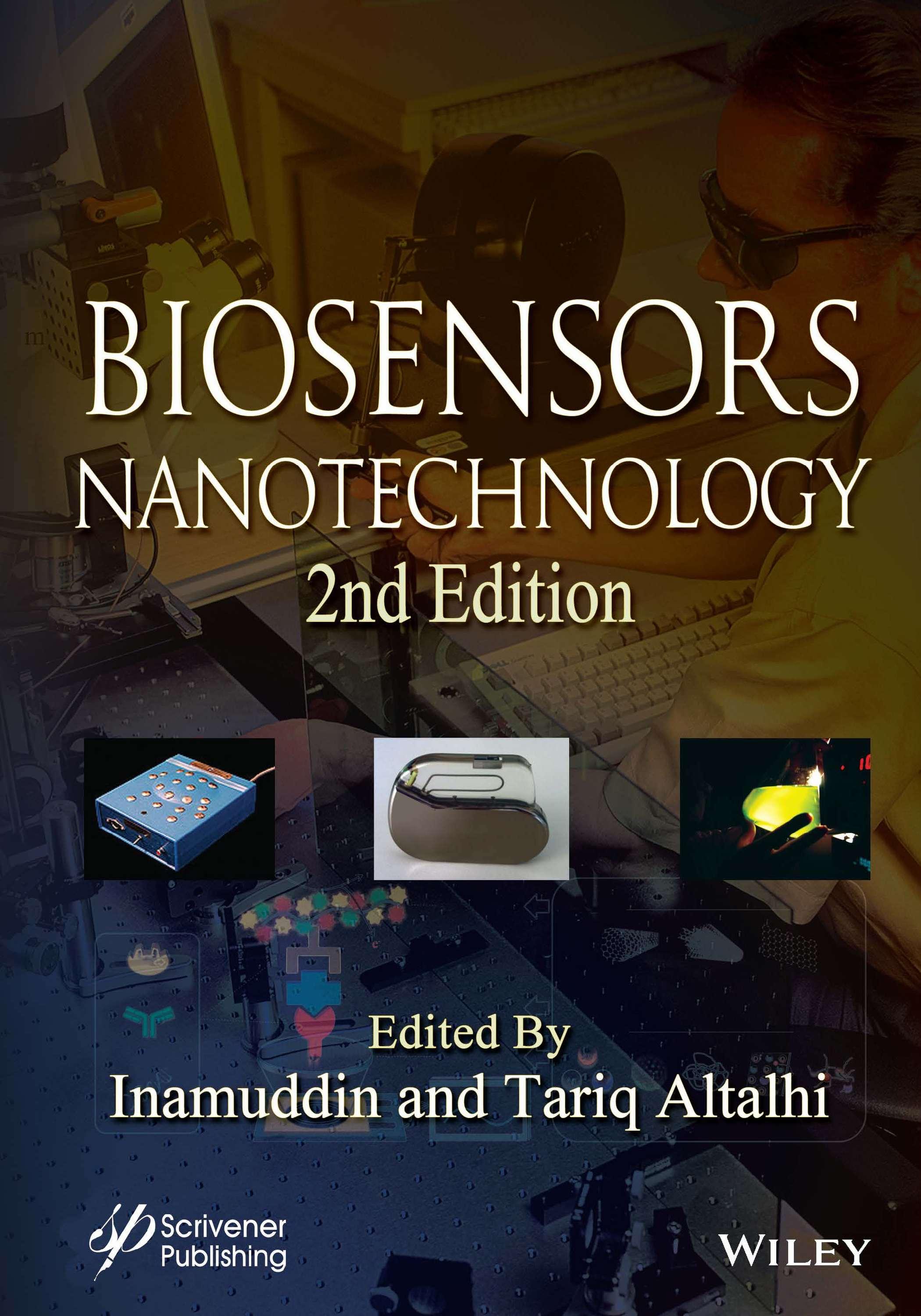
With the greater selectivity to target molecules, bio-elements, such as functional enzymes, serum antibodies, DNA, and other elements, have traditionally been used as bioreceptors in biosensing. Living cells, on the other hand, provide an intriguing alternative to these molecular bioreceptors due to their broad diversity of biomolecular processes [59]. Cell biosensors have been classified by many researchers based on biorecognition element and signal transduction but in this section, in another way they can be classified based on the structural system and extensive work done in the lab to convert a cell into a probe, i.e., cell pretreated in the lab or which utilizes the invasive method of detection and cell without pretreatment or utilizes non-invasive method as shown in Figure 1.1, which is sensitive enough to record and display on-the screen with the maximum level of accuracy.
2.
3.
4.
5.
6.
1.2.1 On the Basis of Cell Origin
1.2.1.1
Mammalian Cell
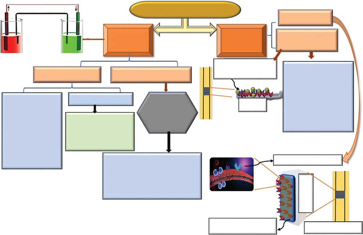
The key transducers for signal creation in mammalian cell-based sensing systems are mammalian cells. When analytes excite living cells, the transducer converts changes in physiological characteristics or biological responses into a measurable and processable signal, fulfilling the detection and analysis goal [60].
Biosensors based on mammalian cells have been touted as potential instruments for pharmacology and toxicology, drug discovery, bioassay of drug substance, pathogen and toxin screening, environmental monitoring,, and biosafety research. The binding of cellular receptors to external substances triggers the cell–analyte interaction. The future of next level of generation biosensing techniques that use natural bio cellular receptors like GPCRs or the nicotinic in cholinergic receptor to measure the ligand-based secondary cellular response have been considered as shown in Figure 1.2 [61].
Hematopoietic and nervous system stem cells have been identified and grown in vitro They develop into cell types, including neurons and myocardiocytes, which spontaneously contract in culture when induced to differentiate. Understanding neural network function from cells structured in vivo has been highly beneficial using brain tissue slices. Using isolated populations of cells as sensors ignores the analyte’s involvement in the in vivo cell metabolism developing coculture systems to expand the operational sensitivity of excitable cells to include metabolites would improve the current capabilities [62].
Figure 1.1 Schematic classification of the biosensor of the cell.
1.2.1.2
Microbial Cell
In the creation of biosensors, microbes are a good substitute for enzymes. They may be manufactured in vast quantities biotechnologically, and microbial cells contain multiple enzymes with the requisite cofactors or coenzymes, allowing them to detect a wide range of substrates. Furthermore, enzymes are very stable in their natural environment and do not require isolation [63].
In a variety of biological systems, biosensors of microbial detection are based on illumination from bacteria that are being used as a sensitive, fast, and non-invasive test. Bioluminescent bacteria may be found in a variety of habitats, from the sea to the land. Bioluminescent-like organism biosensors have also been created for the detection of organic, pesticide, and lead or mercury-like heavy metal pollution utilizing genetically engineered microorganisms (GEM). Cellular organelles can be thought of as multi-oriented biocatalysts that fall between complete cells and enzymes in terms of complexity [64]. In the manufacturing of biosensors, enzymes are the most extensively employed biological sensing element. Although pure enzymes have a high selectivity for their substrate, their use in biosensors may be restricted by the time and expense of enzyme purification, the necessity for numerous enzymes to make the measurable product, or the requirement for a coenzyme [65].
Viable cells and nonviable cells are the two types of cells found in microbes. The assessment of biological oxygen demand (BOD) or the consumption of other growth is two of the
Cascading
Figure 1.2 Pictorial view of detection mechanism of cell biosensors through GPCR receptor.
most common uses of living cells. Pollutants and hazardous substrates can also be detected by viable cells. Both virulent and dead microbial cells are utilized in the immobilization of microbial-based biosensors, although their immobilization needs varied. In case of utilization of an external light source, bacterial-based fluorescence occurs in an entire cell, and the fluorescence emission or radiating intensity is precisely proportional to the amount of analyte detection at very lower concentration [66].
Immobilized yeast was used to detect formaldehyde and assess the toxicity of cholanic acids, with changes in metabolism indicative of the analyte measured by O2 electrode readings or extracellular acidification rates. Organophosphate hydrolase is an enzyme that creates protons during the breakdown of organophosphate insecticides or nerve agents including sarin, soman, and VX. Analyte detection has been proposed using pH changes in the effluent from these immobilized cells [62].
1.2.2 On the Basis of Cell Treatment
1.2.2.1 Cell Pretreated in the Lab or Invasive Detection as Bioreceptor
The cell biosensors can be further classified on the basis of treatment of the cell or modified cell for construction of the biosensor. The methods for cultivating biological live cells on two-dimensional (2D) chip technologies are known as cell culture on-chip. Due to the advent of the scale of the grown environment within the micro-sized chip is tailored to the length of the cells, microchip approaches can give several benefits for cell culture systems.
1.2.2.1.1
Cell Immobilization Techniques
Cell culture of different cell types-based detection involving biosensing techniques use the stimulated response to the external stimuli mediated by a transducer through the modified signal. Immobilized enzymes can be utilized again after the completion of one process and they have higher stability in terms of catalytic activity than mobile enzymes. Although they have a lower catalytic rate when compared to mobile enzymes and require extensive treatment steps, immobilized enzymes are commonly used in medical and industrial plants because of benefits such as fast and efficient control by reusable enzymes i.e., ease of separation of the enzymes from the final product, maintaining purity of the product and high stability of the immobilized enzyme. There are mainly five methods of enzyme immobilization that can be classified as: adsorption, encapsulation, covalent bonding, entrapment as well as cross-linking, among all five, adsorption and covalent bonding method is mainly preferred. Adsorption, encapsulation, and entrapment are termed physical methods of immobilization whereas cross-linked covalent bonding is termed chemical methods or ways of immobilization [31]. Adsorption is one of the simplest techniques of immobilizations, it relies on the only weak force of attraction between enzyme and carrier such as London forces, electrostatic or ionic contacts, and hydrophobic or lipophilic interactions between them. As an extra reagent is not required, the adsorption approach is simple and affordable, moreover it is less damaging to enzyme function than other methods. On the other hand, because of their weak interaction, the enzyme immobilized by this approach are quickly deposited by changes in experimental setups like temperature, pH, or ionization potential.
Furthermore, contamination and signal interference may result from the non-targeted adsorption of various substrates onto a surface. Second, one of the most extensively utilized ways is covalent bonding, which creates stable complex compounds between enzymes and supports or carriers. The covalently immobilized enzymes have stronger binding than the adsorption and this method can provide more stable enzyme immobilization. Despite its advantages, the development of covalent bonds reduces the activity of the immobilized enzymes, and this approach necessitates a considerable amount of bioreagent to be used with it. Third, entrapment is not directly coupled to substrates or products; rather, it is entrapped or encased in polymers, which creates room for substrates and end-products to freely spread. Polymerization can take place in a combination of enzymes and monomeric units to entrap enzymes. Entrapment, like covalent bonding, is a physical interaction that offers the enzymes excellent stability and reduces leaching. Fourth, cross-linking provides strength to the bonded enzymes and ensures the leakage of enzymes during the utilization. It gives one of the very strong and robust connections between enzymes, the immobilization method via cross-linking enhances efficiency and stability. The use of cross-linking reagents like GTA (Glutaraldehyde), on the other hand, can result in a loss of activity due to severe modifications of the functional and non-functional enzymes caused by covalent bonding linkage. Fifth, encapsulation, which is more similar to that of the entrapment process, the only difference, lies in the arrangement of enzymes within the system. The encapsulation method involves the collection of enzymes in the semipermeable membrane and entrapment involves the arrangement of enzymes in the matrix forming structures. These biosensing devices are capable of reliably detecting water toxicity, assuring human safety and aquatic life welfare [32–34].
1.2.2.1.2 Microcontact Printing
The modification of surface technique that uses custom inks to modify surface chemical and biological signals as well as manufacture particular topographical characteristics is widely known as Micro-contact printing (MCP). In 1993, it had been proposed that the combination of MCP as a matter for regulating the concentration and area distribution of proteins will be adsorb onto to the designed self-assembled monolayers (SAMs). SAM is a part of the soft lithography method family, which is one of the most widely utilized surface modification techniques in biomedical applications. MCP has considerable benefits over other surface patterning methods in terms of cheap cost, high dependability, and adaptability. Two of MCP’s main duties in adjusting the surface are the creation of organized geometric or characteristic patterns and 2D surface designing. The similarity with nanoimprinted lithography lies in creating intricate patterns as in the case of MCP. MCP is a flexible surface modification technology that may manipulate surface chemical and biological signals as well as produce specific topographical features using specialized inks.
The primary distinction is that in nanoimprinted lithography, the stamp is made out of surface structured materials that have been created using processes like photolithography and another process known as the femtosecond laser ablation technique. The primary merit of MCP in comparison with the other surface modification methods lies in its good two-dimensional surface patterning capability, which allows varied functional groups or polysaccharides, proteins, and biological signals to be cast or transferred onto the surface of the substrate in precisely specified patterns. MCP has considerable benefits over other
surface patterning methods in terms of cheap cost, high dependability and adaptability. Even though mechanical action regulated by a preset program ensures sample reproducibility and speed, it also allows for large-scale tailored manufacturing. This approach had been studied and implemented in a variety of sectors, and it has shown great promise in facilitating multidisciplinary study in science, engineering and medicine, with new applications being developed all the time. MCP’s operating settings may be gently tweaked and improved to generate excellent surface patterns on a range of substrates, despite of its basic premise [35].
1.2.2.1.3 Fast Ink-Jet Printing
Ink-jet printing is commonly used to accumulate a range of patterns on flexible and tangible substrates for conveniency, environment friendly, high process control, and low-cost electronic devices, among the various nanostructure deposition procedures on electrode surfaces. This method combines spatial resolution, printing speed, repeatability, inexpensive initial investment, and reduced waste into an attractive package. Inkjet printing also eliminates the need for dyes, masks, coloured chemical etchants using etching, and other patterning issues by printing the desired device pattern directly to the substrate. On a number of substrates, inkjet printing has been used to manufacture a range of electrical devices, sometimes in concert with other deposition or patterning processes [36].
Inkjet printing is an old practice that has lately been resurrected for the manufacturing of low-cost and simple electrical sensing and biosensing devices. This technology may be used to create flexible microsystems on ecologically friendly substrates like polymers and paper. Inkjet printing stands out among them because it blends an old-fashioned printing technology with cutting-edge nanoparticle-based inks to provide the printed device important qualities including conductivity, hydrophobicity or hydrophilicity and resistance or insulating properties. The technology was even utilized to use other bio-element such as enzyme, antibody and genetic material-based inks to functionalize the printed devices. This approach has been investigated in the biosensor sector for the past ten years, resulting in a vivid large number of papers as well as intriguing prospects and applications. Inkjet printing has several benefits over other printing processes, including the ability to print on both solid and flexible substrates, the lack of extra components other than inks, and the ability to reduce the time from concept to prototype to a few minutes and therefore it has been produced for electrochemical and optical biosensing by some companies [37].
1.2.2.1.4
Self-Assembled Monolayer
The two-dimensional molecular level structures that form or cast spontaneously on the surface of different kinds of substrates, hence known as Self assembled monolayer (SAMs). Self-assembly is the most versatile concept in nature as such around all biomolecules of larger size to smaller size, such as proteins, peptides, amino acids combine and self-assemble to produce functionally relevant structured and ordered structures. Proteins or short chain polypeptides have been used by nature to build a range of materials, including shells, pearls, and keratin. It may be used to create whole new molecular structures. SAMs are gaining popularity due to their applications in electrical devices such as biosensors, thin film transistors, micropatterning, etc. Molecules by molecules are assembled in self-assembly, thereby “bottom up” approach language is used. The notion of SAMs is gaining prominence
in the field of modification of surface using biomaterials or biological compounds. SAM has a number of advantages over other physical surface modification techniques including UV irradiation and electron beam. Since the SAM surface modification enables for covalent bonding of molecules, one of the core advantages is their long shelf life or functional period. SAMs employing the aforementioned physical approaches, on the other hand it has shown low stability. In some cases, results in a loss of surface chemical activity as a result of irradiation. Furthermore, conventional approaches such as physical or chemical adsorption, London forces, and cross-linking of biomolecules had suffered from some stability issues, but the SAM approach has become widely used for biomolecule immobilization. There are two types of SAM which are small molecule SAM and polymer SAM and also there are two types of SAM production processes, substrate coupled and substrate decoupled, based on interactions between the substrate and the molecule of SAM. For instance, in the first stage, SAM headgroups chemisorb on certain areas of the surface to form an ordered monolayer which have been done experimentally in lab involves saturated thiols on a gold surface. In this case, the substrate’s crystalline structure is crucial, and single crystals such as gold are often used to create the ordered and linear assembly. In a substrate-decoupled process like the synthesis of alkyl siloxane on a substrate of silicon which is in hydrated form, there is indirect contact with the surface molecules, and the building of monolayer process is entirely controlled by intermolecular force of interactions [38–40].
1.2.2.1.5 Microfluidic Technology
Microfluidics are techniques for regulating small-scale fluids in devices and systems, and many microfluidic based devices have been actively and widely used in the replication and evaluation of specific and targeted biological processes in tiny devices with a limited quantity of material. Using microfluidics as an instrument to replace the existing conventional research equipment at a very minimal cost, cell counting, sorting and trapping have been reduced to a good extent. In various research, organizations have developed their specific microfluidic cell culture technique that had imitated like the exact organs of human beings in order to study targeted biological processes or evaluate the efficacy or toxic potential of drugs. Microfluidic based impedance virus biosensors have been intensively investigated because they rely on a simpler and faster method of identifying particular viruses than existing methods. One of the main advantages of microfluidic devices lies in the intrinsic capacity of the device to install many analytical modalities or elements; as a result, MFT biosensors have been created for applications ranging from utilization in an analytical instrument, in research to healthcare and associated industry. Some researchers developed a technique that can be used to test influenza of avian or bird virus with high degree of sensitivity using a portable impedance-based biosensor with twenty-five pairs of microelectrodes were gathered and a microfluidic technology, as well as magnetic nano particles coated with antibodies [24].
1.2.2.2
Cell Without Pre-Treatment or Label-Free or Non-Invasive Detection
Live cells carefully perceive and respond to outside information. For quite some time, cell transmission was thought to be linear, with an ambient stimulus triggering a series of steps that culminated in a well-defined detection response. However, there is compelling