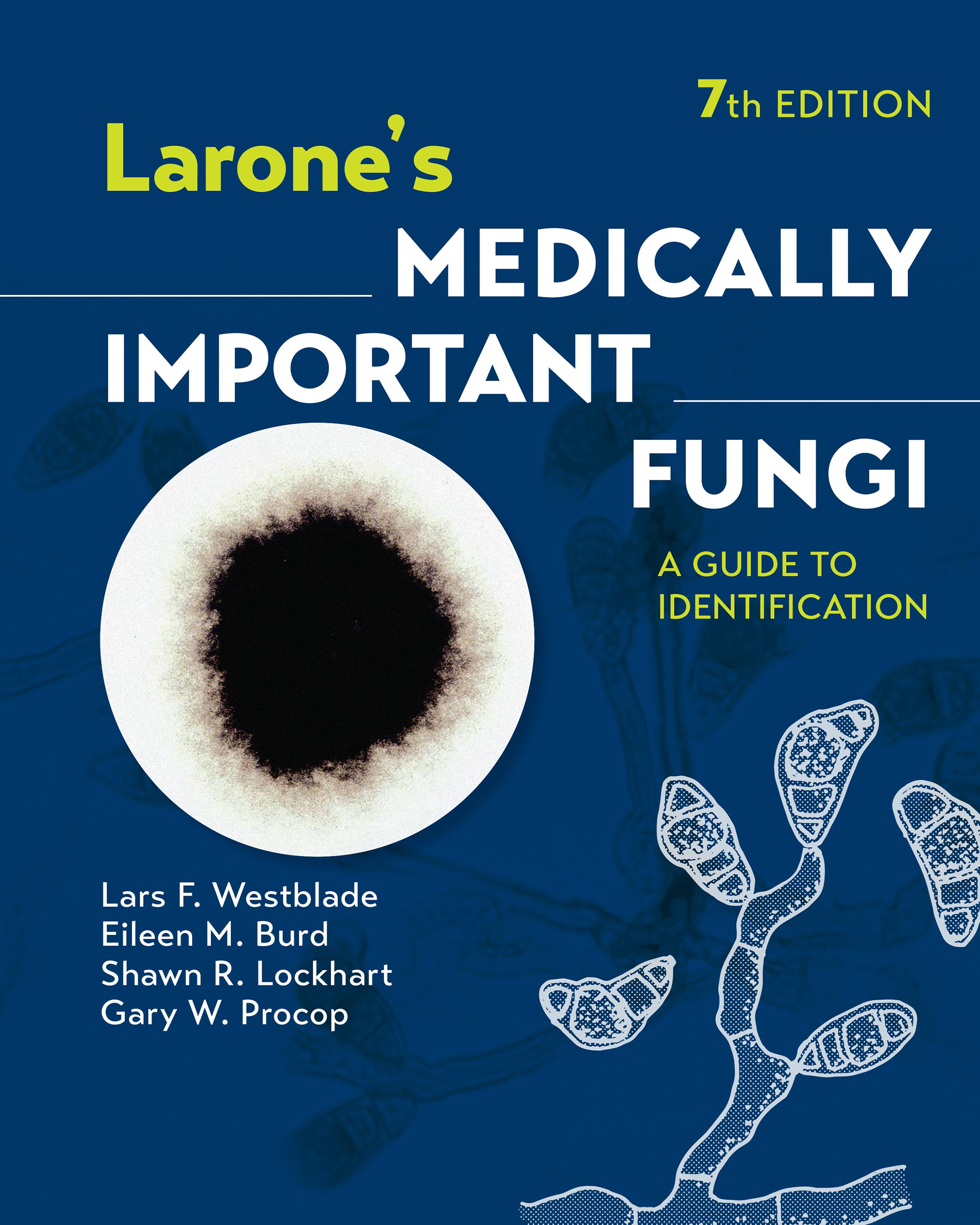
Larone’s Medically Important Fungi
A GUIDE TO IDENTIFICATION
Founding Author
Davise H. Larone, MT(ASCP), PhD, D(ABMM), F(AAM)
Weill Cornell Medicine
New York, New York
Lars F. Westblade, PhD, D(ABMM)
Weill Cornell Medicine
New York, New York
Shawn R. Lockhart, PhD, D(ABMM), F(AAM)
Centers for Disease Control and Prevention Atlanta, Georgia
Eileen M. Burd, PhD, D(ABMM) Emory University Hospital Emory University School of Medicine Atlanta, Georgia
Gary W. Procop, MD, MS American Board of Pathology Tampa, Florida Cleveland Clinic Lerner College of Medicine Cleveland, Ohio
With
Thomas J. Walsh, MD, PhD (hon), F(IDSA), F(AAM), F(ECMM), F(AAAS)
Center for Innovative Therapeutics and Diagnostics Richmond, VA
Copyright © 2023 American Society for Microbiology. All rights reserved.
Copublication by the American Society for Microbiology and John Wiley & Sons, Inc.
No part of this publication may be reproduced, stored in a retrieval system, or transmitted in any form or by any means electronic, mechanical, photocopying, recording, scanning, or otherwise, except as permitted by law. Advice on how to reuse material from this title is available at http://wiley.com/go/permissions.
The right of Lars F. Westblade, Eileen M. Burd, Shawn R. Lockhart, and Gary W. Procop, to be identified as the author(s) of this work/the editorial material in this work has been asserted in accordance with law.
Limit of Liability/Disclaimer of Warranty
While the publisher and author have used their best efforts in preparing this book, they make no representations or warranties with respect to the accuracy of completeness of the contents of this book and specifically disclaim any implied warranties or merchantability of fitness for a particular purpose. No warranty may be created or extended by sales representatives or written sales materials. The publisher is not providing legal, medical, or other professional services. Any reference herein to any specific commercial products, procedures, or services by trade name, trademark, manufacturer, or otherwise does not constitute or imply endorsement, recommendation, or favored status by the American Society for Microbiology (ASM). The views and opinions of the author(s) expressed in this publication do not necessarily state or reflect those of ASM, and they shall not be used to advertise or endorse any product.
Editorial Correspondence: ASM Press, 1752 N Street, NW, Washington, DC 20036‐2904, USA
Registered Offices: John Wiley & Sons, Inc., 111 River Street, Hoboken, NJ 07030, USA
For details of our global editorial offices, customer services, and more information about Wiley products, visit us at www.wiley.com.
Wiley also publishes its books in a variety of electronic formats and by print‐on‐demand. Some content that appears in standard print versions of this book may not be available in other formats.
Library of Congress Cataloging‐in‐Publication Data
Names: Westblade, Lars F. (Lars Frederick), 1976- author. | Westblade, Lars F. (Lars Frederick), 1976- author. | Burd, Eileen M., 1956- author. | Lockhart, Shawn R., 1966- author. | Procop, Gary W., author. | Larone, Davise Honig, 1939- author. | Walsh, Thomas J., M.D. author. | Larone, Davise Honig, 1939- Larone’s medically important fungi.
Title: Larone’s medically important fungi : a guide to identification / Lars F. Westblade, Eileen M. Burd, Shawn R. Lockhart, Gary W. Procop with Davise H. Larone and Thomas J. Walsh.
Other titles: Medically important fungi
Description: 7th edition. | Hoboken, NJ : Wiley-ASM Press, [2023] | Preceded by Larone’s medically important fungi / Thomas J. Walsh, Randall T. Hayden, Davise H. Larone ; illustrated by Davise H. Larone. 6th edition. [2018].
Identifiers: LCCN 2023011203 (print) | LCCN 2023011204 (ebook) | ISBN 9781683674405 (cloth) | ISBN 9781683674412 (adobe pdf) | ISBN 9781683674429 (epub)
Subjects: MESH: Fungi—pathogenicity | Fungi—cytology | Mycology | Laboratory Manual
Classification: LCC QR245 (print) | LCC QR245 (ebook) | NLM QW 25 | DDC 616.9/6901—dc23/eng/20230615
LC record available at https://lccn.loc.gov/2023011203
LC ebook record available at https://lccn.loc.gov/2023011204
Cover: Curvularia sp. on Sabouraud dextrose agar at 30°C for 5 days. Black woolly colony. Microscopic structures consist of septate hyphae and simple or branched conidiophores that are bent or knobby at points of conidium formation. Conidia are large, usually contain four cells, and eventually appear curved due to swelling of a central cell. Illustration by Davise H. Larone.
Cover design: Wiley
Set in 11/13 pt Sabon LT Std by Straive, Chennai, India
Dedicated with love
To Angela, Henry, and Elaine. (LFW)
Dedicated with love
To Rebekah and Larisa. All my heart. (EMB)
Dedicated with love
To my wife Kathleen and to my parents Bob and Dotti for enabling me to become a scientist. (SRL)
In loving memory of Delonda B. Procop (née MacDonald), Mom –When Dad passed too soon, you bore the mantle of mother and father. Thank you for the moral compass, the blue‐ribbon baking, making our house a home, and so much more. (GWP)
Dedicated with love
With all my heart to Marie, Emma, and Laura Walsh, and with loving memory of John, Frances, and Margaret Walsh. (TJW)
Dedicated with love
To Ronit, Jessie, and Beth, and with loving memory to John D. Lawrence. (DHL)
List of Tables xvii
Preface to the Seventh Edition xix
Preface to the First Edition xxi
Acknowledgments xxiii
About the Authors xxv
Basics 1
How To Use the Guide 3
Use of Reference Laboratories and Regulations for Transport 5
Safety Precautions 11
Taxonomy and Nomenclature 13
PART I
Direct Microscopic Examination of Clinical Specimens 15
Introduction 17
Histological Terminology 21
Tissue Reactions to Fungal Infection 25
Stains 29
TABLE 1.1 Histochemical stains for fungi and/or filamentous bacteria in tissue 30
Guide to Interpretation of Direct Microscopic Examination 33
Detailed Descriptions 39
Actinomycosis 40
Mycetoma (Actinomycotic or Eumycotic) 41
Nocardiosis 43
Mucormycosis (Zygomycosis) 44
Aspergillosis 45
Miscellaneous Hyalohyphomycoses (Other than Aspergillosis) 47
Dermatophytosis (Tinea, Ringworm) 49
Tinea versicolor 50
Tinea nigra 51
Phaeohyphomycosis 52
Chromoblastomycosis 53
Sporotrichosis 54
Histoplasmosis 55
Emergomycosis 57
Talaromycosis (Penicilliosis) 58
Blastomycosis 59
Paracoccidioidomycosis 60
Lobomycosis 61
Candidiasis 62
Trichosporonosis 64
Cryptococcosis 65
Pneumocystosis 67
Protothecosis 68
Coccidioidomycosis 69
Rhinosporidiosis 70
Adiaspiromycosis 72
PART II
Identification of Fungi in Culture 73
Guide to Identification of Fungi in Culture 75
Detailed Descriptions 105
Filamentous Bacteria 107
Introduction to Filamentous Bacteria 109
TABLE 2.1 Differentiation of filamentous aerobic actinomycetes encountered in clinical specimens 110
Nocardia spp. 111
Streptomyces spp. 114
Actinomadura spp. 116
Nocardiopsis dassonvillei 117
Yeasts and Yeastlike Organisms 119
Introduction to Yeasts and Yeastlike Organisms 121
Candida albicans 123
TABLE 2.2 Characteristics of the genera of clinically encountered yeasts and yeastlike organisms 124
Candida dubliniensis 125
TABLE 2.3 Characteristics of Candida spp. most commonly encountered in the clinical laboratory 126
TABLE 2.4 Characteristics that assist in differentiating Candida dubliniensis from Candida albicans 128
Candida tropicalis 129
Candida parapsilosis species complex 130
Candida lusitaniae 131
Candida krusei 132
TABLE 2.5 Differentiating characteristics of Magnusiomyces capitatus (formerly Blastoschizomyces capitatus) versus Candida krusei 134
TABLE 2.6 Differentiating characteristics of Candida krusei, Candida inconspicua, and Candida norvegensis 134
Candida kefyr 135
Candida rugosa species complex 136
Candida guilliermondii species complex 138
TABLE 2.7 Differentiating characteristics of Candida guilliermondii versus Candida famata 139
Candida lipolytica 140
Candida zeylanoides 141
Candida glabrata species complex 142
Candida auris 143
Candida haemulonii species complex 144
Candida pelliculosa 145
Cryptococcus neoformans species complex 147
Cryptococcus gattii species complex 149
TABLE 2.8 Characteristics of Cryptococcus spp. and former members of the genus 150
TABLE 2.9 Characteristics of yeasts and yeastlike organisms other than Candida spp. and Cryptococcus spp. 151
Rhodotorula and Cystobasidium spp. 152
Sporobolomyces salmonicolor 154
Saccharomyces cerevisiae 156
Malassezia spp. 158
Malassezia pachydermatis 160
Ustilago spp. 161
Prototheca spp. 162
Trichosporon and Cutaneotrichosporon spp. 163
TABLE 2.10 Key characteristics of the most common clinically encountered Trichosporon spp. and Cutaneotrichosporon spp. 165
Magnusiomyces capitatus (formerly Blastoschizomyces capitatus) 166
Geotrichum candidum 167
Thermally Dimorphic and/or Endemic Fungi 169
Introduction to Thermally Dimorphic and/or Endemic Fungi 171
Histoplasma capsulatum 172
Emergomyces spp. 175
Blastomyces dermatitidis/gilchristii 177
Coccidioides immitis/posadasii 179
Paracoccidioides brasiliensis 181
Talaromyces marneffei (formerly Penicillium marneffei) 183
Sporothrix schenckii species complex 186
Emmonsia crescens 189
Thermally Monomorphic Moulds 191
Mucormycetes 193
Introduction to Mucormycetes 195
TABLE 2.11 Differential characteristics of similar organisms in the class Mucormycetes 197
TABLE 2.12 Differential characteristics of the clinically encountered Rhizopus spp. 197
Rhizopus spp. 198
Mucor spp. 200
Rhizomucor spp. 201
Lichtheimia corymbifera species complex 202
Apophysomyces elegans species complex 204
Saksenaea vasiformis 206
Cokeromyces recurvatus 207
Cunninghamella bertholletiae 209
Syncephalastrum racemosum 211
Basidiobolus spp. 212
Conidiobolus coronatus 213
Dematiaceous Fungi 215
Introduction to Dematiaceous Fungi 217
Fonsecaea spp. 218
Myrmecridium schulzeri 221
Rhinocladiella mackenziei (formerly Ramichloridium mackenziei) 222
Phialophora verrucosa 223
TABLE 2.13 Characteristics of Phialophora, Pleurostoma (formerly Pleurostomophora), Phaeoacremonium, Acremonium and Sarocladium, Phialemonium, and Coniochaeta (formerly Lecythophora) 224
Pleurostoma richardsiae (formerly Pleurostomophora richardsiae) 225
Phaeoacremonium parasiticum 226
Phialemonium spp. 228
Cladosporium spp. 230
TABLE 2.14 Characteristics of Cladosporium spp. and Cladophialophora spp. 232
Cladophialophora carrionii 233
Cladophialophora boppii 235
Cladophialophora bantiana 236
Scedosporium apiospermum species complex 237
TABLE 2.15 Differentiating phenotypic characteristics of the clinically encountered members of Scedosporium spp. and Lomentospora prolificans 241
Lomentospora prolificans (formerly Scedosporium prolificans) 242
Verruconis gallopava (formerly Ochroconis gallopava) 244
TABLE 2.16 Characteristics of some of the “black yeasts” 246
Exophiala jeanselmei species complex 247
Exophiala dermatitidis 249
Hortaea werneckii 251
Madurella mycetomatis 252
Trematosphaeria grisea (formerly Madurella grisea) 253
Piedraia hortae 254
Aureobasidium pullulans 255
TABLE 2.17 Differential characteristics of Aureobasidium pullulans versus Hormonema dematioides 257
Hormonema dematioides 258
Neoscytalidium dimidiatum 259
Botrytis cinerea 261
Stachybotrys chartarum 262
Thermothelomyces thermophilus (formerly Myceliophthora thermophila) 264
Curvularia spp. 265
TABLE 2.18 Characteristics of Curvularia spp. and Exserohilum rostratum 269
Exserohilum rostratum 270
Helminthosporium spp. 272
Alternaria spp. 273
Stemphylium spp. 275
Pseudopithomyces spp. (formerly Pithomyces spp.) 276
Epicoccum spp. 277
Nigrospora spp. 279
Chaetomium spp. 280
Phoma spp. 282
Dermatophytes 285
Introduction to Dermatophytes 287
Latin Terms for Dermatophyte Infections 288
Microsporum audouinii 289
Microsporum canis 290
Paraphyton cookei species complex (formerly Microsporum cookei species complex) 292
Nannizzia gypsea species complex (formerly Microsporum gypseum species complex) 293
Lophophyton gallinae (formerly Microsporum gallinae [zoophilic form] and Microsporum vanbreuseghemii [geophilic form]) 295
Nannizzia nana (formerly Microsporum nanum) 297
Microsporum ferrugineum 299
Trichophyton mentagrophytes species complex 300
TABLE 2.19 Differentiation of similar conidia-producing Trichophyton spp. and Arthroderma spp. 302
Trichophyton rubrum 303
Trichophyton tonsurans 305
Arthroderma terrestre species complex (formerly Trichophyton terrestre species complex) 307
Trichophyton megninii 308
Trichophyton soudanense 309
TABLE 2.20 Growth patterns of Trichophyton spp. and Arthroderma spp. on nutritional test media 310
Trichophyton schoenleinii 311
Trichophyton verrucosum 312
Trichophyton violaceum 313
Arthroderma uncinatum (formerly Trichophyton ajelloi) 314
Epidermophyton floccosum 315
Hyaline Hyphomycetes 317
Introduction to Hyaline Hyphomycetes 319
Fungi in Which Arthroconidia Predominate
TABLE 2.21 Differential characteristics of fungi in which arthroconidia predominate 320
Malbranchea spp. 321
Pseudogymnoascus pannorum (formerly Geomyces pannorum) 323
Arthrographis kalrae 324
Hormographiella aspergillata 326
Common Species of Aspergillus
The genus Aspergillus 327
TABLE 2.22 Differentiating characteristics of the most common Aspergillus spp. 329
Aspergillus fumigatus species complex 331
Aspergillus niger species complex 333
Aspergillus flavus species complex 334
Aspergillus versicolor species complex 336
Aspergillus ustus species complex 338
Aspergillus tanneri 340
Aspergillus nidulans species complex 342
Aspergillus glaucus 344
Aspergillus terreus species complex 345
Aspergillus clavatus 347
Other Common Hyaline Hyphomycetes
Penicillium spp. 348
Paecilomyces variotii 350
Rasamsonia argillacea species complex (formerly Geosmithia argillacea) 351
Purpureocillium lilacinum (formerly Paecilomyces lilacinus) 352
TABLE 2.23 Differential characteristics of Paecilomyces variotii, Rasamsonia argillacea, and Purpureocillium lilacinum 354
Scopulariopsis spp. 355
TABLE 2.24 Differential characteristics of Scopulariopsis brevicaulis versus Scopulariopsis brumptii 357
Gliocladium spp. 358
Trichoderma spp. 359
Metarhizium anisopliae species complex 361
Beauveria bassiana 363
Verticillium spp. 364
Acremonium and Sarocladium spp. 365
Fusarium spp. 367
Coniochaeta spp. (formerly Lecythophora spp.) 369
Trichothecium roseum 370
Chrysosporium spp. 371
TABLE 2.25 Differential characteristics of Chrysosporium versus Sporotrichum 373
Sporotrichum pruinosum 374
Sepedonium spp. 376
Chrysonilia sitophila 377
Schizophyllum commune 378
PART III
Basics of Molecular Methods for Fungal
Identification 379
Introduction 381
Fungal Targets 383
TABLE 3.1 Frequently used fungal molecular targets and primers for sequence-based species identification 385
TABLE 3.2 Examples of fungal molecular targets and primers for multilocus sequencebased species identification 386
Classic Molecular Identification Methods 387
Polymerase Chain Reaction 387
non-Sequencing-Based Identification Methods 389
MALDI-TOF Mass Spectrometry 389
Signal Amplification Methods 390
PnA FISH 390
Nucleic Acid Amplification Methods 390
T2 Magnetic Resonance 390
Broad-Panel Molecular Testing and other Emerging Sample-to-Answer Technologies 391
Sequencing-Based Identification Methods 393
Sanger Sequencing 393
TABLE 3.3 Lane construction for traditional bidirectional Sanger sequencing 394
Massive Parallel or Next-Generation Sequencing 394
Applications of DnA Sequencing 397
Accurate Molecular Identification 397
TABLE 3.4 Commonly used databases for identification of medically important fungi 398
Phylogenetic Analysis 399
Organism Typing 401
Detection of Genetic Determinants of Resistance 401
PART IV
Laboratory Technique 403
Laboratory Procedures 405
Collection and Preparation of Specimens 406
TABLE 4.1 Common clinical sites for laboratory recovery of pathogenic fungi 409 Methods for Direct Microscopic Examination of Specimens 414
Primary Isolation 416
TABLE 4.2 Media for primary isolation of fungi 417
Macroscopic Examination of Cultures 419
Microscopic Examination of Growth 419
Procedure for Identification of Yeasts 421
Isolation of Yeast When Mixed with Bacteria 424
Germ Tube Test for the Presumptive Identification of Candida albicans 425
Rapid Enzyme Tests for the Presumptive Identification of Candida albicans 426
Caffeic Acid Disk Test 426
Olive Oil Disks for Culturing Malassezia spp. 426
Conversion of Thermally Dimorphic Fungi in Culture 427
Sporulation Inducement Method for Apophysomyces and Saksenaea 428
In vitro Hair Perforation Test (for Differentiation of Trichophyton mentagrophytes and Trichophyton rubrum) 428
Temperature Tolerance Testing 429
Maintenance of Stock Fungal Cultures 429
Controlling Mites 430
Staining Methods 431
Acid-Fast Modified Kinyoun Stain for Nocardia spp. 432
Acid-Fast Stain for Ascospores 433
Ascospore Stain 433
Calcofluor White Stain 434
Giemsa Stain 435
Gomori Methenamine Silver (GMS) Stain 435
Gram Stain (Hucker Modification) 438
Lactophenol Cotton Blue 439
Lactophenol Cotton Blue with Polyvinyl Alcohol (PVA) (Huber’s PVA Mounting Medium, Modified) 439
Rehydration of Paraffin-Embedded Tissue (Deparaffination) 440
Media 441
Ascospore Media 443
Assimilation Media (for Yeasts) 444
Birdseed Agar (Niger Seed Agar; Staib Agar) 448
Brain Heart Infusion (BHI) Agar 449
Buffered Charcoal-Yeast Extract (BCYE) Agar 450
Canavanine Glycine Bromothymol Blue (CGB) Agar 450
Casein Agar 451
CHROMagar Candida Medium 452
CHROMagar Candida Plus Medium 453
CHROMID Candida Agar 453
Chromogenic Candida Agar (Brilliance Candida Agar) 454
Cornmeal Agar 454
Dermatophyte Test Medium (DTM) 455
Esculin Agar 456
Fermentation Broth for Yeasts 456
Inhibitory Mould Agar (IMA) 457
Leeming-Notman Agar (Modified) 458
Lysozyme Medium 458
Mycosel Agar 459
Potato Dextrose Agar and Potato Flake Agar 460
Rapid Assimilation of Trehalose (RAT) Broth 460
Sabouraud Brain Heart Infusion Agar (SABHI Agar) 462
Sabouraud Dextrose Agar (SDA) 463
Sabouraud Dextrose Agar with 15% NaCl 464
Sabouraud Dextrose Broth 464
Starch Hydrolysis Agar 464
Trichophyton Agars 465
Tyrosine Agar 466
Urea Agar 467
Water Agar 467
Image Appendix 469
Glossary 477
References Cited 489
Index 507
List of Tables
TABLE 1.1 Histochemical stains for fungi and/or filamentous bacteria in tissue 30
TABLE 2.1 Differentiation of filamentous aerobic actinomycetes encountered in clinical specimens 110
TABLE 2.2 Characteristics of the genera of clinically encountered yeasts and yeastlike organisms 124
TABLE 2.3 Characteristics of Candida spp. most commonly encountered in the clinical laboratory 126
TABLE 2.4 Characteristics that assist in differentiating Candida dubliniensis from Candida albicans 128
TABLE 2.5 Differentiating characteristics of Magnusiomyces capitatus (formerly Blastoschizomyces capitatus) versus Candida krusei 134
TABLE 2.6 Differentiating characteristics of Candida krusei, Candida inconspicua, and Candida norvegensis 134
TABLE 2.7 Differentiating characteristics of Candida guilliermondii versus Candida famata 139
TABLE 2.8 Characteristics of Cryptococcus spp. and former members of the genus 150
TABLE 2.9 Characteristics of yeasts and yeastlike organisms other than Candida spp. and Cryptococcus spp. 151
TABLE 2.10 Key characteristics of the most common clinically encountered Trichosporon spp. and Cutaneotrichosporon spp. 165
TABLE 2.11 Differential characteristics of similar organisms in the class Mucormycetes 197
TABLE 2.12 Differential characteristics of the clinically encountered Rhizopus spp. 197
TABLE 2.13 Characteristics of Phialophora, Pleurostoma (formerly Pleurostomophora), Phaeoacremonium, Acremonium and Sarocladium, Phialemonium, and Coniochaeta (formerly Lecythophora) 224
TABLE 2.14 Characteristics of Cladosporium spp. and Cladophialophora spp. 232
TABLE 2.15 Differentiating phenotypic characteristics of the clinically encountered members of Scedosporium spp. and Lomentospora prolificans 241
TABLE 2.16 Characteristics of some of the “black yeasts” 246
TABLE 2.17 Differential characteristics of Aureobasidium pullulans versus Hormonema dematioides 257
TABLE 2.18 Characteristics of Curvularia spp. and Exserohilum rostratum 269
TABLE 2.19 Differentiation of similar conidia-producing Trichophyton spp. and Arthroderma spp. 302
TABLE 2.20 Growth patterns of Trichophyton spp. and Arthroderma spp. on nutritional test media 310
TABLE 2.21 Differential characteristics of fungi in which arthroconidia predominate 320
TABLE 2.22 Differentiating characteristics of the most common Aspergillus spp. 329
TABLE 2.23 Differential characteristics of Paecilomyces variotii, Rasamsonia argillacea, and Purpureocillium lilacinum 354
TABLE 2.24 Differential characteristics of Scopulariopsis brevicaulis versus Scopulariopsis brumptii 357
TABLE 2.25 Differential characteristics of Chrysosporium versus Sporotrichum 373
TABLE 3.1 Frequently used fungal molecular targets and primers for sequence-based species identification 385
TABLE 3.2 Examples of fungal molecular targets and primers for multilocus sequencebased species identification 386
TABLE 3.3 Lane construction for traditional bidirectional Sanger sequencing 394
TABLE 3.4 Commonly used databases for identification of medically important fungi 398
TABLE 4.1 Common clinical sites for laboratory recovery of pathogenic fungi 409
TABLE 4.2 Media for primary isolation of fungi 417