Equine Behavioral Medicine, First Edition
https://ebookmass.com/product/equine-behavioral-medicine-firstedition-bonnie-v-beaver/
ebookmass.com
Canine and Feline Liver Cytology
Canine and Feline Liver Cytology
Carlo Masserdotti
DVM, Dipl ECVCP, Spec Bioch Clin IAT
Veterinary Clinical Pathologist
Idexx Laboratories, Italy
Copyright © 2024 by John Wiley & Sons, Inc. All rights reserved.
Published by John Wiley & Sons, Inc., Hoboken, New Jersey. Published simultaneously in Canada.
No part of this publication may be reproduced, stored in a retrieval system, or transmitted in any form or by any means, electronic, mechanical, photocopying, recording, scanning, or otherwise, except as permitted under Section 107 or 108 of the 1976 United States Copyright Act, without either the prior written permission of the Publisher, or authorization through payment of the appropriate per-copy fee to the Copyright Clearance Center, Inc., 222 Rosewood Drive, Danvers, MA 01923, (978) 750-8400, fax (978) 750-4470, or on the web at www.copyright.com. Requests to the Publisher for permission should be addressed to the Permissions Department, John Wiley & Sons, Inc., 111 River Street, Hoboken, NJ 07030, (201) 748-6011, fax (201) 748-6008, or online at http://www.wiley.com/go/permission.
Trademarks: Wiley and the Wiley logo are trademarks or registered trademarks of John Wiley & Sons, Inc. and/or its affiliates in the United States and other countries and may not be used without written permission. All other trademarks are the property of their respective owners. John Wiley & Sons, Inc. is not associated with any product or vendor mentioned in this book.
Limit of Liability/Disclaimer of Warranty
While the publisher and author have used their best efforts in preparing this book, they make no representations or warranties with respect to the accuracy or completeness of the contents of this book and specifically disclaim any implied warranties of merchantability or fitness for a particular purpose. No warranty may be created or extended by sales representatives or written sales materials. The advice and strategies contained herein may not be suitable for your situation. You should consult with a professional where appropriate. Further, readers should be aware that websites listed in this work may have changed or disappeared between when this work was written and when it is read. Neither the publisher nor authors shall be liable for any loss of profit or any other commercial damages, including but not limited to special, incidental, consequential, or other damages.
For general information on our other products and services or for technical support, please contact our Customer Care Department within the United States at (800) 762-2974, outside the United States at (317) 572-3993 or fax (317) 572-4002.
Wiley also publishes its books in a variety of electronic formats. Some content that appears in print may not be available in electronic formats. For more information about Wiley products, visit our web site at www.wiley.com.
Library of Congress Cataloging-in-Publication Data
Names: Masserdotti, Carlo, 1965– author.
Title: Canine and feline liver cytology / Carlo Masserdotti.
Description: Hoboken, New Jersey : Wiley-Blackwell, [2024]. | Includes bibliographical references and index.
Identifiers: LCCN 2023007030 (print) | LCCN 2023007031 (ebook) | ISBN 9781119895541 (hardback) | ISBN 9781119895565 (Adobe PDF) | ISBN 9781119895558 (epub)
Subjects: MESH: Liver Diseases–diagnosis | Dog Diseases–diagnosis | Cat Diseases–diagnosis | Cytodiagnosis–veterinary | Liver Diseases–veterinary | Liver–cytology
Classification: LCC SF992.L5 (print) | LCC SF992.L5 (ebook) | NLM SF 992.L5 | DDC 636.089/6362–dc23/eng/20230429
LC record available at https://lccn.loc.gov/2023007030
LC ebook record available at https://lccn.loc.gov/2023007031
Cover Design: Wiley
Cover Image: Courtesy of Carlo Masserdotti Set in 9.5/12.5pt STIXTwoText by Straive, Pondicherry, India
Contents
About the Author ix
Foreword xi
Preface xiii
Acknowledgments xv
1 Before the Analysis: Rules for Interpretation of Hepatic Cytology 1
1.1 The Rules for Cytological Diagnosis of Hepatic Diseases 2
1.1.1 Rule 1 2
1.1.2 Rule 2 2
1.1.3 Rule 3 2
1.1.4 Rule 4 3
1.1.5 Rule 5 3
1.1.6 Rule 6 3
1.1.7 Rule 7 4
1.1.8 Rule 8 4
1.2 Diagnostic Approach to Liver Disease 4
1.2.1 Clinical and Anamnestic Signs 5
1.2.2 Hematochemical Investigation 5
1.2.2.1 Pathological Bases of Liver Damage 5
1.2.2.2 Diagnosis of Liver Damage 8
1.2.2.3 Useful Enzymes for Recognition of Damage to Hepatocytes and Cholangiocytes 9
1.2.2.4 Liver Failure Diagnosis 11
1.2.2.5 Parameters of Liver Failure 12
1.2.3 Ultrasonographic Investigation 14
1.2.4 Cytological and Histopathological Investigation 15
1.2.4.1 Sample Collection 15
1.2.4.2 Cytological Approach to Hepatic Diseases 16
1.3 Key Points 16
References 17
2 Normal Histology and Cytology of the Liver 19
2.1 Normal Histology of the Liver 19
2.2 Normal Cytology of the Liver 27
2.2.1 Hepatocytes 28
2.2.2 Kupffer Cells 30
2.2.3 Stellate Cells (Ito Cells) 31
2.2.4 Cholangiocytes (Biliary Cells) 32
2.2.5 Hepatic Lymphocytes 33
2.2.6 Hepatic Mast Cells 34
2.2.7 Hematopoietic Cells 34
2.2.8 Mesothelial Cells 36
2.3 Key Points 38 References 39
3 Nonspecific and Reversible Hepatocellular Damage 41
3.1 Accumulation of Water 42
3.2 Accumulation of Glycogen 43
3.3 Accumulation of Lipids 46
3.4 Accumulation of Bilirubin and Bile Salts 57
3.5 Hyperplasia of Stellate Cells 57
3.6 Regenerative Changes 59
3.7 Key Points 64 References 64
4 Intracytoplasmic and Extracytoplasmic Pathological Accumulation 67
4.1 Pathological Intracytoplasmic Accumulation 67
4.1.1 Lipofuscin 67
4.1.2 Copper 73
4.1.3 Iron and Hemosiderin 76
4.1.4 Protein Droplets 82
4.1.5 Cytoplasmic Granular Eosinophilic Material 82
4.1.6 Hepatic Lysosomal Storage Disorders 85
4.2 Pathological Extracytoplasmic Accumulation 86
4.2.1 Bile 86
4.2.2 Amyloid 90
4.3 Key Points 96 References 96
5 Irreversible Hepatocellular Damage 101
5.1 Necrosis 101
5.2 Apoptosis 107
5.3 Key Points 110 References 110
6 Inflammation 113
6.1 Presence of Neutrophilic Granulocytes 115
6.2 Presence of Eosinophilic Granulocytes 123
6.3 Presence of Lymphocytes and Plasma Cells 125
6.4 Presence of Macrophages 130
6.5 Presence of Mast Cells 137
6.6 Key Points 139 References 139
7 Nuclear Inclusions 143
7.1 “Brick” Inclusions 143
7.2 Glycogen Pseudo-inclusions 144
7.3 Lead Inclusions 146
7.4 Viral Inclusions 146
7.5 Key Points 147 References 147
8 Cytological Features of Liver Fibrosis 149
8.1 Cytological Features of Liver Fibrosis 150
8.2 Key Points 159 References 160
9 Cytological Features of Biliary Diseases 163
9.1 General Features of Biliary Diseases 165
9.2 Cytological Features of Specific Biliary Diseases 167
9.2.1 Acute and Chronic Cholestasis 167
9.2.2 Acute Cholangitis 170
9.2.3 Chronic Cholangitis 170
9.2.4 Lymphocytic Cholangitis 170
9.3 Key Points 175 References 175
10 Bile and Gallbladder Diseases 177
10.1 Bactibilia and Septic Cholecystitis 179
10.2 Epithelial Hyperplasia 181
10.3 Gallbladder Mucocele 181
10.4 Limy Bile Syndrome 183
10.5 Biliary Sludge 183
10.6 Neoplastic Diseases of Gallbladder 183
10.7 Other Gallbladder Diseases 184
10.8 Key Points 184 References 184
11 Etiological Agents 187
11.1 Viruses 188
11.2 Bacteria 189
11.3 Protozoa 193
11.4 Fungi 194
11.5 Parasites 194
11.6 Key Points 197
References 197
12 Neoplastic Lesions of the Hepatic Parenchyma 199
12.1 Epithelial Neoplasia 200
12.1.1 Nodular Hyperplasia 200
12.1.2 Hepatocellular Adenoma 204
12.1.3 Hepatocellular Carcinoma 206
12.1.4 Cholangioma 215
12.1.5 Cholangiocellular Carcinoma 217
12.1.6 Other Nodular Lesions of Biliary Origin 222
12.1.7 Hepatic Carcinoid 223
12.1.8 Hepatoblastoma 227
12.2 Mesenchymal Neoplasia 227
12.2.1 Malignant Mesenchymal Neoplasms 227
12.3 Hematopoietic Neoplasia 229
12.3.1 Myelolipoma 231
12.3.2 Large Cell Hepatic Lymphoma 232
12.3.3 Small Cell Lymphoma 234
12.3.4 Large Granular Lymphocyte (LGL) Lymphoma 236
12.3.5 Epitheliotropic Lymphoma 239
12.3.6 Other Types of Hepatic Lymphoma 240
12.3.7 Malignant Histiocytic Neoplasms 242
12.3.8 Mast Cell Tumor 245
12.3.9 Hepatic Splenosis 247
12.4 Metastatic Neoplasia 247
12.5 Criteria for Selection of Sampling Methods for Liver Nodular Lesions 248
12.6 Key Points 250
References 250
Index 255
About the Author
Carlo Masserdotti graduated in Veterinary Medicine in 1990 from the University of Milan. From 1993, his scientific interest was mainly focused on clinical pathology, particularly diagnostic cytopathology, and he attended specialist courses and institutions in Italy and abroad. He is the author of scientific papers concerning cytopathology and has presented lectures at national and international meetings.
From 1998 he was a teacher and lecturer on the cytology course organized by SCIVAC (Italian Companion Animal Veterinary Association). From 2001 to 2004 he was President of SICIV (Italian Society of Veterinary Cytology). From 2003 to 2006 he was Vice-president of the European Society of Veterinary Clinical Pathology.
In 2005 he received recognition as a Diplomate of the European College of Veterinary Clinical Pathology. In 2008 he achieved postgraduate specialization in clinical biochemistry, at the University of Brescia.
Currently he is a consultant in anatomic and clinical pathology at IDEXX Laboratories. His research is mainly focused on cytological features of spontaneous tumors and inflammatory diseases of companion animals, mainly in hepatic cytology and histopathology.
He enjoys triathlons and the history, art, and architecture of Brescia, his city; he also loves whales as the greatest expression of grace.
Foreword
Veterinary medicine has progressed dramatically over the recent past. Areas of specialization have become more sophisticated and the relevant information complex. Specialization needs knowledge that is focused on a well-defined field to facilitate, through in-depth study, the acquisition of new data. Clearly, this new book by Dr Masserdotti offers a welcome, focused evaluation of the cytological features of the canine and feline liver in health and disease. This work will be a welcome addition to the library and a useful aid for clinical pathologists, clinicians with an interest in cytology, and anatomic pathologists seeking appropriate correlations between cytology and biopsy results.
Diseases of the liver in the cat and the dog span a wide range of possibilities and accurate interpretation of cytological features and correlation with new knowledge of the underlying mechanisms that lead to cytological changes are essential for new understanding to develop. In clinical practice, cytology is a first-line assessment, providing a relatively less expensive or less invasive early look at changes in the liver. Cytology is widely used to establish the presence of liver disease and to determine its nature and often its etiology. Functional tests of the liver can be less informative than desired. In addition, many hepatic functions are secondarily altered, because either hepatic blood flow is impaired or the liver reacts nonspecifically to primary changes in other organ systems. Thus, liver cytology is an essential element in the diagnosis of liver disease. Elimination of the prevailing confusion in nomenclature by consistent use of the WSAVA standard diagnostic terminology, as followed in this book, will assist in reaching reasonable consensus and better communication between clinicians, clinical pathologists, and anatomic pathologists.
This book clearly illustrates the circumstances in which hepatic cytology is sufficient to obtain a diagnosis and those where cytology is the gateway to additional evaluation, whether that involves imaging, additional clinical testing or histopathological evaluation. The features of a broad variety of hepatic alterations are
Foreword
described in focused detail to aid the investigator in the assessment of known liver diseases and the discovery of new disease processes.
This new text will continue the progress of veterinary medicine to serve the management of the individual patient and to expand and enhance our understanding of diseases of the canine and feline liver.
John M. Cullen, VMD, PhD, DACVP, FIATP Alumni Distinguished Professor of Pathology-Emeritus North Carolina College of Veterinary Medicine, NC, USA
Preface
Liver cytology is one of the murkiest, most complex, controversial, and difficult topics to investigate that I have ever faced in my entire career as a clinical pathologist.
It is one of the most snubbed topics by anatomical pathologists who tend to boast about their histopathological knowledge, which is considered the only source of information concerning liver disease.
It is one of the most frustrating topics – especially for the novice – to approach when attempting to provide useful data for diagnosis.
Despite some almost insurmountable limitations, over the last 30 years I have come to believe that, despite being an incomplete and often inconclusive diagnostic method, liver cytology has excellent potential to complement and complete the histological diagnosis, especially considering its speed of execution and low costs. In some cases, the latter may even be rendered unnecessary, which benefits the patient.
I thought it might be useful to share my 30-year experience in the management of those liver diseases where cytological evaluation has proved to be an excellent –sometimes conclusive – diagnostic aid, describing, listing, and discussing the characteristics of all those conditions for which histopathological investigation was necessary.
I may be labeled irreverent but I believe that most of what has been written about veterinary cytopathology is confusing, superficial, and sometimes misleading –possibly even incorrect. Among such misconceptions and inaccuracies are historically accepted arguments, such as the so-called “vacuolar liver disease,” one of the definitions that have been most abused, often with minimal (if not zero) tangible diagnostic gain, or the claim to apply classic diagnostic criteria –such as anisocytosis or anisokaryosis – to the recognition of hepatocarcinoma. In contrast, in terms of diagnosis of the latter, there is ample evidence of dependence on other morphological criteria.
Before beginning to write this book, I made a promise to myself: “Avoid, as much as possible, any type of psychological subjugation to the data already published and to general beliefs on the subject.” The goal was to put across my point of view in the best way possible, at the risk of being considered extreme, and to try to use simple and clear language for the benefit of those who read this book and for those who, in turn, choose the liver as their field of study and research.
I somewhat held back on this revolutionary attitude when I realized that in order to speak in an organic and orderly manner about the liver, I had to follow this consolidated and shared pattern. Therefore, I described the various aspects of liver disease in the same way as the subdivision provided by the reference text –WSAVA Standards for Clinical and Histological Diagnosis of Canine and Feline Liver Disease – which was drawn up by a group of world experts on liver disease, known as the WSAVA Liver Standardization Group.
Obviously, I have appropriately modulated the subdivision of the arguments on the basis of the purely cytological focus of this book. Because of their purely architectural nature, some chapters that are of broad scope and fundamental importance within histopathology, such as vascular disorders, have been excluded, as they are not subject to cytological investigation.
Aware of a certain tendency towards logorrhea, confusion, and the accumulation of data without the order and linearity necessary to be fully understood, I thought of dividing each chapter into different sections: an introduction, a merely descriptive section of the salient cytological aspects of a certain pathological process; a discussion, where I try to put the ideas in order and compare the cytological data with the knowledge already acquired and published; and useful considerations, which are to be used in the report delivered to the clinician.
I truly hope to have created something useful for those who will read my projections.
Perhaps I have written too much and captured too little of what deserves to be dealt with in the context of pathological alterations of cytological samples of the liver. I have certainly repeated myself, possibly because I care greatly about certain concepts, or because I consider some milestone concepts not exactly solid. Many of the concepts I have expressed may be contradictory or controversial, but the liver is as simple in its repetitive structure as it is complex in its morphological manifestations, as well as in the interpretations based on the alterations of its cells. The liver is a questionable topic, therefore I have decided to paraphrase Walt Whitman and conclude with a laconic:
Do I contradict myself?
Very well then I contradict myself I am large, I contain multitudes (Song of Myself, Walt Whitman).
Carlo Masserdotti
Acknowledgments
I would like to acknowledge the people who have helped me understand, write, and correct everything you will find on these pages. Anything that can improve the knowledge of liver cytology of those who read this book is due to them, while any inaccuracy or error is solely and entirely my responsibility.
My heartfelt thanks go to John Cullen. He has been much more than a great teacher, a point of reference, and an inexhaustible source of knowledge. His support in the revision of this book was invaluable. He has become a true friend, and not just a colleague. I have tried to retain everything he has taught me over the years, and I apologize to him if I have not been able to take my knowledge to the level of his teachings.
I would also like to thank Cinzia Mastrorilli for persuading me to consider different points of view, but also for identifying and correcting several inaccuracies I would have missed. She never failed to provide a touch of irony, comprehensive competence, and unparalleled attention.
Lorenzo Ressel is a truly special friend. The ease with which we understand each other is a blessing. His competence and points of view often pushed me to review, deepen, and improve almost every chapter he delved into.
Alessandra Tosini is not only the colleague we would all love to have, but also the one I – undeservedly – am fortunate to have. She was always there, with her patience and tolerance, showing me how to circumvent and solve small (and big) obstacles. Her support was always precious.
A special thank you to Eleonora Piseddu, Ilaria Cerchiaro and to Marcello Garatti for the never-ending discussions and for all they taught me.
Without the help of Cristina Pana, retrieving cytological cases for pictures and comparisons, this entire work would not have been possible: a special thank you for her invaluable support.
Acknowledgments
Among the many people I need to thank are also those who helped me write this book in many different ways: a simple chat, an opinion, a critical analysis that were very valuable to me in writing this book.
Finally, I thank all those colleagues who, by seeking my advice or opinion or by submitting a case to me, put me in the privileged condition that arises whenever doubts make their way into my head.
Carlo Masserdotti
Dall’esame dei fatti e dal lor confronto ho sempre cercato di discoprire il vero
By the examination of facts
And by their comparison
I’ve always tried
To discover the truth
G. Ragazzoni
Sordo alle ciance, i miei difetti ascolto
Quelle tralascia e dimmi questi in volto.
Pronto a purgarmi o a confessargli io sono
Chè ragion mai, nè il ver non abbandono
Deaf to the chatter, I listen to my defects
Leave the chatter and tell me (my defects) to my face
I’m ready to purge or to confess (my defects)
For this, I abandon neither reason nor truth
G.Turbini
This book is dedicated to all the people that are seeking the truth
1.1 The Rules for Cytological Diagnosis of Hepatic
Diseases
1.1.1 Rule 1
The diagnostic value of cytopathology in evaluation of hepatic diseases ranges from 30.3% to 82.1% agreement with histopathological diagnosis [1, 2]. This discrepancy is mostly due to the fact that the samples used for cytological investigation represent a very small percentage of a potentially pathological liver, and thus may not be indicative of some lesional processes (low diagnostic sensitivity). Moreover, according to Wang, the diagnostic agreement between cytology and histology should be high in cases of so-called “vacuolar hepatopathy,” although this is, in my view, not a specific diagnosis and just morphological evidence of hepatocellular damage. Even a large number of cytological samples from a pathological liver may not detect any alteration, as in some cases of vascular disturbances; in others instances, the tip of the sampling needle collects cells which may not be affected by the primary pathological process occurring or may be affected by aspecific changes. Especially in the course of widespread pathological processes, it is always preferable to sample from many different parts of the liver, as this will increase the chance of collecting samples and therefore data that are morphologically useful for diagnostic purposes. Similarly, when evaluating nodular lesions, comparison between cells from the lesion site and those from the surrounding nonnodular parenchyma may give good diagnostic results (widely described in relevant chapters).
1.1.2 Rule 2
There are some pathological processes, such as amyloidosis or extrahepatocytic cholestasis, whose cytological identification is always extremely useful in terms of diagnosis (very high specificity), even if not corroborated by other tests. For example, amyloidosis in the cat may not be associated with a significant increase in serum amyloid [3]; similarly, cholestasis, in rare instances, may not necessarily be associated with an increase in the concentration of total bilirubin [4]. Indeed, some unmistakable morphological signs may be the result of focal phenomena in a progressive phase and therefore precede certain alterations of other diagnostic parameters.
1.1.3 Rule 3
Many pathological processes can only be successfully interpreted if a histopathological architectural context is available. Furthermore, cytology provides only nonspecific aspects of these processes; for example, fibrosis or inflammation does not provide any information about the causes, extent or distribution but they may
highlight an important morphological aspect, which is a valid and sufficient reason to carry out an in-depth histological analysis.
1.1.4 Rule 4
Liver diseases are often not evaluable by cytology. They are often better assessed by histopathology and, in that case, recognition of a specific disease is the result of a morphological diagnosis based on a biopsy of tissue fragments, carried out according to established, accepted, and shared criteria [5]. Cytology is a diagnostic aid that may render histological examination unnecessary (for example, when recognizing amyloidosis or several neoplastic conditions, such as hepatic large cell lymphoma), but in most cases, the information it provides is nonspecific, as in many cases of mixed inflammation or aspecific reversible change. The role of cytology is often limited to excluding other potential suspected pathologies or to reducing or contextualizing the possible differential diagnoses, which must undergo histopathological evaluation.
1.1.5 Rule 5
Sometimes, it is hard not to feel defeated but I will always be determined to persuade clinical colleagues that a morphological diagnosis of cellular or tissue characteristics is, in many cases, impossible without a comparison with all data resulting from clinical and anamnestic investigations, collateral tests, laboratory and imaging diagnostics. The readers of this book will understand that a specific morphological characteristic may correspond to several different clinical conditions (each with its own therapy and prognosis); furthermore, if they have had firsthand experience in making a diagnosis through cytological morphology, they are also likely to understand the importance of being sufficiently informed about data relating to the lesion being analyzed. Given the above, I call on anyone reading this book to join me in this battle: to accept that collaboration between clinicians and pathologists is essential if we are to succeed in improving the management of a disease.
1.1.6 Rule 6
There is an urgent need for cytologists to translate every morphological characteristic of the sample into a diagnosis that is clinically useful in order to come to terms with the relatively scanty information that a liver sample can provide. Cytological samples must be approached with humility, refraining from drawing any diagnostic conclusion when the signs are insufficient or the correlation with clinical indications is incomplete or missing.
1.1.7 Rule 7
Romanowsky-type stains are represented by a group of different stains, such as Hemacolor®, Diff- Quik®, May Grünwald Giemsa and others [6]. These are normally used as routine stains in veterinary cytology, but chromatic results can differ from one stain to the other; consequently, what can stain deeply basophilic or red with one stain can appear as black or brown with another; for example, bile can appear variously as deeply basophilic, greenish or black on the base of the selected stain. Sometimes a color may appear darker or lighter due to the time the slide is exposed to the stains, mostly in cases when a stain procedure is not standardized; always remember that the colors frequently are subjective and must be interpreted carefully.
I believe these rules provide a solid base on which to start discovering the diagnostic secrets hidden in liver cytology. I have attempted to explore them in this book.
1.1.8 Rule 8
Always consider that, after the therapy is initiated according to cytological features, the pathological process can change and histopathologic examination, when done weeks or months after the cytological examination, can result in different findings. For example, an inflammatory process could initially be evident on cytologic preparations but disappear or appear attenuated on histopathologic examination if this latter is done after adequate therapy has started.
1.2 Diagnostic Approach to Liver Disease
In my view, the diagnosis of liver disease is the result of an algorithm that provides an evaluation of the patient based on:
● historical, clinical, and anamnestic signs
● hematochemical investigation
● ultrasonographic investigation
● cytological investigation
● histopathological investigation.
The above is summarized in the diagnostic pyramid shown in Figure 1.1, which clearly shows that only by going from the bottom up is it possible to refine a diagnostic investigation aimed at identifying the causes. Each step contains the indications necessary to proceed to the next diagnostic phase and, eventually, to reach the diagnostic perception of a specific liver disease. With the exception of the first step, which generally provides nonspecific clinical signs, each step can potentially contribute to acquiring information concerning the liver disease in question.
Histologic examination
Ultrasonography
exCytologic amination Nucleated cells localizedDiffuseordisease
Blood examination: CBC, AST
Clinical signs: anorexia, icterus
Figure 1.1 The “diagnostic pyramid” of hepatic diseases describes cytology and histopathology as the last steps in clinical, laboratory, and ultrasonographic evaluation and that they need the support of all these data to provide a reliable diagnosis.
In the final levels, cytology and especially histology allow identification of the nature of the pathological process.
1.2.1 Clinical and Anamnestic
Signs
The symptomatology of liver disease is generally very nonspecific, as it is represented by generic clinical signs such as malaise, dysorexia or anorexia, vomiting or dysentery. With the exception of jaundice, a consequence of hyperbilirubinemia caused by several liver lesions (may also be caused by prehepatic conditions, such as hemolytic, or posthepatic forms, such as obstructive), hepatic disease has no distinguishing clinical signs. Generally speaking, the symptoms of liver disease are so nonspecific that only an additional assessment (supported by clinical investigation) can confirm with certainty that the ongoing pathological process is localized in the liver.
1.2.2 Hematochemical Investigation
1.2.2.1 Pathological Bases of Liver Damage
When damage to the liver cells occurs, some enzymes contained in the cytoplasm or located on the plasma membrane are released by the damaged cells and
consequently enter the circulatory system, a phenomenon that can be measured and utilized as a diagnostic tool if an increase of the said enzymes is found.
The release of cytoplasmic enzymes can result in reversible or irreversible damage. In reversible damage (Figure 1.2a), there is a release of small portions of cytoplasm containing the diagnostic enzymes (a phenomenon called “blebbing”) even if the cells are not subject to destructive alterations [7]; the small portions of cytoplasm containing the diagnostic enzymes released into the circulatory system undergo lysis and liberation of the enzymes. In contrast, in irreversible damage (Figure 1.2b), the release of cytoplasmic enzymes occurs by destruction of the cell (lethal damage), represented by necrosis or apoptosis, although in smaller degree, which results in total leakage of the cytosol into the extracellular space. Detection of increased cytoplasmic enzyme activity (leakage of enzymes) in the serum indicates the presence of damaged hepatocytes (Figure 1.3). Such increase depends on the number of damaged cells, as well as the extent of the damage, so a given increase may correspond to reversible or widespread damage (mild damage involving many cells), as well as to irreversible but localized damage (involving few cells but with severe damage and leakage of all cytoplasmic contents).
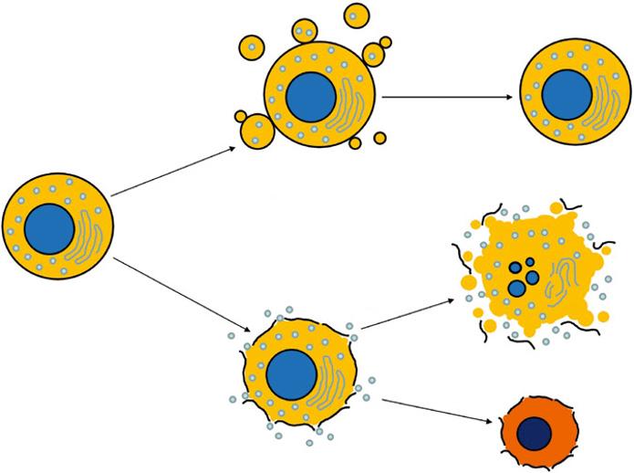
Figure 1.2 (a) Reversible change in the hepatocyte, which recovers completely after the causative process ceases. (b) Irreversible change in the hepatocyte, causing necrosis or apoptosis of the cell.
change
Restitutio ad integrum Apoptosis