SensorsandProbesforBioimaging
Young-TaeChang
Nam-YoungKang
Authors
Prof.Young-TaeChang DepartmentofChemistry
POSTECH,Pohang SouthKorea
Dr.Nam-YoungKang DepartmentofConvergence ITEngineering POSTECH,Pohang SouthKorea
CoverImage: CourtesyofYoung-Tae Chang
Allbookspublishedby WILEY-VCH arecarefully produced.Nevertheless,authors,editors,and publisherdonotwarranttheinformation containedinthesebooks,includingthisbook, tobefreeoferrors.Readersareadvisedtokeep inmindthatstatements,data,illustrations, proceduraldetailsorotheritemsmay inadvertentlybeinaccurate.
LibraryofCongressCardNo.: appliedfor BritishLibraryCataloguing-in-PublicationData Acataloguerecordforthisbookisavailable fromtheBritishLibrary.
Bibliographicinformationpublishedby theDeutscheNationalbibliothek TheDeutscheNationalbibliotheklists thispublicationintheDeutsche Nationalbibliografie;detailedbibliographic dataareavailableontheInternetat <http://dnb.d-nb.de>
©2023WILEY-VCHGmbH,Boschstraße12, 69469Weinheim,Germany
Allrightsreserved(includingthoseof translationintootherlanguages).Nopartof thisbookmaybereproducedinanyform–by photoprinting,microfilm,oranyother means–nortransmittedortranslatedintoa machinelanguagewithoutwrittenpermission fromthepublishers.Registerednames, trademarks,etc.usedinthisbook,evenwhen notspecificallymarkedassuch,arenottobe consideredunprotectedbylaw.
PrintISBN: 978-3-527-34960-9
ePDFISBN: 978-3-527-83434-1
ePubISBN: 978-3-527-83435-8
oBookISBN: 978-3-527-83436-5
Typesetting Straive,Chennai,India
Contents
1IntroductiontoBioimaging 1
1.1Color 1
1.2ColorfulMaterial 4
1.3LightSourceofBioimaging 6
1.4SubcellularImaging 12
1.5Cell-SelectiveImaging 14
1.6TissueandOrganImaging 14
1.7Whole-BodyImaging 15
1.8ProbesinBioimaging 15 References 16
2ChemicalSensorsandProbesforBioimaging 17
2.1HistoryofDyesinBiologicalStains 17
2.2BloodCellStaining 22
2.3BacteriaStainingUsingGramMethod 24
2.4FluorescentSensorsandProbes 25
2.5RepresentativeFluorescentCompoundsforBioimaging 29 References 33
3Organelle-SelectiveProbes 35
3.1Introduction 35
3.2CellPlasmaMembrane 40
3.3EndosomeandLysosome 47
3.4NucleusandDNA 50
3.5NucleolusandRNA 56
3.6ERandGolgiBody 58
3.7Mitochondria 62
3.8LipidDroplet 66
3.9Peroxisome 67
3.10Cytosol 68
3.11ExtracellularVesicle 69
3.12Non-membrane-BoundCondensate 72
3.13OrganelleProbesinLiveCellsandFixedCells 74
3.14ModelingfortheOrganelle-SelectiveProbes 75 References 80
4Live-Cell-SelectiveProbes 85
4.1Protein-OrientedLive-CellDistinction(POLD) 88
4.1.1EmbryonicStemCellProbe:CDy1 93
4.1.2NeuralStemCellProbes 99
4.1.2.1CDr3 99
4.1.2.2CDy5forNeuralStemCellDivisionMonitoring 103
4.1.3Tumor-InitiatingCellProbes 105
4.1.3.1TiY 105
4.1.3.2TiNIR 108
4.1.4MuscleCellProbes 110
4.1.5PancreaticCellProbes 113
4.1.5.1Pancreatic α-CellProbes 114
4.1.5.2Pancreatic β-CellProbes 114
4.1.6AmyloidProbe:CDy11 116
4.2Carbohydrate-OrientedLive-CellDistinction(COLD) 118
4.2.1Lectins 121
4.2.2EmbryonicStemCellProbes:CDg4andCDb8 122
4.2.3Gram-PositiveBacteriaProbe 122
4.2.4BiofilmProbe:CDy14andCDr15 124
4.3Lipid-OrientedLive-CellDistinction(LOLD) 128
4.3.1FilipinasaCholesterolProbe 129
4.3.2LipidDropletProbes 129
4.3.3NeuronProbes 130
4.3.3.1NisslStainsasNeuronBodyProbe 131
4.3.3.2PlasmaMembraneDyesasNeuronalNetworkProbe 131
4.3.3.3NeuOasaUniversalNeuronProbe 132
4.3.4BLymphocyteProbe:CDgB 134
4.3.5ActivatedCD8+ LymphocyteProbe:Probe41 138
4.3.6ApoptoticCellProbe:Apo-15 139
4.4Gating-OrientedLive-CellDistinction(GOLD) 139
4.4.1CellImagingProbesthroughPhagocytosis 140
4.4.2ProbesThroughSLCTransporters 143
4.4.3ProbesThroughGlucoseTransporters 144
4.4.4NaïveEmbryonicStemCellProbe:CDy9 146
4.4.5NeurotransmitterMimeticProbes 147
4.4.6AstrocyteProbe:SR101 149
4.4.7Subtype-SpecificMacrophageProbes:CDg16,CDr17,CDg18 150
4.4.7.1CDg16forActivatedMacrophage 150
4.4.7.2CDr17forM1Macrophage 152
4.4.7.3CDg18forM2Macrophage 153
4.4.8B-Cell-SelectiveProbeThroughGOLDMechanism 153
4.4.9BacteriaProbesThroughTransporters 154
4.4.10ProbesThroughABCTransporters 155
4.4.11Background-FreeTameDye 157
4.5Metabolism-OrientedLive-CellDistinction(MOLD) 160
4.5.1SubstrateforProteasesinExtracellularMatrix 160
4.5.1.1MMP12SubstrateforActivatedMacrophageProbe 162
4.5.1.2CathepsinSSubstrateforTumor-AssociatedMacrophageProbe 162
4.5.1.3ElastaseSubstrateforNeutrophilProbe 163
4.5.1.4GranzymeSubstrateforNaturalKillerandCytotoxicTCellProbe 163
4.5.2MicrogliaProbe:CDr10andCDr20 165
4.5.2.1CDr10aandbforMicrogliaImagingamongBrainCells 165
4.5.2.2MicrogliaProbeCDr20throughUgt1a7c 169
4.5.3NeutrophilProbe:NeutropG 170 References 172
5ExVivoTissueImagingProbes 179
5.1Immunohistochemistry 181
5.2TissueImagingwithNucleicAcidProbes 186
5.3TissueImagingwithSmall-MoleculeProbes 186
5.3.1PancreaticIsletImaging 188
5.3.2NeuronalTissueImaging 191
5.4OrganoidasModelofTissueandOrgan 194
5.4.1BloodVessel3DModel 195
5.4.2TumorOrganoidforDrugScreening 196 References 196
6InVivoWhole-BodyImagingProbes 199
6.1ElaNIRforElastinImaginginMouse 200
6.2ProbesforExposedNeuroninZebrafishEmbryo 201
6.3NeuOforWhole-BodyNeuronImaginginZebrafish 202
6.4LipidGreenforFattyTissueImaginginZebrafish 203
6.5BloodVesselImaginginZebrafish 204
6.6ProbesforBoneImaging 205
6.7ProbesforPancreaticIsletImaging 206
6.8ProbesforEyeImaging 208
6.8.1OpticalCoherenceTomographyforRetina 209
6.8.2FundusPhotographyforBloodVesselImaginginRetina 210
6.8.3NeuronImagingonRetina 210
6.8.4BacterialInfectiononCornea 211
6.9MacrophageImaginginIschemiaandInflammation 213 References 214
7ImagingforBiologicalEnvironmentandFunction 217
7.1pH 218
7.2MetalIons 221
7.2.1Na+ andK+ 222
7.2.2Ca2+ 225
7.2.3Mg2+ 227
7.2.4MetalIonSelectivityofFluorescentSensors 228
7.2.5IronIon 230
7.2.6Zn2+ 230
7.2.7CopperIon 231
7.3Metabolites 232
7.3.1ATP 232
7.3.2NADH 233
7.3.3Histamine 234
7.4Viscosity 234
7.5Temperature 238
7.5.1ERThermometer 238
7.5.2MitochondrialThermometer 240
7.5.3Organelle-SpecificFluorescentThermometers 241
7.6ReactiveOxygenSpeciesandReactiveNitrogenSpecies 241
7.6.1Superoxide 243
7.6.2H2 O2 245
7.6.3ONOO 247
7.6.4HOClandHypochlorite 249
7.6.5HydroxylRadical 251 References 252
8ImagingforDisease 259
8.1Introduction 259
8.2CancerImaging 260
8.2.1ImagingbyCancer-SpecificBiomarkerBinding 261
8.2.2ImagingbyCancer-SpecificMetabolism 263
8.2.3ImagingbyCancer-SpecificTransporter 267
8.2.4ImagingbytheChangedEnvironmentofCancer 268
8.2.5CirculatingTumorCell(CTC) 269
8.2.6CancerCellLineforImaging 269
8.2.7AnimalModelsofTumorImaging 271
8.2.8ExVivo3DTumorCultureModel 275
8.2.9ClinicalImagingofTumorforDiagnosisandPrognosis 277
8.2.10IntraoperativeImagingofTumor 278
8.3NeurodegenerativeDiseaseImaging 278
8.3.1ADImagingThroughAβ AmyloidAggregates 278
8.3.2ADImagingThroughTau 281
8.3.3AnimalModelforAD 283
8.4InflammationImaging 284
8.4.1InflammationImagingbyEnvironmentalChanges 284
8.4.2InflammationImagingThroughImmuneCells 285
8.4.3InflammationAnimalModel 286
8.5DiabetesImaging 286
8.6LiverDiseaseImaging 288
8.7Aging 289
8.8Theranostics 289 References 290
9Non-opticalImagingProbes 297
9.1UltrasoundImagingProbes 297
9.2X-RayContrastAgents 298
9.3MRIContrastAgents 301
9.4SPECTProbes 303
9.5PETProbes 306
9.5.1PETProbesforTumor 307
9.5.2PETProbesforBrainFunction 309
9.6Multimodality 310 References 311
10FluorescenceImagingTechniquesandAnalysisMethods 313
10.1MulticolorImaging 313
10.2RatiometricMeasurement 315
10.3FluorescenceLifetimeImagingMicroscopy 316
10.4ConfocalFluorescenceMicroscopy 316
10.5Two-PhotonExcitationFluorescenceImagingandHarmonic Generation 317
10.6SelectivePlaneIlluminationMicroscopy 318
10.7TotalInternalReflectionFluorescenceMicroscopy 319
10.8Super-ResolutionImaging 320
10.9Single-MoleculeImaging 322
10.10PhotoactivationofCagedMolecule 323
10.11FluorescenceRecoveryAfterPhotobleaching 324
10.12FlowCytometryTechnique 325 References 327
11PerspectivesforFutureProbeDevelopment 329
11.1DesignofSelectiveProbes 329
11.2DiscoveryofSelectiveProbesbyScreening 331
11.3FutureProbeDevelopment 336 References 337
Appendix 339 Index 341
IntroductiontoBioimaging
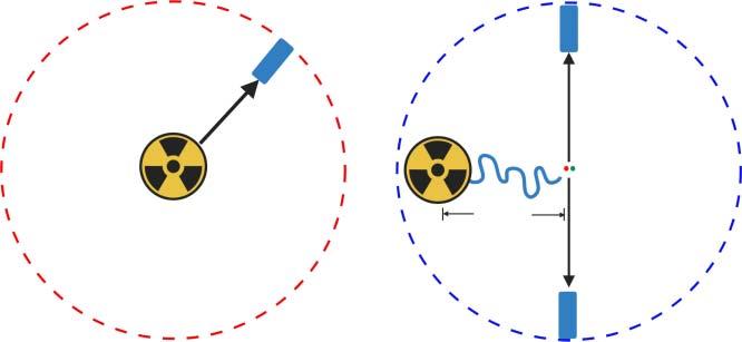
Bioimagingcanbedefinedasvisualizationofabiologicalobject.Themostbasic bioimagingmaybejust“seeing”thelivingobjectusingourowneyes.Thisfunction iscalled“vision”andtheprocedureismediatedbyvisiblelight.Thevisiblelight isapartofelectromagneticwaveinthewavelengthrangeof400–700nm,andthe imageinformationisgeneratedbytheinteractionbetweenlightandobject,suchas reflection,scattering,anddiffraction.Thegeneratedinformation-richlightpackage travelsandreachesoureyes.Thefocusedlightthroughlenswouldbeprojectedto thescreenasinacamera.Theretinainoureyeisthescreenoftheimage,whichis composedofthetwo-dimensionalarrayofopticnerves.Thephotoninlightsignal (containingtheinformationoftheobject)reachestheretinaandactivatesoptical neurons,andthesignalistransferredtothebrainandisreconstructedintotheimage oftheobjectbyneuronalprocessing.Eventhoughthescreenistwo-dimensional, theprocessedimagesviatworetinasprovidethree-dimensionalinformationabout theshapeanddistanceoftheobject.Visiblelighttravelsataso-calledspeed oflight(3 × 10⁸ m/s),sotheinformationtransferinthevisioncouldbealmost instantaneous.Ifthereisapossibledelay,itmaybefromthesignaltransitionstep fromtheopticalnervetothebrainandtheinformationprocessingtimeinthebrain.
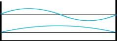
Batsliveinthedarkenvironmentwithoutenoughenvironmentallightfor vision.Instead,theyuseultrasoundforbioimagingplatform.Ifotherconditions aresame,thelightvisioncouldbemilliontimesfasterthanultrasonicsensing (340m/s)(Figure1.1).Amongallthesensors,lightvisionisthefastestandmost information-richsystem.Therefore,theinventionofeye(inmoregeneralterm, photoreceptor)isoneofthemostdramaticeventsintheevolutionoflife.Dueto thehighqualityandalsohugequantityofinformation,visionisthemostimportant sense,easilyaccountingformorethan90%ofinformationwereceivethroughall othersenses,includinghearing,taste,smell,andtouch.
1.1Color
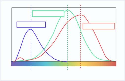
Visiblelightsensingnotonlygeneratesblack-and-whiteimages,butalsocanprovidecolorinformation.Thevisiblelightiscomposedofaspectrumofelectromagneticwaveintherangeof400–700nm.Humaneyehasthreecolorphotoreceptors, SensorsandProbesforBioimaging,FirstEdition.Young-TaeChangandNam-YoungKang. ©2023WILEY-VCHGmbH.Published2023byWILEY-VCHGmbH.
Figure1.1 Visionthroughlightandsoundindifferentspeed.
Figure1.2 Threecolorreceptorsandtheirsensitivitytodifferentwavelengths. ofwhichthemaximumsensitivityisforblue(445nm),green(535nm),andred (575nm)(Figure1.2).Forexample,whenwereceive445nmlight,wesenseitas abluecolor,and575nmlightasaredcolor.Therefore,colorrecognitionistheabilityofsensingdifferentwavelengthsoflight.And,thetermspectroscopyisderived fromspectrum,i.e.spectroscopyisthestudyofthewavelength-dependentinteractionbetweenthelightandtheobject.
Ifwehavethreecolorreceptors,thendowerecognizeonlythreecolors?No,itis not.Atleast,wegivesevennamesofcolortotherainbow!Ourcolorsensorshavethe maximumsensitivitytoaspecificwavelength,butthesensingwavelengthsarebroad andoverlapwitheachother.Iftheeyereceives560nmlight,bothgreenandred receptorsareactivated,andwesenseitasayellowcolor.Thelightwith590nmwill morestronglyactivateredreceptorsandlessstronglygreenreceptors,makingthe colorasorange.Thatmeansourcolorsenseisdeterminedbytheratioofthethree
Blue receptor
Green receptor
Red receptor
Figure1.3 Comparisonbetweenblack-and-whitepictureandcoloredpicture.
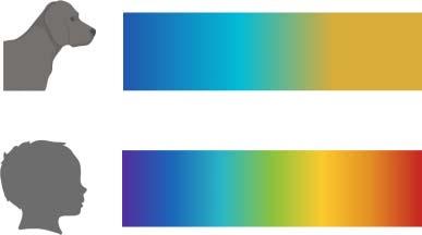
Figure1.4 Thecolorspectrumof dogsandhumans.
dog’s
receptors’activationdegree.Usingthethree-colorreceptors,thedistinguishable colorsbyhumaneyesaremorethan10,000!Withthisability,wecanfindourfood (e.g.redapple)better,alsoourenemy(e.g.redant)faster(Figure1.3).Wecanimaginehowusefulthisabilityistohelpussurvivebetterduringtheevolutionprocess. Interestingly,thisthree-colorrecognitionisnotcommontoallanimals,evento mammals.Onlyourveryclosecousinssuchaschimpanzeesandgorillashavethree colorreceptors,buteventhennotallthemonkeysdo.Includingourremotecousins, dogsandcowshaveonlytwo-colorreceptors.Itsoundstrivialwhetheritistwoor three.But,withtwocolorreceptors,thedistinguishablecolorsarenarroweddown onlytothelevelof100!Itisthedifferencebetweentwo-andthree-dimensionalcombinationpower(Figure1.4).Therefore,thevisionsofcowsanddogswouldbemuch moreboringthanthecolorfulflowersandspectrumofrainbowwesee.Thisiswhy manysensorsaredesignedforcolorchangetoachievemaximumeffecttothenaked eyes.Oureyesareawonderfulcolorsensorysystem!
The
view
The human’s view UV
Thereisafunnystoryinthebullfight.Thefightersuseredclothtostimulatebulls, asredcolormayberelatedtotheimageofblood.Funnythinghereisthebullmay seeredcolormorelikedarkgrayratherthanbloodyred.Theredclothistostimulate theaudience,notthebullsatall!
Colorblindnessariseswhenpartofthecolorreceptorisdefunctionalized.In humans,mostcommontypeisgreen-redblindness,whichoccurswheneither greenorredreceptorhasproblem.Ifyoulookatthereceptorpropertycarefully, youmayrealizethatthemaximumwavelengthsofgreen(535nm)andred(575nm) receptorsarerathercloser,comparedtothatofblue(445nm).Wecallthereceptors greenandred,buttheyaremorelikeyellowandorange.Tomaximizethecombinationpowerincolorcontrast,thisdesignmaynotbetheoptimumchoice.Ifwe designthecolorpixelofacomputerscreen,wemaychoosemoreevendistribution ofthecolors,suchas465,525,and630nm[1].Notsurprisingly,thegreenand redreceptorsarestructurallyclosertoeachother,implyingthattheyevolved fromthecommonancestor.So,wecanimagine,alongtimeago,wealsohadtwo colorreceptorssimilartodogsorbulls(blueandyellow),andtheyellowreceptor divergedtotworeceptors,greenandred.Withoutthisevolutionofcolorreceptors, wemightnotbeabletoenjoythebeautifulsunset!
1.2ColorfulMaterial
Thesyntheticcolorfulmaterialsaremainlyorganicdyesandinorganicpigments. Conventionally,dyeisdefinedasthematerialthatimpartsitscolortoothersubstances,suchasfabricortissue.Usually,dyesaresolubleinsolvents,butpigments areinsolublesolids.Forprintingpurposes,pigmentpowderneedstobedispersed intoaliquidbinderbeforeuse.
Ontheearth,thestrongestlightsourceisthesun.Tominimizethebackground oflightsensing,ourvisionarysystemadjustsoursensorstorecognizethesunlight asabackground,called“purewhite.”Whitelightisnotthestatusofnocolor,but itisthecollectionofallthecolorsincludedinthesunlight.Thecolorsofthewhite lightcanbemanuallyseparatedintoaspectrumbyaprismthroughaprocessof dispersion,whichisthesamemechanismofrainbowformation.Therefore,whiteis thecombinedcolorofallthevisiblelightintherainbow.
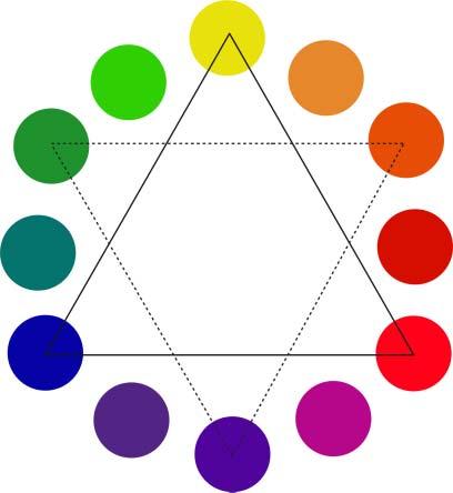
Thecolorofthecolorfulmaterialsisdeterminedbythewavelengthofthe absorbedlight,i.e.leftoverreflectedcolorafterabsorptionofwhitelight.Therefore, theappearedcoloriscomplementarytotheabsorbedcolor.Theconceptofcomplementarycolorhasbeenknownforalongtimeandiswidelyusedinpaintingartfor vividcolorcontrast.Eventhoughthewavelengthofvisiblelightisinlinearscale (400–700nm,violettored),ourcolorreceptorsdeceiveourcolorrecognitiondue tothetiringofreceptors.Therelationshipofthecomplementarycolorinourcolor sensingsystemisdescribedinacolorwheel(Figure1.5).
Achromophoreisthepartofthemoleculewhichisresponsibleforthecolor. Thechromophoreofinorganicpigmentsisusuallyistransitionmetal,whichhasa visiblelightrangeofelectronexcitationenergy.Thechromophoreoforganicdyes
Figure1.5 Colorwheel.
isalong-conjugateddoublebondsystem.Thelightabsorptionhadbeenmodeled inearlyquantummechanicserathroughaparticle-in-a-boxmodel,whichlater ledtoSchrödingerequationforatomicstructureofelectrons.Interestingly,the organicconjugationsystemcouldbedescribedasaparticle-in-a-boxmodel,where the σ bondelectronsdefinethesizeoftheboxand π electronsaretheparticles inthebox.Astheboxsizebecomesbigger,thewavelengthofabsorptionlight getslonger,throughthenarrowingelectrontransitiongap.Whentheabsorption maximumreachestheboundaryofvisiblelight(violetcolor),theappearedcolor ofthematerialwouldbethecomplementarycolorofviolet,yellow(colorsin oppositedirectioninthecolorwheel).Iftheconjugationgetslonger,theabsorption maximummovesfromviolettoblueandthengreen.Accordingly,theappeared colorchangesfromyellowtoorangeandthenbrown.Youmayrecalltheoldbooks turnintotheyellowcolorfirst,andthenchangeintoreddishtone.Thisistheresult oftheextensionofconjugationinthelignincomponentinthepaperpulpandis oneoftheexamplesoftheorganicdyemodelofparticleinabox(Figure1.6).
However,iftheconjugationistoolong,anyoxidationorreductionreactioninthe middleofthechromophorewillbreaktheconjugationbridge,whichistheprincipleofbleachingagents(eitheroxidantsorreductants).Thatmeansthedyewitha longconjugationsystemisweakforthechemicaldamages,andusuallythecolorof naturallyoccurringorganicdyeseasilydiminishovertime.Asaresult,thecolorof simplecarbon-conjugatedsystemsisnotvivid,asshowninthepaperofoldbooks. Inthenineteenthcentury,Germanorganicchemistsopenedthewaytosynthetic dyestoreplacethenaturaldyes.Addingelectron-donatingand-acceptingmotifsat
Secondary color
Primary color
Orange yellow
Orange Orange red
Red
Purple red Purple
Blue red
Blue
Blue green Green Yellow green Yellow
+
Small boxes with short conjugationConjugation-extended bigger box
Figure1.6 Conjugationandthemaximumwavelengthoftheabsorptionlight.
Figure1.7 Representativestructureofdyes.
eachendoftheconjugationsystemprovidesastrongerdipole,whichmakesthe conjugationeffectlongerandalsomakestheabsorptionstrongertogiveavivid color.So,mostofthesyntheticdyesarecomposedoftheconjugationsystemwith electron-donatingand-acceptingmotifsateachend(Figure1.7).Thewavelength ofabsorptionoftheorganicdyescanbepredictedbymolecularorbitalcalculations, andPariser–Parr–Pople(PPP)methodisoneofthebest-knownmodels[2].
1.3LightSourceofBioimaging
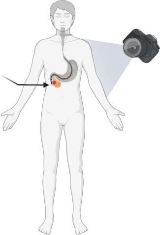
Ifbioimagingisvisualizingabiologicalobject,whichpartofthebodyistheobject? Ifwetakethedaylightphotographyasanexample,wecanuseacasualcameracatchingthevisiblelighttovisualizethesurfaceofourbody.Thesurfaceimagingiseasy, andthedamagebylightexposuretoskinistrivial.However,whatifwewantto visualizetheinsideofourbody?Usuallyweusecatheterscomposedofametaltube withalightsourceandcameraonthetip,andinsertthemthroughthemouthor anustovisualizethegastrointestinaltract(Figure1.8).Whilewecallthistechnique asanendoscopy,isthisreallyinsideofourbody?Well,comparedtotheexposed skinsurface,thegastrointestinaltractseemsmorelikeahiddenpartofourbody. But,topologicallyspeaking,ifweconsiderourbodytobedonutshaped,thesurface ofthegastrointestinaltractshouldbeconsideredaspartoftheoutside,notthereal inside.
Incontrast,ifthecameravisualizesthebeneathskinarea,wemayconsiderit asarealinnerpartofourbodyimaging.Forthisrealendoscopy,onepossibleway maybetoputthecamerapenetratingtheskintoreachthetargettissue.However,
Triphenylmethane
Figure1.8 Regularcameraand endoscopy.
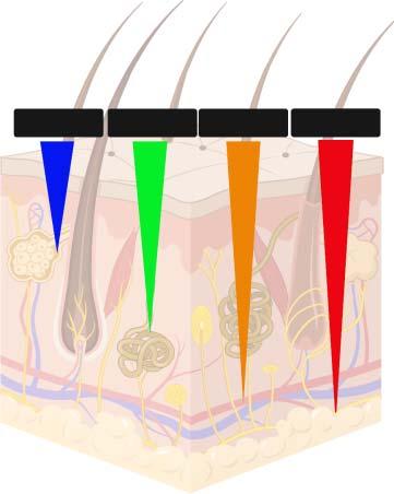
ifitisnotreallynecessaryfortreatmentpurposes,suchaninvasiveapproachmay notbedesirableforhumansoranyliveanimals.Ifitisnotphysicalpenetrationof thecamera,howaboutlightpenetration?Iflightpenetratestheskinreachingthe innerspaceandreturnsbackwiththeinformationofthetargetarea,itwouldbe muchlessdamagingthanphysicalinsertionofthecamera.Inthiscase,thepenetrationdepthoflightwouldbeanimportantfactor.Ifourskinislikeatransparent jellyfish,theinnerworldimagingmaybestraightforward.Theterm“transparency” itselfimpliesfreepenetrationofvisiblelight.Unfortunately,ourbodyskinisnotso transparent,andmostvisiblelightcanpenetrateatmostseveralmillimetersofdepth undertheskin.Then,howcanweincreasethepenetrationdepthoflightthroughthe tissue?
Visiblelightliesinthe400–700nmrange,andthereareotherlightsources outsideofvisiblelight.Theshorterwavelengthofvisiblelightcomprisesultraviolet (UV),X-ray,and γ-ray,etc.Thelongerwavelengthmakesinfrared(IR),terahertz light,microwave,andradiowave.Comingbacktotissueimaging,thepenetration ofelectromagneticwavesisdiminishedbymainlyscatteringandabsorptionofthe lightsourceinthetissue.Absorptionwavelengthisdependentonthetissuecomposition,butthescatteringisusuallyhigherinshorterwavelengthlight.So,longer wavelengthlighttendstopenetratebetterthantheshorterwavelengthlight,by reducedlossbyscattering.Forthisreason,near-infrared(NIR:longerthan700nm lightupto1000nm)lightisapopularopticalimagingsourcefornoninvasivetissue imagingorwhole-bodyimagingofsmallanimalssuchasmice.Recently,even longerwavelengthof1000–1700nmispopularlyusedforbioimagingasthesecond NIRwindoworNIR-II[3].Withfurtherreducedscatteringandnegligibleautofluorescence,NIR-IImayprovideahighersignal-to-noiseratioanddeepertissue
Camera
Endoscopy
Figure1.9 Penetrationoflightintothe skin.
penetrationthantheconventionalNIRimaging(Figure1.9).Asoureyescannot senseNIRdirectly,thedetectedNIRshouldbeconvertedtoartificialvisuallight imageasinthenightvisiongoggleofbattlefield.Thegreencolorinthenightvision goggleimageisaprocessedartificialcolor.Greenistheusualchoiceofcolordue toitsbestsensitivitytooureyes.NIRisbetterthanvisiblelightforthepenetration depth,butstillitisdifficulttoproceedfurtherthanacentimeterintothetissue.
Theevenlongerwavelengthlightsources,suchasmicrowavesorradiowaves, havebetterpenetrationthroughthewholebodyandhavebeenusedinmagnetic resonanceimaging(MRI).MRIrequiresahighmagneticfieldtoseparatethe nuclearspinenergystatusofprotonsinthebody.Theseparatedenergygapof nucleusabsorbsmicrowavesandMRIdetectsthesignaloftherelaxationofthe absorbedmicrowavelight.Protonsindifferentenvironments(suchaswateror lipid)generatedistinguishablesignalsandthroughacomputedtomography(CT), three-dimensionalsectionalimagescouldbeconstructed.MRIisanoninvasiveCT technique,especiallyusefulforsofttissue(whichcontainsprotons)imaging.
Ifwegototheotherdirectionofshorterwavelengthlight,therearestillpossibly differentapplicationsinbioimaging.IntheX-rayrange,thewavelengthoflightisa hundredtimesshorterthanvisiblelight,andthephotonofX-raywouldbesmalland rigid.Ifvisiblelightphotonislikeatennisball,X-rayislikeaneedleandcaneasilypenetratesoftmatter.Thus,X-rayimagesmainlyshowtherigidbonestructure, throughwhichX-raycannotpenetrate(Figure1.10).ByadoptingCTtechniques, X-raythree-dimensionalimaginghasbeenwelldevelopedevenbeforeMRIisintroduced.Duetotheorderofhistoricaldevelopment,conventionallytheterm“CT”is usedfor“X-rayCT”,unlessotherdescriptionisprovided.
AlthoughmostoftheX-raylightdoesnotinteractwithsoftpartofthebody, inamolecularlevel,therecouldbeasmallamount,butstrongdamageto
Hair follicle
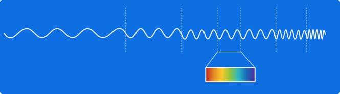
Figure1.10 Electromagneticwavesasthelightsourceofbioimaging.
thebiomoleculescanoccurbybreakingthecovalentbondsorionization.The accumulateddamagesinDNAcancausemutationsofcells,resultingincancerin somaticcellsandmutagenesisinfetus.So,excessamountofexposuretoX-rayis notrecommendedduetohealthconcerns,especiallyforpregnantwomen.
γ-RayhasashorterwavelengththanX-rayandthehigherenergyallowsitspenetrationeventhroughbones,thehardestpartofourbody.Theintensive γ-raycan beusedfortumortreatment,whichiscalledas γ-knifetechnique.Tominimizethe damagetonormaltissue,multiplesourcesof γ-rayareusedfromdifferentdirections,andonlythetumorsiteisfocusedtoaccumulateahighdensityof γ-ray.In principle, γ-rayalsocanbeusedforbioimaginginasimilarwayofX-rayimaging, butitisnotcommoninpractice.Instead, γ-ray-generatingradioactivematerialsare usedasimagingagents.Inthiscase,the γ-rayisnotprovidedfromoutsideasinthe X-raymethod,butisirradiatedfrominsideofthebody,throughanadministered imagingprobeintothetargetsiteofthebody.Thepositionoftheisotopecouldbe imagedthrougha γ-camerasimilartoanX-rayfilm.WhenCTtechniqueiscombined with γ-ray-generatingradioactiveisotope,athree-dimensionalsingle-photonemissioncomputedtomography(SEPCT)imagingisalsopossible.Asimilar,buthigher performancetechniqueispositronemissiontomography(PET).InPET,insteadof adirect γ-ray-generatingisotope,apositron-generatingisotopeisused.Positronis apositivelychangedelectron,akindofanti-particleofelectron.Whenapositron meetsanelectron,theyareannihilated,generatingonepairof γ-rayphotons.Asthe two γ-rayphotonstravelindirect180∘ providingricherinformationfortheoriginal positionofpositron,usuallythespatialresolutionofPETisbetterthanSPECT.Itis noteworthythatX-rayusesanexternallightsourcefortheimaging,butSPECTand PETuseendogenous γ-raygeneratedfromanisotope-labeledprobe(Figure1.11). ThatiswhySPECTandPETarecalledmolecularimagingtechniques,incontrastto X-rayimaging.
Asshownearlier,electromagneticwaveswithdifferentwavelengthsfromvisible lightalsocanbeusedasthelightsourceofvariousimagingtechniques,whencoupledwithaproperdetectororcamerasystem.Thedifferentwavelengthsoflight renderdifferentmodesofinteractionwithmatters,andeachcangenerateunique informationforthetargetobject.Therefore,unexploredareasofelectromagnetic waveswouldprovidenovelchanceofnewimagingtechnologyormodality.Terahertzlightissuchanemergingnewsourceoflight.
Visible light
Figure1.11 Endogenous γ-rayimaginginSPECTandPET.
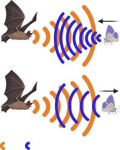
SonarReturning sound waves
Figure1.12 Soundimagingof bats.
Notonlyelectromagneticwaves,soundwavesorseismicwavesalsocangenerate processedimagesthroughinteractionwithmatters.Thebat’svisionthroughultrasonicwaveswouldbeagoodexample.Combiningelectromagneticwavesandsound wavesforimprovedoruniqueimagingtechnique,suchasphotoacousticimaging,is alsoapowerfulvisualizationtechnique(Figure1.12).
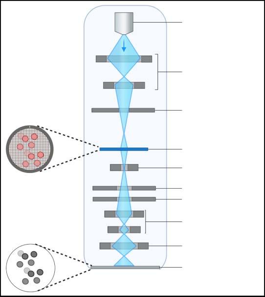
Electronbeamisanothersourcetoprovideultrahigh-resolutionimagingof materials.Thereareseveralmodesofelectronmicroscopy,suchastransmission electronmicroscopy(TEM)andscanningelectronmicroscopy(SEM).InTEM,an ultrathinsampleisirradiatedwithanelectronbeamandthetransmittedelectrons
Electron source
1.3LightSourceofBioimaging
Figure1.13 Transmissionelectronmicroscopy.
Condenser lenses
Condenser aperture
Sample
Objective lens
Objective aperture
Selected area aperture
Intermediate lenses
Projective lens
TEM image
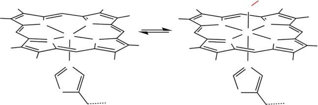
areusedforthetwo-dimensionalimageconstruction,whichissimilartoX-ray imaging.Inthesampleareawithahighelectrondensity,theinputelectronbeam wouldbescatteredandmaynottransmit,generatingthedarkcontrastinTEM image.Therefore,heavymetalsareabsorbedtothesampletoenhancethecontrast, andtheprocedureiscalledelectronstaining.Theelectronstainingcanalsobe achievedbyanorganicdye.Afterdiaminobenzidine(DAB)isstained,through photooxidation,anelectron-denseprecipitatecanbeformedtoincreasetheTEM contrast,whichissimilartodyestainingintheopticalimaging.
InSEM,theincidentelectronbeaminteractswiththesurfaceatomsofthesample andgeneratesbackscatteredelectronsorsecondaryelectrons.Theincidentbeamis focusedonasamplespotandscanthesurface,andthedetectorsarelocatedinthe samesideoftheinputbeam.Asaresult,SEMimageshowsthesurfacemorphologywiththree-dimensionalinformationespeciallyprovidedbysecondaryelectrons. TheresolutionofSEMimageisinnanometerrange,andusuallyTEMhashigher resolutionthanSEM.Whileopticalimagingsuffersfromthediffractionlimitin sub-micrometerrange,electronmicroscopyprovidesmuchhigherresolution.Bythe imagingresolutionscale,electronmicroscopycouldbecalledas“nanoscopy,”rather thanmicroscopy.Bothtechniquesrequirevacuumconditionfortheimagingdueto theelectronbeamusage(Figure1.13).
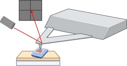
Atomicforcemicroscopy(AFM)isanothernanoscopytechnique.Usingthephysicalcontactforcesensing,thephysicalprobescansthesamplesurface,providingthe informationofsurfacemorphology.TheresultissimilartoSEMimages,butwith amuchhigherspatialresolution.ItisinterestingtocompareAFMwithSEMas AFMdoesnotrequirealightorelectronbeamsource.Also,AFMdoesnotrequire theelectronstain,whichmaychangethesurfacelandscape.However,thephysicalcontactoftheprobewiththesamplesurfacemaypartiallydamagethesample, especiallyduringtheclose-contactmodeprocess.AmodifiedAFMtechniquealso allowsliquidenvironmentinadditiontothevacuumconditionfortheimaging,and morebiologicallyrelevantsamplescouldbeimaged,suchaslive-cellsurfaceimaging(Figure1.14).
1.4SubcellularImaging
Whentheobjectofvisualizationistoofarfromoureyes,weuseatelescope.Ifthe objectistoosmall,weuseamicroscope.Superficially,theymayseemtouseopposite principles,butactuallytheyaresimilarinasensethatthey“magnify”the“toosmall images”toasensiblesizefornakedeyes.Oneisfortoosmallimagesduetothelong distanceoftheobjectandtheotherisfornearby,butphysicallytoosmallobject.If theyaresimilar,canweusetelescopeinsteadofmicroscopeforthesmallobjector viceversa?No,wecannot.Whatisthedifference,then?Thedifferenceliesinfocal distancesdependingonthepositionoftheobject.Inatelescope,thefocusison thelongdistance,andinamicroscope,thefocusisonthesamplesliderightunder thelens.
Now,let’sfocusonthemicroscopeforvisualizingsmallobjectsinbiological systems.Thebasicunitoflifeiscell.Forunicellularorganisms,asinglecellis anindividualorentity.Inmulticellularorganisms,cellsgathertogethertomake tissue,andtissuesmakeorgan,andorgansassembletomakeanindividualbody. Thereasonwhyacellisthebasicunitoflifeisthateachcellcontainsthewhole
Figure1.14 Atomicforcemicroscopy.
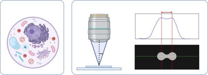
Subcellular imaging
Figure1.15 SubcellularimagingandAbbe’slimit.
genomicinformationoftheindividual.Inotherwords,startingfromanysinglecell, inprinciple,wecanreconstructthewholebody.
Theusualsizeofcellsinanimalsorplantsisaround10 μm,andunicellularbacteriaareabout1 μminsize.Whilebacteriacellstructureisrelativelysimple,animalor plantcellshavecomplexintracellularstructure,calledorganelles,suchasnucleus, mitochondria,lysosomes,Golgibody,andendoplasmicreticulum(ERs).Theintracellularorganellesareusuallyabout1 μmorsmallersize.Whenlightencounters anobjectwithasimilarsizetothewavelength,thelightpathisalteredbydiffraction.Thevisiblelightisin0.4–0.7 μm(400–700nm)andiftheobjectisabouthalf micrometerorsmaller,theimagebecomeblur.ThisisknownasAbbediffraction limit,namedafterErnstAbbewhofounditin1873,andisconsideredasthephysicallimitoftheopticalresolution(Figure1.15).Therefore,thephysicalsizelimitof amicroscopicimageisabout ∼μmrange.
Toovercomethesizelimit,severalopticalandmathematicaltricksweredevelopedinto“super-resolution”techniquesorso-callednanoscopy,whichmeans nanometer-resolutionimaging.Inadditiontothesizelimit,organellesareusually transparent,sotheopticalvisualizationisfurtherchallenging,asitisdifficultto distinguishdifferentorganelles.Thatiswhyorganelle-selectivedyesarewidelyused forvividsubcellularorganellevisualization.Inotherwords,bioimagingisaprocess ofvisualizingabiologicalobject,otherwiseinvisible.Mostofthecellimageswe haveinourmindare“stained”imagesratherthannaturalcellimages.Forexample, chromosome,ascondensedformofDNA,means“colorbody(chromo-some)”asit iseasilystainedbydyes.Youmayhaveseenthechangeofthechromosomeduring thecelldivision,suchascondensation,alignment,anddivisionofDNA.Itimplies thatmostofthechromosomeimagesarealsoobtainedfromDNA-stainedcells ratherthanintactnaturalcells.Bythesametoken,ifthereisaselectivedyefor eachorganelle,itwouldbepossibletoseespecificorganellestandingoutfroma transparentbackground.Theseselectivedyesarecalledorganelle-selectiveprobes, andifthedyeschangetheircolorsuponbindingtothetarget,theycanbecalled assensors.Therefore,thedefinitionofprobesembracessensors.Inotherwords, sensorsarespecialtypeofprobesinbioimaging,providingtheinformationof changeofthetarget.
1.5Cell-SelectiveImaging
Inamulticellularorganismormixedbacteriacommunity,distinctivevisualization ofdifferentcellsorbacteriawouldbecriticalforthestudyofintercellularinteraction. Ifthedifferentcellshaveuniqueshapesandsizes,itwouldbeeasytodiscriminate them.However,inmanycases,distinctionofonetypeofcellfromothersisgenerallydifficultduetotheirsimilarappearanceunderbright-fieldmicroscope.Eventhe samekindofcellsmayhavedifferentstagesofdevelopmentordeathprocess,showingoffdifferentmorphology.Consideringthefactthatallthecellsinthesamebody containexactlythesamegeneticinformation,thediscriminationoftheirphenotypic differenceisthekeyforthestudy.
Toovercometheproblem,cell-selectiveprobeshavebeenexploredforalongtime. Antibodieshavebeenthemostcommonprobesforthecelldistinctionandarewidely used.Hundredsofantibodieshavebeendevelopedandvalidatedforcelldiscriminationandimaging.Whileuseful,duetotheirhighmolecularweightof150kDa,their accesstotheintracellulartargetinlivestatusisintrinsicallylimited.Eventhough thebindingtargetofantibodiesisonthecellsurface,theyareusuallyfunctionallyimportantenzymesorreceptors.Asaresult,antibodiesofteninducefunctional influenceinthetreatedcells,whichisnotdesirablefornormalcellstudy.Alternativesolutionmaybeasmartsmallmoleculeprobe,whichmaycomplementthe antibodies’weakpoints,especiallyfortheintracellulartarget.
Notonlyforourowncells,wealsoneedtodistinguishandvisualizeforeign lifeforms,asourbodyisalwaysinteractingwiththem.Forexample,ourbody hostshugenumbersofbacteriaasguestsinsimilarorevenhighernumberthan ourowncells,whichiscalledthemicrobiome.Thebacteriainthemicrobiome establishedsymbioticrelationshipswithourbodyandmajorityofthemarenot harmfultous.But,ifwegetpathogenicbacterialinfection,figuringouttheidentity ofthebacteriawouldbeurgentandimportantformakingdecisionoftheproper treatment.Themorphologicaldifferencemaynotbeinformativeenoughtogeta gooddiscriminatinginformation.Media-selectiveculturingisastandardtestfor theidentification,buttheprocesstakesdaysoftime,andalsotheidentification islimitedonlytotheknownstrainsfortheirculturecondition.Whilepolymerase chainreaction(PCR)-basedgeneticanalysisisgettingmoreandmorepopularfor highaccuracyandsensitivity,theneedforanin-siteimagingprobeincreases forfasteranalysisandfunctionalmonitoringthroughthevisualimages.Sofar,such anefficientandpracticalcell-selectiveprobeisyettobedeveloped.
1.6TissueandOrganImaging
Whencellsgathertomaketissuesandorgans,atangiblephysicalstructureemerges, andmacroscopicimagingtechniqueisrequired.Fordiagnosisofdiseases,oftena biopsy(tissuesamplingfromlivebody)procedureisrequiredfortissueimagingor biochemicaltesting.Usually,thetissuesarestainedwithdyesandimagedtodeterminethediseasestatus.Asthetestisperformedoutsideofthebody,theprocedure
1.8ProbesinBioimaging 15
iscalledexvivoimaging.Forexample,fromasurgeryforcancer,theexcisedtissue (suspectedasatumor)isprocessedthroughcryo-sectionorparaffintreatment,and thenstainedwithdyesforvisualizingthetumorandhealthytissue.Mostlikely,the sampleissenttoapathologistwhohaslong-termtrainingandexperiencetomake thecallifthetissueisindeedcancerornot.Theproceduretakeseasilyanhour orlonger,anditisquitedifficulttogettheresultsbackbeforethesurgeryprocedureisover.Ifthesamplepreparationprocedurebecomessimplerandfaster,and alsoauser-friendlyprobeisavailable,whichdoesnotrequireapathologistforreading,itwouldbepossibletogettheresultswithinthesurgeryprocedure.Notonly fortumors,anykindofdiseasesymptomssuchasinflammationorinfectioncould benefitbytheselectiveprobes.
1.7Whole-BodyImaging
Ifthetissueimagingcanbeperformedwithoutremovingthetissuefromthebody, itwouldbeevenbetter.Suchanopticalimaginginthelivebodyiscalledintravital microscopy,asakindofinvivoimaging.Theimagingforbloodcellfloworextravasationisanexample,andunliketheexvivoimaging,theintravitalmicroscopyallows repeatedmeasurementwithminimalinvasivenessforlong-termmonitoringofdiseases.Someoftheimagingcouldbeachievedfromthenaturaltissueitself,but sometimesitisnecessarytouseprobestogetaclearcontrast.
Forexample,incancersurgery,itisoftendifficulttodiscriminatetheexactboundarybetweenthetumorandnormaltissue.Ifthereisaselectiveprobeforatumor toshowaclearboundary,itwouldgreatlyhelpthesurgeontodecidetheexcision lineforsavingmaximumhealthytissueforthepatient.Ifthedyewascolorlessbefore bindingtothetumor,butgenerateastrongcolorinthetumor,theprobecouldalsobe asensorforthetumorandcarrieslowbackgroundinthenormaltissue.Theimaging techniqueusedinoperationiscalledintraoperativeimaging.
Theeventualgoalofbioimagingwouldbeanoninvasive(withoutanopen-up surgery)whole-bodyimagingwithoutabiopsysampling(forexvivoimaging).The idealprobecouldactasadiagnostictooltodetectdiseaseoccurrencewithprecise positionandsizeinformationofthetarget.Theprobeshouldnotbetoxicandalso couldbeusedforbodyresponsetodrugtreatmentasaprognosticprocedure.There ishugeroomforimprovementinthecurrentinvivoimagingwithsmartprobesand improvedimageprocess/analysismethod.
1.8ProbesinBioimaging
Probeshelptovisualizetargetorganelles,cells,tissues,andorganswithan outstandingcontrast.Sensorsarepartofprobes,andrespondtotheanalyteor environmentbychangingthecolororintensity.Mostofthebiologicalimagesare physicallystainedimagesorartificiallydrawnpictures,whichreflectthepractical importanceofprobesinthefield.Inthisbook,thehistoryofprobedevelopment,
1IntroductiontoBioimaging theirapplicationsindifferentlevelsofbody,i.e.intracellularorganelles,different cells,tissues,andwholebody.Inlaterchapters,theprobeapplicationinbiological environmentalchangesanddiseases,andvariousimagingtechniquesbothfor nonopticalimagingandfluorescencewillbedescribed.Inperspective,designor discoveryofselectiveprobesandthefuturedirectionwillbesuggested.
References
1 Deckman,I.,Lechêne,P.B.,Pierre,A.,andArias,A.C.(2018). Org.Electron. 56: 139–145.https://doi.org/10.1016/j.orgel.2018.02.009.
2 Griffiths,J.(ed.)(1984). DevelopmentintheChemistryandTechnologyofOrganic Dyes,CriticalReportsonAppliedChemistry,vol.17.Oxford:Blackwell.
3 He,S.,Song,J.,Qu,J.,andCheng,Z.(2018). Chem.Soc.Rev. 47:4258–4278. https://doi.org/10.1039/C8CS00234G.