Andrews’ Diseases of the Skin Clinical Atlas by William James, Dirk Elston, Patrick McMahon Andrews’ Diseases Of The Skin Clinical Atlas By William James
This work is subject to copyright. All rights are reserved by the Publisher, whether the whole or part of the material is concerned, specifcally the rights of translation, reprinting, reuse of illustrations, recitation, broadcasting, reproduction on microflms or in any other physical way, and transmission or information storage and retrieval, electronic adaptation, computer software, or by similar or dissimilar methodology now known or hereafter developed.
The use of general descriptive names, registered names, trademarks, service marks, etc. in this publication does not imply, even in the absence of a specifc statement, that such names are exempt from the relevant protective laws and regulations and therefore free for general use.
The publisher, the authors and the editors are safe to assume that the advice and information in this book are believed to be true and accurate at the date of publication. Neither the publisher nor the authors or the editors give a warranty, expressed or implied, with respect to the material contained herein or for any errors or omissions that may have been made. The publisher remains neutral with regard to jurisdictional claims in published maps and institutional affliations.
This Springer imprint is published by the registered company Springer Nature Switzerland AG
The registered company address is: Gewerbestrasse 11, 6330 Cham, Switzerland
“To the patients for all that they have taught us.”
“To Jackie, my wife and partner in the adventure of life, with love and appreciation.”
John Stern
“To Georges Salamon, who opened many doors for me.”
Noriko Salamon
Preface
The clinical evaluation of epilepsy has evolved over the recent decades to increasingly integrate brain imaging as a complement to seizure semiology and electroencephalography (EEG). Although diagnosing epilepsy can rely entirely upon the history of unprovoked seizures, determining the specifc epilepsy diagnosis, which may be an epilepsy syndrome for generalized epilepsies or a localization for focal epilepsies, depends upon the integration of imaging results and additional diagnostic information. The aim of identifying a specifc epilepsy diagnosis is increasingly important because of the relevance to treatment selection, especially as treatment options have increased. In particular, imaging results can substantially impact treatment recommendations for those with medication-resistant epilepsy who are considering nonpharmacologic treatments. To provide best care, the epilepsy clinician’s skills now include a sophisticated knowledge of epilepsy’s imaging fndings. The challenge in developing this skill arises from the broad range of fndings from abnormalities that are small and subtle to obvious and extensive, and also includes fndings that are either incidental or due to epilepsy treatment.
Achieving expertise in the interpretation of epilepsy imaging is helped by a knowledge of the collection of fndings that are potentially related to epilepsy and the relationships of each to clinical situations. Although numerous types of imaging abnormality may cause epilepsy, the full scope is small within all radiologic abnormalities, and its review is achievable. With this perspective, we created a workshop for the American Epilepsy Society’s annual meeting that reviewed the breadth of imaging fndings related to epilepsy and presented the example images within their clinical contexts. Over several years, this workshop simulated a clinical case conference with clinical review alongside radiologic review. Essentially, we used a framework built upon a synthesis of clinical history and diagnostic modalities, which is the approach in conventional clinical practice, as an interactive educational program.
Transforming the workshop into a book was not initially considered, and we thank the publisher for proposing a reference book version of the workshop. The foundation of this book remains our experience that clinical context is invaluable when interpreting imaging, and this is especially important for epilepsy care with its inherent diagnostic challenges. This book provides a comprehensive review of epilepsy’s imaging fndings with the detailed clinical histories that make the fndings relevant. By understanding patients’ presentations and additional diagnostic testing, this radiologic review’s purpose is to provide an education in imaging fndings alongside insights into the integration of diagnostic information for epilepsy.
The book is divided into parts according to general categories for the imaging fndings. Each imaging fnding is presented as a collection of images across the relevant imaging modalities that relate to one patient, whose history accompanies the images. This detailed presentation of one fnding can be supplemented by a review of the whole category to obtain a broader understanding of the fnding in the context of related fndings. Overall, the case-based organization with clinical history and multiple images is intended to provide a complete visual understanding of the imaging fndings and the corresponding relationship of each fnding to the clinical presentation, treatment, and outcome.
We are appreciative of our colleagues in the UCLA Seizure Disorder Center, who encompass the multiple disciplines needed for epilepsy care, including neurology, radiology, psy-
chology, surgery, psychiatry, nursing, and clinical neurophysiology. We also feel honored by the patients, as they shared their experiences with us and entrusted us with helping them overcome epilepsy.
Los Angeles, CA, USA
John M. Stern, MD
Noriko Salamon, MD, PhD
Key Words List
Epilepsy
Seizure
Focal epilepsy
Neuroradiology
Brain imaging
Epilepsy imaging
MRI (Magnetic Resonance Imaging)
FDG - PET (Fluorodeoxyglucose Positron Emission Tomography)
CT (Computerized Tomography)
DTI (Diffusion Tensor Imaging)
DWI (Diffusion-weighted Imaging)
Porencephaly
Part VI Tumors
53 Astrocytoma
Part I
Hippocampal Sclerosis
The essential MRI fndings for hippocampal sclerosis (HS) are a combination of reduced hippocampal volume, increased signal intensity with T2-weighted imaging, and disrupted internal architecture. Additional fndings that are common but not essential are temporal lobe atrophy, asymmetric enlargement of the lateral ventricle’s temporal horn, loss of the hippocampal head’s digitations, atrophy of the white matter within the parahippocampal gyrus, atrophy of the fornix and mammillary body, and blurring of the junction between the gray matter and white matter within the ipsilateral temporal pole. These additional fndings are less reliable as indicators for HS, and a unilateral, enlarged temporal horn occurs commonly in controls and should not be considered pathologic in isolation. HS does not have contrast enhancement.
The reduced hippocampal volume is more readily visualized with T1-weighted sequences, which provide greater anatomic detail, and the evident volume loss correlates to decreased neuronal cell counts. Classic HS has cell loss in felds CA1 and CA3, but severe HS may have more diffuse cell loss and include CA2. Cell loss that is limited to CA1 is a rare type of HS, and cell loss limited to CA4 can be caused by limbic encephalitis. Diagnosing the subtle intrahippocampal structural changes related to intrahippocampal regional differences requires high resolution MRI and the higher feld strengths of 3 or 7 Tesla.
The increased signal intensity is evident with T2-weighted imaging but is more easily visualized with a fuid-attenuated inversion recovery (FLAIR) pulse sequence; however, the FLAIR sequence fndings are more likely to produce a false-positive interpretation because of the increased signal due to feld inhomogeneity artifacts. Moreover, hippocampal FLAIR hyperintensity without hippocampal atrophy occurs in about one third of normal controls, so it is not an abnormality when alone. Therefore, FLAIR fndings must be confrmed with T2-weighted images, and the T2-weighted images provide a more detailed visualization of the internal architecture for visualization of its integrity. The increased T2 signal probably refects gliosis and is possibly most infuenced by gliosis in the dentate gyrus.
Bilateral HS that is symmetric and not severe can be diffcult to visualize because the asymmetry in volume and signal are used for the visualization. When suspected, volumetric quantifcation can help with the identifcation. Hippocampal volume may be measured manually or with automated methods. Manual measurement of hippocampal volume requires segmenting the hippocampus on serial sections of a volumetric T1-weighted MR scan acquired perpendicular to the long axis of the hippocampus. Expert manual volumetry, which requires special training, is more sensitive than automated methods, but automated methods are a reasonable alternative.
Identifying HS with FDG-PET has greater dependence on visualizing asymmetry than MRI and quantitative approaches can have greater value. Because the mesial temporal lobe’s normal metabolic signal is less than other cerebral regions, comparison to the contralateral temporal lobe is useful but less accurate when the abnormality is bilateral. The decreased metabolic signal of HS includes both the mesial temporal lobe and also the temporal pole and anteriorlateral temporal lobe because of functional integration with the hippocampus. That is, the
spatial extent of hypometabolism does not necessarily indicate the spatial distribution of histopathologic abnormality.
Key References
Berkovic SF, Andermann F, Olivier A, et al. Hippocampal sclerosis in temporal lobe epilepsy demonstrated by magnetic resonance imaging. Ann Neurol. 1991;29:175–82.
Blumcke I, Coras R, Miyata H, Ozkara C. Defning clinico-neuropathological subtypes of mesial temporal lobe epilepsy with hippocampal sclerosis. Brain Pathol. 2012;22:402–11.
Henry TR, Chupin M, Lehericy S, et al. Hippocampal sclerosis in temporal lobe epilepsy: Findings at 7 T. Radiology. 2011;261:199–209.
Malmgren TM. Hippocampal sclerosis: origins and imaging. Epilepsia. 2012;53(Suppl. 4):19–33.
Mild, Unilateral Hippocampal Sclerosis
Clinical History
The frst recognized seizure was bilateral tonic-clonic at age 12 years. Soon after this seizure, episodes of staring and minor confusion were identifed, and these are now believed to have been occurring for several years before the frst recognized seizure.
The habitual seizures manifested as an aura of an abdominal sensation of butterfies or tingling followed by an intense and confusional fear that is described as similar to a disoriented awakening from a nightmare. During the aura, deep breathing was typical, and this progressed to impaired awareness and concentration. Oral automatisms and manual automatisms of the right, left, or both hands often occurred, but seizures also occurred without any motor activity. Sometimes, leftward gaze accompanied the impairment. The seizure duration was typically 30 seconds, and resolution was accompanied by fatigue and an immediate return to verbal ability. Seizures progressing to bilateral tonic-clonic were rare. Treatment included multiple anti-seizure medications with a seizure frequency of 2–3 seizures monthly.
The epilepsy risk factor was a family history of one aunt with epilepsy.
Neurological examination was normal. Neuropsychological testing identifed mild diffculty with visual reasoning, normal verbal reasoning and language, and normal verbal and visual episodic memory.
Interictal EEG depicted epileptiform discharges independently over the right and left temporal lobes. Video-EEG monitoring recorded seizures manifested as restlessness, confused facial expression, right-hand motor automatisms, and sometimes oral automatisms. The ictal EEG’s seizure onset was across the right anterior temporal region.
MRI identifed mild right hippocampal sclerosis. PET identifed right temporal hypometabolism. Functional MRI
of language identifed left hemispheric dominance for language with bilateral basal temporal language areas and Wernicke’s areas. For the epilepsy surgery evaluation, an intracarotid amobarbital test was performed, and it identifed left hemispheric dominance for language with the presence of signifcant right hemispheric language function as evident in continued counting aloud during the left intracarotid injection. Episodic memory was fully intact with both the rightand left-sided injections.
The history and evaluation indicate right mesial temporal lobe epilepsy. The aura has characteristic limbic features of an epigastric sensation and an emotional experience, and limbic epilepsy also is supported by the occurrence of focal seizures with impaired awareness that rarely progress to bilateral tonic-clonic. Lateralization is evident in the EEG’s right temporal seizure onsets, MRI evidence of hippocampal sclerosis, PET evidence of right temporal hypometabolism, and right hemispheric cognitive dysfunction. The leftward head turning can be consistent with a right-sided ictal onset, but head turning is not always a reliable lateralizing feature during focal seizures that do not progress to bilateral tonicclonic. The non-lateralized manual automatisms; the bilateral, independent interictal epileptiform discharges; and the bilaterally intact hippocampal memory systems do not contribute to the lateralization. The intact visual memory function may be related to the hippocampal sclerosis being mild, as evident with MRI.
Because of the medication resistance and diagnostic test results, a right anterior-mesial temporal lobe resection was provided as treatment. Histopathological examination identifed mild hippocampal sclerosis and focal cortical dysplasia, type IIIa, of the adjacent temporal pole. The mild extent of the sclerosis corresponds to the MRI and the integrity of the memory function. Seizures have not occurred for the 2 years since the surgery.
J. M. Stern, N. Salamon, Imaging of Epilepsy, https://doi.org/10.1007/978-3-030-86672-3_1
Fig. 1.1
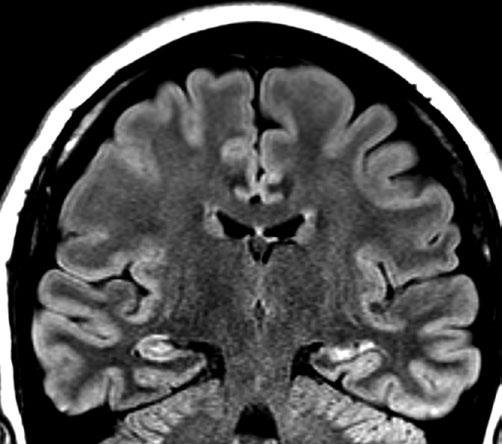
MRI coronal FLAIR sequence at a temporal pole level. The right temporal pole is slightly smaller than the left. Within both temporal poles, gray-white differentiation is normal, and there is no signal abnormality within the gray matter or white matter
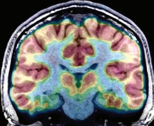
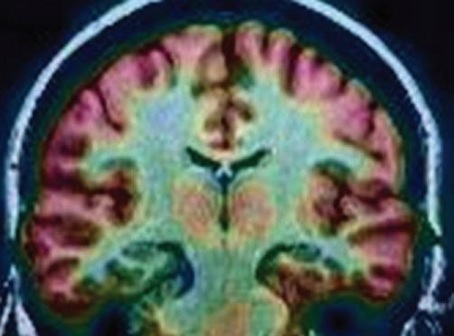
Fig. 1.2 PET-MRI co-registration with color-scale FDG tracer in a coronal plane at the same temporal pole level as Fig. 1.1. The right temporal pole is markedly hypometabolic mesially. Less pronounced hypometabolism is present in the right temporal pole’s lateral and inferior cortices. The left temporal pole is normal
Fig. 1.3 MRI coronal FLAIR sequence at a hippocampal head level. The right hippocampal head is abnormally hyperintense (arrow), and the right temporal lobe is atrophic, as is evident in the larger right temporal horn. The gray-white differentiation along the inferior and lateral temporal lobe is normal bilaterally
Fig. 1.4
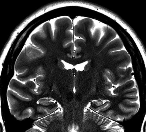
MRI coronal T2 sequence at the same hippocampal head level as Fig. 1.3. The hippocampal heads are asymmetric with the right hippocampus demonstrating subtly increased signal, blurring of the internal architecture, and smoothing of the superior indentations when compared to the left. The gray-white differentiation is normal and symmetric
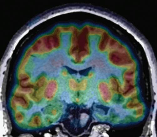
Fig. 1.5 PET-MRI co-registration with color-scale FDG tracer in a coronal plane at the same hippocampal head level as Figs. 1.3 and 1.4. Hypometabolism is evident in the right hippocampal head, parahippocampal gyrus, fusiform gyrus, and inferior temporal gyrus. The left hippocampal head metabolism is normal
Fig. 1.6 MRI coronal FLAIR sequence at a body of the hippocampus level. Increased signal is evident within the right hippocampus (arrow). The gray-white differentiation is normal bilaterally, and the temporal lobes are symmetric in size
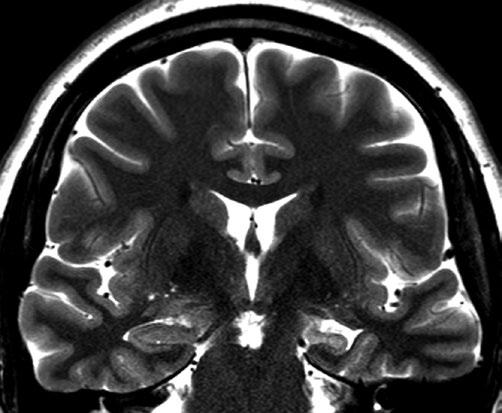
Fig. 1.7 MRI coronal T2 sequence at the same body of the hippocampus level as Fig. 1.6. Mildly increased signal is evident in CA1 of the right hippocampus, and the right hippocampus also is slightly smaller than left. Gray-white differentiation is clear (arrow) in the right mesial temporal lobe
Fig. 1.8 PET-MRI co-registration with color-scale FDG tracer in a coronal plane at a posterior hippocampus level. Hypometabolism is evident in the right mesial temporal lobe, including the hippocampus and inferior temporal cortex. The lateral temporal lobe is normal bilaterally. The right thalamus has mildly decreased metabolism
1 Mild, Unilateral
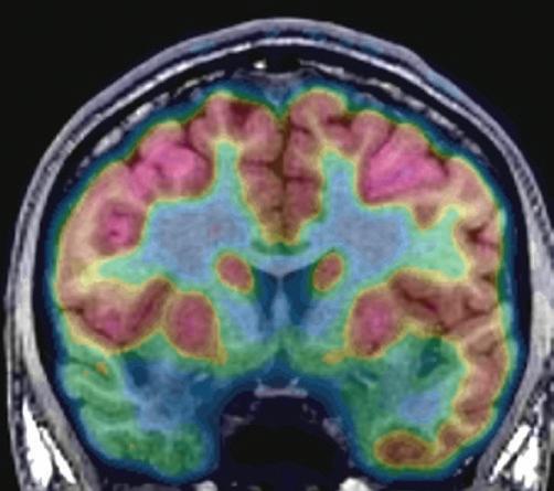
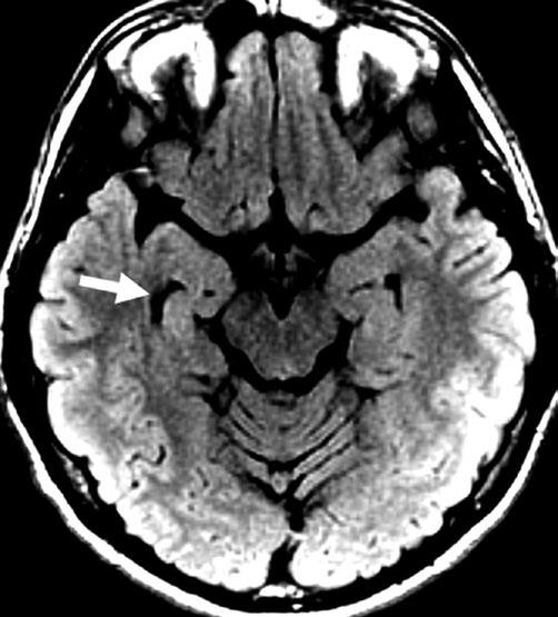
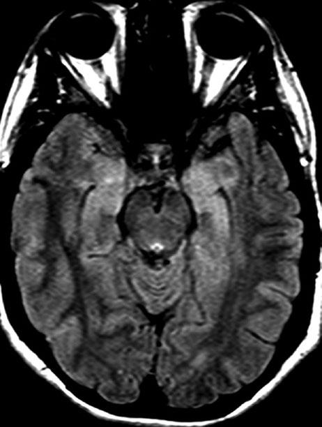
Fig. 1.9 MRI axial FLAIR sequence at a hippocampal level. The right hippocampus is atrophic and has abnormally increased signal. Because of the atrophy, the right temporal horn is larger (arrow)
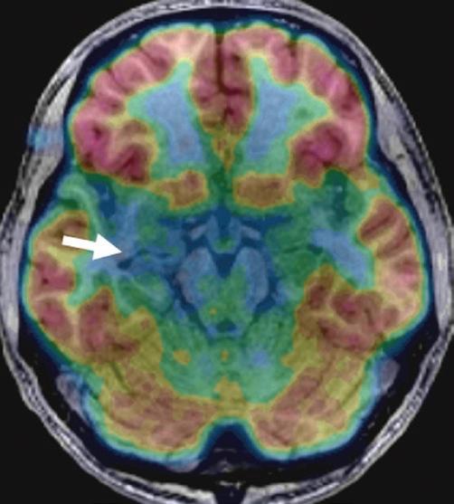
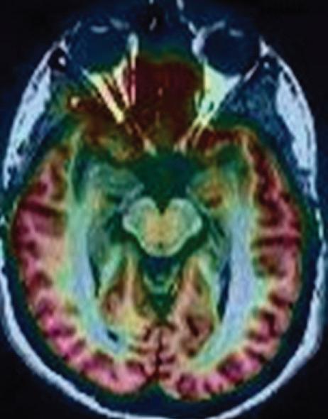
Fig. 1.10 PET-MRI co-registration with color-scale FDG tracer in an axial plane at a hippocampal head level similar to Fig. 1.9. The right hippocampal head is hypometabolic, and marked hypometabolism extends within the right hippocampus (arrow) compared to the left. The lateral temporal metabolism is normal bilaterally
Moderate, Unilateral Hippocampal Sclerosis
Clinical History
The frst recognized seizure occurred at age 7 years as an episode of seeing stars and that progressed to shaking.
The habitual seizures followed an aura of a rising sensation in the abdomen that became shortness of breath and then sometimes progressed to a lapse in awareness and a memory gap with oral automatisms and purposeless wandering. The seizure duration was about 1–2 minutes and postictal fear and headache follows. About half of the seizures were an aura in isolation. In contrast, bilateral tonic-clonic seizures were rare, occurring only when not taking anti-seizure medications. Treatment included multiple anti-seizure medications with a seizure frequency of one seizure weekly. No epilepsy risk factors were present. Neurological examination was normal.
Interictal EEG depicted bilateral, independent anterior temporal epileptiform discharges that were more frequent on the left. Video-EEG monitoring recorded 15 seizures that followed the habitual aura and were manifested as bilateral manual automatisms, non-versive turning to the left, and sometimes dystonic posturing of the left hand. Ictal EEG’s seizure onset was across the left anterior temporal region for
nine seizures and right anterior temporal region for six. The left-hand dystonic posturing occurred during some of the seizures with the right-sided onset and none of the seizures with the left-sided onset.
MRI identifed left hippocampal atrophy and signal abnormality. PET identifed mild left temporal hypometabolism.
The history and evaluation are consistent with mesial temporal lobe epilepsy. The aura includes limbic features that support mesial temporal lobe epilepsy, but the features do not indicate lateralization or whether the epilepsy is unilateral or bilateral. The imaging supports left mesial temporal lobe epilepsy, but the video-EEG monitoring recorded both left- and right-sided seizure onsets. The presence of bilateral independent anterior temporal interictal epileptiform abnormality does not necessarily indicate bilateral mesial temporal lobe, and the greater number of the left- sided discharges is consistent with the imaging in supporting left mesial temporal lobe epilepsy. Additional testing is warranted to determine whether epilepsy surgery would be benefcial for control of the habitual seizures, as the MRI and PET fndings alone cannot establish this.
J. M. Stern, N. Salamon, Imaging of Epilepsy, https://doi.org/10.1007/978-3-030-86672-3_2
2 Moderate, Unilateral Hippocampal Sclerosis
Fig. 2.1 MRI coronal FLAIR sequence at a temporal pole level. The gray-white junction is blurred along the mesial surface of the left temporal pole. The lateral and inferior arborization of the white matter and the gray-white junction is normal bilaterally
Fig. 2.2 PET-MRI co-registration with color-scale FDG tracer in a coronal plane at the same temporal pole level as Fig. 2.1 Hypometabolism is evident in the left superior, middle, inferior temporal gyri. The mesial temporal poles have symmetric metabolic signal
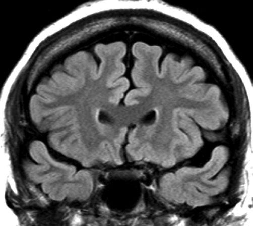
Fig. 2.3 MRI coronal FLAIR sequence at a hippocampal head level. The left hippocampus is smaller and has abnormally increased signal intensity with a fattening of the superior surface. In comparison, the right hippocampus’ superior surface has clear indentations. The graywhite differentiation is preserved bilaterally
Fig. 2.4 MRI coronal T2 sequence at the same hippocampal head level as Fig. 2.3. The left hippocampus is smaller and has increased signal intensity and a fattening of the superior surface. The gray-white differentiation is preserved bilaterally. The fndings are the same as Fig. 2.3 with differences in appearance because of the different MRI sequence
Fig. 2.5 PET-MRI co-registration with color-scale FDG tracer in a coronal plane at a hippocampal head level similar to Figs. 2.3 and 2.4 Asymmetrically greater hypometabolism is evident in the left entorhinal cortex and along the left temporal lobe’s inferior surface. The left thalamus also is hypometabolic
Fig. 2.6 MRI coronal FLAIR sequence at a body of the hippocampus level. The left hippocampus is hyperintense and markedly smaller than the right. The gray-white differentiation is normal bilaterally
Fig. 2.7 MRI coronal T2 sequence at the same body of the hippocampus level as Fig. 2.6. The left hippocampus is small and has increased signal. Its internal architecture is diminished compared to the right hippocampus. Nevertheless, the left hippocampus’ CA1, dentate gyrus, and subiculum are visible, with the left CA1 and dentate gyrus in particular demonstrating higher signal than on the right
Fig. 2.8 PET-MRI co-registration with color-scale FDG tracer in a coronal plane at the same hippocampus level as Figs. 2.6 and 2.7. The left hippocampus and inferior temporal cortex are hypometabolic. The lateral temporal cortex and the thalamus have symmetric metabolic signal
2 Moderate, Unilateral Hippocampal
Fig. 2.9 MRI axial FLAIR sequence at a hippocampal head level. The normal hippocampal indentations are visible across the right hippocampal head. In comparison, abnormal smoothing is evident along the left hippocampal head. The hippocampal signals and volumes are bilaterally symmetric
Fig. 2.10 PET-MRI co-registration with color-scale FDG tracer in an axial plane at a hippocampal head level similar to Fig. 2.9. The left temporal pole, amygdala, and hippocampus are hypometabolic. The lateral temporal lobes have normal, symmetric metabolism
Severe, Unilateral Hippocampal Sclerosis
Clinical History
The frst recognized seizure occurred in early childhood as an episode of staring and mumbling with repetitive opening and closing of the hands. The episode recurred many times in later childhood, but the episodes were not recognized as seizures, so an evaluation and treatment were not undertaken until adulthood.
The habitual seizures did not included an aura. The ictal behavior was similar to the behavior present in childhood except for progression to evident confusion that lasts several minutes. If alone, the patient was unaware of these seizures. Rarely, a seizure progressed to bilateral tonic-clonic. The tonic-clonic seizures also occurred without an aura and they have not been witnessed to include lateralizing features. A total of 4 or 5 were believed to have occurred. Treatment included multiple anti-seizure medications with an estimated seizure frequency of 1 or 2 monthly, but the actual frequency was believed to be higher.
No epilepsy risk factor was present.
Neurological examination was abnormal because of impaired cognitive performance. Neuropsychological testing identifed low borderline global function with asymmetrically greater left hemispheric dysfunction. The language dominant temporal lobe had both mesial and lateral dysfunction with relatively greater lateral temporal abnormality. Medical history included cryptogenic onset hepatic disease with cirrhosis that required a liver transplant in early childhood.
monitoring recorded focal seizures with ictal EEG’s seizure onset across the left temporal region.
MRI identifed marked left hippocampal sclerosis and PET identifed concordant left temporal hypometabolism along with left thalamic hypometabolism. An intracarotid amobarbital test was performed in consideration of epilepsy surgery, and it identifed left hemispheric language dominance, intact memory function with both the right and left injections, and better memory performance with the rightsided injection.
The history and diagnostic evaluation support a diagnosis of left temporal lobe epilepsy, as evident in the ictal EEG’s seizure onset; however, the other diagnostic tests include features of both mesial and lateral temporal lobe epilepsy. The occurrence of focal seizures with impaired awareness as the habitual seizure and rare bilateral tonic-clonic seizures supports mesial temporal lobe epilepsy and this is further supported by the MRI evidence of left hippocampal sclerosis and the concordant PET hypometabolism. In addition, the lack of an aura and the signifcant left lateral temporal dysfunction extend the localization to the temporal neocortex. In contrast, the neuropsychological testing evidence for bilateral temporal dysfunction and the intracarotid amobarbital test identifcation of intact episodic memory function in both right and left hippocampal systems suggest mild, bilateral mesial temporal dysfunction. Because of the uncertainty for the epileptogenic zone and the risk of an anterior temporal lobe resection producing a memory defcit, resective surgery was not offered.
J. M. Stern, N. Salamon, Imaging of Epilepsy, https://doi.org/10.1007/978-3-030-86672-3_3
3 Severe, Unilateral Hippocampal Sclerosis
Fig. 3.1
MRI coronal FLAIR sequence at a temporal pole level. The left temporal pole is smaller with blurring of the gray-white differentiation across the medial-inferior portion. The gray-white differentiation is normal in the right temporal pole
Fig. 3.2 PET-MRI co-registration with color scale fuoro-deoxyglucose (FDG) tracer in a coronal plane at the temporal pole level in Fig. 3.1. The left temporal pole is smaller with marked hypometabolism of its medial and inferior cortex. The left lateral temporal cortex has less prominent hypometabolism as compared to the right
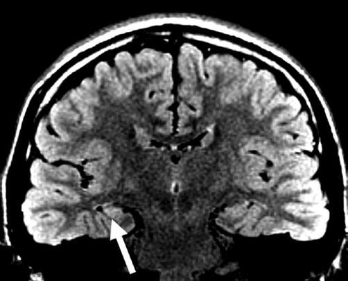
Fig. 3.3 MRI coronal FLAIR sequence at a hippocampal head level. The left hippocampal head has marked atrophy and increased signal with loss of the superior surface indentations. Due to the hippocampal volume loss, the left temporal horn is prominent. The right hippocampal head and the bilateral gray-white differentiation are normal
Fig. 3.4
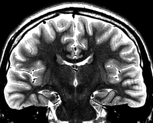
MRI coronal T2 sequence at a hippocampal head level similar to Fig. 3.3. The left hippocampal head has marked atrophy and loss of the internal architecture’s demarcation. With the atrophy, the left temporal horn is prominent. The right hippocampal head demonstrates the normal stripe of the hippocampal internal architecture. The gray-white differentiation is normal bilaterally
Fig. 3.5 PET-MRI co-registration with color scale FDG tracer in a coronal plane at a hippocampal head level similar to Figs. 3.3 and 3.4 Marked hypometabolism of the left hippocampal head is evident with less substantial hypometabolism of the inferior left temporal surface. The left thalamus is slightly hypometabolic when compared to the right
Fig. 3.6 MRI coronal FLAIR sequence at a body of the hippocampus level. The left hippocampus is atrophic and has abnormally increased signal. Its internal architecture is obscured compared to the right hippocampal body. The gray-white differentiation is normal bilaterally
Fig. 3.7 MRI coronal T2 sequence at a body of the hippocampus level similar to Fig. 3.6. The left hippocampus is atrophic and abnormally hyperintense with loss of detailed internal architecture. The subtlety of the right hippocampus’s internal architecture is apparent and normal. The gray-white differentiation is normal bilaterally
Fig. 3.8 PET-MRI co-registration with color scale FDG tracer in a coronal plane at a body of the hippocampus level similar to Figs. 3.6 and 3.7. Marked hypometabolism is evident within the left hippocampal body and the medial and inferior left temporal lobe. The thalamus is symmetric at this level. Metabolism across the right temporal cortex is normal
3 Severe, Unilateral Hippocampal
Fig. 3.9 MRI axial FLAIR sequence at a hippocampal head level. The left hippocampal head is atrophic and hyperintense with a prominent temporal horn of the lateral ventricle. Atrophy of the left amygdala and minor atrophy of the left temporal pole also are evident
Fig. 3.10 PET-MRI co-registration with color scale FDG tracer in an axial plane at a hippocampal head level similar to Fig. 3.9. The left hippocampal head and amygdala are markedly hypometabolic. Hypometabolism also is present across the left temporal pole. The lateral temporal lobes have normal, symmetric metabolism
Mild, Bilateral Hippocampal Sclerosis
Clinical History
The frst recognized seizure was at age 18 years as a bilateral tonic-clonic seizure that occurred months after a major traumatic brain injury.
The habitual seizures manifested with four ictal behaviors. The frst manifestation to have occurred was a bilateral tonic-clonic seizure that did not follow an aura. The duration was 1–3 minutes with postictal sleep for 1 hour. These were infrequent with seizure-free periods of several years. The second manifestation was staring with impaired awareness and turning to the right followed by right-sided shaking and occasionally urinary incontinence. These did not follow an aura and had durations of 30 to 60 seconds with postictal confusion for an hour. The frequency of this seizure was 1–3 weekly except for catamenial clustering of 5–10 daily. The third seizure manifestation was an experience of déjà vu, epigastric rising, and intensifed clarity of perception that produced a brief respiratory arrest. There was limitation in responsiveness with the retained one-word answers, and, occasionally, oral automatisms. Postictally, weariness was present for a few minutes. These seizures occurred one to dozens of times daily. The fourth seizure manifestation frst developed recently. It occurred during sleep and appeared as nonsensical talking with right-hand clenching that lasted 30 seconds. The frequency was 1–2 times weekly. Treatment included multiple anti-seizure medications and the vagus nerve stimulator.
The epilepsy risk factor was a traumatic brain injury from a motor vehicle accident. The injury produced left temporal lobe edema and a left posterior Sylvian fssure hemorrhage. Retrograde amnesia was present for the year prior to the accident.
Neurological examination identifed impairment of recent memory that was further characterized with neuropsychological testing that identifed defcits in executive functioning and verbal and nonverbal learning with intact visuospatial abilities.
Interictal EEG depicted bilateral, independent temporal slowing and bilateral, independent temporal epileptiform discharges that were more often on the left. Video-EEG monitoring recorded all four of the ictal behaviors, and ictal EEG identifed seizure onsets independently across both the right and left temporal regions with the majority occurring across the right.
MRI identifed mild bilateral hippocampal sclerosis, and PET identifed bilateral but asymmetric temporal lobe hypometabolism with greater hypometabolism on the right. In consideration of epilepsy surgery, an intracarotid amobarbital test was performed. It identifed left hemispheric dominance for language and dependence on the left mesial temporal structures for episodic memory.
The history and evaluation indicate an epilepsy that possibly has more than one epileptogenic zone, which is plausible in the context of the major head trauma. The frequent bilateral tonic-clonic seizures and the asymmetric motor seizures are consistent with neocortical onsets. The asymmetric motor seizures may be propagating into medial frontal cortex as a cause of the urinary incontinence. In contrast, the occurrence of auras with limbic features and focal seizures with impairment that do not progress to bilateral tonic-clonic suggests limbic onsets without extensive propagation. The ictal EEG and neuropsychological testing further support the possibility of more than one epileptogenic zone and bilateral epilepsy.
Because of the disability and morbidity associated with the seizures, a palliative epilepsy surgery was considered. The ictal EEG, PET, and intracarotid amobarbital test fndings supported the possibility of beneft and safety from a right anterior-mesial temporal lobe resection, which was performed without the expectation of full seizure control. Histopathological examination of the resected tissue identifed mild focal cortical dysplasia and no evidence of traumatic injury.
Following surgery, memory impairment and anxiety increased. Because of a concern for subtle seizures, videoEEG monitoring was performed, and it identifed ictal EEG’s seizure onsets across the left temporal region that did not have an obvious behavioral change. With education, the family was subsequently able to identify these behavioral seizures,
15
J. M. Stern, N. Salamon, Imaging of Epilepsy, https://doi.org/10.1007/978-3-030-86672-3_4
and the frequency was found to be fve monthly, which was a marked reduction to the frequency prior to epilepsy surgery. Years later, a responsive neurostimulator was implanted with electrodes in the left entorhinal cortex and hippocampal head. Intracranial recordings identifed left anterior temporal seizures, and stimulation has subsequently reduced the seizure frequency to two monthly.
Imaging
4 Mild, Bilateral Hippocampal Sclerosis
Bilateral hippocampal head hypometabolism is present with more pronounced hypometabolism on the right. The right temporal lobe also has asymmetrically greater hypometabolism within the parahippocampal gyrus. Thalamic hypometabolism also is evident, but it is asymmetrically greater on the left
Fig. 4.4 MRI coronal FLAIR sequence at a hippocampal body level. Bilateral hippocampal bodies are abnormally hyperintense and have blurring of their internal architecture
Fig. 4.3 PET-MRI co-registration with color-scale FDG tracer in a coronal plane at the hippocampal head level in Figs. 4.1 and 4.2
Fig. 4.1 MRI coronal FLAIR sequence at a hippocampal head level. Bilateral hippocampal heads demonstrate abnormal hyperintensity without atrophy. However, FLAIR does not depict hippocampal head volumes as clearly as with T2 of T1 sequences. The temporal lobes are normal volume with clear gray-white differentiation
Fig. 4.2 MRI coronal T2 sequence at the hippocampal head level in Fig. 4.1. Bilateral hippocampal heads demonstrate both hyperintensity and atrophy with prominent temporal horns of the lateral ventricles. The internal architecture of both hippocampi is blurred. The temporal lobes are otherwise normal with clear gray-white differentiation
4.5 MRI coronal T2 sequence at a hippocampal body level similar to Fig. 4.4. Bilateral hippocampal atrophy is present with blurring of the internal architecture and a mildly increased signal
Fig. 4.6 PET-MRI co-registration with color-scale FDG tracer in a coronal plane at a hippocampal body level similar to Figs. 4.4 and 4.5 Bilateral hippocampal body hypometabolism is evident with more extensive hypometabolism across the right inferior temporal lobe. The thalamus is symmetric
Fig. 4.7 MRI axial FLAIR sequence at a hippocampal head level. The hippocampal heads and amygdalae are bilaterally hyperintense but are normal in volume. The temporal poles are bilaterally normal in volume and gray-white differentiation
4.8 PET-MRI co-registration with color-scale FDG tracer in an axial plane at a hippocampal head level similar to Fig. 4.7. Bilateral hippocampal hypometabolism and right amygdala hypometabolism are evident. The lateral temporal cortex is normal
Fig.
Fig.
Severe, Bilateral Hippocampal Sclerosis
Clinical History
The frst recognized seizure occurred at age 20 years as an episode of impaired awareness with shaking consistent with tonic-clonic movements.
The habitual seizures manifested as an aura of tingling and chills in bilateral arms with a choking or throat closing sensation. This was followed by staring with oral automatisms and unresponsiveness for about 30 seconds. Progression to forced gaze deviation upward and bilateral tonic-clonic movements did not occur for many years, but it was typical during the first year after the onset of epilepsy. Treatment included multiple anti-seizure medications with a seizure frequency of one to two monthly.
The epilepsy risk factor was one close relative with epilepsy.
Neurological examination was normal. Neuropsychological testing identifed impaired verbal and nonverbal memory with poor learning and rapid forgetting. Visuo-perceptual function also was impaired, suggestive of lateral temporal dysfunction. Medical history includes treatment for depression.
Interictal EEG depicted mildly abnormal generalized slowing with bilateral, independent anterior temporal epileptiform sharp waves. Video-EEG monitoring recorded focal seizures sometimes manifested as minimal behavioral change and sometimes with oral automatisms. Ictal EEG’s seizure onsets were bilaterally independent over the anterior temporal regions.
MRI identifed bilateral hippocampal sclerosis and PET identifed symmetric hypometabolism of bilateral anterior temporal regions.
The history and evaluation indicate bilateral mesial temporal lobe epilepsy. The aura has limbic features, including the possibility of insular involvement with the choking sensation and the diffuse somatosensory experience. The bilaterally independent ictal onsets are concordant with the evidence of bilateral hippocampal abnormality on MRI, PET, and neuropsychological testing. Based on the evaluation, resective epilepsy surgery candidacy was not evident. Neurostimulation was offered with either responsive neurostimulation to bilateral anterior temporal lobes or vagus nerve stimulation. The patient chose vagus nerve stimulation, and this has provided 4 years of a substantial decrease in seizure severity along with improved mood.
Springer Nature Switzerland AG 2022
J. M. Stern, N. Salamon, Imaging of Epilepsy, https://doi.org/10.1007/978-3-030-86672-3_5
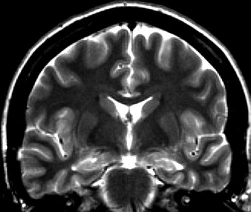
5 Severe, Bilateral Hippocampal Sclerosis
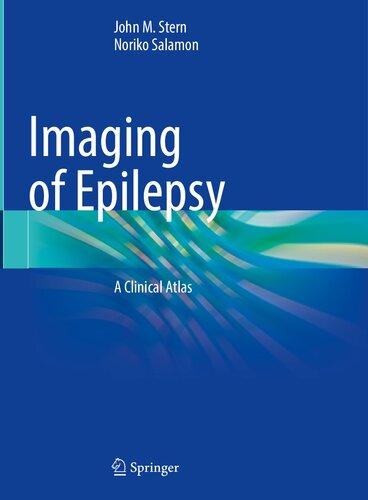
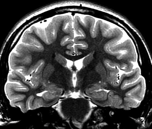
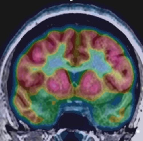
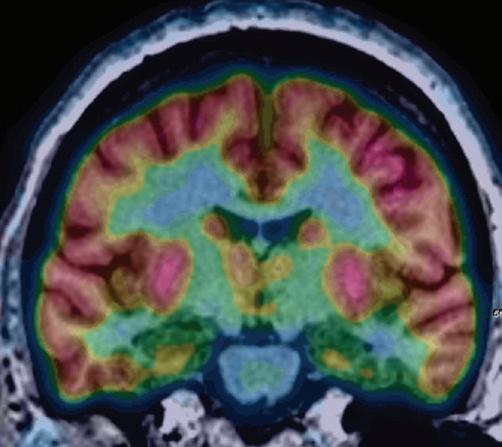
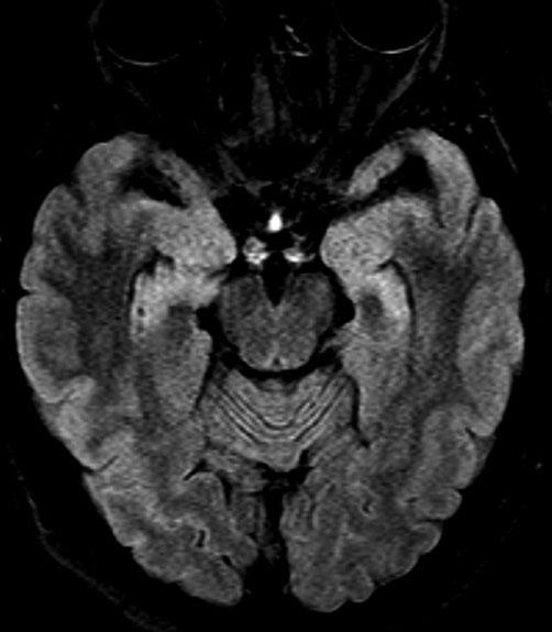
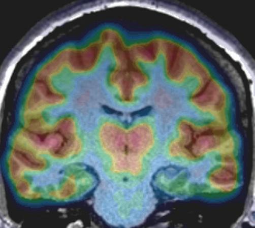
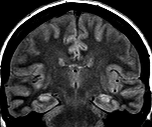
Fig. 5.1 MRI coronal FLAIR sequence at a temporal pole level. The gray-white differentiation is obscured in the medial portion of bilateral temporal poles
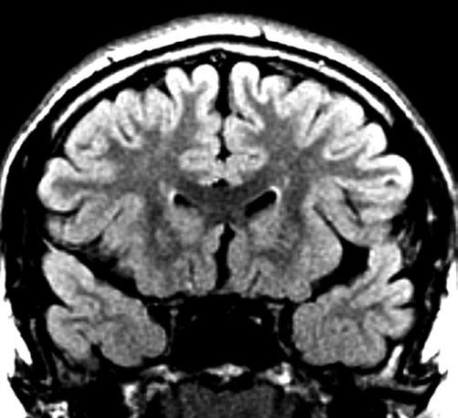
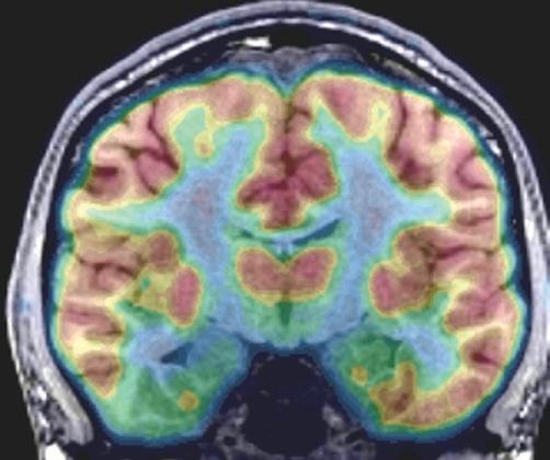
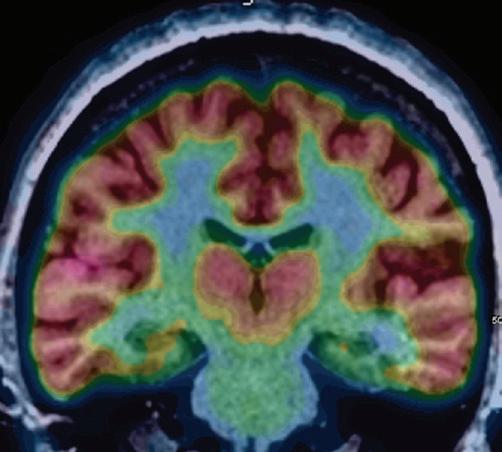
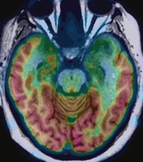
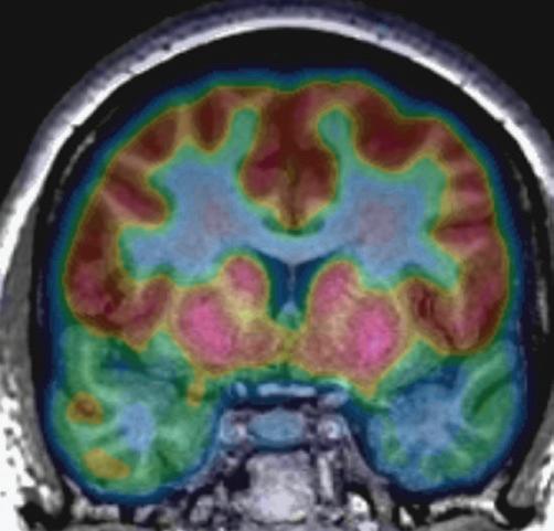
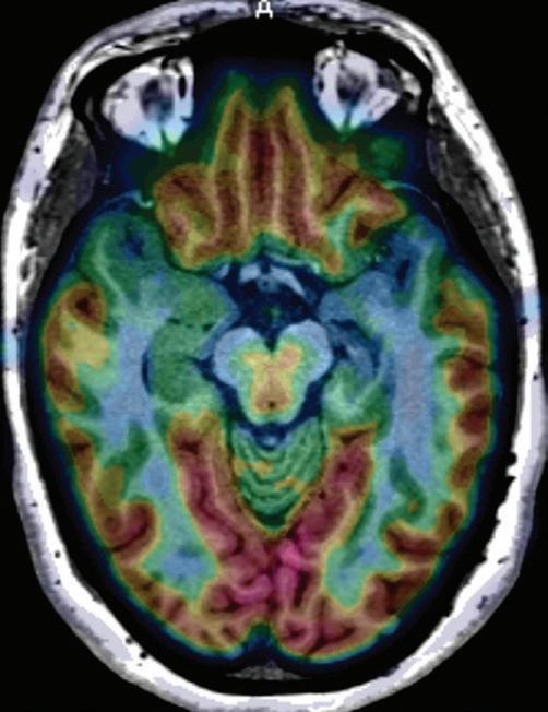
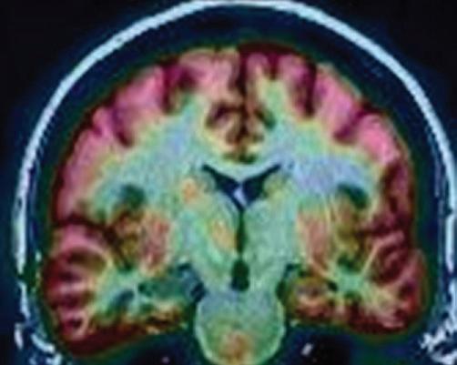
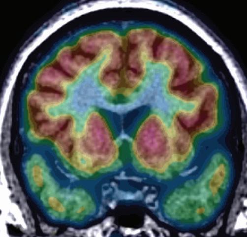
Fig. 5.2 PET-MRI co-registration with color-scale FDG tracer in a coronal plane at the temporal pole level in Fig. 5.1. Bilateral temporal poles demonstrate hypometabolism that includes the temporal lobes’ medial and lateral surfaces
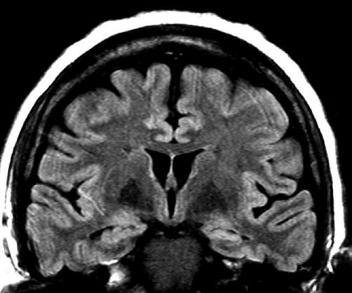
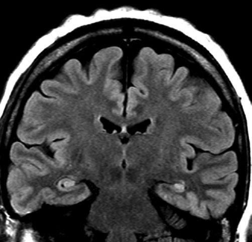
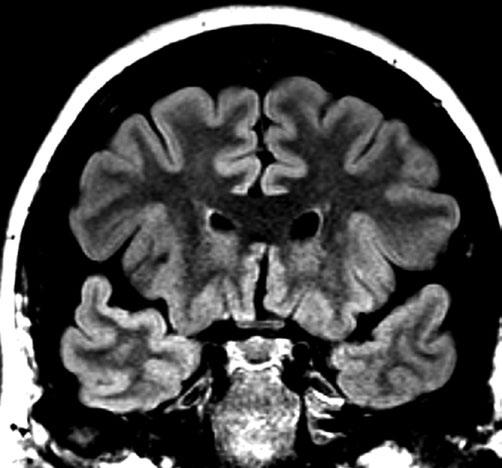
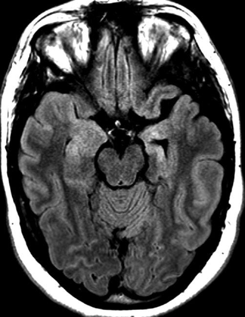
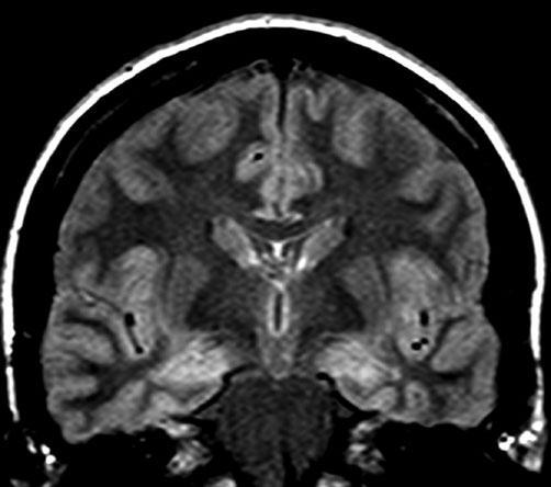
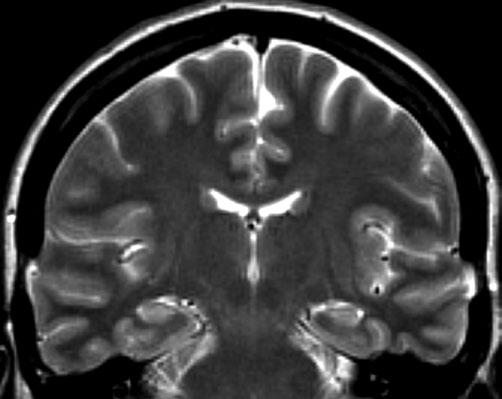
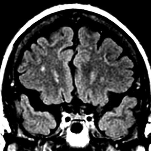
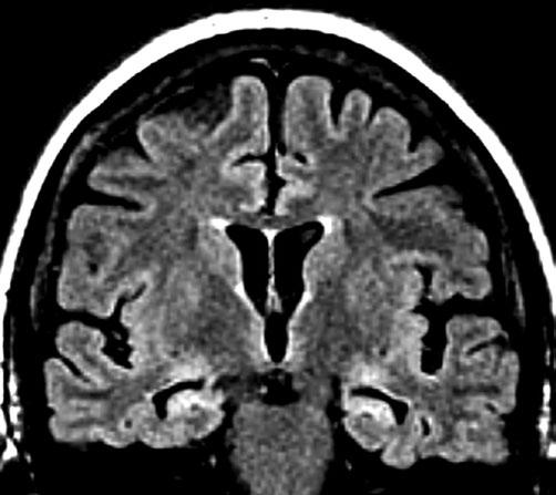
Fig. 5.3 MRI coronal FLAIR sequence at a hippocampal head level. Bilateral hippocampal heads demonstrate marked atrophy and hyperintensity. The superior surfaces of bilateral hippocampal heads are abnormally straight due to loss of the hippocampal indentations. Mild, bilateral cerebral atrophy also is present
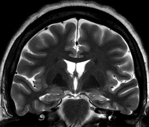
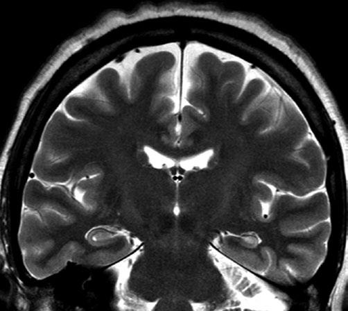
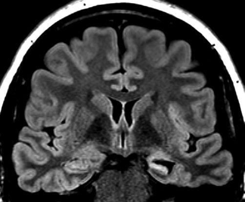
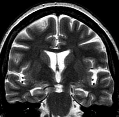
Fig. 5.4 MRI coronal T2 sequence at the hippocampal head level in Fig. 5.3. Bilateral hippocampal head atrophy is present with obscuration of the hippocampi internal architecture. The temporal horns are bilaterally prominent. The extra-hippocampal temporal lobes are normal bilaterally with clear gray-white differentiation. Mild, bilateral cerebral atrophy also is present