Instant download Orthopedics for physician assistant and nurse practitioner students-an introductory
Orthopedics for Physician Assistant and Nurse Practitioner Students-An Introductory Guide (June 14, 2022)_(3031044053)_(Springer).pdf
John A. Gracy
Visit to download the full and correct content document: https://ebookmass.com/product/orthopedics-for-physician-assistant-and-nurse-practiti oner-students-an-introductory-guide-june-14-2022_3031044053_springer-pdf-john-agracy/
More products digital (pdf, epub, mobi) instant download maybe you interests ...
From Classroom to Clinician-How to Practice Medicine
Safely and Confidently as a New Graduate Nurse
Practitioner or Physician Assistant (Aug 1, 2023)_(9798987048139)_(Stangl Medical LLC) John Stangl
This work is subject to copyright. All rights are reserved by the Publisher, whether the whole or part of the material is concerned, specifcally the rights of translation, reprinting, reuse of illustrations, recitation, broadcasting, reproduction on microflms or in any other physical way, and transmission or information storage and retrieval, electronic adaptation, computer software, or by similar or dissimilar methodology now known or hereafter developed.
The use of general descriptive names, registered names, trademarks, service marks, etc. in this publication does not imply, even in the absence of a specifc statement, that such names are exempt from the relevant protective laws and regulations and therefore free for general use.
The publisher, the authors and the editors are safe to assume that the advice and information in this book are believed to be true and accurate at the date of publication. Neither the publisher nor the authors or the editors give a warranty, expressed or implied, with respect to the material contained herein or for any errors or omissions that may have been made. The publisher remains neutral with regard to jurisdictional claims in published maps and institutional affliations.
This Springer imprint is published by the registered company Springer Nature Switzerland AG The registered company address is: Gewerbestrasse 11, 6330 Cham, Switzerland
Acknowledgments
Like any book, the list of people who helped directly or indirectly is long, and I am sure I am going to leave out more than one; so please forgive me. I am grateful to all the patients I have treated over the years, from whom I have learned much. To their credit, not a single one declined to have their photo or radiograph included. I am grateful for the opportunity to teach the PA students from Lincoln Memorial University and the NP students from Southern Adventist University which prompted the writing of this book. Some of them read early drafts and all of them made me go back and review the basics of medicine and orthopedic surgery. Rhonda Chamberlin typed most of the frst draft despite my handwriting. The orthopedic department of the Medical College of Georgia (at the University of Augusta) generously supplied a number of radiographs that I did not have. I greatly appreciate Kristopher Springer’s patience, which is more than that of a saint, as I took way too long to fnish. Finally, thanks to my wife, Julia, for her encouragement and love throughout the years; being married to a surgeon is not an easy life.
5.1.4
5.3.6
5.5.2
Bursitis
6 The Shoulder and Humerus
6.1 Traumatic
6.1.1 Clavicle Fractures
6.1.2 Scapular Fractures
6.1.4 Dislocations
Complications
6.1.6 Other Common Shoulder Pathology
Non-traumatic
6.2.1 Glenohumeral Arthritis
6.2.3 Rotator Cuff
6.2.4 Adhesive Capsulitis
The Foot and Ankle
Traumatic
7.2.1 Specifc Foot and Ankle Fractures
7.2.3 Ankle Sprains
8.2.1
8.3.2 ACL
PCL and LCL
8.4 Meniscal Tears
8.5.1 Prepatellar Bursitis
8.5.2 Patellofemoral Pain aka Anterior
8.5.3 Baker’s Cyst
8.5.7 Osgood-Schlatter
8.5.8 Shin Splints
9.1.2 Acetabular Fractures
9.1.3 Hip Fractures: Proximal Femur
9.1.4 Femoral Shaft Fractures
9.1.5 Supracondylar Femur Fractures
9.2 Hip Dislocations.
Lumbar Spine
10.3 Diseases of the Spine
10.3.1 Arnold-Chiari
10.3.2 Ankylosing Spondylitis (AS)
Scoliosis
10.3.4 Idiopathic Scoliosis
10.3.5 Scheuermann’s Disease
Background
11.2 Benign Bone Tumors
Osteoid Osteoma
Bone Islands
Osteochondroma
Fibrous Tumors
Unicameral Bone Cyst
Enchondroma
11.2.7 Aneurysmal Bone Cysts (ABC)
Giant Cell Tumor
Malignant Bone Tumors
11.3.1 Osteosarcoma
Chondrosarcomas
Ewing’s Sarcoma
11.3.4 Adamantinoma
12.4.3 Fibrous Dysplasia.
12.4.4 Muscular Dystrophy.
Duchenne Muscular Dystrophy
12.5 Neural Tube Defects
12.5.1 Myelomeningocele
12.6 Neurofbromatosis
12.7 Metabolic Disorders of the Bone
12.7.1 Osteogenesis Imperfecta (OI)
12.7.2 Osteopetrosis
12.7.3 Rickets
12.7.4 Scurvy
12.7.5 Paget’s Disease.
Infection
12.9.1 Transient Synovitis of the
Introduction
The purpose of this book is to serve as an introduction to orthopedics. It is not meant to be inclusive of all conditions, nor to give defnitive treatment options but rather to give the advanced practitioner (AP) student a brief overview and insight into what orthopedic surgery can and cannot do, as well as a primer on some of the specialized language and eponyms. To that end I have tried to include the most common conditions as well as those that fall into the category of “don’t you dare miss this.”
Broadly speaking, orthopedic surgery is the specialty dealing with problems in and of the musculoskeletal system including congenital and developmental defects, degenerative disease, tumors, and trauma to both the skeletal system and the soft tissues such as tendons, nerves, ligaments, and muscles. The treatment options vary from simple reassurance that a particular condition is benign and no intervention is needed to major multistage surgery. Related to orthopedics is the feld of rheumatology, and the next to the last chapter will give a brief overview of that feld. Trying to learn even the bare basics of orthopedic surgery during the normal 4-week rotation is like drinking from a fre hose. There are an innumerable number of books, journals, and resources online including videos that go into enormous detail about the conditions presented. Most of the references are particular chapters in textbooks or review articles from major journals; the AP student should read those for further detail as needed or as interested. Each of the review articles will have its own set of references that the student can use for even deeper insight.
After the chapter on orthopedic history taking, physical examination, imaging, and other diagnostic studies, there is a chapter on the operating room, followed by nine chapters each addressing a different anatomic area. Each of those chapters is roughly divided into traumatic and non-traumatic conditions (e.g., ankle sprains and clubfoot, respectively). There will be a brief discussion of clinical presentation, any imaging, or other diagnostic studies (if appropriate), the underlying pertinent pathology, and important differential diagnoses, followed by a broad outline of treatment options. Any signifcant short- or long-term complications of either the condition or the treatment will be mentioned. Exhaustive details about the treatment
will not be discussed as that is both patient and surgeon dependent. Chapter 13 is a brief overview of rheumatology. Finally, Chap. 14 discusses compartment syndrome. Several rules of thumb all medical professionals should follow are:
1. Treat the patient, not the radiograph. The treatment of distal radius fracture in an 88-year-old nursing home patient will be different from that of the 22-year-old college athlete.
2. Most clinicians come to a conclusion about diagnosis and treatment in the frst 30 seconds, tending to ignore or downplay conficting information obtained later. Do not be hasty. For the most part, diagnosis in orthopedics is fairly straightforward, but deciding on the best treatment is not always as easy; be sure to involve the patient in the decision-making process. If all the history and physical and diagnostic studies do not “match,” fgure out why before ignoring or downplaying some of those clues. When a diagnosis is uncertain or if the best method of treatment for that particular patient is in doubt, ask a colleague or your supervising physician.
3. As hard as it is to deal with some of our more long-winded patients, let them tell their story. Not only is it part of the therapeutic process (pain shared is pain divided), many times they will answer your questions and give you clues as to what treatment options to consider. Try to wait at least a minute before interrupting with a question.
4. Ask open-ended questions. Instead of asking “does this cause numbness and tingling?” in the radial aspect of the hand when doing a carpal compression test, ask what kind of symptoms/feelings does this test cause and where.
From here onward, both physician assistants and nurse practitioners will be referred to collectively as advanced practitioners: APs.
Like other specialties and professions, orthopedics is rife with eponyms and abbreviations. As each eponym is introduced, a brief explanation of the associated condition or test will be given. There are various classifcation schemes for both fractures and non-traumatic conditions. It can be diffcult to know which to use or memorize. For example, a literature review in 2018 showed 22 different classifcation schemes for tibial plateau fractures [1]. The most universal fracture classifcation system is the one promulgated by the AO/ASIF short for Association for Osteosynthesis/Association for the Study of Internal Fixation (or the in the original German Arbeitsgemeinschaft für Osteosynthesefragen). It is a cumbersome system mostly for academic use but helpful to be aware of when reading the orthopedic literature. Other abbreviations will be introduced in the appropriate section. Open reduction and internal fxation is ORIF; different types of appropriate ORIFs will be mentioned for each fracture type. Arthroscopy refers to looking into a joint or space with a camera that has the diameter of a pencil and using other specialized instruments of the same size to repair or excise the problem. Arthrodesis means fusing a joint. Arthroplasty refers to replacing the joint which can be with either manufactured material or soft tissue (e.g., total hip arthroplasty or carpometacarpal soft tissue arthroplasty, respectively). Hemiarthroplasty means replacing half the joint and is mostly used when discussing the treatment of femoral neck fractures. It is not
uncommon for these terms to be confusing, and the AP should remember to use plain English, not medicalese, when talking with patients. A few of the standard orthopedic textbooks are as follows:
Weinstein SL, Flynn JM. Lovell and Winter’s Pediatric Orthopedics. 8th ed. Baltimore: Lippincott Williams & Wilkins; 2020.
Herring JA. Tachdjian’s Pediatric Orthopedics: from the Texas Scottish Rite Hospital for Children. 6th ed. Philadelphia: Elsevier; 2021.
Tornetta P, Ricci W, Court-Brown CM, McQueen MM, McKee M. Rockwood and Green’s Fractures in Adults. 9th ed. Baltimore: Lippincott Williams & Wilkins; 2019.
Waters PM, Skaggs DL, Flynn JM. Rockwood and Wilkins Fractures in Children. 9th ed. Baltimore: Lippincott Williams & Wilkins; 2019.
And the number of orthopedic journals is over 200. Although each subspecialty in orthopedic surgery has one or more specifc journals, the most commonly used and referenced are the following:
Journal of the American Academy of Orthopedic Surgeons. Commonly referred to as the yellow journal, it is published twice per month and has a combination of review, technique, and original research articles some of which are only available only online.
Journal of Bone and Joint Surgery. There is both an American and British version and is widely considered the fagship journal of orthopedic surgery. The American edition is published twice per month.
Journal of the Orthopedic Physician Assistant. Published quarterly by the same company that publishes the Journal of Bone and Joint Surgery. As will be mentioned frequently throughout this book, each surgeon may have different ways of handling various diagnoses, but the above references will give more details on the many different ways of caring for patients with orthopedic problems.
Reference
1. Millar SC, Arnold JB, Thewlis D, Fraysse F, Solomon LB. A systematic literature review of the tibial plateau fractures: what classifcations are used and how reliable and useful are they? Injury. 2018;49(3):473–90. Epub 31 Jan 2018.
History, Physical Exam, and Diagnostic Testing
2
2.1 History Taking in Orthopedics
After introducing yourself to the patient and anyone accompanying the patient as well as verifying their relationship, the easiest way to start is to ask how you can help. Then, be quiet and allow the patient to tell his story, interrupting as little as possible. Listen for clues as the patient may answer some of your questions before you ask, as well as listening for items for which you may need more detail.
For acute injuries, the usual where, when, and how are important. Obviously, a fall from the front steps resulting in ankle fracture is very different from a fall off the roof resulting in the same injury. Ask about concurrent injuries, for example, loss of consciousness, and if the injured body part has been injured in the past or has a preexisting disease. For example, did the patient have a previous fracture that did not heal properly leaving the patient with permanent loss of motion, or have a stroke that has left the patient with weakness or numbness, especially in the injured area.
For chronic or non-traumatic problems, the history of present illness is a little longer. The what, when, and how are still applicable, but the details are important. What is the problem? What are the symptoms, e.g. pain, numbness, loss of motion, and weakness? What makes it better? What makes it worse? Any previous tests? What did they show? When and where were they done? Any previous treatment including physical therapy and surgery, and not just by physicians? For example, many patients will use acupuncture, chiropractic care, herbal medicine, over-thecounter medications, or braces, and many patients are reluctant to mention that they tried nontraditional treatments. Did any of those traditional or nontraditional treatments make a difference?
The past medical history is important as it allows the surgeon and the AP to assess risk if surgery is warranted and will also give the AP an idea of the patient’s physiologic age. The obese 35-year-old on three antihypertensives, a H2 inhibitor, and metformin is probably “older” than the normal weight 45-year-old who needs no medication. Rare childhood illnesses such as polio and rheumatic fever may have some impact on overall adult musculoskeletal health. Adult diseases and
J. A. Gracy, Orthopedics for Physician Assistant and Nurse Practitioner Students, https://doi.org/10.1007/978-3-031-04406-9_2
medications must be elicited by specifc questions. Surgical history can give the AP some hints as to how to approach any surgical discussion. Did the patient have problems with pain control or nausea after previous surgery? Past history of addiction may infuence perioperative treatment to minimize the risk of narcotic abuse. Allergies are important, but patients may not think of them until asked by the third or fourth healthcare provider. Because reactions to drugs are not necessarily an allergy, the AP should ask what specifcally happened when the patient had exposure to the drug or food. This will allow the AP to differentiate a true allergy from sensitivity and other reactions. Specifc questions regarding penicillin, NSAIDs, cephalosporins, and latex are helpful.
Social history is not just a way of socializing with the patient. Smoking, vaping, drinking, and illicit drug use impact the patient’s health. Occupation and hobbies help determine treatment and treatment goals. With whom the patient lives may be important in determining who will aid with recovery (household chores, personal hygiene, transportation to appointments, including physical or occupational therapy).
In the current age of electronic health records, the review of systems is usually done by check sheets or bubble sheets. Any positives should be followed up to make sure no important diseases were left out of the past medical history. The check mark by shortness of breath may indicate a recent diagnosis of pneumonia, chronic untreated asthma, or as-yet-undiagnosed congestive heart failure.
2.2 Physical Exam
The physical exam starts off with vitals, especially height and weight, followed by calculation of BMI. Temperature should be obtained for all new patients as well as for those patients who might have an infection as well as those in the immediate postoperative period.
During and after taking vital signs be sure to really look at the patient and ask yourself questions such as: How old does the patient appear versus the patient’s actual age? How much distress? Does he appear chronically ill? Agitated? Depressed? When the patient entered the room, how did he enter: crutches, wheelchair, or limp? How easily can the patient get from the chair to the exam table? Especially with older patients, can the patient stand up without using the upper limbs to assist in pushing up. For some patients, a timed up and go (TUG) test may be appropriate. To perform the TUG test, have the patient stand from a seated position, walk six meters, turn around, walk back, and sit again. Normal is less than 10 seconds, but pay attention to gait, how the patient turns around, and how the patient both sits and stands.
Then, examine the injured body part or painful area as follows. The general rule of thumb is to examine from the joint above to the joint below. Especially with children, try to examine non-painful areas frst (even starting on the opposite side in anxious patients). Check for range of motion (active and passive as well as which parts of the ROM are painful), strength, and when appropriate sensation and refexes. For the area of concern, inspect frst. Look for scars (previously unmentioned injury
or surgery?), abrasions, erythema, ecchymosis, or skin changes. Because of variation in ROM between individuals especially in the hip and shoulder, it is helpful to note side-to-side differences.
Next, palpate the area of concern for tenderness, swelling, and areas of fuctuance. Feeling with the back of the examiner’s hand is the most sensitive way to detect excessive warmth. Crepitus, a grinding sensation, on range of motion should be noted, and when present the AP should ask the patient if the crepitus is painful. Strength should be tested with any side-to-side differences or accompanying pain documented. Any give-way weakness or inconsistencies are important to identify. There are a tremendous number of anatomically specifc tests for a variety of orthopedic conditions, many with associated eponyms which will be covered in the appropriate chapter. For many, the question to ask is not “does this recreate/reproduce your symptoms,” but instead ask what kind of symptoms and where does this test cause them.
For example, when during a carpal compression test (frm pressure over the median nerve at the wrist fexion crease for 30 seconds), do not ask “does this cause your thumb and fngers to go numb,” but rather ask “what kind of symptoms, if any, does this cause?” or you can ask, “does this make things better, worse, or the same?” If the answer is worse, “how so?”
There are a dozen or so books available that describe orthopedic examinations, most of which are written by physical therapists including Ortho Notes: Clinical Examination Pocket Guide by Gulick [1] and Orthopedic and Trauma Findings by Roth [2]. The latter book reminds the examiner what questions to ask when examining each body part in addition to reviewing specifc physical tests. In addition, there are innumerable resources in textbooks, in journals [3], and on the Internet if the student is just looking for one exam in particular. Remember the Internet is not always right.
2.3 Testing
Testing includes radiographs, CT, bone scan, MRI, ultrasound, laboratory work for infection and autoimmune diseases, and EMG/NCS.
In these cost-conscious days, it is important to think before blindly ordering tests. Routine ordering foot and ankle flms for every patient with a “sprained” ankle that comes through the door is not only costly but exposes the patient to unnecessary radiation. If the history and physical examination indicate both ankle and foot flms are necessary, by all means order both. With most tests, the AP should ask the following: Will this test help the patient? Does it confrm a diagnosis? Does it rule out certain diagnoses? Will it change management of the patient? Some patients may be insistent on having certain tests for emotional rather than logical reasons. For example, a patient may insist on having a radiograph to rule out bone cancer because a parent or relative died of bone cancer at the same age. Depending on the anxiety level of the patient, it may be of psychological beneft to obtain the radiograph to reassure the patient. 2.3
2.3.1
Imaging Studies
All APs should make it a habit to review all images prior to reading what the radiologist saw so as to read the images without bias. There are two reasons to personally review all diagnostic imaging. First, they may see something that the radiologist missed or misinterpreted. After all, the AP has the beneft of a thorough history and physical examination of the patient. Second, although modern technology and teleradiology provide radiology readings quickly, there will be times and places when and where a radiologist’s interpretation is not available in a timely fashion.
2.3.1.1
Radiographs
Radiographs (also referred to as “X-rays,” although that term more specifcally refers to the ionizing radiation that creates the images) are simply pictures made up of a variety of black, white, and gray overlaid shadows. They come in three forms:
• The traditional way has been exposing a piece of flm to the ionizing radiation and then developing it just like a photographic negative. This method is becoming obsolete as most frst world facilities have one of the two types of computergenerated images: CR and DR.
• Computed radiography (CR) involves exposing a cassette, which contains a special plate, to ionizing radiation and then placing the cassette into a “reader” which in turn sends the digitized image to a computer screen. It then erases the cassette using bright white light to ready it for the next radiograph.
• Digital radiography (DR) is a slight advance to CR where the image is instantly transmitted via a wired or wireless cassette directly to the computer screen. There is no difference in image quality between CR and DR; however, with digital radiography the radiology technologist will know almost instantaneously if the radiograph taken is adequate.
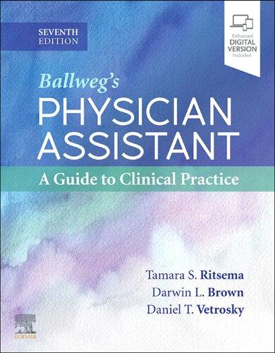
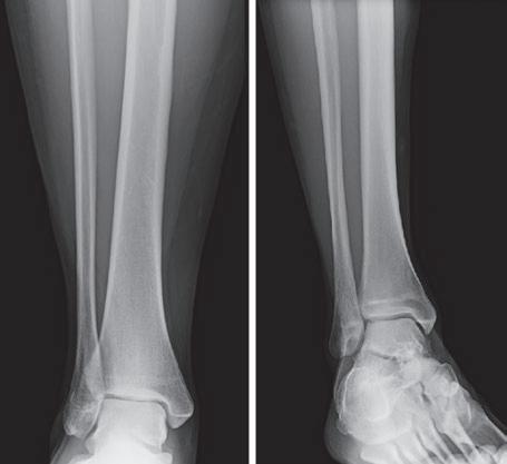
Because radiographs are shadows and not three dimensional, it is important to take at least two images at right angles (orthogonal) to each other to avoid missing a fracture or dislocation (Fig. 2.1). Interpreting a radiograph is both simple and diffcult. All the AP has to do is describe what he sees. When reviewing a radiograph, the frst thing is to look at all the views and identify which views are present. For example, there are three views of the left ankle – AP, lateral, and mortise. Then, describe the abnormalities as simply but as accurately as possible. For example, there is a non-displaced oblique fracture through the lateral malleolus, but the ankle mortise and syndesmosis are intact. Fractures should be described in terms of location, comminution (i.e., number of fragments), displacement, direction of the displacement, angulation, and rotation if applicable. Any involvement of the articular surface or growth plates should be mentioned as well.
2.3.1.2 CT Scans
To get more bony detail, a CT scan is an option. Computed tomography (CT) scans are obtained by placing the patient on a gantry and taking a high number of pictures while rotating the source of ionizing radiation and receiver circumferentially around
Fig. 2.1 Three views of a normal left ankle. The left radiograph shows an AP view of the ankle with the toes pointed straight up. The middle radiograph shows the mortise view which is taken with the ankle internally rotated 15°. Note the even spacing of the joint along both sides of the talus and the tibiotalar joint. The right radiograph shows a lateral view of the ankle
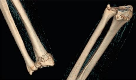
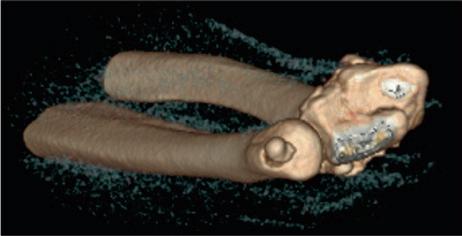
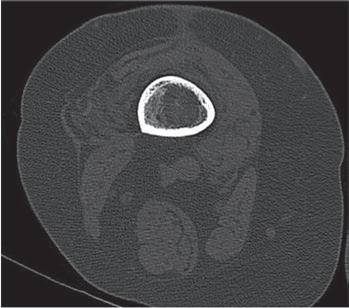
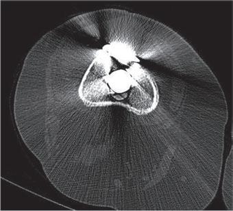
the patient. The patient is then moved fractionally (in increments as small as 1 mm) and the process repeated until the area of interest has been completely scanned. Marked detail of the bone and soft tissue can be obtained. For tumors and complex fractures, 3D reconstructions can be performed to allow better diagnostic images and surgical planning. With 3D reconstruction, the soft tissue is “subtracted” allowing only the bone to show (Fig. 2.2). Metal implants both prevent transmission of the ionizing radiation and cause defection of the beam to alternate receptors causing degradation of the image (Fig. 2.3).
2.3.1.3 Nuclear Imaging
In certain instances, knowledge of the activity of the bone is helpful. Bone scans can be done with either labeled WBC scans or technetium scans; the latter is more common. Although rarely used, indium-labeled WBC scans are helpful to diagnose and localize infection. It is done by drawing off a sample of the patient’s blood, adding indium 111 (radioactive), and then injecting the cells back into the patient. Images taken by a passive receptor several hours later show where the white cells have accumulated presumably in the area of infection.
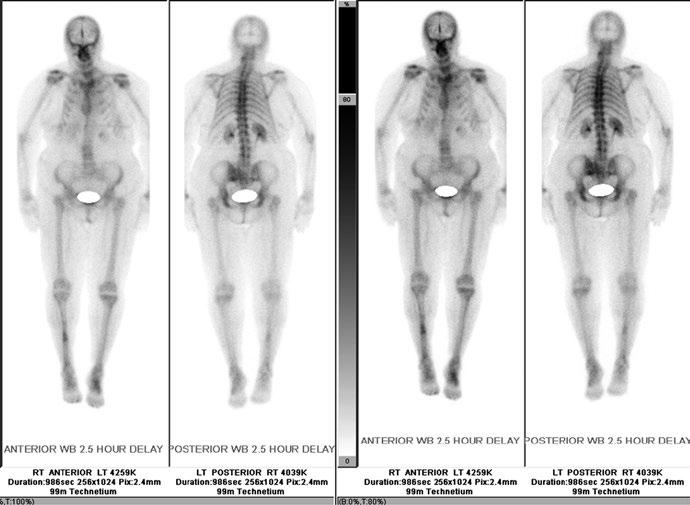
Technetium bone scans are done in three phases. After injecting a small amount of radioactive dye (about as much radiation as in one chest radiograph), two sets of pictures are taken almost immediately. The frst is the “blood fow” stage, and the second, the “blood pool” stage, both of which are of necessity, are limited to one region of the body. The third phase images are taken several hours later when the technetium has settled in the bone. More technetium settles in the area of bone that is more metabolically active as a result of infection, fracture (old or new), or
Fig. 2.2 These three images show a 3D reconstruction of a forearm CT showing the details of a complex distal radius fracture with an ulnar styloid fracture. Note that the soft tissues and carpal bones have been “subtracted” allowing detailed visualization of the bone
Fig. 2.3 The CT scan on the left shows the midshaft of the femur. Note the bone in the middle as well as the differing soft tissue densities of the muscle and adipose tissue. The CT scan on the right is of the same femur but through a more distal section of the thigh with a metal revision stem from a knee arthroplasty. Note how the metal artifact obscures not only the bone outline but the soft tissue as well
arthritis. The third phase can be done on the whole body if desired but that may need to be specifed when ordering the bone scan. Depending on the pattern of uptake in the three different phases, the disease process and extent can be identifed, or the differential narrowed (Fig. 2.4).
Fig. 2.4 Whole-body technetium bone scan in the third phase. Note the increased uptake in the midshaft of the right tibia consistent with an old fracture and the increased uptake in the left midfoot consistent with arthritis when both are correlated with the patient’s history. As the dye is excreted through the kidneys, a shield is placed over the bladder to prevent scatter radiation from obscuring the pelvis
Although not used much except in orthopedic oncology, positron emission tomography (PET) scans use injected fuorodeoxyglucose (FDG). Since cancer cells are more metabolically active, the PET scan will show greater activity in areas of malignancy [4].
2.3.1.4
MRI
Although plain radiographs and CT are excellent at showing bone and bony lesions, they only give hints to soft tissue lesions, primarily swelling and blurring of tissue boundaries on high-quality radiographs. The best way to obtain images of soft tissue is an MRI (magnetic resonance imaging) (Fig. 2.5). Simplifying the actual process, the patient is placed in a high-strength magnetic feld and the magnet turned on and off (thus accounting for the loud clicking the patient hears). When the magnet is turned on, the hydrogen ions associated with the water molecules line up in the same direction. When the magnet is turned off, the hydrogen ions go back to their random directions but release a tiny radio signal which is picked up by a receiver and in turn processed by a computer which produces an image. Depending on magnetic feld strength, how long the magnet is on, how long the signals are
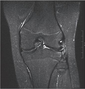
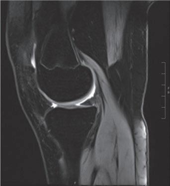
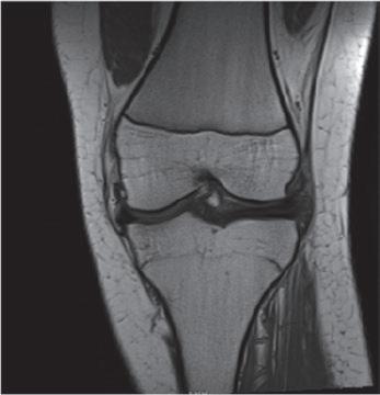
Fig. 2.5 MRI views of the knee. The upper images are coronal or frontal views, while the lower are sagittal views. The images on the right are T1 weighted which highlights the fat. The images on the left are STIR (short tau inversion recovery) based which highlights water as white and suppresses signal from fatty tissue. Note the presence of the distal femoral physeal plate
received, and how the signals are processed all determine the type and quality of images. Resolution can be as small as 1 mm depending on the magnet’s strength which is measured in Teslas. The higher the strength, the better the signal to noise ratio with most MRI’s currently having 1.5–3.0 T strength [5].
Contrast (typically gadolinium) can outline structure, making interpretation easier. The contrast can be injected intra-articularly, for example, in the shoulder making identifcation of labral or rotator cuff tears easier; the contrast can also be injected intravenously, for example, to make differentiation between herniated disc material and scar tissue on previously operated spines easier. Because gadolinium can be nephrotoxic in some patients, contrast should only be used when necessary [6].
2.3 Testing
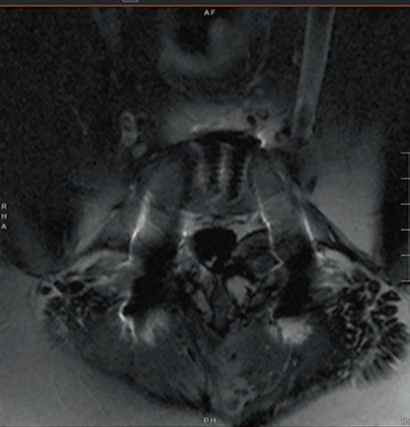
Fig. 2.6 MRI of the spine. This is a transverse image showing the diffculty of visualization in the presence of metal, even with titanium and specialized metal subtraction software
Because it is magnetic, any regional and implanted ferrous metal will make images much more diffcult to interpret, although there is software that will “subtract” some of the metal artifact improving the imaging (Fig. 2.6).
2.3.1.5
Ultrasound
MRI machines, however, are expensive (~$500,000), and the clinician may want something quicker or is just looking for a specifc soft tissue lesion. In the right circumstances, ultrasound can be useful for diagnostic or therapeutic use. Ultrasound machines for orthopedic use run from $15,000 to $40,000. As described by the name, the transducer bounces an ultrasound wave off the tissue and picks up the echo (much like sonar or radar). Generally, with higher frequencies deeper tissue can be seen, but the image degrades.
Ultrasound interpretation is very user dependent and is best done “live” as opposed to reviewing the static images. The most common use is to diagnose deep venous thrombosis. Other uses include diagnosing rotator cuff pathology (Fig. 2.7), subluxing peroneal tendons, or determining whether an infant’s hip is reduced, subluxable, or dislocated (developmental hip dysplasia). From a therapeutic standpoint, it is invaluable in placing intra-articular injections in deeper joints such as the hip as well as placing a scalene block prior to upper limb surgery [7, 8].
2.3.2 Lab Tests
Preoperative laboratory testing should be patient specifc and not general. If the AP is prepping an otherwise healthy 22-year-old male for arthroscopy, then no
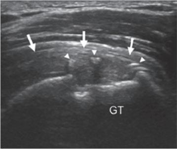
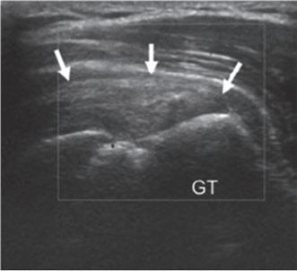
Fig. 2.7 Ultrasound of a repaired rotator cuff tear. GT: greater tuberosity. The arrows in both (a) and (b) point to the supraspinatus portion of the rotator cuff. The arrow heads in (a) mark the sutures placed for the rotator cuff repair (From Susan et al. [12])
laboratory tests are necessary. But if the patient is female, then a serum pregnancy and hematocrit would be indicated. EKGs for older patients and those with heart disease are appropriate. If on a diuretic, then the AP should order a basic metabolic profle. Depending on the patient, other laboratory tests may be necessary prior to surgery such as PT/PTT if on blood thinners or HbA1c to assess long-term diabetic control.
In addition to appropriate preoperative lab tests, there are two sets of labs that are of interest to the orthopedic AP. First there is the set of labs used to help diagnose infection. These typically include a complete blood count with differential (CBC with diff), erythrocyte sedimentation rate (ESR), C-reactive protein (CRP) (not to be confused with a hastily scrawled creatinine), and the most recent addition: alphadefensin protein (ADP). The ESR is generally thought of as indicating overall infammation which can result from infection or autoimmune disease, while CRP is much more specifc for infection. ADP is a relative newcomer and is likewise very specifc for infection; the drawback of ADP is that the test can only be run on synovial fuid. All have a specifc range of normal depending on the lab, with false positives and false negatives being possible. They should be reviewed in context of the whole clinical picture. If elevated, they are especially useful to track trends [9]. The second set of labs, less useful for the orthopedist and more useful to a rheumatologist, are those used to aid in diagnosing a variety of autoimmune diseases such as rheumatoid arthritis, systemic lupus erythematosus, psoriatic arthritis, and others. Keep in mind however that many patients will have negative lab tests despite clear evidence that the patient has disease (e.g., seronegative rheumatoid arthritis). These tests include complete blood count with differential, ESR, antinuclear antibody (ANA), anti-citrullinated protein antibody (ACPA), rheumatoid factor (RF), and others. In addition, tracking liver or kidney function may be necessary before starting drug therapy or while taking chronic medications including NSAIDs.
2.3.3 EMG/NCS
Finally, an EMG/NCS test can help evaluate the status of specifc muscles and nerves, but like any test there can be false negatives and false positives, and to a certain extent, the interpretation is operator dependent. Nerve conduction studies (NCS) are exactly what the term describes; a specifc nerve is stimulated and how fast the impulse travels is measured; the normal velocity is greater than 50 m per second. Various nerves are tested at the usual sites of entrapment or potential compression such as the median nerve at the wrist which may indicate carpal tunnel syndrome or the ulnar nerve at the elbow for cubital tunnel syndrome. Both motor and sensory nerves can be evaluated.
Electromyograms (EMG) are the most painful part of the test. A small-gauge needle is inserted into a specifc muscle. How that muscle responds to needle insertion, direct stimulation, and nerve impulses can reveal different pathologies. The EMG/NCS is normally done by a neurologist or physiatrist [10, 11].
2. Roth A. Orthopedic and trauma fndings: examination techniques, clinical evaluation, clinical presentation. 1st ed. Translated by GF Preller. Berlin: Springer; 2017.
3. Hippensteel KJ, Brophy R, Smith MV, Wright RW. A comprehensive review of physical examination tests of the cervical spine, scapula, and rotator cuff. J Am Acad Orthop Surg. 2019;27(11):385–94.
4. Hsu W, Hearty TM. Radionuclide imaging in the diagnosis and management of orthopaedic disease. J Am Acad Orthop Surg. 2012;20(3):151–9.
5. Hartley KG, Damon BM, Patterson GT, More. MRI techniques: a review and update. Orthopaed Surg J Am Acad Orthop Surg. 2012;20(12):775–87.
6. Llamas M. Gadolinium side effects [Internet]. 2019 [cited 21 Jan 2020]. Available from: https://www.drugwatch.com/gadolinium/side-effects/.
7. Saranteas T, Igoumenou VG, Megaloikonomos PD, Mavrogenis AF. Ultrasonography in trauma: physics, practice, and training. J Bone Joint Surg Rev. 2018;6(4):E12.
8. Li X, Yi PH, Curry EJ, et al. Ultrasonography as a diagnostic, therapeutic, and research tool in orthopaedic surgery. J Am Acad Orthop Surg. 2018;26(6):187–96.
9. Fehring TK, Fehring KA, Hewlett A, Higuera CA, et al. What’s new in musculoskeletal infection. J Bone Joint Surg. 2019;100(14):1237–44.
10. Lee DH, Claussen GC, Oh S. Clinical nerve conduction and needle electromyography studies. J Am Acad Orthop Surg. 2004;12(4):276–87.
11. Dy CJ, Colorado BS, Landau AJ. Interpretation of electrodiagnostic studies: how to apply it to the practice of orthopaedic surgery. J Am Acad Orthop Surg. 2021;29(13):e646–54.
12. Lee SC, Williams D, Endo Y. The repaired rotator cuff: MRI and ultrasound evaluation. Curr Rev Musculoskelet Med. 2018;11(1):92–101. https://doi.org/10.1007/s12178-018-9463-6. Published online 24 Jan 2018. https://link.springer.com/article/10.1007/s12178-018-9463-6.
The Operating Room
3.1 Infection and Preoperative Evaluation
Orthopedic surgery obviously takes place in the operating room. Like other specialties, one of the biggest concerns is postoperative infections. Unlike other surgical specialties, orthopedics frequently involves large metal implants which makes eliminating postoperative infections extremely diffcult. The bioflm (the glycocalyx) that is formed on the metal implant by the bacteria prevents penetration by the body’s defense, i.e., the white blood cells, as well as preventing the antibiotics from reaching the bacteria. In addition, because of cortical bones’ limited vascularity, bacteria can “hide” in the Haversian canals. After lying quiescent for years, the bacteria can re-emerge when the patient’s immune system weakens.
For elective operations many conditions can be corrected preoperatively, thus reducing but not eliminating the chance of infection. This includes optimizing comorbid conditions such as diabetes, hypertension, urinary tract infections, and especially dental caries. Regarding diabetes, the HgBA1c level can give the AP a good idea of the patient’s long-term compliance with diet and medication. Glucose levels the morning of surgery of greater than 180–200 portend a higher risk of infection [1]. Other factors can infuence the risk of infection including the length of the procedure, emergency/trauma surgery, and even the size of the operating room [2].
Urinary catheters are necessary in some cases to monitor intraoperative hydration as well as easing the patient’s ability to void in the immediate postoperative period. Early removal of the Foley either in the postanesthesia care unit (recovery room) or on the foor the evening of or the morning after surgery reduces the risk of urinary tract infection in addition to forcing the patient to move earlier. This in turn reduces the risk of deep venous thrombosis, pulmonary emboli, and pneumonia [3–5].
An excellent review of how to reduce the chance (not prevent) of infection in patients undergoing total knee arthroplasty can be found in “Infection Prevention in Total Knee Arthroplasty” [6].
J. A. Gracy, Orthopedics for Physician Assistant and Nurse Practitioner Students, https://doi.org/10.1007/978-3-031-04406-9_3
3
17
3 The Operating Room
3.2 Positioning
Unlike other specialties, orthopedic operations often require specifc positioning of the patient. This requires planning ahead not only to make sure the appropriate table is available including tables for the morbidly obese but also that all the accessory equipment is working as well.
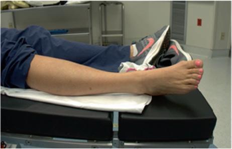
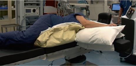
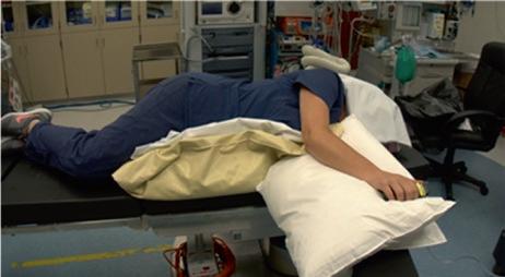
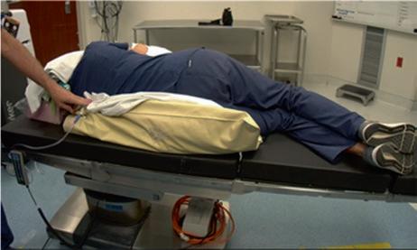
The simplest position is supine for hand operations and most foot and ankle surgeries. The latter may require a bump under the hip on the affected side allowing easy access to the lateral ankle (Fig. 3.1). Operations on the posterior ankle such as Achilles tendon repair usually require prone positioning.
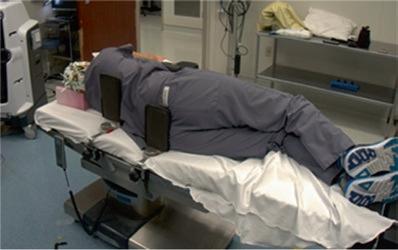
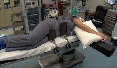
The lateral decubitus position is extremely common and used for both lateral and posterior approaches to the hip. Either a bean bag (Fig. 3.2) or some type of pelvic positioner (Fig. 3.3) (e.g., a “Montreal” positioner) can be used. The surgical assistant should check, prior to scrubbing, that once the hip is prepped and draped, the full range of motion needed intraoperatively will be available without destabilizing the pelvis. In addition to making sure the downside bony prominences are appropriately padded (greater trochanter, knee, and lateral malleolus), an axillary roll placed under the downside of the upper thorax will ensure blood fow to the downside arm. Checking the radial pulse and using a pulse oximeter on the downside fngers is helpful. For shoulders done in the lateral decubitus position, some type of traction
Fig. 3.1 (a, b) The ankle in (a) is without a “bump” under the hip which results in excessive external rotation of the ankle making access to the lateral malleolus more diffcult. The same ankle (b) shows the much easier access when a “bump” is placed under the ipsilateral buttock. The bump is usually a 5 pound sandbag but can be several towels or even a bag of IV fuid
Fig. 3.2 The pelvic positioner stabilizes the pelvis so that appropriate orientation will not be lost when working on the proximal femur or acetabulum. (a) shows the two posterior posts to prevent the patient from rolling to the supine position during surgery. (b) shows the pelvis perpendicular to the foor and tabl. (c) shows the view from the front where the more caudal positioner is short allowing for full fexion of the hip
to distract the glenohumeral joint will be necessary, taking care not to contaminate the operative feld in the process. The lateral decubitus position is also used for some elbow and upper arm surgeries (e.g., triceps avulsion, olecranon fractures).
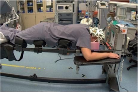
The prone position is almost exclusively used for spine surgeries (Fig. 3.4). Again, appropriate padding for bony prominences such as the anterior-superior iliac crest is necessary, but the AP should also ensure a female’s breasts are not pinched, a male’s genitals are not pinched, and the abdomen hangs free. If the abdomen has excessive pressure, it makes breathing more diffcult and may contribute to the development of deep vein thrombosis. Checking the position of the upper limb is important; a traction nerve palsy can develop quite easily with prolonged abduction or external rotation of the shoulders [7].
The beach chair position (Fig. 3.5) is used for various shoulder operations including clavicle or humeral fracture repair, arthroplasty, and arthroscopy; the operative side can either be draped free or be attached to a mechanical arm to hold the arm in a steady position. This frees up the assistant’s hands. In the beach chair position, the position of the cervical spine needs to be as close to neutral as possible to avoid traction injuries to the brachial plexus. The ears should be free of compression with shielding placed over the eyes. In addition some type of bolster under the thighs may be necessary to prevent the patient from sliding down – this may be diffcult in
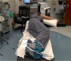
Fig. 3.3 For surgery where the lateral decubitus position is necessary, e.g. posterior approaches to the humerus, a bean bag can be used. Once the patient is positioned suction is applied which results in a stable torso with the pressure symmetrically distributed. It can be used for hip fractures if necessary but the pelvis will not be nearly as stable as with the positioner shown in Fig. 3.2
particularly tall or short patients. Finally beach chair positioners typically have an upper weight limit of 250–300 lbs. (110–140 kg), so heavier patients will require alternate positioning [8, 9].
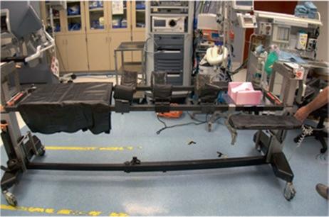
There are two types of fracture tables used for lower limb fractures including hip and femur fractures. Both allow excellent access to the thigh and allow the C-arm to be utilized in both the AP and lateral planes. The Amsco-type table is the most common (Fig. 3.6). The operative limb is placed in some type of traction, either foot traction or via a transtibial pin. The nonoperative limb must be positioned to allow the C-arm access to all necessary areas of the operative limb in orthogonal planes. Avoidance of excessive traction being placed in the perineal area and ensuring appropriate padding of the fbular head with its associated superfcial peroneal nerve on the nonoperative side are all important points to check before draping. The Hana
Fig. 3.4 (a, b) The Jackson table has multiple padded support positions for the prone position but allows the abdomen to hang free decreasing the risk of deep venous thrombosis and aiding in ease of respiration for long spine cases in addition to allowing access for the C-arm. Note the three isolated pads seen in a are for the pelvis, the lower ribs, and the shoulder as seen in b
table is similar with the most common use being for anterior hip replacement but can also be used for hip and femur fracture fxation.
Once the patient is positioned on the fracture table, the AP should ensure that the C-arm has access to the operative site in all necessary planes. Care should be taken that the nonoperative limb (right in the photos above) is padded over the peroneal nerve at the knee, that there is not pressure that would compress the popliteal vessels, and that the hip is not excessively fexed [10].
Finally, when it comes to positioning, in addition to making sure the C-arm can acquire appropriate images, the viewing monitors for fuoroscopy and arthroscopy should be where the surgeon and assistant can easily see them without excessive contortions. Some hospitals will have more than one monitor allowing both the surgeon and the assistant to view what is going on without either having to look too far from the operative feld.
Preparation of the surgical site after ensuring proper positioning starts with clipping (not shaving) excess hair that may contaminate the operative site as well as hair that may cause pain when the dressing is removed. The most common solutions used to clean the site are povidone-iodine (Betadine scrub and solution) and chlorhexidine gluconate (ChloraPrep) both of which require a short (1–3 minutes)