Sperm Morphology of Domestic Animals
J.H. Koziol, DVM, MS, DACT
Associate Professor of Food Animal Medicine and Surgery
Texas Tech University School of Veterinary Medicine Amarillo, TX, USA
C.L. Armstrong, DVM, MS, DACT
Associate Veterinarian
Elgin Veterinary Hospital Elgin, TX, USA
This edition first published 2022 © 2022 John Wiley & Sons, Inc.
All rights reserved. No part of this publication may be reproduced, stored in a retrieval system, or transmitted, in any form or by any means, electronic, mechanical, photocopying, recording or otherwise, except as permitted by law. Advice on how to obtain permission to reuse material from this title is available at http://www.wiley.com/go/permissions.
The right of J.H. Koziol and C.L. Armstrong to be identified as the authors of this work has been asserted in accordance with law.
Registered Office
John Wiley & Sons, Inc., 111 River Street, Hoboken, NJ 07030, USA
Editorial Office
111 River Street, Hoboken, NJ 07030, USA
For details of our global editorial offices, customer services, and more information about Wiley products visit us at www.wiley.com.
Wiley also publishes its books in a variety of electronic formats and by print-on-demand. Some content that appears in standard print versions of this book may not be available in other formats.
Limit of Liability/Disclaimer of Warranty
The contents of this work are intended to further general scientific research, understanding, and discussion only and are not intended and should not be relied upon as recommending or promoting scientific method, diagnosis, or treatment by physicians for any particular patient. In view of ongoing research, equipment modifications, changes in governmental regulations, and the constant flow of information relating to the use of medicines, equipment, and devices, the reader is urged to review and evaluate the information provided in the package insert or instructions for each medicine, equipment, or device for, among other things, any changes in the instructions or indication of usage and for added warnings and precautions. While the publisher and authors have used their best efforts in preparing this work, they make no representations or warranties with respect to the accuracy or completeness of the contents of this work and specifically disclaim all warranties, including without limitation any implied warranties of merchantability or fitness for a particular purpose. No warranty may be created or extended by sales representatives, written sales materials or promotional statements for this work. The fact that an organization, website, or product is referred to in this work as a citation and/or potential source of further information does not mean that the publisher and authors endorse the information or services the organization, website, or product may provide or recommendations it may make. This work is sold with the understanding that the publisher is not engaged in rendering professional services. The advice and strategies contained herein may not be suitable for your situation. You should consult with a specialist where appropriate. Further, readers should be aware that websites listed in this work may have changed or disappeared between when this work was written and when it is read. Neither the publisher nor authors shall be liable for any loss of profit or any other commercial damages, including but not limited to special, incidental, consequential, or other damages.
Library of Congress Cataloging-in-Publication Data applied for
ISBN: 9781119769767 (hardback)
Cover Design: Wiley
Cover Image: Courtesy of J.H. Koziol, Westend61/Getty Images
Set in 9.5/12.5pt STIXTwoText by Straive, Pondicherry, India 10 9 8 7 6 5 4 3 2 1
For RLC and DFW who taught us to never be satisfied and complacent with the status quo
Preface
Sperm Morphology of Domestic Animals has been written to further the understanding of morphologic assessment in the dog, stallion, bull, buck, and ram. This text is directed toward veterinary students and practitioners. Morphologic evaluation of sperm is a confusing part of the fertility examination. Ample images and figures have been added to provide real-time support during semen evaluation. The authors sincerely hope the added imagery will help provide clarity to a complex and often confusing portion of fertility assessment. Morphologic evaluation of sperm is an ever-evolving area of theriogenology, and this text has been written with the latest information from the current literature.
The authors would like to thank their mentors and other leaders in the field of theriogenology that inspire excellence in all that they set out to accomplish. This text would not have been possible without the generous support from Wiley.
This book is accompanied by a companion website: www.wiley.com/go/koziol/sperm
The website includes:
● Teaching PowerPoints
Introduction
Sperm evaluation provides a noninvasive method to evaluate testicular and epididymal function, providing information similar to that gained by a testicular biopsy. An abnormal spermiogram with supporting evidence from the history and physical exam can give insights into reasons for abnormal testicular function, and consequently allow formation of a prognosis for recovery or potential treatment. When an abnormal spermiogram is found, the types and number of abnormalities combined with history regarding environment, nutrition, and health status can be used to compile a reason for spermiogram disturbances noted. The veterinarian can then use that information to make a diagnosis and prognosis for recovery.
An awareness of the mechanism of sperm transport in the female reproductive tract aids in understanding the relationship between sperm quality and fertility and consequently the level of defects that can be tolerated and still maintain reasonable pregnancy rates. The cervix, uterus, and uterotubal junction all reduce the number of abnormal sperm that can reach the site of fertilization. The cervix filters sperm with tail defects, while severely deformed heads are filtered at the level of the cervix, uterus, and uterine tubes. However, the filter system is not perfect and some sperm abnormalities may still reach the oocyte and induce fertilization [1, 2].
Three main criteria should be taken into account when evaluating abnormal sperm morphology: (i) abnormalities of the nucleus that allow ovum penetration and zona reaction but not fertilization or embryonic development cannot be tolerated at levels >15–20% without subsequent decrease in pregnancy rate; (ii) abnormalities of the acrosome and sperm tail do not interfere with the ability of normal sperm to fertilize ova and therefore can be tolerated at levels up to 25%; (iii) a least 70–75% of spermatozoa should be normal [3]. The types and percentages of defects should be taken into consideration in both natural and artificial mating systems.
I.1 Spermatogenesis
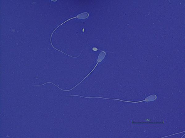
A thorough understanding of normal and abnormal spermatogenesis facilitates understanding and interpretation of a spermiogram (description of sperm morphology noted during evaluation) (Figures I.1–I.4). The testis is largely composed of seminiferous tubules and interstitial tissue. The latter is located between the seminiferous tubules and consists primarily of Leydig cells, which produce and secrete steroid hormones, as well as vascular and lymph vessels that supply the testicular parenchyma. Seminiferous tubules arise from primary sex cords and contain germinal cells, as well as Sertoli cells, which support and nurture production of sperm. Sertoli cells form tight junctions,
Sperm Morphology of Domestic Animals, First Edition. J.H. Koziol and C.L. Armstrong. © 2022 John Wiley & Sons, Inc. Published 2022 by John Wiley & Sons, Inc. Companion website: www.wiley.com/go/koziol/sperm
creating the most important portion of the blood–testis barrier [4]. Spermatogenesis occurs primarily in the tubulus contortus section of the seminiferous tubules. These tubules are surrounded by contractile peritubular cells that promote flow out of the tubulus contortus into the rectus (straight portion) of the seminiferous tubule. Both ends of each tubule are connected with the rete testis.
Spermatogenesis can be divided into three phases. The first phase, proliferation, consists of six mitotic divisions of spermatogonia, increasing the number of A- spermatogonia then B-spermatogonia. An important part of this phase is stem cell renewal. Loss of intercellular bridges allow some spermatogonia to revert to stem cells. B-spermatogonia then undergo several mitotic divisions, with the last division resulting in primary spermatocytes. The meiotic phase begins with diploid primary spermatocytes. During meiosis I, genetic diversity is ensured by DNA replication and crossing over during production of secondary spermatocytes, and as a result, from a genetic perspective, no two sperm are identical. The last phase, known as the differentiation phase or spermiogenesis, is differentiation of a spherical undifferentiated spermatid into a fully differentiated sperm with a head, a flagellum which includes the midpiece, and the principal piece. The length of spermiogenesis differs between species. An example is that it takes the last 18 days for the spermiogenesis stage of spermatogenesis to be completed in the bull.
Correct clinical interpretation of spermiograms also requires understanding of the specific timing of spermatogenesis in the species being evaluated. An understanding of the cycle of the seminiferous epithelium aids in this mission. The cycle of seminiferous epithelium is the progression through a complete series of stages at one location along a seminiferous tubule. On cross-sectional evaluation of the seminiferous tubule, four to five concentric layers of germ cells are present with each layer representing a generation. Each cross-section along the length of the seminiferous tubule will have a distinct appearance. Each cross section with its four to five generations of germ cells represents a stage of the seminiferous epithelium cycle. For example, a cross section in stage 1 will have a base layer of A-spermatogonium followed by primary spermatocytes, and another layer of primary spermatocytes, followed by a layer of immature spermatids. A stage 8 cross section will begin with a layer of A- spermatogonium, followed by B- spermatogonium, primary
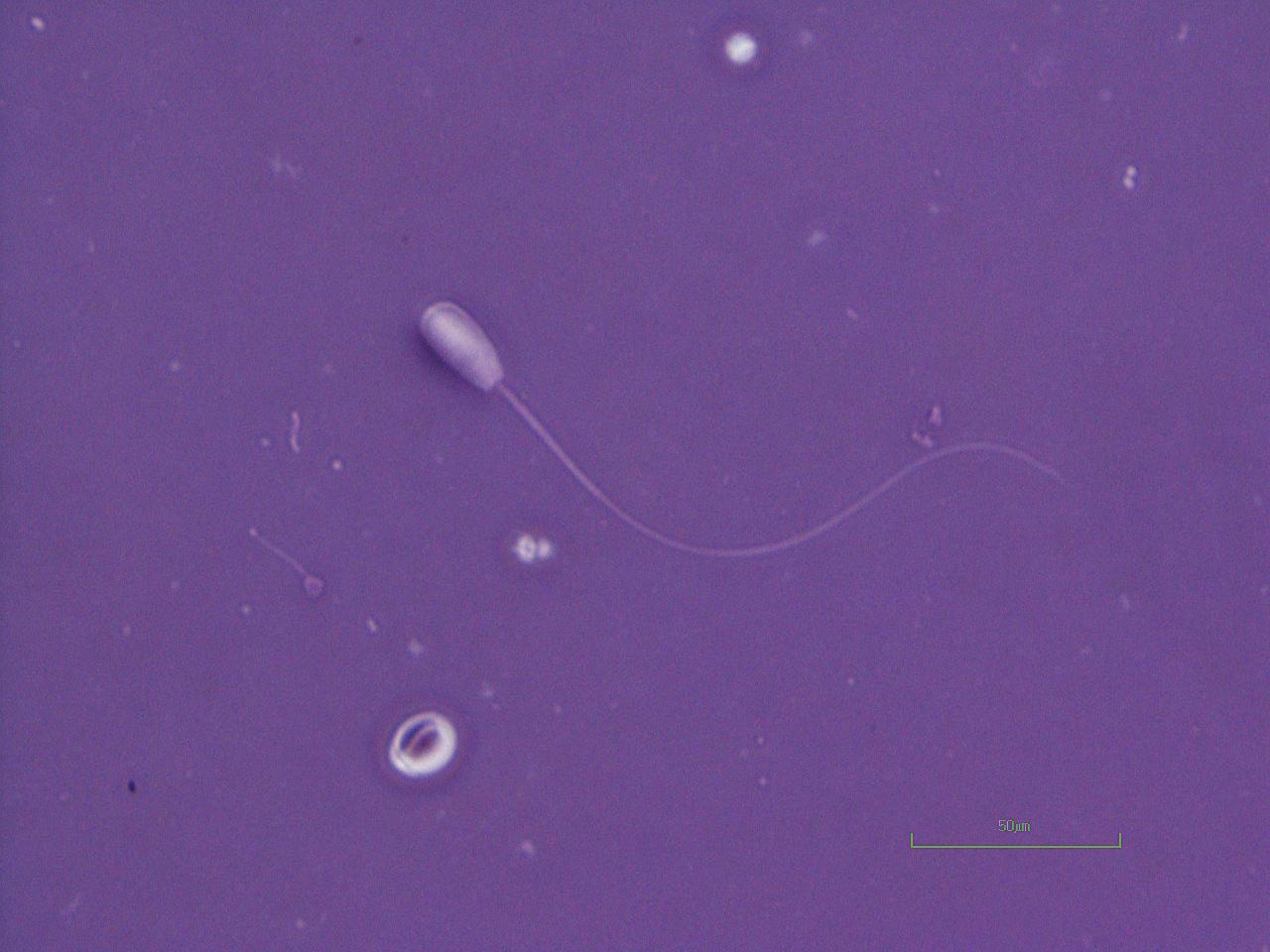
Figure I.1 Morphologically normal sperm from a bull (eosin–nigrosin, 1000×).
spermatocytes, immature spermatids, and mature spermatids that are ready to be released into the lumen. Along the length of any given seminiferous tubule, there are many zones or cross sections in different stages with only a few zones at an appropriate stage to release mature spermatid into the lumen of the seminiferous tubule, which will then travel through the rete testis to be transported to the head of the epididymis. These different zones allow for the creation of a spermatogenic wave or constant release of sperm along the length of each tubule. With zones in multiple
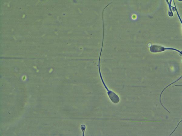
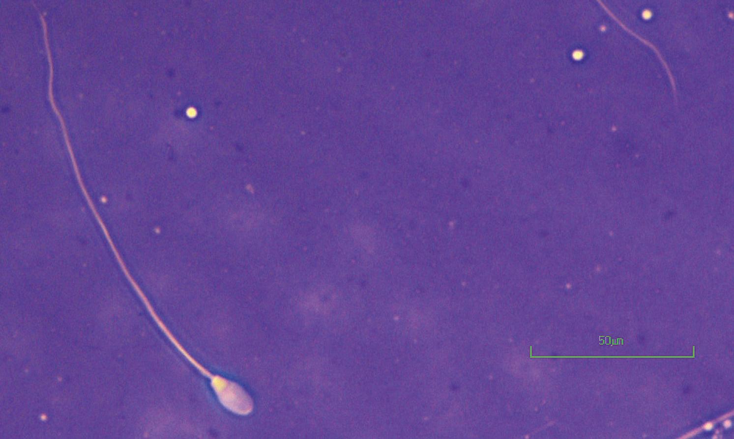
Figure I.2 Morphologically normal sperm from a bull (phase contrast wet mount, 1000×).
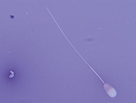
Figure I.3 Morphologically normal sperm from a buck (eosin–nigrosin, 1000×).
stages along the length of the tubule, there is a constant flow of spermiation, which allows for a constant flow of sperm to the epididymis.
Normal testicular function is dependent on appropriate endocrine, autocrine, and paracrine control. In the male, gonadotropin releasing hormone (GnRH) is secreted from the hypothalamus in a pulsatile nature resulting in waves of luteinizing hormone (LH) and follicle stimulating hormone (FSH) release. LH acts on the Leydig cells within the testes, which are responsible for synthesizing progesterone, the majority of which is converted to testosterone. This pulsatile nature of LH is necessary for normal testicular function as sustained LH can lead to a refractory nature of the Leydig cells secondary to a downregulation in the number of receptors. This culminates in reduced secretion of testosterone from the Leydig cells. High concentrations of testosterone must be maintained for normal spermatogenesis to occur. The pulsatile release of LH and consequently testosterone prevents negative inhibition of FSH, which is needed for maintenance of Sertoli cells. Sertoli cells convert testosterone to dihydrotestosterone and estradiol along with secreting androgen binding protein (ABP). Approximately 80% of the secreted ABP goes into the lumen of the seminiferous tubule and travels to the epididymis, while the remainder is secreted into the interstitial compartment and is absorbed into the systemic circulation. ABP binds testosterone and is responsible for not only maintaining a high concentration of testosterone within testes but also carries testosterone to the epididymis.
The most common causes of abnormal spermatogenesis in males include abnormal testicular thermoregulation; hormonal imbalances, particularly those associated with stress; and effect(s) of toxins or expression of deleterious genes [5]. Stress typically elevates systemic cortisol concentrations, profoundly decreasing release of LH and testosterone [6–8]. Stress has many origins, including environment, illness, or injury, causing changes in the spermiogram similar to those induced by disruption of thermoregulation. The primary spermatocyte is extremely sensitive to alterations in the hormonal milieu secondary to stress or illness. For example, in cases of testicular degeneration, the changes appear first in the cytoplasm, centrosomes, and spindles at the level of the primary spermatocyte, which predispose to disturbances in the developing spermatid [3, 9].
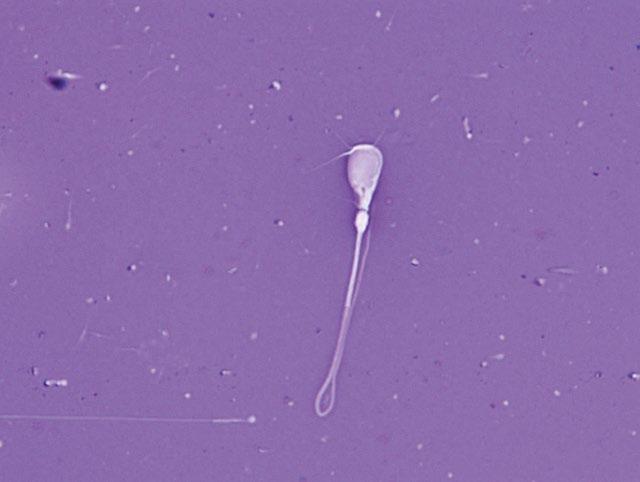
Figure I.4 Morphologically normal sperm from a stallion (eosin–nigrosin, 1000×).
I.2 Length of Spermatogenesis in Domestic Species
I.3 Maturation of Spermatozoa in the Epididymis
From the rete testis, sperm travel to the efferent duct and into the head of the epididymis. Grossly, the epididymis can be divided into three segments: the head (caput), body (corpus), and tail (cauda). The epididymis is responsible for transport, concentration, storage, and maturation of sperm.
During epididymal transport, maturation of sperm continues with many biochemical and morphological modifications occurring. All sperm that enter the head of the epididymis do so with a proximal cytoplasmic droplet; as they move through the epididymis, the droplet normally migrates down the midpiece and is present at the distal end of the midpiece when sperm arrive in the tail of the epididymis.
Along the entire length of epididymis, the lumen is bordered by epithelium that synthesizes and secretes proteins under androgen stimulation [12]. Intraluminal fluids released sequentially along the epididymal lumen are key for maturation of sperm [13]. As the sperm cell travels along the lumen many biochemical modifications occur. These processes include nucleus chromatin condensation, changes in plasma membrane composition and relocation of surface antigens, and the ability to respond to hypo-osmotic stress [14]. Circulating and intraluminal androgens are critical for epididymal gene expression and secretion. Androgens in the epididymis are synthesized by enzymatic reduction of testicular testosterone. Estrogen produced by the testicular Leydig cells also plays a role in the epididymis particularly in the role of water reduction in the proximal regions of the epididymis [14].
Following ejaculation, distal droplets should be shed after mixing with seminal fluid. Epididymal transport time is species dependent (Table I.1). The tail of the epididymis and ductus deferens serve as short-term sperm storage. However, in the absence of ejaculation, senescent sperm should be continually expelled into the urinary tract, ensuring that viable sperm are always available, even following long periods of sexual rest [15].
I.4 Staining Techniques and Evaluation of Morphology
Microscopic evaluation of sperm morphology is a pillar of successful sperm evaluation and breeding soundness examinations of any species. The three basic types of light microscopy are brightfield, phase contrast, and differential interface contrast (DIC). Most conventional microscopes have a brightfield function and many also have phase contrast capability. However, DIC is expensive and is generally restricted to use in research or andrology laboratories.
Brightfield microscopy is suitable for examination of morphology utilizing stained semen smears. Morphology evaluations must always be performed using the oil immersion (100×) objective lens; with eyepieces of 10–12.5×, the total magnification is 1000–1250× (hereafter referred to as 1000×). Morphology evaluation at lower magnification is not recommended as it results in failure to recognize many sperm abnormalities.
Utilization of phase contrast microscopy allows examination of unstained, cover-slipped, preparations prepared by mixing semen with warmed 10% neutral buffered formalin on a microscope slide, as well as slides stained with Feulgen stain [20]. Evaluation of sperm morphology utilizing phase contrast at 1000× is preferred over the use of stained preparations by some practitioners, as defects such as nuclear vacuoles, [21] acrosomal defects, and abnormal DNA in the case of Feulgenstained slides are easier to identify. However, these defects can also readily be identified with highquality brightfield microscopes.
Morphologically abnormal sperm have long been associated with male infertility, and assessment of these abnormalities is a fundamental component of analysis of semen quality. [3, 22–24]. Sperm morphology is an excellent predictor of the outcome of natural mating, artificial insemination, and in vitro fertilization, [3, 25, 26] and there is a relationship between morphologically abnormal sperm and poor DNA quality [24].
I.5 Vital Staining
Live/dead stains, also referred to as supravital stains, such as eosin–nigrosin and eosin–aniline blue, are commonly used for routine morphological examination of sperm [27]. In a study by Tanghe et al., eosin–nigrosin staining of sperm was the only bull sperm quality parameter evaluated that demonstrated a significant association with pronucleus formation in in vitro fertilization [28]. When utilizing live/dead stains, nigrosin or aniline blue provides a background, whereas eosin acts as a vital stain. Functional plasma membranes of viable sperm prevent penetration of eosin into the cell; therefore, viable sperm appear white. In contrast, eosin penetrates damaged cell membranes, staining injured or nonviable sperm pink.
Table I.1 Mean time of sperm epididymal passage in domestic
Species Average time (days)
Dog 15
Boar 9–14
Bull 8–11
Stallion 8–11
Ram 16.4
References
Linde-Forsberg [16]
Franca and Cardoso [17]
Barth and Oko [3]
Swierstra [18]
Amann [19]
Figure I.5 Par tially stained sperm with a pink posterior and a white anterior (eosin–nigrosin, bull, 1000×).
Partially stained sperm, with a pink posterior portion and a white anterior portion of the sperm head, are sometimes observed. These sperm have suffered membrane damage, yet the acrosome remained intact. This staining pattern occurs because the acrosomal membrane is tightly applied to the nucleus at the equatorial region and penetration of the eosin stain under the acrosome is initially impeded at the equatorial region [29]. Partially stained sperm are more common when semen samples are processed under less than ideal conditions, suggesting that sperm membrane damage or cell death occurred very recently (Figure I.5).
When assessing the proportion of sperm staining alive, it may be relevant to only count sperm stained completely white. When semen has been handled properly and stained correctly, the percentage excluding eosin, and therefore considered “alive” is highly correlated to progressive motility. This is not to suggest that only the “alive” cells be counted during evaluation of a sperm morphology slide; 100 cells should be counted regardless of vital status to give an accurate representation of the current state of the ejaculate. The accuracy of the assessment of marginal bulls can be improved by counting 300 cells.
I.6 Preparation of Semen Smears or Coverslip Slides
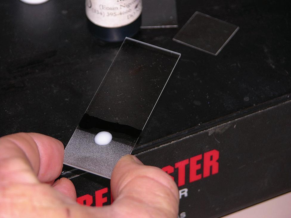
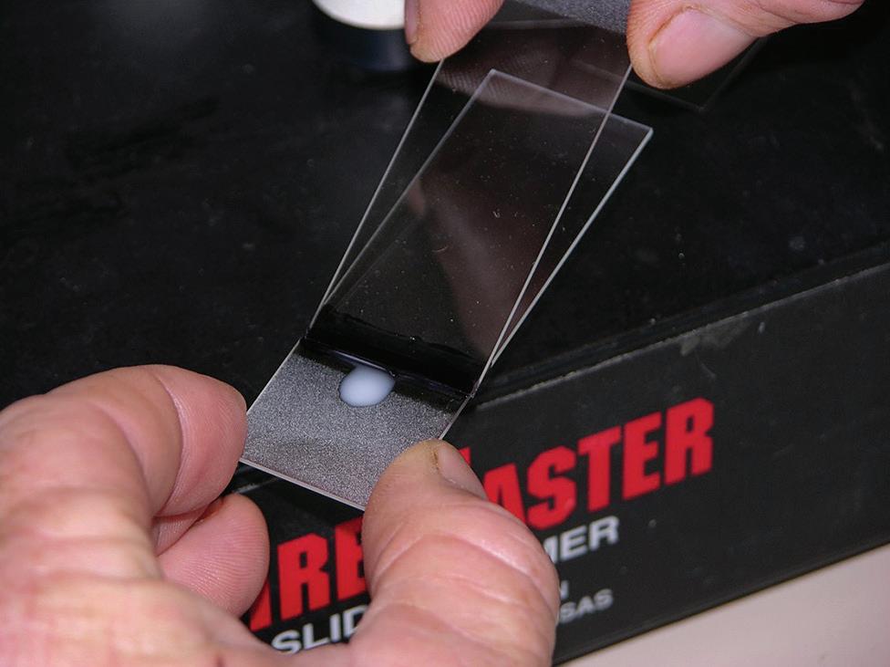
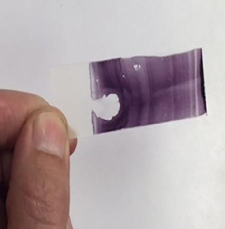
Slide preparation is crucial for proper evaluation of sperm morphology and requires practice and attention to detail. Common errors include too many sperm per slide (Figure I.6); slides stained too dark or too light; and slow drying resulting in cracks and stain artifacts. There are multiple ways to create stained slides. Some may prefer to streak out slides using another glass slide in a manner similar to producing a blood smear while others prefer to use a wood applicator stick to roll the sample out (Figures I.7–I.9). Some practitioners believe that the wood applicator stick results in less mechanical damage to the sperm (detached heads, damaged acrosomes). However, no studies have proven this clinical suspicion. Slides can be viewed anywhere along the length but usually half way down the slide is a good place to start as the cells are usually evenly distributed with good background stain. Wet mounts can be made for evaluation of sperm morphology by phase-contrast or DIC microscopy at 1000×. Freshly ejaculated semen must be diluted and motility stopped to allow for accurate evaluate. This can be accomplished with the addition of 0.2% glutaraldehyde in phosphate buffered saline (PBS), buffered formal saline, or with neutral buffered formalin. To prepare a wet mount slide, a 2–3 mm drop of diluted semen is placed on a warm clean microscope slide and a coverslip is placed over top. Allow the drop to evenly distribute under the coverslip before placing a drop of oil
I.6 Poorly prepared slide with too many sperm per field, which impeded the evaluators ability to evaluate each individual sperm (eosin–nigrosin, 500×).
Figure I.7 A small line of stain is placed along the slide followed by a small drop of semen.
Figure I.8 Using a second slide, one can start in the stain backing up to pick up a small line of semen and then proceed to streak out the slide. There are many different methods than can be used for streaking out slides.
Figure
Figure I.9 Slides should neither be too dark or too light, and when viewed under the microscope, the cells should be spaced in such a manner that they can each be individually examined.
on the coverslip for examination at 1000×. The preparation should be very thin to avoid sperm laying on their edges rather than flat. When using glutaraldehyde or neutral-buffered formalin, clumping of cells may occur. Wet mount slides can be viewed at any location along the coverslip with the exception of near the edges, as this is where a higher number of dead cells tend to accumulate [3].
I.7 Differential Counting of Sperm Morphology
Differential counts of sperm morphology must be performed using 1000× magnification (oil immersion) as lower magnifications result in failure to recognize some defects. The goal for examining sperm morphology is to determine the percentage and types of sperm abnormalities present in a sample and thereby construct a spermiogram. The spermiogram may be used to interpret potential fertility of semen at the time of sampling, as well as determine potential causes of an abnormal spermiogram. Knowledge of probable cause of abnormalities may allow the practitioner to develop a prognosis for recovery, which will aid the client in making an informed decision about the male in question [29].
Determination of a spermiogram is facilitated by the use of a differential cell counter. Mechanical cell counters often allow one to press two or more keys simultaneously to record more than one defect within the same sperm, while counting the individual sperm only once. With this system, a more accurate spermiogram may be produced.
When sperm morphology is good, differential counting of 100 sperm is adequate to determine a spermiogram. However, when the percentage of abnormal sperm present is high, counting at least 300 sperm may be necessary to produce an accurate spermiogram.
Section I
Head Abnormalities
Pyriform and Tapered Heads
The prevalence of pyriform head defects are relatively high and are either the most or second most prevalent defect in an ejaculate [3, 30–34]. The classical pyriform sperm has a pear-shaped head, a normal acrosome, and a narrow post-acrosomal region [3, 35–37]. Sperm with irregularity in head shape and size generally have good motility [3].
Several variants of pyriform head defects have been reported, ranging from slightly affected to severely tapered through the acrosome and post-acrosomal regions. The main initiating factor for this deformity appears to be adverse environmental conditions impacting male health and fitness [3, 38]. Pyriform sperm have been noted in ejaculates three to four weeks after initiation of dexamethasone injections to simulate stress, following scrotal insulation, or exposure to high ambient temperatures in the case of rams [3, 5, 38]. Pyriform or tapered heads have also been noted in those suffering from obesity resulting in scrotal or inguinal deposition of fat, frostbite or dermatitis of the scrotum, varicoceles, or other injuries that impede proper testicular thermoregulation [3]. Disruptions in endocrine hormone balance occurring either systemically or locally may also be implicated. Local disruption is caused by abnormalities in testicular thermoregulation while systemic disruption descends from unfavorable conditions or events that cause stress to the animal. These stressful events can include chronic pain, joint pain, conditions affecting the hooves including lameness or laminitis, extremes in weather, housing or social stress, improper nutrition, or travel [3].
Differences in the severity of response between males with similar treatments suggest that some males may be genetically predisposed to develop pyriform heads in response to adverse environmental influences [3, 5, 29]. In most instances, there will be many other sperm abnormalities present in the ejaculate, indicating a disturbance in spermatogenesis has occurred. However, after removal of stress factors, males often return to normal sperm production [3].
Small numbers of pyriform or tapered defects can be found in the semen of most bulls, even in bulls of good fertility [3]. In a study of bulls used for natural service and artificial insemination, percentage of bulls producing affected sperm were 9 and 16%, respectively. The percentage of pyriform sperm in an ejaculate from affected individual varies from 10 to >50% [3]. When pyriform defects are in high numbers, their impact on fertility has been documented in both in vivo and in vitro studies. Thundathil et al. [39] reported that in vitro fertilization rates were lower for semen containing high numbers of pyriform heads than for ejaculates containing low numbers, 68.5 versus 84.4%, respectively. Sixty-day pregnancy rates in estrus-synchronized artificially inseminated heifers using semen from these same bulls were 37 and 61%, respectively, for bulls with pyriform sperm and the control bull. In the same study, rates of embryo/fetal loss
Sperm Morphology of Domestic Animals, First Edition. J.H. Koziol and C.L. Armstrong. © 2022 John Wiley & Sons, Inc. Published 2022 by John Wiley & Sons, Inc. Companion website: www.wiley.com/go/koziol/sperm
between rates of embryo/fetal loss between days 22 and 60 of pregnancy were 23 and 8% for the pyriform head group and the control group, respectively [39].
Pyriform sperm disrupt various stages of the fertilization process including: sperm transport, [40–42] oocyte binding and zona penetration, [43] along with postfertilization events [39]. Furthermore, the prognosis for recovery is highly variable among animals. Mature bulls with high levels (25–75%) of pyriform sperm have a poor prognosis for recovery when there is no
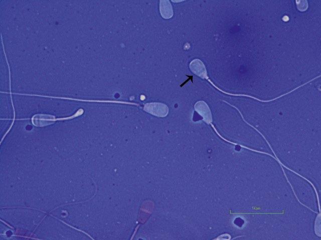
Figure 1.1 Pyriform head in a bull (eosin–nigrosin, 1000×).
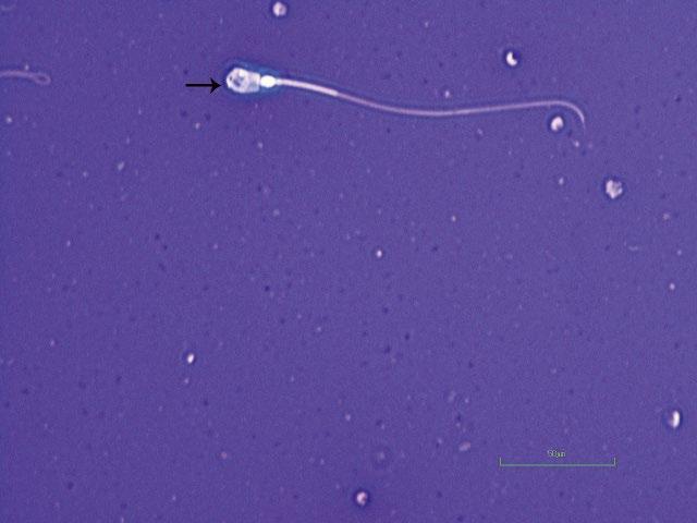
Figure 1.2 Pyriform head in a bull as indicated by arrow (eosin–nigrosin, 1000×).
apparent reason for a disturbance of spermatogenesis [21]. Return to normal sperm production by bulls known to have suffered an adverse condition predisposing them to disturbed spermatogenesis is dependent on the severity and duration of the insult. Younger overconditioned bulls often produce an ejaculate with a high level of pyriform heads and recover after weight loss [3].
In stallions, Jasko and Love both noted a negative correlation between the percentage of sperm head defects and fertility [32, 33]. Jasko et al. noted that head defects accounted for the large proportion of variation in per cycle pregnancy rates [33]. Love et al. similarly noted that a 1% increase in percentage of head defects resulted in a 0.67% reduction in per cycle pregnancy rates.
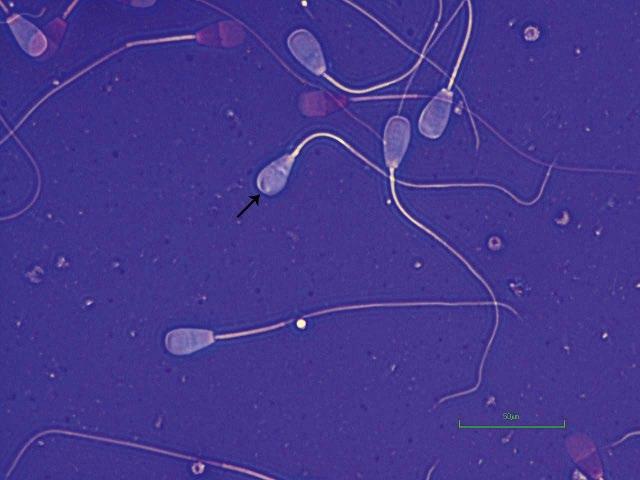
Figure 1.3 Pyriform head in a bull as indicated by arrow (eosin–nigrosin, 1000×).
Figure 1.4 Detached pyriform head in a bull indicated by arrow (eosin–nigrosin, 1000×).
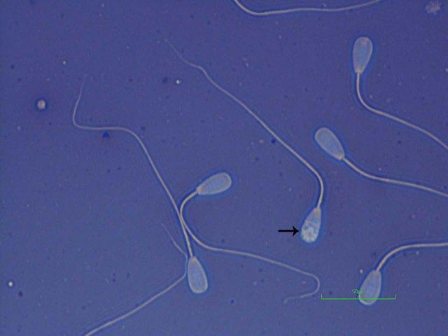
Figure 1.5 Pyriform head in bull as indicated by arrow (eosin–nigrosin, 1000×).
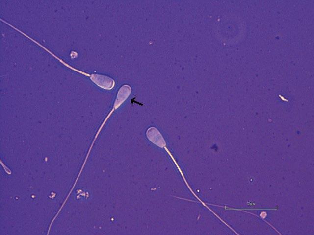
Figure 1.6 Pyriform head in a bull as indicated by arrow (eosin–nigrosin, 1000×).
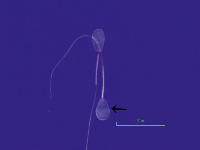
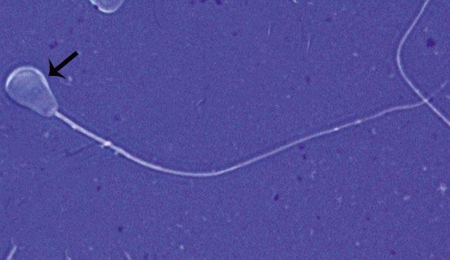
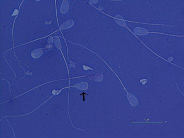
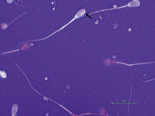
Pyriform head in bull as indicated by arrow also note the proximal droplet and terminally coiled tail (eosin–nigrosin, 1000×).
Figure 1.7
Figure 1.8 Pyriform head in a bull (eosin–nigrosin, 1000×).
Figure 1.9 Pyriform head in a ram (eosin–nigrosin, 1000×).
Figure 1.10 Pyriform head in a bull (eosin–nigrosin, 1000×).
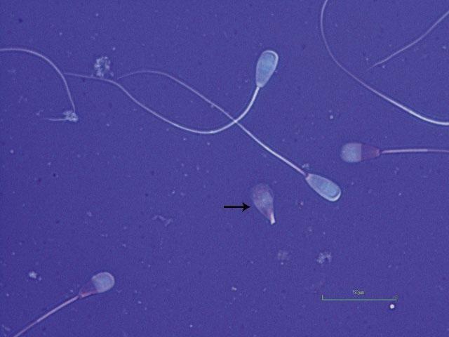
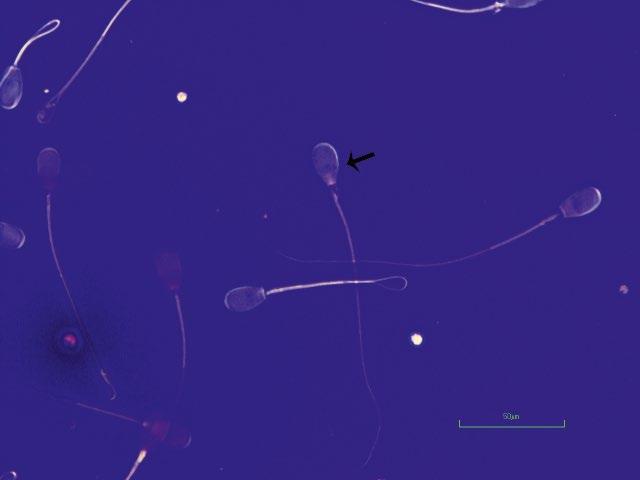
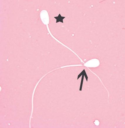
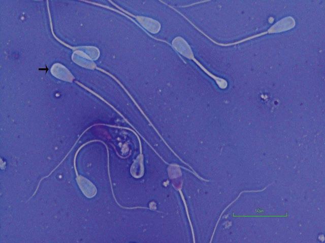
Figure 1.11 Pyriform head defect in a bull as indicated by arrow compared to normal-shaped head as indicated by star (eosin–nigrosin, 1000×).
and Tapered Heads
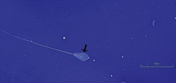
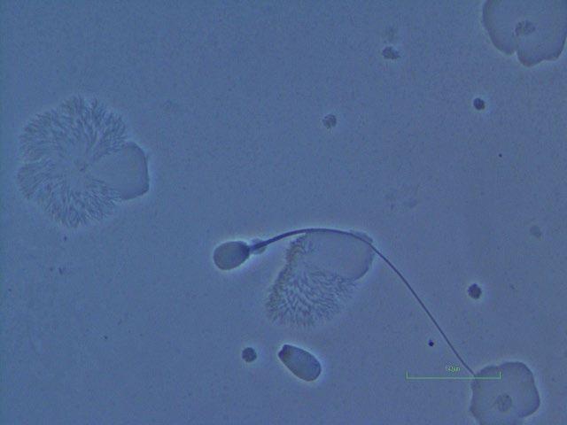
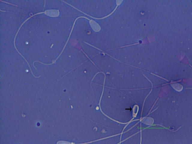
Figure 1.12 Pyriform-shaped head in a bull with proximal droplet (phase contrast wet mount, 1000×).
Nuclear Vacuolation, Including the Diadem Defect
Nuclear vacuoles have been described in sperm heads of all domestic species [44–50]. Historically, this defect has also been referred to as pouches or craters [49–52]. The defect is characterized by an invagination of the nuclear membrane into the nucleoplasm of the sperm head [53]. Nuclear vacuole formation has been identified as beginning as early as step 6 of spermiogenesis and has been identified in early and late spermatids. This coincides with the onset of condensation of the spermatid nucleoplasm and lengthening and flattening of the nucleus [54]. Vacuoles usually contain inclusions apparently derived from spermatid cytoplasm [29]. Various initiators of nuclear vacuolation have been postulated, including stress, aberrant testicular thermoregulation, viral infections, toxins, improper nutrition, and inheritance [29]. Dexamethasone treatments to mimic effects of stress in bulls acutely increased percentage of vacuoles, followed by a decline over several weeks [3, 5, 55]. Maximal levels of vacuoles occurred two to four weeks after dexamethasone treatment (20 mg/day for seven days given IM). In addition, scrotal insulation induced production of sperm head vacuolation in some bulls [5].
The incidence of nuclear vacuolation in the spermiogram of affected males can vary from <1% to nearly 100% [44]. When viewed using eosin–nigrosin morphology stain, vacuoles appear as dark spots in the nucleus and must be differentiated from artifacts such as stain crystals or other debris. Nuclear vacuoles are most easily spotted with phase contrast or differential interference microscopy. Feulgen staining will also reveal defects in the nuclear DNA [3]. Single or multiple vacuoles can be present anywhere in the sperm nucleus. However, they tend to be most commonly observed at the postnuclear cap-acrosome junction or near the apical ridge [55].
Nuclear vacuoles have three forms: single apical vacuoles present anywhere in the sperm head; large confluent vacuoles; or multiple pinpoint nuclear vacuoles at the acrosome post-acrosomal junction, resembling a string of pearls known as diadem vacuoles [44, 45, 56–58].
There are multiple reports that sperm with nuclear vacuoles impair fertility [44, 57, 59–61]. In one study, superovulated cows bred with semen from a bull with 80% nuclear vacuoles had a fertilization rate of 18% compared to that of 72% for control bulls [57]. Saacke demonstrated that sperm with nuclear vacuoles could transverse the female reproductive tract and access the ovum, thereby competing for fertilization [1]. However, vacuolated sperm were deficient in their ability to bind and penetrate the zona pellucida of in vitro-matured bovine oocytes [62]. High levels of nuclear vacuoles have also been shown to impair fertility in the stallion with semen from a stallion with 57% nuclear vacuoles producing no pregnancies over three years [63].
Sperm vacuoles have been noted to disappear in a few weeks following insult or persist for multiple years [3, 57, 61]. Collective results of available reports indicate a genetic predisposition to
Sperm Morphology of Domestic Animals, First Edition. J.H. Koziol and C.L. Armstrong. © 2022 John Wiley & Sons, Inc. Published 2022 by John Wiley & Sons, Inc. Companion website: www.wiley.com/go/koziol/sperm
2 Nuclear Vacuolation, Including the Diadem Defect 21 formation of nuclear vacuoles following disruptions in thermoregulation or stress; however, levels of vacuolated sperm can fluctuate dramatically over short intervals without apparent reason, and response to stressors is very unpredictable. Animals producing sperm severely affected by nuclear vacuoles should be closely monitored for the rest of their reproductive careers [29]. Considering recovery of the defect prognosis primarily depends on etiology of the problem.
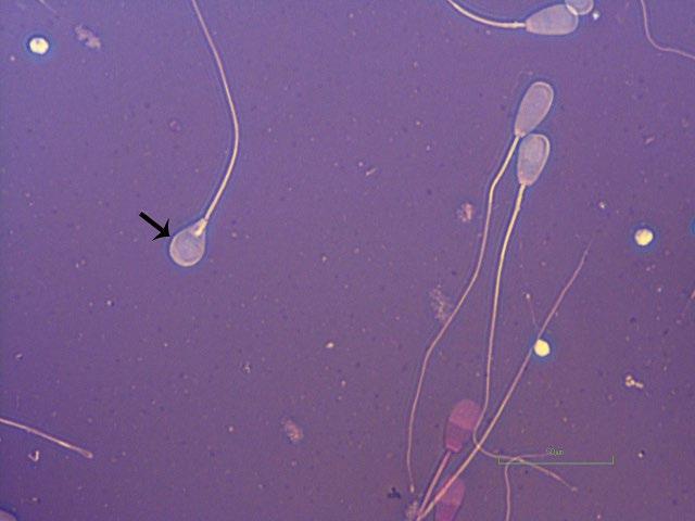
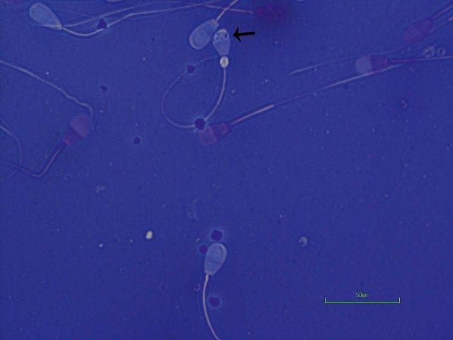
Figure 2.1 Nuclear vacuoles as indicated with arrow also note the proximal droplet (eosin–nigrosin, 1000×).
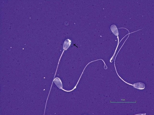
Figure 2.2 Small vacuoles in head of sperm as indicated with arrow (eosin–nigrosin, 1000×).
Figure 2.3 Vacuole in the head of sperm from a bull (eosin–nigrosin, 1000×).
Figure 2.4 Nuclear vacuoles in a bull also note the proximal droplet (eosin–nigrosin, bull, 1000×).
Figure 2.5 Large nuclear vacuole in a bull (eosin–nigrosin, 1000×).
Figure 2.6 Multiple small nuclear vacuoles in the head of a sperm from a bull (eosin–nigrosin, 1000×).
Figure 2.7 Multiple small nuclear vacuoles in the head of a sperm from a bull (eosin–nigrosin, 1000×).
Figure 2.8 Large confluent nuclear vacuole from a bull (eosin–nigrosin, 1000×).