Rossi's principles of transfusion medicine, 6th edition toby l. simon 2024 scribd download
Rossi's Principles of Transfusion Medicine,
6th Edition Toby L. Simon
Visit to download the full and correct content document: https://ebookmass.com/product/rossis-principles-of-transfusion-medicine-6th-edition-t oby-l-simon/
More products digital (pdf, epub, mobi) instant download maybe you interests ...
Harrison's Principles of Internal Medicine 21st Edition
Senior Medical Director, Plasma and Plasma Safety, CSL Plasma, Boca Raton, FL, USA; and Clinical Professor of Pathology, University of New Mexico School of Medicine, Albuquerque, New Mexico, USA
Eric A. Gehrie, MD
Executive Physician Director of Direct Patient Care and Emerging Cell and Gene Therapy Solutions, American Red Cross, Baltimore, Maryland, USA
Jeffrey McCullough, MD
Professor Emeritus, University of Minnesota Medical School, Minneapolis, Minnesota, USA
John D. Roback, MD, PhD
Professor of Pathology and Laboratory Medicine; Director, Center for Transfusion and Cellular Therapy, Department of Pathology & Laboratory Medicine, Emory University School of Medicine, Atlanta, Georgia, USA
Edward L. Snyder, MD, FACP
Professor, Department of Laboratory Medicine, Yale University School of Medicine; Blood Bank/Apheresis Service, Yale-New Haven Hospital, New Haven, Connecticut, USA
John Wiley & Sons Ltd (5e, 2016); John Wiley, Blackwell Publishing Ltd (4e, 2009).
All rights reserved. No part of this publication may be reproduced, stored in a retrieval system, or transmitted, in any form or by any means, electronic, mechanical, photocopying, recording or otherwise, except as permitted by law. Advice on how to obtain permission to reuse material from this title is available at http://www.wiley.com/go/permissions.
The right of Toby L. Simon, Eric A. Gehrie, Jeffrey McCullough, John D. Roback, and Edward L. Snyder to be identified as the authors of the editorial material in this work has been asserted in accordance with law.
Registered Offices
John Wiley & Sons, Inc., 111 River Street, Hoboken, NJ 07030, USA
John Wiley & Sons Ltd, The Atrium, Southern Gate, Chichester, West Sussex, PO19 8SQ, UK
Editorial Office
The Atrium, Southern Gate, Chichester, West Sussex, PO19 8SQ, UK
For details of our global editorial offices, customer services, and more information about Wiley products visit us at www.wiley.com.
Wiley also publishes its books in a variety of electronic formats and by print-on-demand. Some content that appears in standard print versions of this book may not be available in other formats.
Limit of Liability/Disclaimer of Warranty
The contents of this work are intended to further general scientific research, understanding, and discussion only and are not intended and should not be relied upon as recommending or promoting scientific method, diagnosis, or treatment by physicians for any particular patient. In view of ongoing research, equipment modifications, changes in governmental regulations, and the constant flow of information relating to the use of medicines, equipment, and devices, the reader is urged to review and evaluate the information provided in the package insert or instructions for each medicine, equipment, or device for, among other things, any changes in the instructions or indication of usage and for added warnings and precautions. While the publisher and authors have used their best efforts in preparing this work, they make no representations or warranties with respect to the accuracy or completeness of the contents of this work and specifically disclaim all warranties, including without limitation any implied warranties of merchantability or fitness for a particular purpose. No warranty may be created or extended by sales representatives, written sales materials or promotional statements for this work. The fact that an organization, website, or product is referred to in this work as a citation and/or potential source of further information does not mean that the publisher and authors endorse the information or services the organization, website, or product may provide or recommendations it may make. This work is sold with the understanding that the publisher is not engaged in rendering professional services. The advice and strategies contained herein may not be suitable for your situation. You should consult with a specialist where appropriate. Further, readers should be aware that websites listed in this work may have changed or disappeared between when this work was written and when it is read. Neither the publisher nor authors shall be liable for any loss of profit or any other commercial damages, including but not limited to special, incidental, consequential, or other damages.
Library of Congress Cataloging-in-Publication Data
Names: Simon, Toby L., editor. | Gehrie, Eric A., editor. | McCullough, Jeffrey, 1938- editor. | Roback, John D., 1961- editor. | Snyder, Edward L. (Edward Leonard), 1946- editor.
Title: Rossi’s principles of transfusion medicine / edited by Toby L. Simon, Eric A. Gehrie, Jeffrey McCullough, John D. Roback, Edward L. Snyder.
Section VIII: Cellular and tissue transplant technologies
52 Hematopoietic growth factors, 601
David J. Kuter
53 Hematopoietic stems cells and transplantation, 617
Garrett S. Booth
54 HLA antigens, alleles, and antibodies, 624
Scott M. Krummey, Robert A. Bray, Howard M. Gebel, and H. Clifford Sullivan
55 Chimeric antigen receptor T cells and other cellular immunotherapies, 633
Andrew D. Fesnak and Don L. Siegel
56 Gene therapy applications to transfusion medicine, 642
Seena Tabibi, Eric A. Gehrie, Emanuela M. Bruscia, and Diane S. Krause
57 Tissue engineering and regenerative medicine, 648
Cecilia Motta, Qi Xing, Cynthia Wilkins, and Julie Allickson
58 Human tissue allografts: responsibilities in understanding the path from donor to recipient, 660
Cassandra D. Josephson and Matthew J. Kuehnert
Index, 674
List of contributors
Julie Allickson
Mayo Clinic, Center for Regenerative Medicine, Rochester, MN, USA
Torunn Oveland Apelseth
Department of Immunology and Transfusion Medicine, Haukeland University Hospital, Bergen; and Norwegian Armed Forces Medical Ser vices, Oslo, Norway
Donald M. Arnold
Department of Medicine, Michael G. DeGroote School of Medicine, McMaster University, Hamilton, ON, Canada
Jeffrey A. Bailey
Department of Pathology and Laboratory Medicine, Warren Alpert Medical School, Brown University, Providence, RI, USA
Natalie Bavli
Department of Internal Medicine (Hematology/Oncology), University of Texas Southwestern, Dallas, TX, USA
Richard J. Benjamin
Cerus, Concord, CA, USA and Department of Laboratory Medicine, Georgetown University Washington, DC, USA
Melvin Berger
Departments of Pediatrics and Pathology, Case Western Reserve University, Cleveland, OH, USA
Thomas C. Binns
Departments of Pathology and Laboratory Medicine, Yale School of Medicine and Yale New Haven Hospital, New Haven, CT, USA
Lionel Blanc
Division of Pediatrics Hematology/Oncology, Cohen Children’s Medical Center, Zucker School of Medicine at Hofstra/Northwell and Feinstein Institutes for Medical Research, Manhasset, NY, USA
Evan M. Bloch
Division of Transfusion Medicine, Department of Pathology, Johns Hopkins University School of Medicine, Baltimore, MD, USA
Garrett S. Booth
Department of Pathology, Microbiology, and Immunology, Vanderbilt University Medical Center, Nashville, TN, USA
Robert A. Bray
Department of Pathology, Emory University Hospital, Atlanta, GA, USA
Nathan Brinkman
CSL Behring, Bradley, IL, USA
Patricia A. R. Brunker
Blood Transfusion Service, Department of Pathology, Massachusetts General Hospital, Harvard University, Boston, MA, USA
Emanuela M. Bruscia
Department of Pediatrics, Yale School of Medicine, Yale University, New Haven, CT, USA
Eileen M. Bulger
Trauma Center, Department of Surgery, Harborview Medical Center, University of Washington, Seattle, WA, USA
Edwin A. Burgstaler
Therapeutic Apheresis Treatment Unit, Mayo Clinic, Rochester, MN, USA
Jeffrey L. Carson
Division of General Internal Medicine, Rutgers Biomedical Health Sciences, Rutgers Robert Wood Johnson Medical School, New Brunswick, NJ, USA
Satheesh Chonat
Aflac Cancer and Blood Disorders Center, Children’s Healthcare of Atlanta, Department of Pediatrics, Emory University School of Medicine, Atlanta, GA, USA
Laura Cooling
Department of Pathology, University of Michigan, Ann Arbor, MI, USA
Angelo D’Alessandro
Department of Biochemistry and Molecular Genetics; and Metabolomics Core (SOM and Cancer Center), University of Colorado Denver–Anschutz Medical Campus, Aurora, CO, USA
Meghan Delaney
Children’s National Hospital, and Departments of Pediatrics and Pathology, The George Washington University School of Medicine and Health Sciences, Washington, DC, USA
E. Alexander Dent
Department of Pathology and Laboratory Medicine, Emory University Hospital, Atlanta, GA, USA
Anne F. Eder
Center for Biologics Evaluation and Research, Food and Drug Administration, Silver Spring, MD; and Georgetown University Medical Center, Washington, DC, USA
Lise J. Estcourt
NHS Blood and Transplant, Department of Haematology, Oxford University Hospitals and University of Oxford, Oxford, UK
Emmanuel A. Fadeyi
Department of Pathology, Wake Forest University School of Medicine, Winston-Salem, NC, USA
Ross M. Fasano
Center for Transfusion and Cellular Therapies, Department of Pathology and Laboratory Medicine, Emory University School of Medicine; and Aflac Cancer and Blood Disorders Center, Children’s Healthcare of Atlanta, Department of Pediatrics, Emory University School of Medicine, Atlanta, GA, USA
Andrew D. Fesnak
Division of Transfusion Medicine and Therapeutic Pathology, Department of Pathology and Laboratory Medicine, Perelman School of Medicine at the University of Pennsylvania, Philadelphia, PA, USA
Aline Floch
Univ Paris Est Créteil, INSERM U955, IMRB, Etablissement français du sang Ile-de-France, Créteil, France
Caroline Gabe
Department of Medicine, Michael G. DeGroote School of Medicine, McMaster University, Hamilton, ON, Canada; Faculty of Medicine, University of São Paulo, São Paulo, Brazil
Howard M. Gebel
Department of Pathology, Emory University Hospital, Atlanta, GA, USA
Eric A. Gehrie
The American Red Cross, Biomedical Services Division, Baltimore, MD, USA
Corinne Goldberg
The American Red Cross, Biomedical Services Division, Durham, NC, USA
Barry Gooch
CSL Behring, King of Prussia, PA, USA
Jed B. Gorlin
Innovative Blood Resources and Community Blood Center of Greater Kansas City, Divisions of New York Blood Center Enterprises, St. Paul, MN, USA
Laura Green
NHS Blood and Transplant and Barts Health NHS trust, London; Queen Mary University of London, London, UK
Jennifer Ricci Hagman
Department of Clinical Immunology and Transfusion Medicine, Division of Laboratory Medicine, Office for Medical Services; and Division of Hematology and Transfusion Medicine, Department of Laboratory Medicine, Lund University, Lund, Sweden
Paul C. Hébert
Medicine and Palliative Care, Department of Medicine, University of Ottawa, Ottawa, ON, Canada
Peter Hellstern
Center of Hemostasis and Thrombosis Zurich, Zurich, Switzerland
Jeanne E. Hendrickson
Departments of Laboratory Medicine and Pediatrics, Yale University School of Medicine, New Haven, CT, USA
John R. Hess
Laboratory Medicine and Pathology and Hematology, University of Washington School of Medicine; and Transfusion Service, Harborview Medical Center, Seattle, WA, USA
Benjamin R. Huebner
Surgical Critical Care, Harborview Medical Center, University of Washington, Seattle, WA, USA
Cassandra D. Josephson
Departments of Pathology and Pediatrics, Center for Transfusion and Cellular Therapies, Aflac Cancer and Blood Disorders Center, Emory University School of Medicine, Atlanta, GA, USA
Louis M. Katz
ImpactLife Blood Services, Davenport, IA and Medicine and Infectious Diseases, Roy and Lucille Carver College of Medicine, University of Iowa, Iowa City, IA, USA
Allan M. Klompas
Department of Anesthesiology, Mayo Clinic, Rochester, MN, USA
Scott Koepsell
University of Nebraska Medical Center, Omaha, NE, USA
Daryl J. Kor
Department of Anesthesiology, Mayo Clinic, Rochester, MN, USA
Mark J. Koury
Division of Hematology/Oncology, Vanderbilt University Medical Center, Nashville, TN, USA
Diane S. Krause
Department of Laboratory Medicine and the Yale Stem Cell Center, Yale School of Medicine, Yale University, New Haven, CT, USA
Scott M. Krummey
Department of Pathology, Johns Hopkins Hospital, Baltimore, MD, USA
Matthew J. Kuehnert
Musculoskeletal Transplant Foundation, Edison, NJ, USA; and Department of Medicine, Hackensack Meridian School of Medicine, Hackensack, NJ, USA
David J. Kuter
Hematology Division, Massachusetts General Hospital, Harvard Medical School, Boston, MA, USA
Courtney E. Lawrence
Department of Pediatrics and Department of Pathology, Johns Hopkins University School of Medicine, Baltimore, MD, USA
Edward S. Lee
Department of Laboratory Medicine, Yale University School of Medicine; and Blood Bank, Yale–New Haven Hospital, New Haven, CT, USA
Valery J. Li
Bloodworks Northwest Research Institute, Seattle; University of Washington School of Medicine, Seattle, WA, USA
Wen Lu
Department of Pathology, Brigham and Women’s Hospital, Boston, MA, USA
Johnathan P. Mack
The Ottawa Hospital/Ottawa Hospital Research Institute, University of Ottawa, ON, Canada
Cheryl L. Maier
Center for Transfusion and Cellular Therapies, Department of Pathology and Laboratory Medicine, Emory University School of Medicine, Atlanta, GA, USA
Samuel P. Mandell
Department of Surgery, Harborview Medical Center, University of Washington, Seattle, WA, USA
Peter W. Marks
Center for Biologics Evaluation and Research, U.S. Food and Drug Administration, Silver Spring, MD, USA
Karl McCann
CSL Behring, Broadmeadows, Victoria, Australia
Jeffrey McCullough
University of Minnesota Medical School, Minneapolis, MN, USA
Emily Riehm Meier
Indiana Hemophilia and Thrombosis Center, Indianapolis, IN, USA
Asher A. Mendelson
Section of Critical Care Medicine, Department of Medicine, Rady Faculty of Health Sciences, University of Manitoba, Winnipeg, MB, Canada
Celina Montemayor
Medical, Laboratory and Stem Cell Services, Canadian Blood Services, Toronto, ON, Canada
Cecilia Motta
Wake Forest School of Medicine, Wake Forest Institute for Regenerative Medicine, Wake Forest University, Winston Salem, NC, USA
Debra L. Mraz
Department of Laboratory Medicine, Yale School of Medicine; and Blood Bank, Yale–New Haven Hospital, New Haven, CT, USA
Michael F. Murphy
NHS Blood and Transplant, Department of Haematology, Oxford University Hospitals and University of Oxford, Oxford, UK
Monica B. Pagano
Department of Laboratory Medicine and Pathology, University of Washington, Seattle, WA, USA
Sandhya R. Panch
Department of Transfusion Medicine, National Institutes of Health Clinical Center, Bethesda, MD, USA
Seema R. Patel
Aflac Cancer and Blood Disorders Center, Children’s Healthcare of Atlanta/Emory University School of Medicine, Atlanta, GA, USA
Jenny Petkova
Weill Cornell department of Medicine, Hematology Division, Houston Methodist Hospital, Houston, TX, USA
Gregory J. Pomper
Department of Pathology, Wake Forest University School of Medicine, Winston-Salem, NC, USA
Eva D. Quinley
Regulatory Affairs and Laboratory Operations, CSL Plasma, Knoxville, TN, USA
Lynsi Rahorst
Laboratory of Immunohematology and Genomics, New York Blood Center Enterprises, New York, NY, USA
Sandra Ramirez- Arcos
Division of Medical Affairs and Innovation, Canadian Blood Services; and Department of Biochemistry, Microbiology and Immunology, University of Ottawa, Ottawa, ON, Canada
Lawrence Rice
Department of Medicine and Cancer Center, Houston Methodist Hospital and Weill Cornell Medical College, Houston, TX, USA
Henry M. Rinder
Department of Laboratory Medicine and Internal Medicine (Hematology), Yale School of Medicine, New Haven, CT, USA
John D. Roback
Center for Transfusion and Cellular Therapies, Department of Pathology and Laboratory Medicine, Emory University School of Medicine, Atlanta, GA, USA
Ennio C. Rossi
Northwestern University School of Medicine, Chicago, IL, USA (deceased)
Susan N. Rossmann
Gulf Coast Regional Blood Center, Houston, TX, USA
Paula P. Saá
American Red Cross Scientific Affairs, Gaithersburg, MD, USA
Eric Salazar
Department of Pathology and Laboratory Medicine, UT Health San Antonio, San Antonio, TX, USA
Ravi Sarode
Director of Transfusion Medicine and Hemostasis, Department of Pathology, University of Texas Southwestern, Dallas, TX, USA
Wade L. Schulz
Department of Laboratory Medicine, Yale School of Medicine, Yale University; and Blood Bank, Yale–New Haven Hospital, New Haven, CT, USA
Jansen N. Seheult
Department of Laboratory Medicine and Pathology, Mayo Clinic, Rochester, MN, USA
Sujit Sheth
Department of Pediatrics, Weill Cornell Medical College, New York, NY, USA
Alexa J. Siddon
Department of Laboratory Medicine, Yale School of Medicine; and Department of Pathology, Yale School of Medicine, New Haven, CT, USA
Robert F. Sidonio, Jr.
Aflac Cancer and Blood Disorders Center, Children’s Healthcare of Atlanta, Emory University School of Medicine, Atlanta, GA, USA
Don L. Siegel
Division of Transfusion Medicine and Therapeutic Pathology, Department of Pathology and Laboratory Medicine, Perelman School of Medicine at the University of Pennsylvania, Philadelphia, PA, USA
James Sikora
Center for Transfusion and Cellular Therapies, Department of Pathology and Laboratory Medicine, Emory University School of Medicine, Atlanta, GA, USA
Toby L. Simon
CSL Plasma, Boca Raton, FL, USA; and Department of Pathology, University of New Mexico School of Medicine, Albuquerque, NM, USA
James W. Smith
Department of Medicine, Michael G. DeGroote School of Medicine, McMaster University, Hamilton, ON, Canada
Edward L. Snyder
Department of Laboratory Medicine, Yale School of Medicine; Blood Bank, Yale–New Haven Hospital, New Haven, CT, USA
Bryan R. Spencer
American Red Cross Scientific Affairs, Dedham, MA, USA
Simon J. Stanworth
NHS Blood and Transplant and Oxford University Hospitals, and Departments of Haematology, Oxford University NHS Trust, Oxford, UK
Kristin M. Stendahl
Department of Laboratory Medicine, Yale School of Medicine, Yale University; and Blood Bank, Yale–New Haven Hospital, New Haven, CT, USA
Moritz Stolla
Bloodworks Northwest Research Institute, University of Washington School of Medicine, Seattle, WA, USA
Jill R. Storry
Department of Clinical Immunology and Transfusion Medicine, Division of Laboratory Medicine, Office for Medical Services, and Division of Hematology and Transfusion Medicine, Department of Laboratory Medicine, Lund University, Lund, Sweden
James R. Stubbs
Division of Transfusion Medicine, Department of Laboratory Medicine and Pathology, Mayo Clinic, Rochester, MN, USA
H. Clifford Sullivan
Department of Pathology and Laboratory Medicine, Emory University School of Medicine, Atlanta, GA, USA
Ruth Sylvester
USAF (ret), Omaha, NE, USA
Seena Tabibi
Department of Pathology, The Johns Hopkins University School of Medicine, Baltimore, MD, USA
Leanne Thalji
Department of Anesthesiology, Mayo Clinic, Rochester, MN, USA
Christopher A. Tormey
Department of Laboratory Medicine, Yale School of Medicine, and Transfusion Medicine, Yale New Haven Hospital, New Haven, CT, USA
Mary Townsend
Vitalant Inc., Scottsdale, AZ, USA
Darrell J. Triulzi
Department of Pathology, University of Pittsburgh Medical Center, Pittsburgh, PA, USA
Nancy L. Van Buren
Innovative Blood Resources and Community Blood Center of Greater Kansas City, MO; and Divisions of New York Blood Center Enterprises, St. Paul, MN, USA
Theodore E. Warkentin
Department of Medicine, Michael G. DeGroote School of Medicine, McMaster University; and Department of Pathology and Molecular Medicine, Michael G. DeGroote School of Medicine, McMaster University, Hamilton, ON, Canada
Matthew A. Warner
Department of Anesthesiology, Mayo Clinic, Rochester, MN, USA
Jonathan H. Waters
Departments of Anesthesiology and Bioengineering, University of Pittsburgh, Pittsburgh, PA, USA
Jennifer Webb
Children’s National Hospital, and Departments of Pediatrics and Pathology, The George Washington University School of Medicine and Health Sciences, Washington, DC, USA
Connie M. Westhoff
Laboratory of Immunohematology and Genomics, New York Blood Center Enterprises, New York, NY, USA
Cynthia Wilkins
Mayo Clinic, Center for Regenerative Medicine, Rochester, MN, USA
Jeffrey L. Winters
Therapeutic Apheresis Treatment Unit, Mayo Clinic, Rochester, MN, USA
Gary M. Woods
Aflac Cancer and Blood Disorders Center, Children’s Healthcare of Atlanta, Emory University School of Medicine, Atlanta, GA, USA
Qi Xing
Wake Forest School of Medicine, Wake Forest Institute for Regenerative Medicine, Wake Forest University, Winston Salem, NC, USA
Mark H. Yazer
Department of Pathology, University of Pittsburgh Medical Center, Pittsburgh, PA, USA
Preface
In 1983, the National Heart, Lung and Blood Institute (NHLBI) awarded five medical school faculty the first of what were to be a host of Transfusion Medicine Academic Awards. The purpose of the program was to enhance instruction in and exposure to the essential principles related to transfusion of blood into patients. This was considered a neglected area in medical education. Embedded in that decision was the idea that blood banking was part of a broader medical field now termed transfusion medicine.
Dr. Ennio C. Rossi was one of these first five awardees. At that time, he was a professor at Northwestern University School of Medicine in Chicago and director of its apheresis unit. Dr. Rossi was approached by Williams and Wilkins to put together a major textbook in this newly identified field of transfusion medicine. Dr. Rossi subsequently recruited two coeditors: Dr. Toby Simon, a board certified transfusion medicine physician who was also one of the first five NHLBI awardees, and Dr. Gerald Moss, a prominent surgeon who had notable research achievements in oxygen transport. Thus, the first edition of Principles of Transfusion Medicine was launched by two hematologists and a surgeon. After the second edition, Dr. Rossi retired and Dr. Toby Simon assumed the senior editor role. It was decided to add Dr. Rossi’s name to the title in recognition of his conception of the role of the book and to establish continuity for subsequent editions. Sadly, Dr. Rossi passed away on September 3, 2021. We dedicate this book to his memory and quote as follows from the first two paragraphs of the Preface to the first edition published in 1991:
Blood transfusion is an essential part of medical care and indispensable for the support of increasingly more sophisticated surgery. In the past, transfusion decisions were simple because therapeutic options were few. Now, decisions are more complicated. Transplantation biology and immunohematology are tightly intertwined, and transplantation surgery is frequently contingent upon special transfusion support. Advances in the technology of plasma fractionation and apheresis now provide a broad array of services for a large variety of clinical problems. Balanced against these benefits are the risks of blood-transmitted diseases, which have been underscored in the public consciousness by the emergence of acquired immunodeficiency syndrome (AIDS). Autologous transfusion and products of genetic engineering, such as hematopoietic growth factors, are being made available to diminish the risk, albeit small, of transfusiontransmitted disease by homologous blood. As these and other innovations render transfusion therapy more complex, blood banking has developed a clinical arm, transfusion medicine, to deal with these complexities. Principles of Transfusion Medicine will attempt to define the proper use of blood in clinical care. It is intended for the clinicians who prescribe blood, for the students who expect to enter clinical practice, for the scientists, physicians, nurses, technologists, and others who ensure the quality of our blood services. Many diverse sciences are applied to the preparation of blood for transfusion, and virtually all medical and surgical specialties must employ transfusion, from time to time, in care of their patients. For this reason, transfusion medicine is, of necessity, multidisciplinary.
In preparation of this sixth edition, we have also been challenged by a pandemic. In response to this pandemic, two chapters in the
early part of the book have been added, detailing how our specialty responded to the emergency and the lessons learned. In addition, we have chapters focusing on other “megatrends”: the application of molecular biology to the basics of matching donor and recipient, the use of apheresis to support new cellular approaches to cancer, the application of pathogen reduction for blood safety, the growth in plasma fractionation to meet the growing use of immune globulin preparations and other plasma-derived derivatives, as well as new approaches to support patients with massive bleeding, coagulopathy, and malignancy.
When the first edition was published, the transition away from whole blood to components and from cold-stored platelets to room-temperature stored platelets was nearly complete. Now we are seeing a reverse trend with recognition of potential benefits for the bleeding patient when treated with whole blood and cold-stored platelets. In the period since 1991, hemophilia care has gone from blood components such as cryoprecipitate, to plasma-derived factor concentrates, to recombinant products, to nonreplacement recombinant treatments, and finally to gene therapy to correct the defect that causes the disease. This is but one example of the evolution of the broader field of transfusion medicine we are capturing in this new sixth edition. We have assembled chapters and authors to guide the reader in understanding the changes that are occurring. At the same time, we have retained a significant amount of stillrelevant material from earlier editions.
Contributors for this edition have once again been drawn from various scientific, medical, and surgical disciplines. Thus, this book encompasses topics including encouraging and managing donors, collecting and preserving donated blood, and matching each component to the appropriate recipient, based on the patient’s clinical needs. The text also extends these concepts to tissue and goes beyond the field’s basic tenets to address new applications.
We can think of no better way to honor Dr. Rossi’s legacy than to present a sixth edition that blends transfusion science with clinical medicine, thus facilitating the thoughtful and measured prescription of blood, blood components, and their alternatives. Both the laboratory practice of blood banking and the clinical practice of transfusion medicine remain as important as ever. We proudly attribute the long-term influence of this field to its early leaders, who organized the discipline of transfusion medicine for success by anticipating future practice. We thank our new and returning contributors and the editorial staff at Wiley Blackwell for making possible a sixth edition of Rossi’s Principles of Transfusion Medicine in this new pandemic-influenced world of transfusion practice.
SDS-PAGE sodium dodecyl sulfate-polyacrylamide gel electrophoresis
Se secretor-positive
SHDAg short Hepatitis D antigen
SHOT Serious Hazards of Transfusion program (UK)
shRNA short hairpin RNA
SID Sda antigen
SID secondary immune deficiencies
SINV Sindbis virus
siRNA small interfering RNA
SIRS systemic inflammatory response syndrome
SIS small intestinal submucosa
SIT trial Silent Infarct Transfusion trial
Sl Swain-Langley antigen
SLE systemic lupus erythematosus
SMC smooth muscle cell
SNO-Hb S-nitrosohemoglobin
SNP single nucleotide polymorphism
SNV single nucleotide variation
SoGAT International Working Group on the Standardization of Genomic Amplification Techniques for the Virological Safety Testing of Blood and Blood Products
WHIM (syndrome) warts, hypogammaglobulinemia, infections, and myelokathexis
WHO World Health Organization
WNV West Nile virus
WOMAN World Maternal Antifibrinolytic trial
ZFN zinc finger nuclease
ZIKV Zika virus
ZnPP zinc protoporphyrin
About the companion website
This book is accompanied by a companion website. www.wiley.com/go/simon/Rossi6
The website features:
• The figures from the book in downloadable PowerPoint slides.
• Downloadable PDFs of all chapters.
• Downloadable PDFs of the complete reference lists from the book.
The password for the website is the first word of Chapter 1. Please use all lowercase.
SECTION I
Transfusion medicine from ancient times to the current pandemic
CHAPTER 1
Transfusion in the new millennium
Ennio C. Rossi1 & Toby L. Simon2
1Northwestern University School of Medicine (deceased), Chicago, IL, USA
2CSL Plasma, Boca Raton, FL, USA; and Department of Pathology, University of New Mexico School of Medicine, Albuquerque, NM, USA
Prehistoric man left drawings of himself pierced by arrows.1 This means he was as aware of blood as he was of his own limbs. The flint implements he used as tools and weapons distinguished him from other creatures and contributed to the violence of his era. As he hunted food and fought enemies, he observed bleeding and the properties of blood. A cut, received or inflicted, yielded a vivid red color. If the cut was shallow, there was little blood. But if the cut was deep, a red torrent flowing from the stricken victim quickly led to death, with shed blood congealed and darkening in the sun. Fatal hemorrhage was commonplace. Nonetheless, the sight must have been fearful and possibly existential as life flowed red out of the body of an enemy or a wounded animal.2 It is no wonder, then, that at the dawn of recorded history, blood was already celebrated in religious rites and rituals as a life-giving force.
The cultural expressions of primitive and ancient societies, although separated by time or space, can be strikingly similar. Whether these expressions emerged independently or were diffused about the world by unknown voyagers will probably always remain clouded in mystery.2 Nonetheless, there is a common thread in the ancient rituals that celebrate blood as a mystical vital principle. In Leviticus 17:11, “the life of the flesh is in the blood,” and the Chinese Neiching (circa 1000 bce) claims the blood contains the soul.2 Pre-Columbian North American Indians bled their bodies “of its greatest power” as self-punishment,3 Egyptians took blood baths as a recuperative measure, and Romans drank the blood of fallen gladiators in an effort to cure epilepsy.4
The Romans also practiced a ceremony called taurobolium—a blood bath for spiritual restoration. A citizen seeking spiritual rebirth descended into a pit or fossa sanguinis. Above him on a platform, a priest sacrificed a bull, and the animal’s blood cascaded down in a shower upon the beneficiary. Then, in a powerful visual image, the subject emerged up from the other end of the pit, covered with blood and reborn.1
The legend of Medea and Aeson taken from Ovid’s Metamorphoses and quoted in Bulfinch’s Mythology5 also ascribed rejuvenating powers to blood. Jason asked Medea to “take some years off his life and add them to those of his father Aeson.” Medea, however, pursued an alternative course. She prepared a cauldron with the blood of a sacrificed black sheep. To this, she added magic herbs, hoarfrost gathered by moonlight, the entrails of a wolf, and many other things “without a name.” The boiling cauldron was stirred
with a withered olive branch, which became green and full of leaves and young olives when it was withdrawn. Seeing that all was ready,
Medea cut the throat of the old man and let out all his blood, and poured into his mouth and into his wound the juices of her cauldron. As soon as he had imbibed them, his hair and beard laid by their whiteness and assumed the blackness of youth; his paleness and emaciation were gone; his veins were full of blood, his limbs of vigour and robustness. Aeson is amazed at himself and remembers that such as he now is, he was in his youthful days, 40 years before.
This legend seems to echo the apocryphal story of Pope Innocent VIII, who is said to have received the blood of three young boys in 1492 while on his deathbed. As the story goes, a physician attempted to save the pope’s life by using blood drawn from three boys 10 years of age, all of whom died soon thereafter. Some nineteenth-century versions of this tale suggest the blood was transfused. However, earlier renditions more plausibly suggest that the blood was intended for a potion to be taken by mouth. In any event, there is no evidence the pope actually received any blood in any form.6,7
The folklore that flowed with blood was not accompanied by a great deal of accurate information. The ancient Greeks believed that blood formed in the heart and passed through the veins to the rest of the body, where it was consumed. Arteries were part of an independent system transporting air from the lungs. Although Erasistratos (circa 270 bce) had imagined the heart as a pump, his idea was ahead of its time. As long as veins and arteries were dead-end channels transporting blood and air, there was little need for a pump in the system. Although Galen (131–201 ce) finally proved that arteries contain blood, communication with the venous system was not suspected. Blood, formed in the liver, merely passed through the blood vessels and heart on its way to the periphery.1 These teachings remained in place for 1400 years until they were swept away in 1628 by Harvey’s discovery of the circulation.
The realization that blood moved in a circulating stream opened the way to experiments on vascular infusion. In 1642, George von Wahrendorff injected wine8 and, in 1656, Christopher Wren and Robert Boyle injected opium and other drugs9 intravenously into dogs. The latter studies, performed at Oxford, were the inspiration for Richard Lower’s experiments in animal transfusion.
Richard Lower (1631–1691) was a student at Oxford when Christopher Wren and Robert Boyle began their experiments on infusion. In due course, Lower joined their scientific group and studied the intravenous injection of opiates, emetics, and other substances into living animals.10 In time, the transfusion of blood itself became the objective. The announcement of the first successful transfusion, performed by Richard Lower at Oxford in February 1665, was published on November 19, 1666, in the Philosophical Transactions of the Royal Society in a short notation titled, “The Success of the Experiment of Transfusing the Blood of One Animal into Another.”11 The entire notation is as follows:11
This experiment, hitherto look’d upon to be of an almost insurmountable difficulty, hath been of late very successfully perform’d not only at Oxford, by the directions of that expert anatomist Dr. Lower, but also in London, by order of the R. Society, at their publick meeting in Gresham Colledge: the Description of the particulars whereof, and the Method of Operation is referred to the next opportunity.
The December 17, 1666, issue of the Transactions contained the full description as promised.12 It was taken from a letter13 written by Lower to Robert Boyle on July 6, 1666, in which Lower described direct transfusion from the carotid artery of one dog to the jugular vein of another. After describing the insertion of quills into the blood vessels of the donor and recipient dogs, Lower wrote:13
When you have done this you may lay the dogs on their side and fasten them densely together as best you may to insure the connection of the two quills. Quickly tighten the noose around the neck of the receiving animal as in venasection, or at all events compress the vein on the opposite side of the neck with your finger, then take out the stopper and open the upper jugular quill so that while the foreign blood is flowing into the lower quill, the animal’s own blood flows out from the upper into suitable receptacles—until at last the second animal, amid howls, faintings, and spasms, finally loses its life together with its vital fluid. When the tragedy is over, take both quills out of the jugular vein of the surviving animal, tie tightly with the former slipknots, and divide the vein. After the vessel has been divided, sew up the skin, slacken the cords binding the dog, and let it jump down from the table. It shakes itself a little, as though aroused from sleep, and runs away lively and strong, more active and vigorous perhaps, with the blood of its fellow than its own.
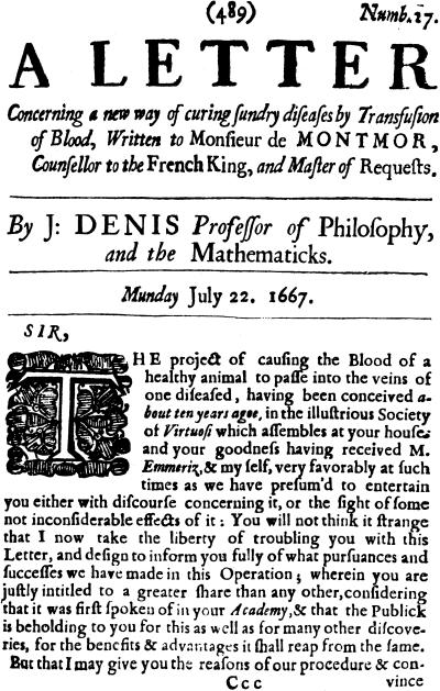
These studies inevitably led to the transfusion of animal blood to humans. In England, this occurred on November 23, 1667, when Lower and Edmund King transfused sheep blood into a man named Arthur Coga.14 Described by Samuel Pepys as “a little frantic,” Coga was paid 20 shillings to accept this transfusion, with the expectation that it might have a beneficial “cooling” effect. One week later, Coga appeared before the Society and claimed to be a new man, although Pepys concluded he was “cracked a little in the head.”13 However, this was not the first transfusion performed in a human. The credit for that accomplishment belongs to Jean-Baptiste Denis (1635–1704), who had performed the first human transfusion several months earlier in Paris.
The first animal-to-human transfusion
The founding of the Royal Society in London in 1662 was followed in 1666 by the establishment of the Academie des Sciences in Paris under the patronage of King Louis XIV. The new Academie reviewed the English reports on transfusion with great interest. Denis probably read of Lower’s experiments in the Journal des Savants on January 31, 1667, and he began his own studies approximately one month later.15,16 The first human transfusion was
Figure 1.1 The first human transfusion. Source: Denis (1967).17 Figure 01, p 01 / With permission of The Royal Society.
then performed on June 15, 1667, when Denis administered the blood of a lamb to a 15-year-old boy (Figure 1.1).
Although discovery of the circulation had suggested the idea of transfusion, indications for the procedure remained uninformed. Transfusion was still thought to alter behavior and possibly achieve rejuvenation. The blood of young dogs made old dogs seem frisky; the blood of lions was proposed as a cure for cowardice; and, five months later, Arthur Coga would receive a transfusion of sheep blood because of its presumed “cooling” effect. Denis used animal blood for transfusion because he thought it was “less full of impurities”:17
Sadness, Envy, Anger, Melancholy, Disquiet and generally all the Passions, are as so many causes which trouble the life of man, and corrupt the whole substance of the blood: Whereas the life of Brutes is much more regular, and less subject to all these miseries.
It is thus ironic that the symptoms of the first transfusion recipient may have been explained in part by profound anemia; the single transfusion of lamb blood may have produced temporary amelioration owing to increased oxygen transport. Denis described the case as follows:17
On the 15 of this Moneth, we hapned upon a Youth aged between 15 and 16 years, who had for above two moneths bin tormented with a contumacious and violent fever, which obliged his Physitians to bleed him 20 times, in order to asswage the excessive heat.
Before this disease, he was not observed to be of a lumpish dull spirit, his memory was happy enough, and he seem’d cheerful and nimble enough in body; but since the violence of this fever, his wit seem’d wholly sunk, his memory perfectly lost, and his body so heavy and drowsie that he was not fit for anything. I beheld him fall asleep as he sate at dinner, as he was eating his Breakfast, and in all occurrences where men seem most unlikely to sleep. If he went to bed at nine of the clock in the Evening, he needed to be wakened several times before he could be got to rise by nine the next morning, and he pass’d the rest of the day in an incredible stupidity.
I attributed all these changes to the great evacuations of blood, the Physitians had been oblig’d to make for saving his life.
Three ounces of the boy’s blood were exchanged for 9 ounces of lamb arterial blood. Several hours later the boy arose, and “for the rest of the day, he spent it with much more liveliness than ordinary.” Thus, the first human transfusion, which was heterologous, was accomplished without any evident unfavorable effect.
This report stimulated a firestorm of controversy over priority of discovery.18,19 The letter by Denis was published in the Transactions on July 22, 1667, while the editor, Henry Oldenburg, was imprisoned in the Tower of London. Oldenburg, following some critical comments concerning the Anglo-Dutch War then in progress (1665–1667), had been arrested under a warrant issued on June 20, 1667. After his release two months later, Oldenburg returned to his editorial post and found the letter published in his absence. He took offense at Denis’s opening statement, which claimed that the French had conceived of transfusion “about ten years ago, in the illustrious Society of Virtuosi” (Figure 1.1). This seemed to deny the English contributions to the field. Oldenburg cited these omissions in an issue of the Transactions published September 23, 1667, “for the Months of July, August, and September.” By numbering this issue 27 and beginning pagination with 489, Oldenburg attempted to suppress the letter by Denis.18 However, as is evident, this did not ultimately succeed. Nonetheless, subsequent events created even greater difficulties for Denis.
Although the first two subjects who underwent transfusion by Denis were not adversely affected, the third and fourth recipients died. The death of the third subject was easily attributable to other causes. However, the fourth case initiated a sequence of events that put an end to transfusion for 150 years.
Anthony du Mauroy was a 34-year-old man who suffered from intermittent bouts of maniacal behavior. On December 19, 1667, Denis and his assistant Paul Emmerez removed 10 ounces of the man’s blood and replaced it with 5 or 6 ounces of blood from the femoral artery of a calf. Failing to note any apparent improvement, they repeated the transfusion 2 days later. After the second transfusion, du Mauroy experienced a classic transfusion reaction:20
His pulse rose presently, and soon after we observ’d a plentiful sweat over all his face. His pulse varied extremely at this instant, and he complain’d of great pains in his kidneys and that he was not well in his stomach.
Du Mauroy fell asleep at about 10 o’clock in the evening. He awoke the following morning and “made a great glass full of urine, of a color as black, as if it had been mixed with the soot of chimneys.”20 Two months later, the patient again became maniacal, and his wife again sought transfusion therapy. Denis was reluctant but finally gave in to her urgings. However, the transfusion could not be accomplished, and du Mauroy died the next evening.
The physicians of Paris strongly disapproved of the experiments in transfusion. Three of them approached du Mauroy’s widow and encouraged her to lodge a malpractice complaint against Denis. She instead went to Denis and attempted to extort money from him in return for her silence. Denis refused and filed a complaint before
the Lieutenant in Criminal Causes. During the subsequent hearing, evidence was introduced to indicate that Madame du Mauroy had poisoned her husband with arsenic. In a judgment handed down at the Chatelet in Paris on April 17, 1668, Denis was exonerated, and the woman was held for trial. The court also stipulated “that for the future no transfusion should be made upon any human body but by the approbation of the Physicians of the Parisian Faculty.”21 At this point, transfusion research went into decline, and within 10 years it was prohibited in both France and England.
The beginnings of modern transfusion
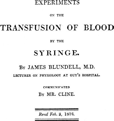
After the edict that ended transfusion in the seventeenth century, the technique lay dormant for 150 years. Stimulated by earlier experiments by Leacock, transfusion was “resuscitated” and placed on a rational basis by James Blundell (1790–1877), a London obstetrician who had received his medical degree from the University of Edinburgh.22 Soon after graduation, Blundell accepted a post in physiology and midwifery at Guy’s Hospital. It was there that he began the experiments on transfusion that led to its rebirth. The frequency of postpartum hemorrhage and death troubled Blundell. In 1818, he wrote:23
A few months ago I was requested to visit a woman who was sinking under uterine hemorrhage. . . . Her fate was decided, and notwithstanding every exertion of the medical attendants, she died in the course of two hours.
Reflecting afterwards on this melancholy scene . . . I could not forbear considering, that the patient might very probably have been saved by transfusion; and that . . . the vessels might have been replenished by means of the syringe with facility and prompitude.
This opening statement introduced Blundell’s epoch-ma king study titled “Experiments on the Transfusion of Blood by the Syringe”23 (see Figure 1.2). Blundell described in detail a series
Figure 1.2 The beginnings of modern transfusion. Source: Blundell (1818).23 Figure 01, p 01 / With permission of The Royal Society of Medicine.
of animal experiments. He demonstrated that a syringe could be used effectively to perform transfusion, that the lethal effects of arterial exsanguination could be reversed by the transfusion of either venous or arterial blood, and that the injection of 5 drams (20 cc) of air into the veins of a small dog was not fatal but transfusion across species ultimately was lethal to the recipient.23 Thus, Blundell was the first to clearly state that only human blood should be used for human transfusion. The latter conclusion was confirmed in France by Dumas and Prevost, who demonstrated that the infusion of heterologous blood into an exsanguinated animal produced only temporary improvement and was followed by death within six days.24 These scientific studies provided the basis for Blundell’s subsequent efforts in clinical transfusion.
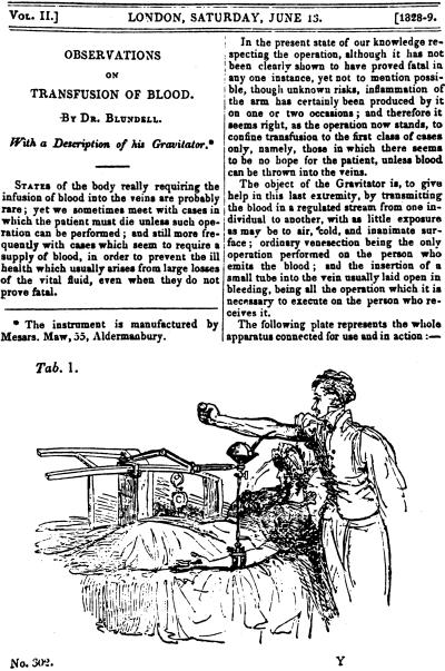
The first well-doc umented transfusion with human blood took place on September 26, 1818.25 The patient was an extremely emaciated man in his mid-thir ties who had pyloric obstruction caused by carcinoma. He received 12 to 14 ounces of blood in the course of 30 or 40 minutes. Despite initial apparent improvement, the patient died two days later. Transfusion in the treatment of women with postpartum hemorrhage was more successful. In all, Blundell performed 10 transfusions, of which 5 were successful. Three of the unsuccessful transfusions were performed on moribund patients, the fourth was performed on a patient with puerperal sepsis, and the fifth was performed on the aforementioned patient with terminal carcinoma. Four of the successful transfusions were given for postpartum hemorrhage, and the fifth was administered to a boy who bled after amputation.22 Blundell also devised various instruments for the performance of transfusion. They included an “impellor,” which collected blood in a warmed cup and “impelled” the blood into the recipient via an attached syringe, and a “gravitator”26 (Figure 1.3), which received blood and delivered it by gravity through a long vertical cannula.
The writings of Blundell provided evidence against the use of animal blood in humans and established rational indications for transfusion. However, the gravitator (Figure 1.3) graphically demonstrated the technical problems that remained to be solved. Blood from the donor, typically the patient’s husband, flowed into a funnel-like device and down a flexible cannula into the patient’s vein “with as little exposure as possible to air, cold, and inanimate surface.”25 The amount of blood transfused was estimated from the amount spilled into the apparatus by the donor. In this clinical atmosphere, charged with apprehension and anxiety, the amount of blood issuing from a donor easily could be overstated. Clotting within the apparatus then ensured that only a portion of that blood actually reached the patient. Thus, the amount of blood actually transfused may have been seriously overestimated. This may explain the apparent absence of transfusion reactions. Alternatively, reactions may have been unrecognized. Patients who underwent transfusion frequently were agonal. As Blundell stated, “It seems right, as the operation now stands, to confine transfusion to the first class of cases only, namely, those in which there seems to be no hope for the patient, unless blood can be thrown into the veins.”26 Under these circumstances, “symptoms” associated with an “unsuccessful” transfusion might be ascribed to the agonal state rather than the transfusion itself. For a time, the problem of coagulation during transfusion was circumvented by the use of defibrinated blood. This undoubtedly increased the amount of blood actually transfused. However, there were numerous deaths. Interestingly, these deaths were attributed to intravascular coagulation when in actuality they
were probably fatal hemolytic reactions caused by the infusion of incompatible blood.27
Transfusion at the end of the nineteenth century, therefore, was neither safe nor efficient. The following description, written in 1884, illustrates this point:28
Students, with smiling faces, are rapidly leaving the theatre of one of our metropolitan hospitals. The most brilliant operator of the day has just performed immediate transfusion with the greatest success. By means of a very beautiful instrument, the most complex and ingenious that modern science has yet produced, a skilful surgeon has transfused half a pint, or perhaps a pint, of blood from a healthy individual to a fellow creature profoundly collapsed from the effects of severe hemorrhage. Some little difficulty was experienced prior to the operation, as one of the many stop-cocks of the transfusion apparatus was found to work stiffly; but this error was quickly rectified by a mechanic in attendance. Towards the close of the operation the blood-donor, a powerful and heavy young man, swooned. Two porters carried him on a stretcher into an adjoining room.
In the latter half of the nineteenth century, there were many attempts to render transfusion a more predictable and less arduous procedure. In 1869, Braxton-Hicks,29 using blood anticoagulated with phosphate solutions, performed a number of transfusions on women with obstetric bleeding. Many of the
Figure 1.3 Blundell’s gravitator. Source: Blundell (1828).26 With permission of Jeremy Norman & Co., Inc.
patients were in extremis and ultimately all died. Unfortunately, a detailed description of terminal symptoms was not provided.29 Some investigators attempted to rejuvenate animal-to-human transfusion, and Oscar Hasse persisted in this approach despite disastrous results. Studies by Emil Ponfick and by Leonard Landois finally put an end to this practice. Ponfick, in carefully controlled studies, confirmed the lethality of heterologous transfusion and identified the resulting hemoglobinuria along with its donor erythrocyte source. Landois documented the poor results of animal-to-human transfusion and demonstrated the lysis of sheep erythrocytes by human serum in vitro.8
Frustration with blood as a transfusion product led to even more bizarre innovations. From 1873 to 1880, cow, goat, and even human milk was transfused as a blood substitute.30 The rationale derived from an earlier suggestion that the fat particles of milk could be converted into blood cells. Milk transfusion was particularly popular in the United States,30 where the practice of animal-to-human transfusion was recorded as late as 1890.31 Fortunately, these astonishing practices were discontinued when saline solutions were introduced as “a life-saving measure” and “a substitute for the transfusion of blood.”32 A passage from an article written by Bull in 188432 is particularly instructive:
The danger from loss of blood, even to two-thirds of its whole volume, lies in the disturbed relationship between the calibre of the vessels and the quantity of the blood contained therein, and not in the diminished number of red blood-corpuscles; and. . . . This danger concerns the volume of the injected fluids also, it being a matter of indifference whether they be albuminous or containing blood corpuscles or not.
Mercifully, volume replacement with saline solutions deflected attention from the unpredictable and still dangerous practice of blood transfusion. Accordingly, transfusions were abandoned until interest was rekindled by the scientific and technical advances of the early twentieth century.
The twentieth century
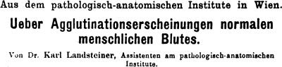
The twentieth century was ushered in by a truly monumental discovery. In 1900, Karl Landsteiner (1868–1943) observed that the sera of some persons agglutinated the red blood cells of others. This study, published in 1901 in the Wiener Klinische Wochenschrift33 (Figure 1.4), showed for the first time the cellular differences in individuals from the same species. In his article, Landsteiner wrote:34
In a number of cases (Group A) the serum reacts on the corpuscles of another group (B), but not, however, on those of group A, while, again, the corpuscles of A will be influenced likewise by serum B. The serum of the third group (C) agglutinates the corpuscles of A and B, while the corpuscles of C will not be influenced by the sera of A and B. The corpuscles are naturally apparently insensitive to the agglutinins which exist in the same serum.
With the identification of blood groups A, B, and C (subsequently renamed group O) by Landsteiner and of group AB by Decastello and Sturli,35 the stage was set for the performance of safe transfusion. For this work, Landsteiner somewhat belatedly received the Nobel Prize in 1930. But even that high recognition does not adequately express the true magnitude of Landsteiner’s discovery. His work was like a burst of light in a darkened room. He gave us our first glimpse of immunohematology and transplantation biology and provided the tools for important discoveries in genetics, anthropology, and forensic medicine. Viewed from this perspective, the identification of human blood groups is one of only a few scientific discoveries of the twentieth century that changed all of our lives.34 Yet the translation of Landsteiner’s discovery into transfusion practice took many years.
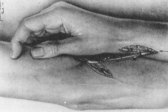
At the turn of the twentieth century, the effective transfer of blood from one person to another remained a formidable task. Clotting, still uncontrolled, quickly occluded transfusion devices and frustrated most efforts. In 1901, the methods used in transfusion were too primitive to demonstrate the importance of Landsteiner’s discovery. Indeed, the study of in vitro red cell agglutination may have seemed rather remote from the technical problems that demanded attention. An intermediate step was needed before the importance of Landsteiner’s breakthrough could be perceived and the appropriate changes could be incorporated into practice. This process was initiated by Alexis Carrel (1873–1944), another Nobel laureate, who developed a surgical procedure that allowed direct transfusion through an arteriovenous anastomosis. Carrel36 introduced the technique of end-toend vascular anastomosis with triple-threaded suture material. This procedure brought the ends of vessels in close apposition and preserved luminal continuity, thus avoiding leakage or thrombosis. This technique paved the way for successful organ transplantation and brought Carrel the Nobel Prize in 1912. It was also adapted by Carrel37 and others38,39 to the performance of transfusion. Crile38 introduced the use of a metal tube to facilitate placement of sutures, and Bernheim39 used a two-piece cannula to unite the artery to the vein (Figure 1.5). Because all of these procedures usually culminated in the sacrifice of the two vessels, they were not performed frequently. Direct transfusion was also fraught with danger. In a passage written two decades later, the procedure was recalled in the following manner:40
Figure 1.4 Landsteiner’s description of blood groups. Source: Landsteiner (1901).33 With permission of ScienceOpen.
Figure 1.5 Direct transfusion by means of arteriovenous anastomosis through the two-pieced cannula of Bernheim. Source: Bernheim (1917).39 With permission of J. B. Lippincott & Company.
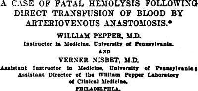
Figure 1.6 Report of a fatal transfusion reaction. Source: Pepper and Nisbet (1907).41 With permission of American Medical Association.
The direct artery to vein anastomosis was the best method available but was often very difficult or even unsuccessful. And, what was almost as bad, one never knew how much blood one had transfused at any moment or when to stop (unless the donor collapsed). (I remember one such collapse in which the donor almost died—and the surgeon needed to be revived.)
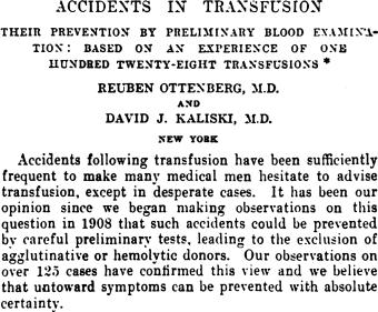
Despite these many difficulties, direct transfusion through an arteriovenous anastomosis for the first time efficiently transferred blood from one person to another. The process also disclosed fatal hemolytic reactions that were undeniably caused by transfusion41 (Figure 1.6). However, the relation of these fatal reactions to Landsteiner’s discovery was not recognized until Reuben Ottenberg (1882–1959) demonstrated the importance of compatibility testing.
Ottenberg’s interest in transfusion began in 1906 while he was an intern at German (now Lenox Hill) Hospital in New York. There, Ottenberg learned of Landsteiner’s discovery and in 1907 began pretransfusion compatibility testing.42 Ottenberg accepted an appointment at Mount Sinai Hospital the next year and continued his studies on transfusion. In 1913, Ottenberg published the report that conclusively demonstrated the importance of preliminary blood testing for the prevention of transfusion “accidents”43 (Figure 1.7). This was not Ottenberg’s only contribution. He observed the Mendelian inheritance of blood groups,44 and he was the first to recognize the relative unimportance of donor
Figure 1.7 Report of the importance of testing before transfusion. Source: Ottenberg and Kaliski (1913).43 Figure 01, p 01 / With permission of American Medical Association.
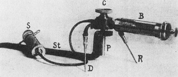
Figure 1.8 Apparatus for Unger’s two-syringe, four-way stopcock method of indirect transfusion. Source: Unger (1915).49 Figure 01, p 02 / With permission of American Medical Association.
antibodies and consequently the “universal” utility of type O blood donors.45
Further advances in immunohematology were to occur in succeeding decades. The M, N, and P systems were described in the period between 1927 and 1947.46 The Rh system was discovered in connection with an unusual transfusion reaction. In 1939, Levine and Stetson47 described an immediate reaction in a group O woman who had received her husband’s group O blood soon after delivery of a stillborn fetus with erythroblastosis. This sequence of events suggested that the infant had inherited a red cell agglutinogen from the father that was foreign to the mother. At about the same time, Landsteiner and Wiener48 harvested a rhesus monkey red cell antibody from immunized guinea pigs and rabbits. This antibody agglutinated 85% of human red cell samples (Rh-positive) and left 15% (Rh-negative) unaffected. When the experimentally induced antibody was tested in parallel with the serum from Levine’s patient, a similar positive and negative distribution was observed, and the Rh system had been discovered. Other red cell antigen systems were subsequently described, but when Rh immunoglobulin was introduced as a preventive measure for hemolytic disease of the newborn, it became one of the major public health advances of the century. Despite the introduction of compatibility testing by Ottenberg, transfusion could not be performed frequently as long as arteriovenous anastomosis remained the procedure of choice. Using this method, Ottenberg needed five years (Figure 1.7) to accumulate the 128 transfusions he reported in his study on pretransfusion testing.43 New techniques, such as Unger’s two-syringe method introduced in 191549 (Figure 1.8), eventually put an end to transfusion by means of arteriovenous anastomosis. However, transfusions did not become commonplace until anticoagulants were developed and direct methods of transfusion were rendered obsolete.
Anticoagulants, the blood bank, and component therapy
The anticoagulant action of sodium citrate completely transformed the practice of transfusion. Early reports from Belgium50 and Argentina51 were followed by the work of Lewisohn52 that established the optimal citrate concentration for anticoagulation. The work of Weil53 then demonstrated the feasibility of refrigerated storage. Subsequently, Rous and Turner54 developed the anticoagulant solution that was used during World War I.55 Despite its very large volume, this solution remained the anticoagulant of choice until World War II, when Loutit and Mollison56 developed an acid–citrate–dextrose (ACD) solution. Used in a ratio of 70 mL ACD to 450 mL blood, ACD provided 3–4 weeks of preservation of a more concentrated red
cell infusion. Thus, the two world wars were the stimuli for the development of citrate anticoagulants and the introduction of indirect transfusion.46 For the first time, the donation process could be separated, in time and place, from the actual transfusion. Blood drawn and set aside now awaited the emergence of systems of storage and distribution. Again, it was the provision of medical support during armed conflict that stimulated these developments.
A blood transfusion service, organized by the Republican Army during the Spanish Civil War (1936–1939), collected 9000 L of blood in citrate–dextrose anticoagulant for the treatment of battle casualties.57 At about that same time, Fantus58 began operation of the first hospital blood bank at Cook County Hospital in Chicago. His interest had been stimulated by Yudin’s report59 on the use of cadaveric blood in Russia. Apart from certain scruples attached to the use of cadaveric blood, Fantus reasoned that a transfusion service based on such a limited source of supply would be impractical. Accordingly, he established the principle of a “blood bank” from which blood could be withdrawn, provided it had previously been deposited. As Fantus58 himself stated, “[J]ust as one cannot draw money from a bank unless one has deposited some, so the blood preservation department cannot supply blood unless as much comes in as goes out. The term ‘blood bank’ is not a mere metaphor.” The development of anticoagulants and the concept of blood banks provided an infrastructure upon which a more elaborate blood services organization could be built. World War II was the catalyst for these further developments.
At the beginning of World War II, blood procurement programs were greatly expanded.46 In Great Britain, an efficient system had been developed through the organization of regional centers. When the war started, these centers, already in place, were able to increase their level of operation. In the United States, the use of plasma in the management of shock had led to the development of plasma collection facilities.60 The efficient long-term storage of plasma had been further facilitated by the process of lyophilization developed by Flosdorf and Mudd and the introduction of ABO-independent “universal” plasma produced by pooling several thousand units of plasma.61 In 1940, the United States organized a program for the collection of blood and the shipment of plasma to Europe. The American Red Cross, through its local chapters, participated in the project, which collected 13 million units by the end of the war.46
The national program of the American Red Cross ceased at the end of the war. However, many of the local chapters continued to help recruit donors for local blood banks, and in 1948, the first regional Red Cross blood center was begun in Rochester, New York. By 1949–1950 in the United States, the blood procurement system included 1500 hospital blood banks, 1100 of which performed all blood bank functions. There were 46 nonhospital blood banks and 31 Red Cross regional blood centers. By 1962, these numbers had grown to 4400 hospital blood banks, 123 nonhospital blood banks, and 55 American Red Cross regional blood centers, and the number of units collected had grown to between 5 and 6 million per year.62
During this time, blood was collected through steel needles and rubber tubing into rubber-stoppered bottles. After washing and resterilization, the materials were reused. On occasion, “vacuum bottles” were used to speed up the collection. However, the high incidence of pyrogenic reactions soon led to the development of disposable plastic blood collection equipment.
In a classic article written in 1952, Walter and Murphy63 described a closed, gravity technique for whole blood preservation. They used a laminar flow phlebotomy needle, an interval donor tube, and a collapsible bag of polyvinyl resin designed so that the unit could be
assembled and ready for use after sterilization with steam. The polyvinyl resin was chemically inert to biologic fluids and nonirritating to tissue. Soon thereafter, Gibson et al. 64 demonstrated that plastic systems were more flexible and allowed removal of plasma after sedimentation or centrifugation. In time, glass was replaced with plastic, and component therapy began to emerge. This development was enhanced by the US military’s need to reduce the weight and breakage of blood bottles during shipment in the Korean War.
Component and derivative therapy began during World War II, when Edwin J. Cohn and his collaborators developed the cold ethanol method of plasma fractionation.65 As a result of their work, albumin, globulin, and fibrinogen became available for clinical use. As plastic equipment replaced glass, component separation became a more widespread practice, and the introduction of automated cell separators provided even greater capabilities in this area.
Clotting factor concentrates for the treatment of patients with hemophilia, and other hemorrhagic disorders were also developed during the postwar era. Although antihemophilic globulin had been described in 1937,66 unconcentrated plasma was the only therapeutic material until Pool discovered that Factor VIII could be harvested in the cryoprecipitable fraction of blood.67 This resulted in the development of cryoprecipitate, which was introduced in 1965 for the management of hemophilia. Pool showed that cryoprecipitate could be made in a closed-bag system and urged its harvest from as many donations as possible. The development of cryoprecipitate and other concentrates was the dawn of a golden age in the care of patients with hemophilia. Self-infusion programs, made possible by technologic advances in plasma fractionation, allowed early therapy and greatly reduced disability and unemployment. This golden age came abruptly to an end with the appearance of the AIDS virus.
Transfusion in the age of technology
In contrast to the long ledger of lives lost in previous centuries because of the lack of blood, transfusion in the twentieth century saved countless lives. In 1937, during those early halcyon days of transfusion, Ottenberg wrote:40
Today transfusion has become so safe and so easy to do that it is seldom omitted in any case in which it may be of benefit. Indeed the chief problem it presents is the finding of the large sums of money needed for the professional donors who now provide most of the blood.
It is ironic that Ottenberg’s statement should refer to paid donors and foreshadow difficulties yet to come. However, experience to that point had not revealed the problem of viral disease transmission. More transfusions would have to be administered before that problem would be perceived.
After the introduction of anticoagulants, blood transfusions were given in progressively increasing numbers. At Mount Sinai Hospital in New York, the number of blood transfusions administered between 1923 and 1953 increased 20-fold68,69 (Table 1.1). This increase was particularly notable after the establishment of blood banks. It was during this period that Beeson wrote his classic description of transfusion-transmitted hepatitis.70 He had been alerted to the problem by the outbreaks of jaundice that followed inoculation programs with human serum during World War II. Thus, blood transfusion entered a new era. Blood components not only saved lives but also transmitted disease. The discovery of the Australian antigen71 and the subsequent definition of hepatitis A virus and B virus (HBV) still left residual non-A and non-B diseases,72 a gap that has been largely filled by the discovery of the
Table 1.1 Increase in the Number of Blood Transfusions at Mount Sinai Hospital, New York, 1923–1953
Source: Lewisohn (1955).68 Reprinted with permission of Elsevier.
hepatitis C virus (HCV).73 However, it was the outbreak of AIDS that galvanized public attention to blood transfusion.
The AIDS epidemic was first recognized in the United States, and the first case of AIDS associated with transfusion was observed in a 20-month-old infant.74 Subsequently, the suspicion that AIDS could be transmitted by means of transfusion was confirmed.75
The human immunodeficiency virus (HIV) was identified,76 and an effective test to detect the HIV antibody was developed.76
Concern for blood safety
Since 1943, transfusion therapy has been shadowed by the specter of disease transmission. In that year, Beeson described posttransfusion hepatitis and unveiled a problem that has grown with time. As transfusion increased, so did disease transmission. In 1962, the connection between paid donations and post-transfusion hepatitis was made.77 A decade later, the National Blood Policy mandated a voluntary donation system in the United States. And yet, blood usage continued to increase.
Concern about post-transfusion hepatitis was not sufficient to decrease the number of transfusions. Although the use of whole blood declined as blood components became more popular, total blood use in the United States doubled between 1971 and 1980
(Table 1.2).78–85 This pattern changed as the emergence of AIDS exposed all segments of society to a revealing light.
AIDS probably arose in Africa in the 1960s and spread quietly for years before it was detected. By 1980, an estimated 100,000 persons were infected, and by 1981, when the first cases were reported, a worldwide pandemic lay just beneath the surface. The initial response of the public and officials seemed trifling and insufficient as the outbreak grew to proportions few had foreseen. Criticism was levied against the news media for initially ignoring the story, the government for delay in acknowledging the problem, gay civil rights groups for resistance to epidemiologic measures, research scientists for unseemly competition, blood services for delayed response in a time of crisis, and the US Food and Drug Administration (FDA) for inadequate regulatory activity. Historians with the perspective of time will determine whether there really were more villains than the virus itself.89
The realization that transfusion can transmit an almost invariably fatal disease had a chilling effect on the public. Two major changes in blood services have occurred in the aftermath of the AIDS epidemic. The FDA, using pharmaceutical manufacturing criteria not “tailored to . . . blood banks,” became more aggressive in regulatory actions against blood collection establishments.90 And, finally, blood use moderated for approximately 10 years. Through the 1980s and early 1990s, red cell and plasma transfusion peaked and began to stabilize (Table 1.2). Only platelet use and human progenitor cell transplantation, driven by the demands of cancer chemotherapy, continued to increase.78–85 Educational programs to encourage judicious use of blood have been initiated, and they have been favorably received by practicing physicians. Use of red cells and plasma fell from 2008 to 2011. This was a combined impact of the great recession reducing healthcare utilization and the widespread use of patient blood management programs intended to reduce blood transfusion. This represents the first time since the end of the Second World War that the growth in transfusion of blood products in the United States and other developed countries stopped and was reversed for a sustained period.
Relentless public pressure for a “zero-risk” blood supply resulted in dividends through continued scientific and technologic improvements. Enhanced sensitivity and better use of serologic testing, along with improved scrutiny of donors, resulted in major reductions in risk of transmitted disease by the mid-1990s.91 Discovery that pools of units subjected to nucleic acid testing almost closed the window for HIV and HCV virus resulted in the application of this testing for both whole blood and plasma donations beginning between 1998 and 2000.91,92 This, combined with virus reduction and inactivation of the final product, resulted in plasma derivatives that have not transmitted AIDS or hepatitis since 1994.93 For whole blood and platelet components, risks have become low. Pathogen reduction has driven the risks lower for plasma and platelets, and very low for plasma-derived therapies such as immune globulin.
* Platelets reported in apheresis units in this and subsequent years.
Source: Adapted from Surgenor and Schnitzer (1985)78; Surgenor et al. (1998)79; Wallace et al. (1989)80; Wallace et al. (1990)81; Sullivan et al. (2002)82; Sullivan et al (2005)83; US Department of Health and Human Services (2008)84; AABB (2012)85; Chung et al. (2016)86; Ellingson et al. (2017)87; Jones et al. (2020)88
With the reduction in the risk of viral transmission, the focus in the developed world has shifted to transfusion-related acute lung injury (TRALI)—possibly from recipient-directed leukocyte antibodies and lipid mediators in transfused plasma—and bacterial infection primarily occurring in room-temperature stored platelets. In order to make incremental gains against these risks, the use of male plasma only and the culture of platelets before they are released have been implemented. The 2011 blood utilization survey indicated a 28.8% reduction in TRALI, suggesting a significant breakthrough with regard to this risk.85 Geographic exclusions have been aimed at reducing the potential for variant
Table 1.2 Transfusions in the United States (in Millions of Units)78–88