Equine Anesthesia and Co-Existing Disease
First Edition
Stuart Clark-Price, DVM, MS
Diplomate, American College of Veterinary Internal Medicine, Large Animal Diplomate, American College of Veterinary Anesthesia and Analgesia Associate Professor College of Veterinary Medicine Auburn University Auburn, Alabama, USA
Khursheed Mama, DVM
Diplomate, American College of Veterinary Anesthesia and Analgesia Professor, Veterinary Anesthesiology College of Veterinary Medicine and Biomedical Sciences Colorado State University Fort Collins, Colorado, USA
This first edition first published 2022 © 2022 John Wiley & Sons, Inc.
All rights reserved. No part of this publication may be reproduced, stored in a retrieval system, or transmitted, in any form or by any means, electronic, mechanical, photocopying, recording or otherwise, except as permitted by law. Advice on how to obtain permission to reuse material from this title is available at http://www.wiley.com/go/permissions.
The right of Stuart Clark-Price and Khursheed Mama to be identified as the authors of the editorial material in this work has been asserted in accordance with law.
Registered Office
John Wiley & Sons, Inc., 111 River Street, Hoboken, NJ 07030, USA
Editorial Office
111 River Street, Hoboken, NJ 07030, USA
For details of our global editorial offices, customer services, and more information about Wiley products visit us at www.wiley.com.
Wiley also publishes its books in a variety of electronic formats and by print-on-demand. Some content that appears in standard print versions of this book may not be available in other formats.
Limit of Liability/Disclaimer of Warranty
The contents of this work are intended to further general scientific research, understanding, and discussion only and are not intended and should not be relied upon as recommending or promoting scientific method, diagnosis, or treatment by physicians for any particular patient. In view of ongoing research, equipment modifications, changes in governmental regulations, and the constant flow of information relating to the use of medicines, equipment, and devices, the reader is urged to review and evaluate the information provided in the package insert or instructions for each medicine, equipment, or device for, among other things, any changes in the instructions or indication of usage and for added warnings and precautions. While the publisher and authors have used their best efforts in preparing this work, they make no representations or warranties with respect to the accuracy or completeness of the contents of this work and specifically disclaim all warranties, including without limitation any implied warranties of merchantability or fitness for a particular purpose. No warranty may be created or extended by sales representatives, written sales materials or promotional statements for this work. The fact that an organization, website, or product is referred to in this work as a citation and/or potential source of further information does not mean that the publisher and authors endorse the information or services the organization, website, or product may provide or recommendations it may make. This work is sold with the understanding that the publisher is not engaged in rendering professional services. The advice and strategies contained herein may not be suitable for your situation. You should consult with a specialist where appropriate. Further, readers should be aware that websites listed in this work may have changed or disappeared between when this work was written and when it is read. Neither the publisher nor authors shall be liable for any loss of profit or any other commercial damages, including but not limited to special, incidental, consequential, or other damages.
Library of Congress Cataloging-in-Publication Data
Names: Clark-Price, Stuart, 1972- editor. | Mama, Khursheed, 1964- editor.
Title: Equine anesthesia and co-existing disease / [edited by] Stuart Clark-Price, Khursheed Mama.
Description: First edition. | Hoboken, NJ : Wiley-Blackwell, 2022. | Includes bibliographical references and index.
Identifiers: LCCN 2021048465 (print) | LCCN 2021048466 (ebook) | ISBN 9781119307150 (paperback) | ISBN 9781119307396 (adobe pdf) | ISBN 9781119307419 (epub)
Subjects: MESH: Horse Diseases–surgery | Anesthesia–veterinary | Anesthesia–adverse effects | Anesthetics–adverse effects | Intraoperative Complications–veterinary
Classification: LCC SF951 (print) | LCC SF951 (ebook) | NLM SF 951 | DDC 636.1/0896796–dc23/eng/20211005
LC record available at https://lccn.loc.gov/2021048465
LC ebook record available at https://lccn.loc.gov/2021048466
Cover Design: Wiley
Cover Image: Courtesy of Stuart Clark-Price and Khursheed Mama
Set in 9.5/12.5pt STIXTwoText by Straive, Pondicherry, India
The path to veterinary medicine for me is unique and personal, as I suspect it is for most veterinarians. There are a great many people that mentored me along the way, and, to all of them, I say thank you! The following individuals were particularly influential in helping me reach my goals. I would like to sincerely thank Dr. Joseph Coli and Dr. Stephen Damonte for edifying integrity and dedication; Dr. Alan Reich, Dr. Kathy Yvorchuk, and Dr. Roger Warren for inspiring me; and Dr. Christine Schweizer, Dr. Bonne Rush, and Dr. Robin Gleed for taking a chance on me. Finally, I would like to dedicate this textbook to my father, Charles Price. It was his suggestion that I pursue veterinary medicine. While he was far from a prefect human, he was my dad.
Stuart Clark-Price
This is dedicated to family, mentors, colleagues, trainees, and friends who have enhanced my career. You encouraged me to pursue my passion, challenged me to continually strive to provide outstanding care, supported me in accomplishing my goals, and encouraged me when I wavered in my commitment. I remain grateful to you all. Gene, you are a constant source of support and, through your actions, remind me that excellence is a worthy goal. I consider it a privilege to be entrusted with the anesthesia care of these amazing and sometimes fragile animals and acknowledge all who are dedicated to advancing their management.
Khursheed Mama
Content
Contributing Authors viii
Preface xi
1 Anesthetic Management for Dental and Sinus Surgery 1
2 Anesthetic Management for Ocular Interventions 16
3 Anesthetic Management for Inflammatory or Infectious Respiratory Diseases 35
4 Anesthetic Management for Surgery of the Respiratory Tract 73
5 Anesthetic Management for Interventional Cardiac Procedures 84
6 Anesthetic Management for Medical and Surgical Neurologic Conditions 116
7 Anesthetic Management for Orthopedic Conditions 137
8 Anesthetic Management for Muscular Conditions 159
9 Anesthetic Management for Laparoscopic and Thoracoscopic Procedures 195
10 Anesthetic Management for Gastrointestinal Diseases 216
11 Anesthetic Management for Endocrine Diseases and Geriatric Horses 229
12 Anesthetic Management for Urogenital Interventions 260
13 Anesthetic Management of Foals 292
14 Anesthetic Management of Other Domesticated and Non-Domesticated Equids 337
15 Accident and Error Management 352
Index 385
Contributing Authors
Jennifer Carter, DVM, MClinEd
Diplomate, American College of Veterinary
Anesthesia and Analgesia
Senior Lecturer
University of Melbourne Melbourne, Australia
Sathya Chinnadurai, DVM, MS
Diplomate, American College of Zoological Medicine
Diplomate, American College of Veterinary
Anesthesia and Analgesia
Diplomate, American College of Animal Welfare
Director of Animal Health
Saint Louis Zoo
Saint Louis, Missouri
Stuart Clark-Price, DVM, MS
Diplomate, American College of Veterinary
Internal Medicine, Large Animal
Diplomate, American College of Veterinary
Anesthesia and Analgesia
Associate Professor
College of Veterinary Medicine
Auburn University
Auburn, Alabama
Jeremiah Easley, DVM
Diplomate, American College of Veterinary Surgeons
Associate Professor
College of Veterinary Medicine and Biomedical Sciences
Colorado State University
Fort Collins, Colorado
Ryan Fries, DVM
Diplomate, American College of Veterinary Internal Medicine, Cardiology
Assistant Professor College of Veterinary Medicine
University of Illinois Urbana, Illinois
Kirsty Gallacher, BVMS
Diplomate, American College of Theriogenologists. Lecturer
School of Animal and Veterinary Sciences
The University of Adelaide
Roseworthy campus, Australia
Santiago Gutierrez-Nibeyro, DVM, MS
Diplomate, American College of Veterinary Surgeons
Diplomate, American College of Veterinary Sports Medicine and Rehabilitation
Clinical Associate Professor College of Veterinary Medicine
University of Illinois Urbana, Illinois
Eileen Hackett, DVM, PhD
Diplomate, American College of Veterinary Surgeons,
Diplomate, American College of Veterinary Emergency and Critical Care, American College of Veterinary Surgeons
Founding Fellow Minimally Invasive Surgery (Large Animal Soft Tissue) Professor
College of Veterinary Medicine and Biomedical Sciences
Colorado State University Fort Collins, Colorado
Diana Hassel, DVM, PhD
Diplomate, American College of Veterinary Surgeons
Diplomate, American College of Veterinary
Emergency & Critical Care
Professor
College of Veterinary Medicine and Biomedical Sciences
Colorado State University Fort Collins, Colorado
Bonnie Hay Kraus, DVM
Diplomate, American College of Veterinary Surgeons
Diplomate, American College of Veterinary
Anesthesia and Analgesia
Associate Professor
College of Veterinary Medicine
Iowa State University Ames, Iowa
Rachel Hector, DVM, MS
Diplomate, American College of Veterinary
Anesthesia and Analgesia
Assistant Professor
College of Veterinary Medicine and Biomedical Sciences
Colorado State University Fort Collins, Colorado
Dean Hendrickson, DVM, MS
Diplomate, American College of Veterinary Surgeons
ACVS Founding Fellow, Minimally Invasive
Surgery (Large Animal Soft Tissue)
Professor
College of Veterinary Medicine and Biomedical Sciences
Colorado State University Fort Collins, Colorado
Klaus Hopster, DVM
Diplomate, European College of Veterinary
Anaesthesia and Analgesia
Assistant Professor
School of Veterinary Medicine
University of Pennsylvania Kennett Square, Pennsylvania
Philip Johnson, BVSc (Hons), MS, MRCVS
Diplomate, American College of Veterinary Internal Medicine, Large Animal Diplomate, European College of Equine Internal Medicine
Professor
College of Veterinary Medicine
University of Missouri Columbia, Missouri
Stephanie Keating, DVM, DVSc
Diplomate, American College of Veterinary Anesthesia and Analgesia
Clinical Assistant Professor College of Veterinary Medicine University of Illinois Urbana, Illinois
Kara Lascola, DVM, MS
Diplomate, American College of Veterinary Internal Medicine, Large Animal Associate Professor College of Veterinary Medicine Auburn University Auburn, Alabama
Khursheed Mama, BVSc, DVM
Diplomate, American College of Veterinary Anesthesia and Analgesia.
Professor College of Veterinary Medicine and Biomedical Sciences
Colorado State University Fort Collins, Colorado
Bianca Martins, DVM, MS, PhD
Diplomate, American College of Veterinary Ophthalmology
Associate Professor
University of California, Davis School of Veterinary Medicine Davis, California
Contributing Authors x
Manuel Martin-Flores, MV
Diplomate, American College of Veterinary
Anesthesia and Analgesia
Associate Professor
College of Veterinary Medicine
Cornell University
Ithaca, New York
Nora Matthews, DVM
Diplomate, American College of Veterinary
Anesthesia and Analgesia
Professor Emeritus College of Veterinary Medicine & Biomedical Sciences
Texas A & M University
College Station, Texas
Adjunct Professor College of Veterinary Medicine
Cornell University
Ithaca, New York
Erica McKenzie, BSc, BVMS, PhD
Diplomate, American College of Veterinary
Internal Medicine, Large Animal
Diplomate, American College of Veterinary
Sports Medicine and Rehabilitation
Professor College of Veterinary Medicine
Oregon State University Corvallis, Oregon
Valerie Moorman, DVM, PhD
Diplomate, American College of Veterinary Surgeons (Large Animal)
Clinical Associate Professor College of Veterinary Medicine University of Georgia Athens, Georgia
Daniel Pang, BVSc, MSc, PhD, MRCVS
Diplomate, American College of Veterinary
Anesthesia and Analgesia
Diplomate, European College of Veterinary
Anaesthesia and Analgesia
EBVS European Specialist in Veterinary Anaesthesia & Analgesia
Associate Professor
Faculty of Veterinary Medicine
University of Calgary
Calgary, Alberta, Canada
Adjunct Professor
Faculty of Veterinary Medicine
Université de Montréal
St-Hyacinthe, Quebec, Canada
Marlis Rezende, DVM, MS, PhD
Diplomate, American College of Veterinary
Anesthesia and Analgesia
Associate Professor College of Veterinary Medicine and Biomedical Sciences
Colorado State University
Fort Collins, Colorado
Eugene Steffey, VMD, PhD, MRCVS (hon) and Dr.h.c.(U Bern).
Diplomate, American College of Veterinary Anesthesia and Analgesia
Diplomate, European College of Veterinary
Anaesthesia and Analgesia
Professor Emeritus School of Veterinary Medicine
University of California, Davis Davis, California
Affiliate Faculty College of Veterinary Medicine and Biomedical Sciences
Colorado State University
Fort Collins, Colorado
Alexander Valverde, DVM, DVSc
Diplomate, American College of Veterinary
Anesthesia and Analgesia
Professor
Ontario Veterinary College University of Guelph Ontario, Canada
Tom Yarbrough DVM
Diplomate, American College of Veterinary Surgeons
Senior Veterinarian
Dubai Equine Hospital Dubai, UAE
Preface
Textbooks solely dedicated to veterinary anesthesia became widely available in the early 1960s, and many have been published since. More recent textbooks contain detailed information on clinical disease and management of small animal patients with specific conditions. A few excellent books related to anesthetic management of equine patients have also been published. However, there are no comprehensive textbooks addressing anesthetic management of horses for specific surgical procedures and diseases. The editors are excited to present this book, which aims to fill that void by providing both a review of the pathogenesis of specific diseases, and procedural considerations relevant to equine anesthesia management.
Recognizing that teamwork is important when providing medical care, most chapters are co-authored by anesthesiologists and known experts in their field including internal medicine, surgery, dentistry, ophthalmology, cardiology, reproduction and zoological medicine. Each chapter combines traditional and cutting-edge knowledge with practical information related to peri-anesthetic management to provide the reader with unparalleled information in a single source. Our hope is that specialists, general practitioners, residents, trainees, and students will find this textbook
helpful when managing their equine patients. In addition to chapters focusing on gastrointestinal and orthopedic diseases, considerations for horses undergoing laparoscopy, thoracoscopy, and interventional cardiac procedures, as well as those with co-morbidities unrelated to the need of anesthesia such as inherited muscular diseases, endocrinopathies, and inflammatory respiratory diseases, are included. Considerations for neonatal foals, domestic and non-domestic equids, and a discussion of accidents and error management round out the compilation. The goal is for this to be a broad-based and comprehensive resource relevant to the advances in anesthesia in both healthy and compromised horses.
No such work is possible without the involvement of many. The editors wish to thank the contributing authors for their time, experience, and dedication to this project. The completion of this work is particularly notable given that much of it was accomplished during a global pandemic that had an immeasurable impact on personal and working lives. The editors also thank Merryl Le Roux and the team at Wiley for their assistance, support, and patience.
Stuart Clark-Price Khursheed Mama
1
Anesthetic Management for Dental and Sinus Surgery
Santiago Gutierrez-Nibeyro1 and Jennifer Carter2
1 Department of Veterinary Clinical Medicine, College of Veterinary Medicine, University of Illinois, 1008 W. Hazelwood Dr., Urbana, IL, 61802, USA
2 Faculty of Veterinary and Agricultural Sciences, Melbourne Veterinary School, University of Melbourne, 250 Princes Highway, Werribee, VIC, 3030, Australia
Introduction
Most adult horses have 36–44 teeth by the time they reach 5 years of age. In general, the dental arcades are composed of 12 incisors, 12 premolars, and 12 molars (some horses will also have additional teeth including canine and wolf teeth). Due to the grinding nature of eating, horse teeth must continue to grow at approximately 1/8″ per year until the individual horse reaches old age where teeth can then be completely shed. Throughout the maxillary and frontal areas of the skull, air-filled sinus cavities developed to allow for a large number of premolar and molar teeth without adding significant weight. The linings of the sinuses are rich in vasculature and may play a role in thermoregulation. Significant disease requiring surgical intervention can occur in the teeth or sinus.
Relevant Anatomy
The nasal cavity is a voluminous cavity divided by the nasal septum and vomer bone (Hillmann 1975). The nasal cavity contains the reserve crowns of the maxillary cheek teeth and a portion of the paranasal sinuses of which the major clinically significant sinuses are the
frontal and maxillary sinuses (Hillmann 1975). Two major nasal conchae in each nasal cavity divide the nasal passage into the dorsal, middle, ventral, and common meatus.
The frontal sinus has a large communication with the dorsal conchal sinus, and thereby both are known as the conchofrontal sinus (Hillmann 1975). The ventral conchal sinus communicates with the rostral maxillary sinus over the infraorbital canal and is separated from the caudal maxillary sinus by a thin osseous sheet, the caudal bulla of the ventral conchal sinus. The conchae (or turbinates) are delicate scrolls of bone that are attached laterally in the nasal passage and contain the conchal sinuses (Hillmann 1975).
The maxillary sinus is divided by a thin septum into rostral and caudal compartments or rostral and caudal maxillary sinuses, respectively (Hillmann 1975). The rostral maxillary sinus contains the root of the maxillary first molar and the caudal maxillary sinus contains the roots of the second and third molars (Dixon 2005). The caudal maxillary sinus is partially divided by the infraorbital canal, which may be distorted by a disease process within the sinus. The caudal and rostral maxillary sinuses have separate openings into the middle nasal meatus and the caudal maxillary
sinus communicates with the frontal sinus through the large frontomaxillary opening (Hillmann 1975).
Diagnostic Techniques
Complete history and physical examination of the horse, including assessment of mental status, cardiopulmonary functions, hydration status, and body temperature are mandatory prior to sedation, anesthesia, and/or local anesthetic techniques for dental and sinus surgery. Frontonasal and maxillary bone flaps are indicated to remove of a wide variety of lesions that may develop in the paranasal sinuses or turbinates, such as paranasal sinus cysts, neoplasia, progressive ethmoid hematomas, and apical infections of maxillary cheek teeth (Nickels 2012). The lesions typically cause unilateral epistaxis or mucopurulent nasal drainage, in contrast with diseases of the pharynx or lungs in which the drainage is typically bilateral. However, appropriate diagnostic techniques are indicated to rule out concurrent diseases of the pharynx and lungs that may affect patient management either under general anesthesia or under standing sedation.
On endoscopy, narrowed nasal meati, purulent material, masses, or blood can be seen in
the nasal passage and/or draining from the sinus openings (Nickels 2012). Radiography of the skull may reveal free fluid lines, radiodense masses, paranasal sinus cysts, and lucency and/or proliferation associated with dental disease (Figure 1.1). Sinocentesis can be used to obtain fluid sample for culture and cytological examination. Sinuscopy with the horse standing and sedated is useful for the examination, diagnosis, and treatment of some disorders of the paranasal sinuses (Nickels 2012).
Local Anesthetic Techniques for the Equine Teeth and Sinuses
Locoregional anesthesia can be performed prior to many dental and surgical procedures for horses under both standing sedation and general anesthesia. It is routinely accomplished using either lidocaine 2% or mepivacaine 2% solutions with mepivacaine providing a longer duration of action compared to lidocaine (two to four hours versus one to two hours). It is generally advisable to infuse a small amount (1–2 ml) of local anesthetic into the skin at the site of the nerve block to desensitize the skin prior to attempting the locoregional block. This is especially important under standing sedation conditions. Approximately 5–10 minutes should

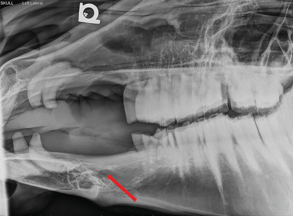
Figure 1.1 Lateral radiograph of a horse skull showing a fluid line (blue arrows) running through the caudal maxillary sinuses. The horse’s nose was angled downward resulting in the gravity-dependent fluid line being parallel to the ground. The sinusitis likely resulted from a periapical infection of a cheek tooth (red arrow). Maxillary nerve blockade can be used to desensitize the area for surgical removal of the tooth and drainage and lavage of the sinus.
be allowed to elapse after administration of the local anesthetic to achieve desensitization of the region.
Infraorbital Block
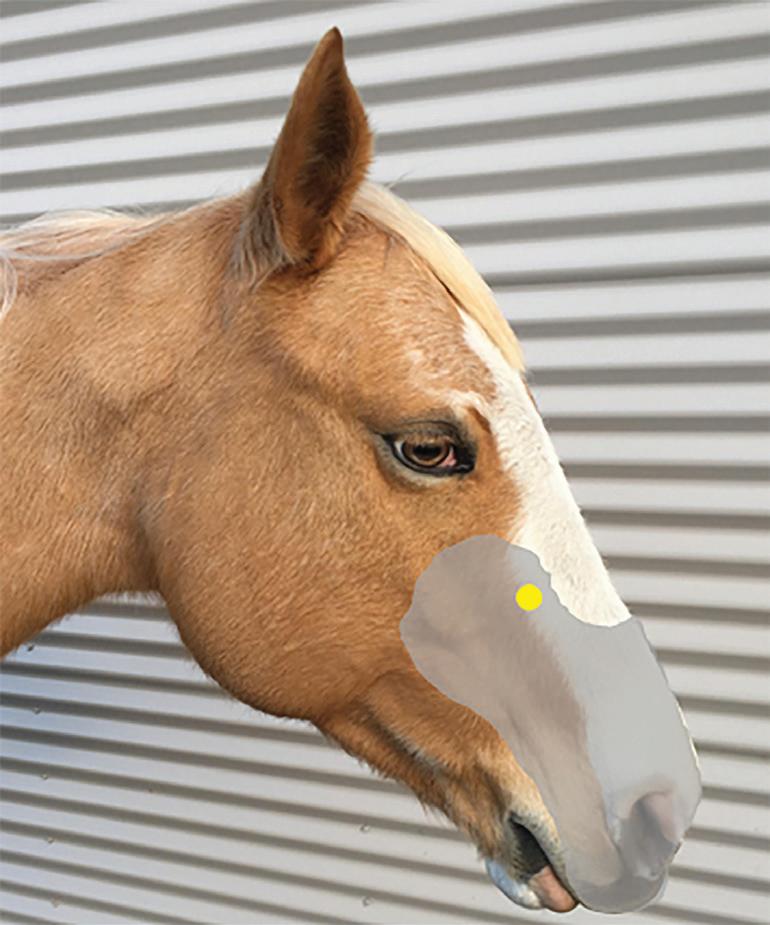
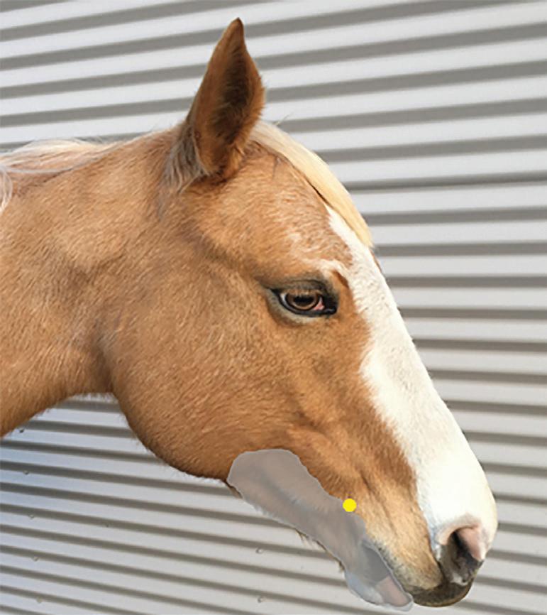
The infraorbital block desensitizes the maxillary teeth to the level of the first molar, the maxillary sinus, the skin from the lip nearly to the medial canthus, and the rostral nose as well as the roof of the nose (Skarda et al. 2010) (Figure 1.2). The infraorbital canal is palpated as the midpoint on a line between the nasoincisive notch and the rostral most aspect of the facial crest (Rice 2017). The levator labii superioris muscle must be manually elevated to facilitate placement of the needle into the canal (Rice 2017). For local anesthesia of the upper lip and nose, a 20 gauge, 2.5 cm needle can be advanced perpendicularly to the skin at the opening of the infraorbital canal using 5 ml of local anesthetic (Skarda et al. 2010). For
The infraorbital block in the horse. The yellow circle indicates the location of the infraorbital canal, and stippling indicates the area of desensitization following administration of local anesthetic.
blockade of the maxillary teeth and sinus, a 25–20 gauge, 3.8–5 cm needle should be advanced into the canal and 3–5 ml of local anesthetic should be injected (Rice 2017; Skarda et al. 2010).
Maxillary Nerve Block
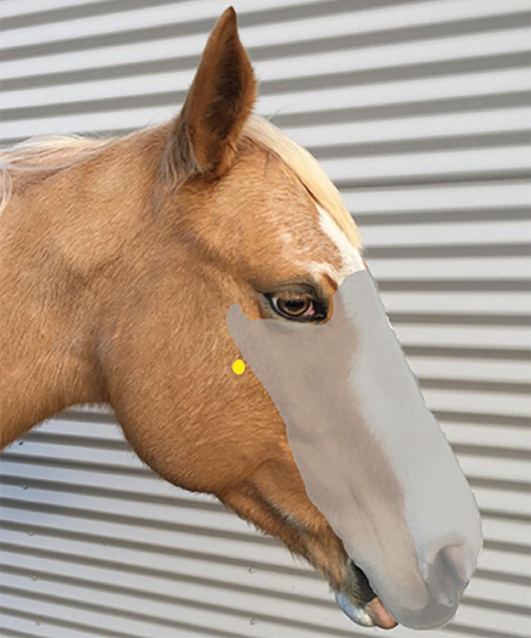
Blocking the maxillary nerve within the pterygopalatine fossa results in blockade of the maxillary teeth, the paranasal sinus, and the nasal cavity (Woodie 2013) (Figure 1.3). Multiple techniques have been described for performing this block, owing in part to relatively vague surface landmarks for injection. The first involves the injection of local anesthetic into the extraperiorbital fat body (Staszyk et al. 2008). This technique uses a 18 gauge, 3.5″ spinal needle to inject approximately 10 ml of local anesthetic into the fat body surrounding the maxillary nerve (Staszyk et al. 2008). The injection site is made
Figure 1.3 The maxillary nerve block in the horse. The yellow circle indicates the location of the infraorbital canal, and stippling indicates the area of desensitization following administration of local anesthetic.
Figure 1.2
Anesthetic Management for Dental and Sinus Surgery 4
perpendicular to the skin at a point located 10 mm ventral to the zygomatic arch transverse to the plane between the middle and caudal 1/3 of the eye and the needle is advanced until it pops through the masseter muscle for a total depth of approximately 4.5–5 cm (Staszyk et al. 2008). The technique was used in horses under standing sedation using a 20 gauge, 3.5″ spinal needle and reported generally successful blockade with no reaction of mechanical or thermal stimulus with mild chewing, bleeding, swelling, and turgor at the injection site as the only complications (Rieder et al. 2016a). Another study evaluating the volume of lidocaine necessary to produce anesthesia with the extraperiorbital fat body technique and found that 2 ml/100 kg of body weight should result in sufficient local anesthesia while minimizing side effects (Rieder et al. 2016b).
Another technique involves the use of a 19 gauge, 2.5″ spinal needle with an injection site along the ventral border of the zygomatic arch at the narrowest point of the arch (Newton et al. 2000). The needle is inserted into the skin on a rostromedial and ventral angle and is directed along this angle toward the 6th cheek tooth on the contralateral side to a depth of approximately 2″ at which 5 ml of local anesthetic is injected (Newton et al. 2000). At a landmark slightly rostral to that described by Newton, an injection can be made perpendicular to the skin, ventral to the zygomatic process at a point on the skin found on the line running perpendicular to the dorsal head contour and through the temporal canthus of the eye (Bemis 1917). These two techniques were compared using cadaver heads and new methylene blue dye and failed to elucidate a significant difference between the two, with both techniques resulting in at least partial success in “blockade” of the maxillary nerve approximately 80% of the time (Bardell et al. 2010). It is worth noting that the authors reported inadvertent deposition of dye into the deep facial vein on two instances (Bardell et al. 2010). This reinforces the need to aspirate the needle at the
injection site prior to injection for any local anesthesia technique.
Another approach to locoregional anesthesia of the maxillary nerve is accomplished by directing a long (8–9 cm, 21–19 gauge) Touhy spinal needle through the infraorbital canal, using the landmarks described previously and injecting 10 ml of local anesthetic (Nannarone et al. 2016). This technique was evaluated using CT of cadaver heads and contrast medium and noted that the needle placement and injection were reasonably easy and that the contrast medium reached the maxillary nerve sufficiently such that it would be expected to result in blockade of the nerve (Nannarone et al. 2016).
In an attempt to minimize complications, including inadvertent puncture of vascular structures in the pterygopalatine fossa, a technique for ultrasound-guided perineural injections of the maxillary nerve has been described (O’Neill et al. 2014). Using a 6 mHz ultrasound probe facilitated visual identification of all relevant anatomical structures, allowing the operator to position the 18 gauge, 3.5″ spinal needle tip in close proximity to the maxillary nerve (O’Neill et al. 2014). In cadavers injected with new methylene blue, ultrasound guidance resulted in successful staining of the maxillary nerve with all injections while cutaneous desensitization of the ipsilateral nose was achieved in all live horses injected with mepivacaine (O’Neill et al. 2014).
Lastly, a recent study evaluated veterinary students performing contrast injection maxillary nerve blocks using the Bemis (1917) or Newton et al. (2000) techniques for surface landmark locations with O’Neill’s et al. (2014) ultrasound-guided technique and a technique using a new needle guidance tool (SonixGPS) (Stauffer et al. 2017). Compared to a success rate of 50% with surface landmark techniques, ultrasound guidance resulted in 65.4% success and the GPS tool increased the success rate to 83.3%; however, there was no difference in complication rates between the three (53.9%) (Stauffer et al. 2017).
It is important to note that both the infraorbital and maxillary nerves arise from the trigeminal nerve and are responsible for sensation. However, the facial nerve supplies motor function to the muscle of the face, and branches can run in proximity to the infraorbital and maxillary nerves. Inadvertent blockade of the facial nerve can result in paralysis of the levator labii superioris, levator nasolabialis, and levator anguli oris muscles. As a result, a horse may lose the ability to “flair” its nostrils during inspiration and can even result in nasal collapse during inspiration leading to upper airway obstruction, particularly if blockade is bilateral. Short endotracheal tubes or cut syringe cases can be inserted into the nostrils to provide stenting of the nasal passage until the nerve blockade wears off and normal nerve function resumes.
Mental Nerve Blocks
Blockade of the mental nerve at the mental foramen results in desensitization of the lower lip while advancement of a needle into the mandibular canal results in blockade of the mandibular alveolar nerve leading to desensitization of the ipsilateral incisors and premolars (Skarda et al. 2010; Rice 2017) (Figures 1.4 and 1.5). The block is achieved by elevating the depressor labii inferioris muscle and depositing
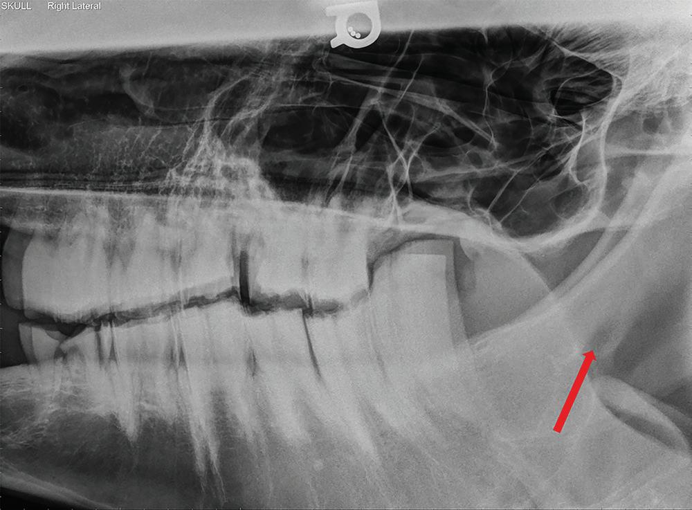
Figure 1.5 Lateral radiograph of a horse skull with severe dental disease. The mental foramen (red arrow) can be seen where the mental nerve exits to innervate the rostral aspect of the mandible. Mental nerve blockade can be used to desensitize the mandible rostral to the mental foramen to the level of the mandibular symphysis.
yellow circle indicates the location of the infraorbital canal, and stippling indicates the area of desensitization following administration of local anesthetic.
approximately 5 ml of local anesthetic with a 22 gauge, 1″ needle at the palpable ridge along the mandible at approximately the middle of the interdental space (Skarda et al. 2010).
A 25–20 gauge, 1–2.5″ needle and 3–10 ml of local anesthetic is described blockade of the mandibular alveolar nerve within the mandibular canal (Skarda et al. 2010; Rice 2017).
Figure 1.4 The mental nerve block in the horse. The
Inferior Alveolar (Mandibular) Nerve Blocks
Desensitization of the inferior alveolar nerve prior proximal to its entrance into the mandibular canal results in blockade of the entire hemi-mandible including teeth, mandibular bone, skin, and gingiva (Figures 1.6 and 1.7). Due to the close anatomical proximity of the lingual branch of the trigeminal nerve, it is also possible to inadvertently desensitize the tongue, potentially resulting in self-trauma on recovery (Caldwell and Easley 2012; Harding et al. 2012). Smaller volumes of local anesthetic may minimize this risk (Harding et al. 2012). Much like the maxillary nerve, multiple techniques have been proposed to achieve desensitization of this nerve in the horse. The earliest was by Bemis who suggested that a vertical line be drawn from the lateral canthus of the eye ventrally to the mandible and a horizontal line be drawn from the occlusal surface of the mandibular molars caudally to the ramus of the mandible with the mandibular foramen located on the medial aspect of the mandible at the junction of the two lines (1917). The technique suggested inserting a spinal needle 3 cm ventral to the temporomandibular junction and advancing it medially to the mandibular foramen (Bemis 1917). Modifications of this technique include approaching the foramen
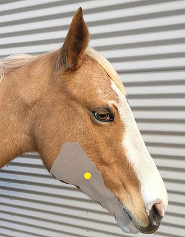
Figure 1.6 The inferior alveolar/mandibular nerve block in the horse. The yellow circle indicates the location of the infraorbital canal, and stippling indicates the area of desensitization following administration of local anesthetic.
from the ventral border of the ramus and inserting a long spinal needle along the imaginary horizontal line from the caudal aspect of the vertical ramus, medially along the mandible to a depth of 9 cm and injecting
Figure 1.7 Lateral radiograph of a horse skull with severe dental disease. The mandibular foramen (red arrow) can be seen where the mandibular nerve enters on the medial aspect to innervate the entire hemi-mandible. Mandibular nerve blockade can be used to desensitize the entire hemi-mandible.
10 ml of local anesthetic (Fletcher 2004; Harding et al. 2012).
A recent cadaver study compared inferior alveolar nerve blocks performed using the landmarks suggested by Bemis and advancing an 18 gauge, 20 cm spinal needle either from the ventral border of the vertical ramus at Bemis’ imaginary vertical line to the mandibular foramen or rostrodorsal from the angle where the horizontal and vertical rami meet the foramen (Harding et al. 2012). The study reported successful dye staining of the nerve in 59% of the vertical injections and 73% of the angled injections; however, there was no significant difference between the two techniques (Harding et al. 2012).
Lastly, another recent study has described the use of an intraoral approach to the inferior alveolar nerve block in the horse. The intraoral approach is used quite frequently in other species; however, the anatomy and relatively narrow gape of the horse make manual palpation of the mandibular foramen impossible. The study described the use of a custom-made tool that was essentially a 20-gauge needle attached to a length of extension tubing and secured to a bent metal rod to allow the needle to be directed through the mouth and to the medial mandibular location of the foramen (Henry et al. 2014). In the study, a total of 51 blocks using 5 ml of 2% mepivacaine were administered and procedures including endodontics, mucosal elevation, and dental extractions were performed successfully following all blocks (Henry et al. 2014). One horse was reported to develop an abscess in the pterygoid fossa two weeks after the procedure (Henry et al. 2014).
Dental Extractions and Repulsions
Extractions and repulsions of teeth can be done either with standing sedation and local anesthesia techniques or under a general anesthetic. The choice is a matter of the horse’s personality and health status, the perceived invasiveness of the procedure, as well as the
surgeon’s preference; however, the obvious benefit of performing the procedure in a standing horse is the avoidance of the inherent risk and complications associated with general anesthesia. In addition, standing sedation avoids having an orotracheal tube in the mouth where it may obstruct the surgeon’s view and/ or make access to the tooth with extraction equipment difficult. Multiple studies have described the successful use of a combination of standing sedation and local anesthesia blocks in order to accomplish oral extraction or sinus repulsion of both retained fragments and intact teeth (MacDonald et al. 2006; Coomer et al. 2011; Dixon et al. 2005). There are no procedure-specific considerations when choosing a general anesthesia protocol for dental procedures in the horse. To facilitate adequate access to the oral cavity, a total intravenous (TIVA) protocol such as “triple-drip” (guaifenesin, ketamine, xylazine) can be used while supplementing oxygen via a nasotracheal tube. TIVA techniques should be reserved for procedures in healthy horses lasting less than 45 minutes to 1 hour. During the general anesthetic, care should be taken when positioning the horse for the extraction or repulsion to avoid pressure on the contralateral facial nerve. Lastly, recovery should be facilitated with either a nasotracheal or orotracheal tube left in place to maintain the airway and partially guard the airway against any remaining bleeding from the procedure.
Standing sedation protocols have been reviewed elsewhere in this book; however, it is worth highlighted a recent study describing the use of romifidine-based standing sedation for cheek tooth extraction (Müller et al. 2017). The authors evaluated the use of a romifidine continuous rate infusion alone (0.05 mg/kg/h) and in combination with butorphanol (0.04 mg/ kg/h), midazolam (0.06 mg/kg/h), or ketamine (1.2 mg/kg/h). All drugs received appropriate loading doses (romifidine 0.03 mg/kg; butorphanol 0.02 mg/kg; midazolam 0.02 mg/kg; ketamine 0.5 mg/kg) prior to commencement of the CRI and lidocaine-based maxillary or
mandibular nerve blocks. The protocol achieved sufficient sedation for completion of the extractions in all groups other than group receiving romifidine alone. The combination of romifidine with midazolam produced substantial ataxia compared to combination with ketamine although both produced good surgical conditions. Combination with butorphanol resulted in a reduced cortisol stress response (Müller et al. 2017). The study concludes that romifidine should not be used alone for standing sedation for dental extractions (Müller et al. 2017).
Surgical Diseases of Paranasal Sinuses
Primary Sinusitis
Primary sinusitis is caused by an upper respiratory tract infection (most commonly, Streptococcus species) that has involved the paranasal sinuses, and secondary sinusitis is caused by an apical infection of maxillary cheek teeth (Tremaine and Dixon 2001a). Systemic antibiotic therapy is very effective but sinus lavage once or twice daily with an indwelling catheter introduced into the infected sinus through a trephine opening may be clinically necessary to fully resolve the infection or remove inspissated pus. The skin over the trephination site is locally infiltrated with 2–3 ml of 2% mepivacaine prior to surgery (Tremaine 2007).
Paranasal Sinus Cysts
Paranasal sinus cysts are single or loculated fluid-filled lesions that typically develop in the maxillary sinuses and ventral concha and can extend into the conchofrontal sinus (Woodford and Lane 2006). The etiology and pathogenesis are unknown (Tremaine et al. 1999); however, extirpation of the cyst and the involved conchal lining through a frontonasal or maxillary bone flap is curative (Woodford and Lane 2006).
Ethmoid Hematoma
Ethmoid hematoma is a progressive and locally destructive idiopathic mass that may arise from the ethmoid labyrinth or the floor of the paranasal sinuses. These lesions are characterized by endoturbinate or a sinus submucosal hemorrhage and concurrent stretching and thickening of the mucosa that becomes the capsule of the hematoma (Tremaine et al. 1999). Ethmoid hematomas can extend into the frontal sinus, the maxillary sinus, the nasal cavity, or the sphenopalatine sinus by disrupting the tectorial plate (Nickels 2012). If the ethmoid hematoma extends into one of the paranasal sinuses, a frontonasal bone flap is indicated to remove the lesion, but it can be associated with profuse intraoperative hemorrhage (Nickels 2012). Typically, ethmoid hematoma causes mild and spontaneous intermittent epistaxis; however, anemia is very rare.
Neoplasia
Neoplasia in the nasal cavity or paranasal sinuses are uncommon (Tremaine and Dixon 2001a). Squamous cell carcinoma is the most common neoplasia of the paranasal sinuses; however, other types of sarcoma tumors (osteogenic sarcoma, lymphosarcoma, poorly differentiated carcinoma, fibrosarcoma, hemangiosarcoma, and adenocarcinoma) have been reported.
Surgery of the Paranasal Sinuses
Sinoscopy
Sinoscopy of the paranasal sinuses is usually performed with the horse standing and adequately sedated. The examination is performed with a flexible endoscope using portals created with a 15-mm Galt trephine following local infiltration of 3–4 ml of 2% mepivacaine at the surgery sites. The endoscopic portal for the conchofrontal sinus is located 60% of the distance from midline toward the medial canthus and 0.5 cm caudal to the medial canthus of the
eye, whereas the endoscopic portal for the maxillary sinuses is located 2 cm rostral and 2 cm ventral to the medial canthus of the eye (caudal maxillary sinus) or 50% of the distance from the rostral end of the facial crest to the level of the medial canthus and 1 cm ventral to a line joining the infraorbital foramen and the medial canthus (rostral maxillary sinus).
Frontonasal and Maxillary Bone Flap Technique
A frontonasal bone flap approach is used to gain access to the conchofrontal and caudal maxillary sinuses and, by additional steps, the rostral maxillary and ventral conchal sinuses. If there is bilateral disease, a tracheotomy should be performed prior to induction and the horse should be intubated through the tracheostomy incision; the animal is otherwise intubated routinely. In addition, sufficient analgesia of the cheek teeth and sinus is a prerequisite for procedures performed on standing horses under chemical sedation. To provide anesthesia of the paranasal sinuses and maxillary cheek teeth, the maxillary branch of the trigeminal nerve in the pterygopalatine fossa should be desensitized using one of the locoregional maxillary nerve block techniques described in the previous section. This block should be performed regardless of whether the surgical procedure is done under standing sedation or general anesthesia.
The frontonasal or maxillary bone flap procedure consists of incising the skin, periosteum, and bone on three sides. For a frontonasal bone flap, the caudal margin is a line drawn at right angle to the dorsal midline and midway between the supraorbital foramen and the medial canthus of the eye, the lateral margin is a line 2.5 cm medial to the medial canthus of the eye that runs slightly dorsal to another line from the medial canthus to the nasoincisive notch, and the rostral margin is caudal to the point at which the nasal bones become parallel. For a maxillary bone flap, the rostral margin of the maxillary bone flap is a line drawn
from the rostral end of the facial crest to the infraorbital foramen, the dorsal margin is a line from the infraorbital foramen to the medial canthus of the eye, the caudal margin is a line (parallel to the rostral margin) from the medial canthus of the eye to the caudal aspect of the facial crest, and the ventral margin is the facial crest (Nickels 2012). The bone flap is then elevated, the affected cheek teeth are repelled by placing a dental punch onto the roots or the lesion (paranasal cyst, ethmoid hematoma, neoplasia, etc.) and removed while controlling intraoperative bleeding.
Following a frontonasal or maxillary bone flap, a surgical opening into the nasal cavity is created and then saline-soaked rolled gauze is inserted immediately into the sinus and nasal passages to control hemorrhage and the end of the gauze is pulled through the sinuses and out the nostril. The packing is removed at 48–72 hours after surgery. Historically, frontonasal and maxillary bone flaps have been associated with profuse hemorrhage so that having a potential blood donor identified by crossmatch is recommended. However, in recent publications of the surgical procedure this complication was infrequent (Tremaine and Dixon 2001a, b; Woodford and Lane 2006; Hart and Sullins 2011).
Surgical Diseases of the Nasal Cavity
Nasal Septum Deformity
Although it is clinically infrequent, nasal septum resection is indicated for treatment of malformations, cystic degeneration, fungal infections, traumatic thickening secondary to septal fracture, and neoplasia among others (Valdez et al. 1978; Watt 1970; Doyle and Freeman 2005; Schumacher et al. 2008). The cartilage of the nasal septum responds in an exaggerated fashion to trauma and heals with deformity, thickening, and deviation which produces decreased airflow or complete unilateral obstruction and
nasal stertor. Diagnosis can generally be made with endoscopic and radiographic examination. The abnormality may be rostral enough to palpate digitally; however, advanced diagnostic imaging may be useful in same cases to plan the surgical treatment (Auer 2012).
Campylorrhinus Lateralis (Wry Nose)
Campylorrhinus lateralis is a congenital abnormality consisting of dysplasia of one side of the maxilla and premaxilla that results in deviation of the maxillae, premaxillae, nasal bones, vomer, and the nasal septum to the dysplastic side (Schumacher et al. 2008). The deviation usually results in malocclusion of the incisors of the mandible and maxilla. Depending on the degree of deviation foals may have difficulty breathing and stridor due to progressive airway obstruction secondary to deviation of the nasal septum to the convex side of the deformity (Schumacher et al. 2008). Radiography supports the clinical diagnosis; however, computed tomography may be useful to assess if there is rotational component in the deviation (Auer 2012). Slight nasal deviation may straighten with growth, but horses with moderate or severe deviation require surgical treatment to resolve respiratory obstruction and to improve incisor occlusion and cosmetic appearance (Auer 2012).
Choanal Atresia
This is a rare maxillofacial malformation in which one or both nasal cavities fail to communicate with the nasopharynx due to persistence of the buccopharyngeal septum, which separates the nasal cavities from the nasopharynx during the embryonic development (James et al. 2006; Hogan et al. 1995). Although there are multiple theories proposed for its embryological origin, it appears that buccopharyngeal membrane persistence is due to misdirection of mesodermal flow caused by errors in neural crest migration in the nasal cavities during embryogenesis.
Affected animals may have partial (unilateral atresia) or complete (bilateral atresia) airflow obstruction through the nasal cavity (James et al. 2006; Hogan et al. 1995; Richardson et al. 1994). Given that horses are obligate nasal breathers, bilateral choanal atresia in foals can cause immediate asphyxia upon birth unless an airway is immediately established by a temporary tracheostomy (James et al. 2006). When the atresia occurs unilaterally, foals exhibit loud respiratory noise, exercise intolerance, and asymmetry of airflow from the nostrils can be detected (James et al. 2006; Hogan et al. 1995, Richardson et al. 1994). The diagnosis is typically made by endoscopic examination but other modalities such as skull radiography, contrast radiography, and computed tomography can be helpful to plan the surgical approach (Gerros and Stone 1994; Nykamp et al. 2003). The treatment is resection of the buccopharyngeal membrane.
Surgery of the Nasal Cavity
Nasal Septum Resection
The nasal septum is covered with a highly vascular mucosa, consequently excision of the septum may cause severe intraoperative hemorrhage, hypotension, and hypoxia. Administration of large volumes of intravenous fluids during surgery is recommended to help alleviate the hypotension. However, it is advisable to identify a suitable blood donor and collect 4–8 l of blood before surgery in case a blood transfusion is necessary. A tracheotomy should be performed before surgery for maintenance of ventilation and inhalational agent delivery thereby allowing adequate surgical access to the oral cavity during surgery.
This procedure should be performed under general anesthesia; however, no specific protocol is indicated and, instead, should be chosen based on the needs of the horse. As the horse will be placed in lateral recumbency for the procedure, administration of a bilateral maxillary block can be challenging. The anesthetist should
consider positioning the horse in dorsal recumbency on the surgical table initially to facilitate blockade of the “down” nerve prior to repositioning to lateral recumbency. Alternatively, consideration could be made for performing the bilateral maxillary blocks in the standing horse under sedation/premedication and possibly at the same time as the temporary tracheostomy is performed.
The horse is positioned in lateral recumbency, and the obstetrical wire is passed up one nasal passage. The most commonly used technique involves transecting the nasal septum dorsally, ventrally, and caudally with obstetrical wire and the most rostral incision is made with a scalpel leaving 5 cm of septum to support the nostrils and alar folds (Doyle and Freeman 2005). The mouth is held open with a speculum, a hand passed back into the oral cavity, and the wire is retrieved around the edge of the soft palate. Another wire is introduced in similar manner up the other nasal passage and is also brought out through the mouth. The ends of the wires are connected outside the mouth, and one is drawn back through a nasal passage until the splice is brought out of the nostril. The wires are then disconnected and the one is left in place so that it passes around the ventral edge of the vomer bone and out both nostrils; this wire is used to transect the nasal septum ventrally.
Next, a trephine hole is placed in the nasal bones immediately rostral to the point at which these bones diverge on the face toward the eyes. A Chamber’s mare catheter is introduced into the nasal passage to exit through the trephine hole. A length of obstetrical wire is then threaded down this catheter and the catheter is removed so that the wire runs from the trephine hole to exit at the nostril. The catheter is introduced into the other nostril and the same wire is threaded down it so that it loops around the septum at the level of the trephine hole and both ends exit the nostril; this wire is used to transect the septum dorsally.
Another length of obstetrical wire is now passed through the trephine hole and into one
nasal passage, in a direction toward the caudal edge of the soft palate. The mouth is held open with a speculum, a hand passed back into the oral cavity, and the wire is retrieved around the edge of the soft palate. Another wire is introduced in similar manner through the trephine hole into the other nasal passage and is also brought out the mouth. The ends of the wires are then taped together outside the mouth, and one is drawn back through a nasal passage until the splice is brought out of the trephine hole. The remaining wire is left in place so that it passes around the ventral edge of the vomer bone and out the trephine hole; this wire is used to transect the septum caudally. The ventral and dorsal cuts are made simultaneously taking care and completed rapidly once started so that hemorrhage is minimal before packing is inserted. The septum is extracted by grasping it with long forceps at the rostral end and pulling it through one of the nostrils. The nasal cavity is packed similarly as for sinuses following surgery and the horse should be allowed to recover with a temporary tracheostomy tube secured in place. The nasal packing and temporary tracheostomy tube may be removed after 48 or 72 hours.
Wry Nose (Campylorrhinus Lateralis)
Successful surgical treatment can improve cosmetic appearance and alleviate airway obstruction (Schumacher et al. 2008). This reconstructive surgery is recommended when the foal is two to three months of age to allow the facial bones to gain enough strength to support implant. The foal is anesthetized using a routine protocol and positioned in dorsal or lateral recumbency on the surgery table (Schumacher et al. 2008). The endotracheal tube is inserted ideally through a tracheostomy approach, the cuff is inflated, and the mouth is thoroughly washed. A bilateral maxillary block can be performed at this time; however, there are currently no studies reporting on this technique in foals.
The first step consists of resecting the nasal septum leaving sufficient septum to support the nostrils and alar folds; the section of
septum to be removed is centered on the middle of the bend in the nasal septum. The nasal passages are packed with rolled gauze to control the hemorrhage and the nostrils are sutured closed to retain the packing. The second step consists of transecting the rami of the maxilla at the level of the interalveolar space to allow re-alignment of the maxillae and premaxillae followed by stabilization with bone plates or Steinmann pins. During surgery, symmetry of the upper and lower jaws is maintained by temporarily wiring the incisors of the maxilla and mandible. The third step consists of correcting the nasal bone deviation and stabilize with reconstruction bone plates. Some surgeons use an autogenous cortical bone graft in the form of a rib to fit the gap in the concave side of the premaxilla; therefore, a section of rib can be harvested before straightening the maxillae/premaxillae, with the horse positioned in left or right lateral recumbency, or in dorsal recumbency (Schumacher et al. 2008).
Choanal Atresia
Treatment options for choanal atresia include transendoscopic laser excision of the buccopharyngeal membrane, or via bilateral frontonasal bone flap (if the buccopharyngeal membrane is osseous, or the condition is bilateral), or via laryngotomy with endoscopic assistance to gain access to the obstructed choanae (Aylor et al. 1984; James et al. 2006; Ducharme 2012).
In horses with unilateral choanal atresia, transendoscopic laser excision can be done with the patient standing and seated with detomidine and butorphanol, and topical anesthesia with phenylephrine (2% lidocaine or mepivacaine hydrochloride and 10 ml of 0.15% phenylephrine) applied to the buccopharyngeal membrane and nasal cavity. Choanal stenosis is a frequent complication, so the use of stents is mandatory post-operatively to prevent stricture (James et al. 2006). This can be done with a silicone endotracheal tube threaded over the
wire, pushed through the puncture site in the choanal membrane, and left in place to serve as a stent. If resection of the buccopharyngeal membrane via frontonasal bone flap with the animal anesthetized is elected, a temporary tracheostomy should be performed to allow tracheal intubation during surgical manipulations of the head and should be left in place with the cuff inflated during recovery to assure an adequate airway and protect it from surgical site bleeding.
Equine Blood Typing and Transfusion Considerations
As stated earlier in this chapter, significant acute hemorrhage is a potential risk associated with surgical procedures of the nose. Acute blood loss of >20% of the blood volume and/or clinical signs of hypovolemia should be used to assess the need for emergency transfusions as changes in packed cell volume (PCV) or total protein may lag behind by several hours due to splenic contraction (Hardy 2009). Transfusions of whole blood should be reserved for lifesaving situations however as transfused red blood cells only last in recipient circulation for two to six days (Hardy 2009; Kallfelz et al. 1978). Transfusion medicine in horses is more complicated than in humans or small companion animals owing to the lack of a universal blood type in horses. Horses have eight different blood groups, A, C, D, K, P, Q, U, and T as well as more than 30 different surface marker factors on the red blood cells. As a result, the likelihood of a perfect match between donor and recipient is estimated to be upward of 1 in 400 000. Fortunately, the vast majority of transfusion reactions are attributable to two specific blood types, Aa and Qa, and horses that possess Ca antigens also pose a theoretical risk (Schmotzer 1985). As stored equine blood samples are only stable for a few weeks, blood banking is not common, and donors must typically be either kept on site or available at short
notice (Mudge et al. 2004). Ideal donors must be healthy adult horses, up to date on vaccines and free from any diseases. Unrelated geldings of the same breed or mares who have never foaled can be chosen for blood donors as long as their PCV is greater than 35% and they test negative for Aa, Qa, and Ca antigens. Related horses or mares who have foaled previously are more likely to possess antibodies that could lead to transfusion reactions. In larger practice settings where donor horses are maintained, blood typing should be performed prior to donation to avoid Aa, Qa, and Ca antigens; however, Quarter horses and standardbreds are reported as breeds less likely to possess Aa or Qa antigens if prior blood typing is not possible (Nolen-Walston, n.d.).
Cross-matching should be performed, if possible, prior to blood transfusion administration. A major cross-match involves combining washed donor red blood cells with recipient serum while a minor cross-match combines donor serum with washed recipient red blood cells. Signs of incompatibility include autoagglutination and hemolysis.
Donor horses should have a large gauge, short catheter placed aseptically in the jugular vein and connected to a bag containing anticoagulant. Adult horses greater than 450 kg can donate between 15 and 20 ml/kg of whole blood which should be replaced with at least an equivalent volume of crystalloid fluids after donation (Malikides et al. 2001; Nolen-Walston, n.d.).
References
Auer, J.A. (2012). Craniomaxillofacial disorders. In: Equine Surgery, 4e (eds. J.A. Auer and J.A. Stick), 1456–1482. St. Louis, MO: Elsevier Saunders.
Aylor, M.K., Campbell, M.L., Goring, R.L., and Hillidge, C.J. (1984). Congenital bilateral choanal atresia in a standardbred foal. Equine Veterinary Journal 16 (4): 396–398.
When estimating the amount of donor blood needed for transfusion, one can calculate with the following general estimate:
Donor blood needed = Estimated Recipient blood volume xDesireed PCV% Actual PCV%/Donor PCV%
A more general estimate assumes that 2–3 ml/ kg of whole blood from a donor with a PCV of 40% will raise the recipient’s PCV by 1% (Hardy 2009). In general, adult horse’s blood volumes can be estimated as 80 ml/kg. The technique for administration of the actual transfusion is not dissimilar to that of small animals or humans where the transfusion is started at a slow rate for the first 15 minutes to closely observe for transfusion reactions after while time the remainder of the volume can be administered at 5–20 ml/kg/h with the goal of completing the transfusion in less than four hours to maintain sterility (Hardy 2009; Nolen-Walston, n.d.). Signs of transfusion reactions include hypotension, skin wheals, sweating, tachycardia, fever, diarrhea, colic, and piloerection (Hardy 2009; NolenWalson, n.d.). Transfusions should be stopped if signs of reaction occur and treatment, including fluids and epinephrine if needed, should be instituted. If the transfusion reaction is mild and the signs resolve with discontinuation of the infusion, restarting the infusion at a slower rate can be considered, especially if no other suitable donor is available.
Bardell, D., Iff, I., and Mosing, M. (2010). A cadaver study comparing two approaches to perform a maxillary nerve block in the horse. Equine Veterinary Journal 42 (8): 721–725.
Bemis, H.E. (1917). Local anaesthesia in animal dentistry. American Veterinary Journal 51: 188. Caldwell, F.J. and Easley, K.J. (2012). Selfinflicted lingual trauma secondary to inferior
alveolar nerve block in 3 horses. Equine Veterinary Education 24: 119–123.
Coomer, R.P., Fowke, G.S., and McKane, S. (2011). Repulsion of the maxillary and mandibular cheek teeth in standing horses. Veterinary Surgery 40: 590–595.
Dixon, P.M. (2005). Dental anatomy. In: Equine dentistry, 2e (eds. G.J. Baker and J. Easley), 25–66. St. Louis MO: Elsevier.
Dixon, P.M., Dacre, I., Dacre, K. et al. (2005). Standing oral extraction of cheek teeth in 100 horses (1998–2003). Equine Veterinary Journal 37: 105–112.
Doyle, A.J. and Freeman, D.E. (2005). Extensive nasal septum resection in horses using a 3-wire method. Veterinary Surgery 34 (2): 167–173.
Ducharme, N.G. (2012). Pharynx. In: Equine Surgery, 4e (eds. J.A. Auer and J.A. Stick), 569–591. St. Louis, MO: Elsevier Saunders.
Fletcher, B.W. (2004). How to perform effective equine dental nerve blocks. In: Proceedings of the Convention of the American Association of Equine Practitioners, Denver, CO.
Gerros, T.C. and Stone, W.C. (1994). What is your diagnosis? Complete bilateral choanal atresia. Journal of the American Veterinary Medical Association 205 (2): 179–180.
Harding, P.G., Smith, R.L., and Barakzai, S.Z. (2012). Comparison of two approaches to performing an inferior alveolar nerve block in the horse. Australian Veterinary Journal 90: 146–150.
Hardy, J. (2009). Venous and arterial catheterization and fluid therapy. In: Equine Anesthesia: Monitoring and Emergency Therapy, 2e (eds. W.W. Muir and J.A.E. Hubbell), 131–148. St. Louis, MO: Saunders. Hart, S.K. and Sullins, K.E. (2011). Evaluation of a novel postoperative treatment for sinonasal disease in the horse (1996–2007). Equine Veterinary Journal 43 (1): 24–29.
Henry, T., Pusterla, N., Guedes, A.G.P., and Vertraete, F.J.M. (2014). Evaluation and clinical use of an intraoral inferior alveolar nerve block in the horse. Equine Veterinary Journal 46: 706–710.
Hillmann, D.J. (1975). Skull. In: Sisson and Grossman’s the Anatomy of the Domestic Animals, 5e (ed. R. Getty), 255–348. Philadelphia, PA: W. B. Saunders Company. Hogan, P.M., Embertson, R.M., and Hunt, R.J. (1995). Unilateral choanal atresia in a foal. Journal of the American Veterinary Medical Association 207 (4): 471–473.
James, F.M., Parente, E.J., and Palmer, J.E. (2006). Management of bilateral choanal atresia in a foal. Journal of the American Veterinary Medical Association 229 (11): 1784–1789.
Kallfelz, F.A., Whitlock, R.H., and Schultz, R.D. (1978). Survival of 59 Fe-labeled erythrocytes in cross-transfused equine blood. American Journal of Veterinary Research 39: 617–620.
MacDonald, M.H., Basile, T., Wilson, W.D. et al. (2006). Removal of maxillary tooth fragments and root remnants in standing horses. In: American Association of Equine Practitioners Dental Focus Meeting Proceedings, 148–155. Indianapolis, IN.
Malikides, N., Hodgson, J.L., Rose, R.J., and Hodgson, D.R. (2001). Cardiovascular, haematological and biochemical responses after large volume blood collection in horses. Veterinary Journal 162 (1): 44–55.
Mudge, M.C., MacDonald, M.H., Owens, S.D., and Tablin, F. (2004). Comparison of 4 blood storage methods in a protocol for equine pre-operative autologous donation. Veterinary Surgery 33 (5): 475–486.
Müller, T.M., Hopster, K., Bienert-Zeit, A. et al. (2017). Effect of butorphanol, midazolam or ketamine on romifidine based sedation in horses during standing cheek tooth removal. BMC Veterinary Research 13: 381.
Nannarone, S., Bini, G., Vuerich, M. et al. (2016). Retrograde maxillary nerve perineural injection: a tomographic and anatomical evaluation of the infraorbital canal and evaluation of needle size in equine cadavers. The Veterinary Journal 217: 33–39.
Newton, S.A., Knottenbelt, D.C., and Eldridge, P.R. (2000). Headshaking in horses: possible
aetiopathogenesis suggested by the results of diagnostic tests and several treatment regimes used in 20 cases. Equine Veterinary Journal 32 (2): 208–216.
Nickels, F.A. (2012). Nasal passages and paranasal sinuses. In: Equine Surgery, 4e (eds. J.A. Auer and J.A. Stick), 557–568. St. Louis, MO: Elsevier Saunders.
Nolen-Walston, R.D. (n.d.) Equine blood transfusion- what you need to know to get the job done. https://vvma.org/resources/ Conferences/2016%20VVC%20Notes/ Nolen-Walston-%20Blood%20Transfusions.pdf
Nykamp, S.G., Dykes, N.L., Cook, V.L. et al. (2003). Computed tomographic appearance of choanal atresia in an alpaca cria. Veterinary Radiology & Ultrasound 44 (5): 534–536.
O’Neill, H.D., Garcia-Pereira, F.L., and Mohankumar, P.S. (2014). Ultrasound-guided injection of the maxillary nerve in the horse. Equine Veterinary Journal 46 (2): 180–184.
Rice, M.K. (2017). Regional nerve blocks for equine dentistry. Journal of Veterinary Dentistry 34: 106–109.
Richardson, J.L., Lane, J.G., and Day, M.J. (1994). Congenital choanal restriction in 3 horses. Equine Veterinary Journal 26: 162–165.
Rieder, C.M., Zwick, T., Hopster, K. et al. (2016a). Maxillary nerve block within the pterygopalatine fossa for oral extraction of cheek teeth in 80 horses. Pferdeheilkunde 32: 587–594.
Rieder, C.M., Staszyk, C., Hopster, K., and Bienert-Zeit, A. (2016b). Maxillary nerve block within the equine pterygopalatine fossa with different volumes: practicability, efficacy and side-effects. Pferdeheilkunde 32 (2): 132–140.
Schmotzer, W.B. (1985). Time-saving techniques for the collection, storage, and administration of equine blood and plasma. Veterinary Medicine 80: 89–94.
Schumacher, J., Brink, P., Easley, J., and Pollock, P. (2008). Surgical correction of wry nose in four horses. Veterinary Surgery 37 (2): 142–148.
Skarda, R.T., Muir, W.W., and Hubbell, J.A.E. (2010). Local anesthetic drugs and techniques. In: Equine Anesthesia: Monitoring and Emergency Therapy, 2e (eds. W.W. Muir
and J.A.E. Hubbell), 210–242. St. Louis, MO: Saunders.
Staszyk, C., Bienert, A., Bäumer, W. et al. (2008). Simulation of local anaesthetic nerve block of the infraorbital nerve within the pterygopalatine fossa: anatomical landmarks defined by computed tomography. Research in Veterinary Science 85: 399–406.
Stauffer, S., Cordner, B., Dixon, J., and Witte, T. (2017). Maxillary nerve blocks in horses: an experimental comparison of surface landmark and ultrasound-guided techniques. Veterinary Anaesthesia and Analgesia 44: 951–958.
Tremaine, W.H. (2007). Local analgesic techniques for the equine head. Equine Veterinary Education 19 (9): 495–503.
Tremaine, W.H. and Dixon, P.M. (2001a). A longterm study of 277 cases of equine sinonasal disease. Part 1: details of horses, historical, clinical and ancillary diagnostic findings. Equine Veterinary Journal 33 (3): 274–282.
Tremaine, W.H. and Dixon, P.M. (2001b). A long-term study of 277 cases of equine sinonasal disease. Part 2: treatments and results of treatments. Equine Veterinary Journal 33 (3): 283–289.
Tremaine, W.H., Clarke, C.J., and Dixon, P.M. (1999). Histopathological findings in equine sinonasal disorders. Equine Veterinary Journal 31 (4): 296–303.
Valdez, H., McMullan, W.C., Hobson, H.P., and Hanselka, D.V. (1978). Surgical correction of deviated nasal septum and premaxilla in a colt. Journal of the American Veterinary Medical Association 173 (8): 1001–1004.
Watt, D.A. (1970). A case of cryptococcal granuloma in the horse. Australian Veterinary Journal 46: 493.
Woodford, N.S. and Lane, J.G. (2006). Long-term retrospective study of 52 horses with sinunasal cysts. Equine Veterinary Journal 38 (3): 198–202.
Woodie, J.B. (2013). How to use local and regional anesthesia for procedures of the head and perineum in the horse. Proceedings of the American Association of Equine Practitioners Annual Conference, 59, 464–466.
Anesthetic Management for Ocular Interventions
Bianca Martins1 and Manuel Martin-Flores2
1 Department of Surgical and Radiological Sciences, School of Veterinary Medicine, University of California, One Garrod Drive, Davis, CA, 95616, USA
2 Department of Clinical Sciences, College of Veterinary Medicine, Cornell University, 930 Campus Road, Ithaca, NY, 14853, USA
Introduction
Resolution of ocular procedures in horses can present several particular challenges to the anesthesia provider: many surgeries that are performed under general anesthesia require a central and immobile eye, which is best achieved by the use of neuromuscular blockers. The introduction of these agents to the anesthetic protocol adds a new level of complexity, as it will be reviewed in following paragraphs. In some instances, the procedure might be completed with the horse awake and standing, facilitated by the use of sedatives and locoregional anesthesia. In that case, the anesthetist must be experienced in the use of sedatives to provide a state of “conscious sedation” that allows sufficient cooperation from the animal (or tolerance to the surgery) while retaining the ability to stand with as minimal ataxia as possible. For this approach, knowledge of the relevant anatomy and locoregional techniques is necessary. Aside from these technical challenges, complications might arise from manipulation of the eye during surgery (i.e. cardiac dysrhythmias), or secondary to topical ophthalmic medication. Therefore, a basic understanding of ocular physiology and therapeutics is also necessary.
Several ocular pathologies are resolved through surgical interventions in horses. These involve extraocular procedures, such as those involving the eyelids, removal of the globe, and ocular/intraocular surgeries.
Anatomy and Physiology of the Eye
The globe is located within the orbit and surrounded by periorbital tissues (extraocular muscles [EOMs], retrobulbar fat, among others) and periocular structures (such as the conjunctiva and eyelids). The eyelids are modified upper and lower folds of skin, which form the palpebral fissure (opening), provide mechanical protection for the globe, and spread the tear film. Several muscles are responsible for the closure and opening of the eyelids (Table 2.1). However, in a simplistic way, closure of the eyelids is achieved by contraction of the orbicularis oculi muscle, while the opening of the eyelids is done by a combination of relaxation of the orbicularis oculi muscle, and contraction of the levator palpebrae superioris muscle. All other muscles aid in those movements. In general, motor innervation to the eyelids is provided by the oculomotor nerve (CN III) and
Table 2.1 Muscles of the eyelids, associated function and cranial nerve (CN) innervation.
Eyelid muscle
Orbicularis oculi
Levator palpebrae superioris
Retractor anguli oculi
Corrugator supercilli
Malaris
Function
Closure of eyelids
Elevation of upper eyelid
Lengthening palpebral fissure
Innervation
Facial (CN VII)
Oculomotor (CN III)
Facial (CN VII)
Assist in elevation of upper eyelidFacial (CN VII)
Depression of lower eyelid
Levator anguli oculi medialisLengthening palpebral fissure
Müller
Elevate upper eyelid
facial nerve (CN VII), while the sensory innervation to all aspects of the eyelids is provided by the ophthalmic branch of the trigeminal nerve (CN V).
The orbit is a bony fossa that separates the globe from the cranial cavity. In addition to mechanical protection, the orbit provides several foramina and fissures, which are pathways for blood vessels and nerves involved with the ocular maintenance. Within the orbit, a total of seven EOMs are responsible for providing ocular motility – four recti (dorsal, ventral, lateral, and medial rectus), two obliques (dorsal oblique and ventral oblique), and one retractor bulbi muscle (Table 2.2). The four rectus muscles (dorsal, ventral, lateral, and medial rectus) move the globe in the direction of their respective names; the dorsal oblique muscle pulls the dorsal aspect of the globe medially and ventrally; the ventral oblique muscle moves the
Facial (CN VII)
Facial (CN VII)
Sympathetic fibers
globe medially and dorsally; and the retractor bulbi, which forms a “cone shape” behind the eye, is responsible for retracting the globe into the orbit. The dorsal, ventral, and medial rectus muscles, as well as the ventral oblique muscle, are innervated by cranial nerve III (oculomotor). The dorsal oblique muscle is innervated by cranial nerve IV (trochlear), while the lateral rectus and the retractor bulbi muscles are innervated by cranial nerve VI (abducens). The globe itself is separated into three layers or tunics. The external or outmost wall of the globe consists of the fibrous tunic, composed by the cornea and sclera, which provides the shape and mechanical support for the intraocular structures. The second, middle layer is the highly vascular uveal tunic or tract, composed by the iris, ciliary body, and the choroid. The third and innermost layer is the neural tunic of the eye, composed by the retina. The lens is
Table 2.2 Extraocular muscle responsible for movement of the globe, associated movement and cranial nerve (CN) innervation.
Extraocular muscle
Dorsal rectus
Ventral rectus
Medial rectus
Lateral rectus
Dorsal oblique
Ventral oblique
Retractor bulbi
Function
Pull globe upward
Pull globe downward
Pull globe medially
Pull globe laterally
Innervation
Oculomotor (CN III)
Oculomotor (CN III)
Oculomotor (CN III)
Abducens (CN VI)
Move globe medially and ventrallyTrochlear (CN IV)
Move glove medially and dorsallyOculomotor (CN III)
Retract globe
Abducens (CN VI)
located just posterior to the iris and is attached to the ciliary body by the lenticular zonules. The anterior segment comprises all structures from the cornea to the lens (including the cornea itself, the iris, ciliary body, and the lens), and is bathed by the aqueous humor. The anterior segment is divided into two spaces: the anterior chamber (from the cornea to the iris), and the posterior chamber (from the iris to the lens).
The posterior segment comprises all structures from the lens to the retina, including the vitreous humor, the choroid, and the retina itself.
The cornea, the anterior outermost structure of the globe, is one of the most densely innervated tissues in the body, richly supplied in sensory nerves (mainly pain receptors) to provide corneal protection, with a neuronal density that is 300–600 times greater than the skin epithelium (Rozsa and Beuerman 1982; Brooks et al. 2000). Sensory innervation of the cornea originates from the long ciliary nerves, which are derived from the ophthalmic branch of the trigeminal nerve (CN V). Most of the nerve endings are located on the subepithelial and anterior stromal layers of the cornea and, in the horse, the cornea appears to be most sensitive in the center and less so toward the periphery.
The innervation of the uveal tract is made by both sympathetic and parasympathetic fibers. The iris sphincter muscle is partly responsible for controlling the pupil size, while the ciliary body muscle is partly responsible for the lens accommodation and focus. The ciliary body muscle and iris sphincter muscle are supplied by parasympathetic fibers from the oculomotor nerve (CN III) via the short ciliary nerves, and by sympathetic nerve fibers via the long ciliary nerves. Medications that may stimulate or paralyze those neuronal pathways may alter the pupil size, either facilitating or precluding some intraocular procedures, such as cataract removal.
Tear Production
The preocular tear film (POTF) is responsible for maintaining an optically uniform ocular surface, lubricating the cornea and conjunctiva,
and providing nutrients to the cornea. The tears are mainly composed of three layers: (i) the outer lipid layer, produced by the meibomian glands, and responsible for preventing the evaporation of the tear film; (ii) the intermediate aqueous layer, produced by the orbital (or principal) gland and by the third eyelid gland, composed mainly by water, plus electrolytes, glucose, urea, globulins, lysozymes, and other solids, and responsible for providing the lubrication and nutrients to the cornea; (iii) the inner mucin layer, produced by the conjunctival goblet cells, responsible for anchoring the aqueous film to the corneal epithelium. Also, the POTF contains antibacterial cytokines and exerts some control over the ocular surface flora; therefore, the incidence of ocular surface infections is elevated in tear-deficient patients.
Even though the innervation to the lacrimal gland is not yet completely understood, it is known that the lacrimal gland receives sensory input from the lacrimal nerve, which is a branch of the trigeminal nerve (CN V). Also, the lacrimal branch of the facial nerve (CN VII), sympathetic and parasympathetic fibers are involved in the process. In general, some cholinergic drugs stimulate lacrimation, while anticholinergic drugs decrease the tear production. Even though keratoconjunctivitis sicca (“dry eye”) is rare in horses (and usually related to trauma), it is important to remember that general anesthesia may decrease or completely temporarily stop tear production in horses, increasing the risk of corneal erosions and ulcers. Tear replenishment with lacrimomimetics during anesthetic procedures is, therefore, recommended. Artificial tears in ointment or gel formulations are preferable over drops or solutions since those forms stay longer on the eye.
Aqueous Humor and Intraocular Pressure
The aqueous humor is a transparent fluid that resembles an ultrafiltration of plasma and fills and nourishes the anterior segment structures. This fluid is produced by the ciliary body, flows
from the posterior chamber through the pupil, into the anterior chamber, and drains out of the eye via the iridocorneal angle into the intrascleral venous plexus (conventional outflow) or via the supraciliary-suprachoroidal space into the scleral vessels (unconventional outflow). In healthy conditions, aqueous humor is constantly produced and drained, establishing a normal intraocular pressure (IOP). Decreased IOPs are usually associated with intraocular inflammation (uveitis), while elevated IOP is one of the main and most devastating components of the glaucoma syndrome, a progressive condition that leads to blindness.
The aqueous humor is formed by three mechanisms: (i) diffusion of solutes following a down concentration gradient; (ii) ultrafiltration from plasma by hydrostatic force; (iii) active secretion by the ciliary body epithelium, utilizing energy to secrete solutes against a concentration gradient. This active process is catalyzed by the carbonic anhydrase enzyme, leading to entry of sodium (Na+) and bicarbonate (HCO3 ) into the aqueous, establishing an osmotic gradient leading to the entry of water into the posterior chamber. Topical and systemic carbonic anhydrase inhibitors will thus decrease the rate of aqueous humor formation, resulting in lower IOP values. Even though no significant diurnal IOP variation has been detected in horses, the effect of head position on IOP is well documented, with increased IOPs detected when the head is below heart level (Komaromy et al. 2006). This phenomenon is particularly relevant during hoisting after induction of anesthesia, with IOP values up to 80 mmHg obtained while the horse is hoisted. This should be taken into consideration when anesthetizing and hoisting patients where an increase in IOP could be detrimental, such as those suffering from glaucoma, deep corneal ulcers, descemetocele, or corneal rupture (Monk et al. 2017).
Oculocardiac Reflex
Any sensory stimulation to the eye and its periocular structures may result in stimulation of
the vagal nucleus, leading to a slowing of the heart. This oculocardiac reflex has been documented secondary to traction of the globe during enucleations, and during intraocular surgeries in the dog, cat, horses, humans, and other species. Sensory fibers of the ophthalmic branch of the trigeminal nerve (CN V) to its nucleus make up the afferent pathway of this reflex, while the efferent pathway is via the vagus nerve (CN X) to the heart. The most common clinical sign associated with this phenomenon is bradycardia; however, cardiac arrest and ventricular fibrillation have also been documented (Raffe et al. 1986; Oel et al. 2014).
Sedation for Ocular Procedures
Several ophthalmic procedures, such as surgical correction of defects in the eyelids (often of traumatic origin), placement of episcleral cyclosporine implants for immune-mediated keratitis, intra-stromal injections for corneal abscesses, and even removal of the globe, can be performed under sedation and regional anesthesia. Sedation is tailored to produce a cooperative horse that can tolerate surgical manipulation while standing as stable as possible. Regional anesthesia is also provided to achieve sensory blockade, motor blockade, or both.
Sedation in horses is most commonly achieved with the use of ɑ2-agonist agents. These agents provide reliable sedation, analgesia, and result in an animal that typically stands with minimal ataxia. While some minor clinical differences are reported between the use of different agents [e.g. less ataxia reported with romifidine (England et al. 1992)], all ɑ2agonists in clinical use are suitable sedatives for these purposes, and all can be administered either as repeated boluses or infused over time to prolong the duration of sedation. If sedation is insufficient during stimulating periods of the procedure, boluses of the agent can be administered without altering the infusion rate. Alternatively, the infusion can be titrated according to a horse’s response to surgical
Table 2.3 Alpha 2-agonist drugs loading dosages and infusion rates for standing sedation for ophthalmic procedures.
Drug Loading dosage
Xylazine 0.5–1.0 mg/kg 0.7 mg/kg/h
Detomidine0.008–0.01 mg/kg 0.006–0.02 mg/kg/h
Romifidine0.08 mg/kg 0.03 mg/kg/h
References
Ringer et al. (2012)
Gozalo-Marcilla et al. (2019)a
Marly et al. (2014)
a The range has been expanded based on the dosages and experiences at the author’s institution.
stimuli. Doses for constant rate infusions of these agents are summarized in Table 2.3. The author commonly uses opioids such as morphine (0.05–0.1 mg/kg) or butorphanol (0.01–0.02 mg/kg), or the phenothyazine agent acepromazine (0.005–0.01 mg/kg), to enhance sedation from ɑ2-agonists in horses that respond to surgical stimulation.
When choosing which a2-agonist to use, it is important to discuss with the ophthalmologist or surgeon the expected duration of the procedure. In general, detomidine and romifidine are most commonly used for longer procedures (>60 minutes), while xylazine is usually chosen for shorter procedures.
While ketamine can be utilized at lower dosages to provide standing sedation, it has been shown to increase the IOP in horses (Ferreira et al. 2013). On the other hand, sedation with xylazine and detomidine decreases the IOP (Holve 2012) and are likely safer options for patients with increased IOP, such as those with glaucoma or deep corneal ulcers.
Locoregional Anesthesia of the Eye and Surrounding Structures
Locoregional Anesthesia of the Eyelid
Anesthesia of the eye and/or surrounding structures is also necessary, in addition to sedation, for performing ocular procedures in the standing horse. Moreover, a combination of sedation and local anesthesia might be used to facilitate ocular exams by decreasing muscular tone of the eyelid and allowing better exposure of the globe.
Akinesia of the eyelids is achieved by the desensitization of the auriculopalpebral nerve, a branch of the facial nerve, which innervates the orbicularis oculi muscle. Desensitization of this nerve facilitates examination of the eye and improves the surgical field, but does not provide anesthesia (sensory block) to the lids. The auriculopalpebral nerve runs superficial over the temporalis muscle and dorsal orbit and can be palpated dorsal to the zygomatic process of the temporal bone (Figure 2.1). It can be desensitized at this location, or alternatively, at the depression found caudally to the mandible and ventral to the temporal portion of the zygomatic arch. Surgical procedures involving the lids that are expected to produce pain also require sensory blockade, which may be achieved by desensitizing four nerves that surround the eye (Figure 2.2). The supraorbital (or frontal) nerve can be desensitized as it emerges from the supraorbital foramen, on the dorsolateral aspect of the orbit (Figure 2.1). Desensitization of this nerve provides sensory block to the central portion of the upper eyelid. Anesthesia of the lacrimal nerve, located dorsolateral to the lateral canthus, provides sensory block to the lateral canthus and lateral portion of the eyelid. The medial canthus and medial aspect of the lids are desensitized by anesthetizing the infratrochlear nerve, located dorsomedial to the medial canthus of the eye. The zygomatic nerve, located on the ventrolateral aspect of the orbit, provides sensory block to the central portion of the lower lid. Each nerve may be blocked individually to provide complete sensory block of these structures. Alternatively, a ring block may be performed

Figure 2.1 Lateral view of an equine skull. The blue line indicates where the auriculopalpebral nerve can be palpated and blocked as it courses over the zygomatic arch. The red arrow indicated the supraorbital foramen where the supraorbital (frontal) nerve emerges and can be blocked. The green circle indicated the bony orbit.
so that all four nerves are involved. Excessive use of local anesthetics during infiltration, however, can disrupt the anatomy and result in edema, which may complicate the conditions of the surgical field.
Locoregional Anesthesia of the Globe
Procedures involving not only the surrounding structures but also the globe itself can be performed in many horses under sedation. Foreign bodies might be removed under a combination of sedative and locoregional techniques as mentioned above. In addition, topical desensitization of the cornea should be performed and can be achieved with topical ophthalmic
Figure 2.2 Lateral view of an equine head. Colored dots indicate the location where blockade can be performed to provide local anesthesia of the skin and structures surrounding the eye. Green is the supraorbital (frontal) nerve, blue is the lacrimal nerve, orange is the infratrochlear nerve, and red is the zygomatic nerve.
anesthetics, such as tetracaine 0.5% and proparacaine 0.5% ophthalmic solutions, applied directly to the ocular surface. In general, the maximum anesthetic effect is achieved within 5–10 minutes when either tetracaine or proparacaine are used, and last approximately 20–25 minutes. If one drop is used, full desensitization of the cornea is not achieved with aqueous solutions of proparacaine and tetracaine, and some sensation (though minimal) might still be present. However, the instillation of two drops of aqueous 0.5% tetracaine, one minute apart, results in full desensitization of the cornea, with the same duration of action (Monclin et al. 2011). A viscous preparation of 0.5% tetracaine is currently available. It