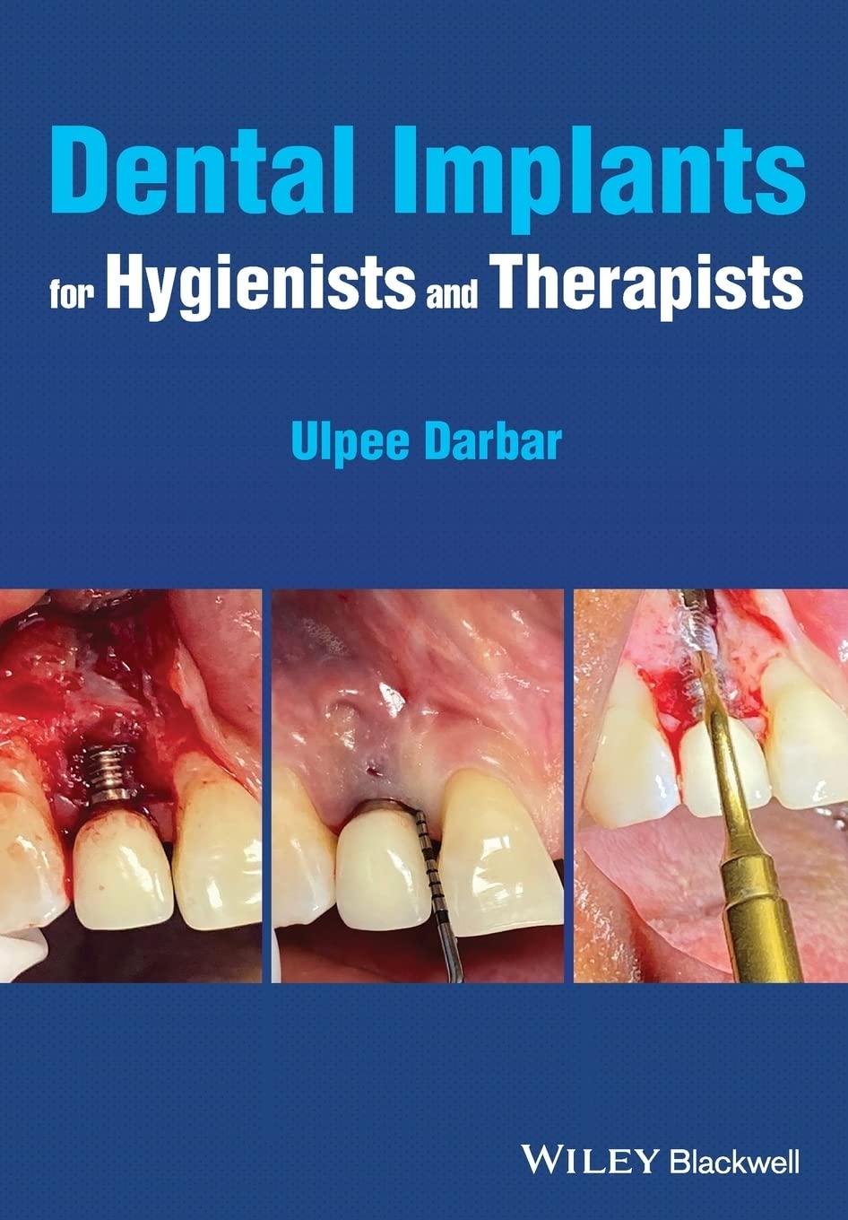
Dental Implants for Hygienists and Therapists
Ulpee Darbar BDS, MSc, FDS(Rest Dent)RCS, FHEA
Consultant in Restorative Dentistry
Eastman Dental Hospital London, UK
This edition first published 2022 © 2022 John Wiley and Sons Ltd
All rights reserved. No part of this publication may be reproduced, stored in a retrieval system, or transmitted, in any form or by any means, electronic, mechanical, photocopying, recording or otherwise, except as permitted by law. Advice on how to obtain permission to reuse material from this title is available at http://www.wiley.com/go/permissions.
The right of Ulpee Darbar to be identified as the author of this work has been asserted in accordance with law.
Registered Offices
John Wiley & Sons, Inc., 111 River Street, Hoboken, NJ 07030, USA
John Wiley & Sons Ltd, The Atrium, Southern Gate, Chichester, West Sussex, PO19 8SQ, UK
Editorial Office
9600 Garsington Road, Oxford, OX4 2DQ, UK
For details of our global editorial offices, customer services, and more information about Wiley products visit us at www. wiley.com.
Wiley also publishes its books in a variety of electronic formats and by print-on-demand. Some content that appears in standard print versions of this book may not be available in other formats.
Limit of Liability/Disclaimer of Warranty
The contents of this work are intended to further general scientific research, understanding, and discussion only and are not intended and should not be relied upon as recommending or promoting scientific method, diagnosis, or treatment by physicians for any particular patient. In view of ongoing research, equipment modifications, changes in governmental regulations, and the constant flow of information relating to the use of medicines, equipment, and devices, the reader is urged to review and evaluate the information provided in the package insert or instructions for each medicine, equipment, or device for, among other things, any changes in the instructions or indication of usage and for added warnings and precautions. While the publisher and authors have used their best efforts in preparing this work, they make no representations or warranties with respect to the accuracy or completeness of the contents of this work and specifically disclaim all warranties, including without limitation any implied warranties of merchantability or fitness for a particular purpose. No warranty may be created or extended by sales representatives, written sales materials or promotional statements for this work. The fact that an organization, website, or product is referred to in this work as a citation and/or potential source of further information does not mean that the publisher and authors endorse the information or services the organization, website, or product may provide or recommendations it may make. This work is sold with the understanding that the publisher is not engaged in rendering professional services. The advice and strategies contained herein may not be suitable for your situation. You should consult with a specialist where appropriate. Further, readers should be aware that websites listed in this work may have changed or disappeared between when this work was written and when it is read. Neither the publisher nor authors shall be liable for any loss of profit or any other commercial damages, including but not limited to special, incidental, consequential, or other damages.
The work presented has been either been undertaken directly by the editor or the editor has been involved in overseeing the work.
Library of Congress Cataloging-in-Publication Data
Names: Darbar, Ulpee R., author.
Title: Dental implants for hygienists and therapists / Ulpee Darbar, Eastman Dental Hospital, London, UK.
Description: Hoboken, NJ : John Wiley & Sons, 2022. | Includes bibliographical references and index.
Identifiers: LCCN 2021053096 (print) | LCCN 2021053097 (ebook) | ISBN 9781119763826 (paperback) | ISBN 9781119763833 (pdf) | ISBN 9781119763840 (epub)
Subjects: LCSH: Dental implants. | Teeth--Transplantation.
Classification: LCC RK667.I45 D37 2022 (print) | LCC RK667.I45 (ebook) | DDC 617.6/93--dc23/eng/20211220
LC record available at https://lccn.loc.gov/2021053096
LC ebook record available at https://lccn.loc.gov/2021053097
Cover image: Courtesy of Ulpee Darbar
Cover design by Wiley
Set in 9.5/12.5pt STIXTwoText by Integra Software Services Pvt. Ltd, Pondicherry, India
10 9 8 7 6 5 4 3 2 1
Glossary xii
1 History of Dental Implants 1
2 Osseointegration 7
Concept 7
Factors affecting Osseointegration 8
- Implant Related 8
- Environment Related 19
Success vs Survival 22
New Concepts 24
3 Implant Systems 28 Components 28
Fixture 28
Transmucosal 32
Implant Types 33
Prosthetic 36
- Abutments 38 - Screws 39 - Torque Wrench 39 Laboratory 39
Implant systems 40
4 Patient Selection and Indications for Treatment 43
Patient Selection 44
- Generic Planning 45
- Site Specific Planning 48
Indications for implant Treatment 54
Follow up care 58 Contents
5 Surgical and Prosthodontic Protocols 60
Surgical Protocols 60
- Timing of Implant Fixture Placement 61
- Computer Guided placement 65
Augmentation Procedures 66
- Timing 67
- Techniques 70
- Materials 73
Prosthodontic Protocols 78
- Type of Prosthesis 79
- Complications 83
Loading Protocols 85
Constructing the Prosthesis 85
Fitting the Prosthesis 87
Post insertion Instructions 87
Outcomes 88
6 Peri-Implant Tissues 90
Anatomy 90 Health 93
Disease 94
- Factors to consider 94
- Periimplant Mucositis 95
- Peri-implantitis 96
Assessment 99
Treatment 99
7 Maintenance Care around Dental Implants 109
Definition 109 Principles 110 Components 112
Intervals 118
Treatment During Maintenance care 122
Criteria for Success and Failure Dental Implant Retained Restorations 127
8 Role of the Hygienists/Therapists 129
Role as a Clinician 131
Role as an Educator 134
Implant Systems 138
Implant Restoration Types 138
Surgical Procedures 138
Index 141
Glossary
Abutment: The component of an implant that interfaces with the implant fixture and the prosthetic entity. Retained with a screw or can be adapted for a prosthesis to be cemented. Made of titanium, alloyed metals, gold; zirconia; ceramic. They can be preformed and come as straight or angled or custom made.
Analogue: Replica of the implant fixture or the abutment which is used by the laboratory to make the prosthesis.
Abutment driver: Instrument used to connect the abutment to the fixture.
Abutment healing cap: A temporary cover used to protect the implant fixture head during the healing period.
Abutment–implant interface: The surface of contact between the implant fixture and the abutment.
Abutment-level impression: Impression of the abutment taken once the abutment is connected to the implant fixture either directly through conventional impressions or indirectly through an impression coping.
Abutment Screw: The screw used to connect the abutment to the implant fixture and has different features depending if it is a single crown or a bridge. It is torqued to the final position.
Allogenic bone: Bone from the same species.
Alloplastic material: Material of synthetic origin that does not have human or animal origin.
Anti-rotation: A feature that prevents rotational movement
Barrier membrane: A material used to exclude cells from invading into the defect allowing the preferred cells to grow into the defect. When used technique is called guided bone or tissue regeneration. Membrane can be resorbable or non-resorbable. Made of collagen or synthetic derivatives which are resorbable or titanium or polytetrafluorethylene (PTFE) which are non resorbable.
Bicortical stabilisation: Used when both the superior and inferior cortices of bone are used to obtain stability of the implant.
Bisphosphonate-related osteonecrosis of the jaw (BRONJ): Also called medication-related necrosis, it is the necrosis of bone related to bisphosphonates.
Bone to implant contact: A term used to describe the direct contact of bone to the implant.
Bone to implant interface: The line of separation between the living bone and implant fixture surface.
CAD-CAM: Computer-aided design computer-aided manufacture used to plan, design and construct implant restorations. It forms part of the digital workflow.
Connective tissue attachment: The mechanism by which the connective tissue attaches to the implant.
Countersinking: Bone preparation of the crestal aspect using special drivers to allow subcrestal (below the bone) placement of the implant shoulder.
Cover screw: Fits over the implant head to protect it when the gum tissue is closed over it and the fixture is submerged.
Dental implant: A screw made of titanium that is screwed into the jawbone using specialised and specified techniques to resemble a tooth root.
Diagnostic wax up: Procedure in which the teeth are created to match the planned restoration and used in planning and also for construction of a radiographic and surgical guide.
Digital workflow: A workflow that uses digital technology to convert analogue structures into a digital format.
External connection: The connection that protrudes on top of the implant fixture platform and connects the prosthesis to the fixture.
Fixed prosthesis: A prosthesis that is fixed to the implant fixture which the patient cannot remove for cleaning.
Fixation screws and tacks: Used to stabilise membranes or block grafts to the underlying bone.
Fixture: Endosteal dental implant.
Guide drill: The first drill used to open the cortical bone at the implant site during implant surgery.
Guided bone regeneration: Technique used to selectively allow bone cells to populate the defect.
Healing abutment/cap: Used after the first- or second-stage surgery to connect the implant fixture to the oral cavity.
Implant stability: Clinical evaluation of the implant assessing its degree of stability.
Implant substructure: The metal framework onto which the crown or prosthesis is connected.
Impression coping: A device used to register the position of the dental implant or abutment.
Immediate loading: The prosthesis is placed underload at the same time as implant fixture placement.
Internal connection: The connection which sits inside the body of the implant fixture and links the implant fixture to the prosthesis. It comes in different configurations.
Peri-implant diseases: Include peri-implant mucositis where there is reversible inflammation of the gingival tissues and peri-implantitis where there is irreversible loss of bone with inflammation.
Prosthetic screw: Screw used to connect the prosthesis to the abutment.
Primary stability: Mechanical stability achieved when the implant fixture is placed. Also knows as the initial stability.
Provisional restoration: Temporary restoration placed whilst the tissues are healing.
Radiographic marker: A radio-opaque material incorporated into the radiographic guide to show the position.
Radiographic stent/guide: Used to direct the position of the tooth in relation to the underlying bone. Worn by the patient when having the radiograph or CT scan.
Regeneration: Technique used to reconstitute tissues lost through disease.
Surgical guide/template: Used during the surgical implant placement to guide the placement of the implant fixture to be placed in the correct restoratively driven position and angulation.
Torque driver: Instrument used to apply the correct level of tightening force (torque) to the screws.
Two-stage surgery: When the implant fixture is covered over by the soft tissue and a minor procedure is undertaken to uncover the fixture.
History of Dental Implants
The concept of dental implants dates as far back as 2000 BC when carved bamboo pegs were originally used to replace missing teeth. A dental implant is a prosthetic device made of alloplastic material implanted either into the oral tissues beneath the mucosal and/or the periosteal layer and/or within the bone to provide retention and support for a fixed or removable prosthesis. When inserted into the bone, the implants are called endo-osseous implants.
Around 3000 years ago, Egyptians used metal pegs to replace teeth, and it was not until the 1930s the concept of modern implantology came into existence with progressive development of methods used to replace missing teeth (Table 1.1). The materials from which dental implants are made should be biocompatible, corrosion resistant, and encourage bone ingrowth and biofunctionality.
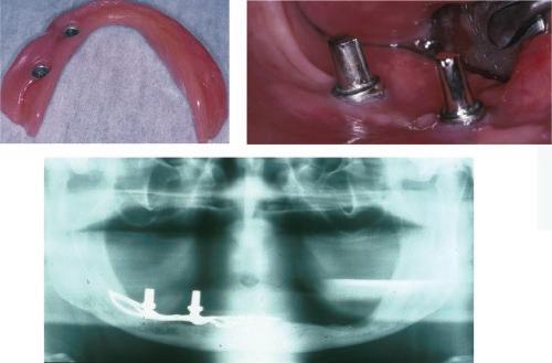
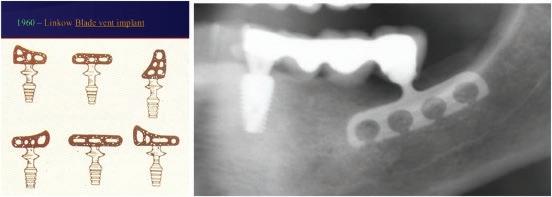
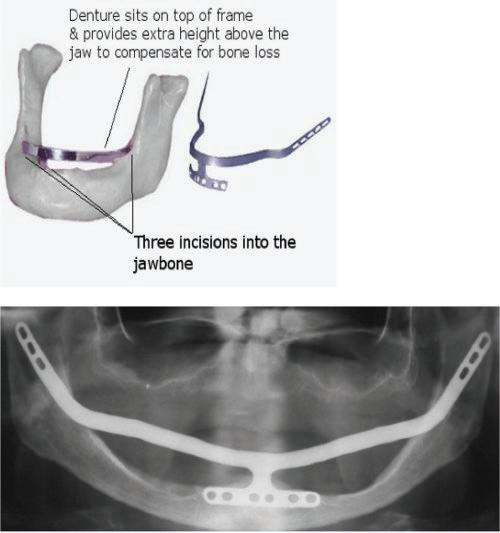
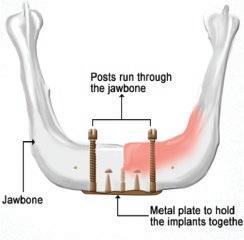
During 1939–60s the concept of the ‘in the bone’ (endosteal) implant arose with the first cylindrical endo-osseous solid screw implant with threads both internally and externally with a smooth gingival collar and healing cap being placed. Following this during the 1940s, a spiral stainless steel post type endosseous implant with a design that allowed bone to grow into the implant emerged and Dahl in Germany, around the same time, introduced the concept of the subperiosteal implant with mucosal inserts (Figure 1.1). This implant was made of cobalt-chromium molybedenum with a direct impression of the struts on the ridge crest taken to construct the denture. Throughout the 1940s–50s variations on the original Dahl design emerged in an attempt to make the provision of implants simpler and included the use of vitallium implants in 1948, the Linkow endoosseous blade vent implant in 1966 with different designs for the maxilla and mandible (Figure 1.2), the ramus frame implant in 1970, made of stainless steel (Figure 1.3) and mandibular transossteal implant which engaged the lower border of the mandible with inserts projecting into the mouth to support a prosthesis (Figure 1.4). The ramus frame and tranossteal implants were predominantly designed for patients with atrophic mandibles who had difficulties wearing dentures and were used to aide denture retention to improve function.
The key challenge with these older implant systems was biocompatibility, the lack of fusion to the jawbone resulting in recurring infections after a period of time and the complex surgical techniques needed to insert the implants leading to limited use aimed at
Dental Implants for Hygienists and Therapists. Ulpee Darbar BDS, MSc, FDS(Rest Dent)RCS, FHEA
© 2022 John Wiley and Sons Ltd
ISBN: 1
Table 1.1 Progressive Development of Methods used for Tooth Replacement.
500–2500 BC 300–600 AD 800 AD 1500–1800s 1809 1913
– Egyptians tried splinting teeth using gold ligature wires – Eustracians used customised soldered gold bands from animals and oxen bone
– Phoenicians used Ivory to carve teeth used as bridge replacements – Mayans introduced the concept of implants when they tried to use ‘Pieces of Shells’ as implants to replace mandibular teeth; Radiographs taken in the 1970s of such mandibles show compact bone formation around the implants (bone similar to that around blade implants)
Hondurans used a stone implant and placed this in the mandible Europeans used cadaver teeth for allotransplantation J Maggiolo inserted a gold implant tube into a fresh extraction socket and after healing a crown was added; other materials used were silver capsules, corrugated porcelain
Dr Greenfield placed a ‘24-gauge hollow latticed cylinder of iridioplatinum soldered with 24-karat gold’ as an artificial root to ‘fit exactly the circular incision made for it in the jawbone of the patient’
Figure 1.1 Subperiosteal implants in the mouth.
Figure 1.2 Blade vent implants.
Figure 1.3 Ramus implants.
specific patient groups. Additionally, the infections led to secondary issues with bone resorption compounding the existing issues.
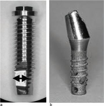
In the 1950s, an orthopaedic surgeon, Per Ingvar Branemark, accidentally found, during the study of bone healing and regeneration, that a titanium cylinder fused together with the bone in the femur of a rabbit. He hypothesised that this fusion could be utilised in field of dental implants and placed the first dental implant made of commercially pure titanium in a human volunteer in 1965. This finding introduced the concept of osseointegration which forms the basis of today’s endo-osseous dental implants. Osseointegration became accepted as a worldwide phenomenon when the concept was launched at the Toronto World Conference in 1982. At around the same time, whilst Branemark was looking at a two-stage threaded implant, Schroeder and his group were independently evaluating the use of a one-piece root form implant made with a hollow design and a roughened surface (Figure 1.5a, b).
Since the introduction of osseointegration, in the late 1980s, as a predictable method of tooth replacement, growing confidence and predictability has led to the widespread use of dental implants moving from edentulism to partial edentulism including single teeth and those with extensive tissue and tooth loss usually seen in patients who have suffered traumatic injuries and congenital anomalies (e.g. Hypodontia). This progressive change has led to the focus changing from improving function to include aesthetics and psychological well-being alongside the need to address patient expectations.
Dental implantology continues to evolve with concomitant modification of implant screw designs, surfaces and techniques used for implant placement and restoration aimed at reducing integration healing times, optimising function and aesthetics alongside predictability. These changes have led to newer concepts for tooth replacement being considered which include the use of zygomatic implants in those with atrophic maxillae, the mini implants and the ‘All-on-Four’ concept whereby the teeth are extracted and implants placed and restored all on the same day. Additionally, the advent
Figure 1.4 Mandibular tranossteal implant.
of digital technology has enabled clinicians and technicians to push this clinical envelop even further with digital systems being used for planning, surgical placement and restoration without any analogue interfaces being used. Whilst, we live in a fast-moving world driven by technology and systems geared to meet patient demands, the biological envelop in which we as clinicians have to work has seen little change and as clinicians we need to be cognizant of this challenge commonly referred to as ‘patient and site’ related factors.
Today there are in excess of 250 implant systems on the market with varying design features, many of which resemble either one or more features of the eight mainstream implant systems. Table 1.2 shows the development of different key implant systems since 1982.
Key Learning Points
● Be able to describe the older systems, as patients may attend for treatment with these systems
● Being able to recognise the older systems to assist with management
● Be able to explain to patients possible problems and issues with infections
● Be aware of challenges associated with evolution of the concept of dental implants
Figure 1.5 a, b: The Branemark two-piece implant fixture and the Shroeder one-piece hollow cylinder implant.
Table 1.2 Some of the Mainstream Dental Implant Systems.
1977 Branemark Implants
1982 Launch of Osseointegration
1982 Non-submerged implant system: ITI
Corevent implant system
1985 Biocon
1987 IMZ
1989 3i
1990 Astra
1999 Straumann Synocta
Late 1990s Frident (Frialit 2, Xive)
Early 2000
Mid-2000 onwards
References
Ankylos and similar
Modification of the earlier implant systems with newer surfaces, shapes and designs
1 Abraham, C. (2014). A brief historical perspectice on dental implants, their surface coatings and their treatments. Open Dentistry Journal 8: 50–55.
2 Branemark, P.I. and Zarb, G. (1985). Tissue Integrated Prosthesis: Osseointegration in Clinical Dentistry. Quintessence Publishing.
3 Rajput, R., Chouhan, Z., Sindhu, M., Sundararajan, S., and Chouhan, R. (2016). A brief chronological review of dental implant history. International Dental Journal of Student's Research 4 (3): 105–107.
2
Osseointegration
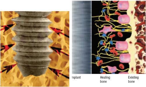
Osseointegration (OI) was first defined at the light microscopic level as a ‘direct structural and functional connection between ordered living bone and the surface of a load carrying implant’ (Branemark 1983). The phenomenon involves a complex interaction between the implant, bone and tissue interface (Figure 2.1). The process involves two stages with an initial biomechanical interlocking between the implant body and alveolar bone, called primary stability, followed by the second stage which involves the biological fixation of the implant through bone remodelling and apposition, called secondary stability, with each stage being influenced by various factors and steps being followed during the placement of the implant (Figure 2.2). Primary implant stability plays an essential role in successful osseointegration and is dependent on the implant geometry, the bone quality and quantity as well as the site preparation technique.
Concept
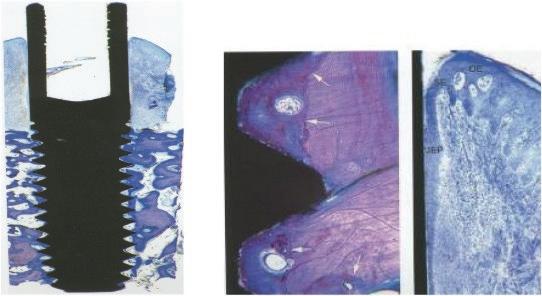
The original concept of OI was redefined at the clinical level by Zarb and Albrektsson (1983) as a ‘time dependent healing process whereby clinically asymptomatic rigid fixation of alloplastic materials is achieved and maintained in bone during functional loading’. The histologic appearance resembled a functional ankylosis with no intervening fibrous or connective tissue between bone and implant surface (Figure 2.3). The first implants, known as ‘Branemark Implants’, were made of pure machined titanium surfaces and had to be covered over by the gum flap after surgical placement for at least six months to allow the screw to integrate into the jaw bone. Today with an improved understanding of the concept and changes in implant screw designs and surface modifications, the healing times have reduced with implants being restored at the same time as placement with healing periods of 0–8 weeks. Figure 2.4 shows the different healing time lines for dental implants and their restorations. The original clinical definition of osseointegration thus needs updating with removal of the word ‘time dependent’ as a fixed period of healing before loading is no longer an absolute requirement.
Dental Implants for Hygienists and Therapists. Ulpee Darbar BDS, MSc, FDS(Rest Dent)RCS, FHEA
© 2022 John Wiley and Sons Ltd
ISBN: 1
Stabilit y (per cent)
Primar y Stability (old bone)
Secondar y Stability (new bone)
Implant-related
Figure 2.2 Primary (mechanical) stability vs secondary (biological) stability.
Factors Affecting Osseointegration
Factors affecting osseointegration can be divided into implant related and environment related (Table 2.1).
● Implant-related factors:
These factors primarily affect the mechanical interlocking of the implant and are thus fundamental to facilitating OI. They include the material of which the implant is made (biocompatibility), the design features of the implant and the macroscopic and
Figure 2.1 Bone to implant contact in osseointegration.
Figure 2.3 Osseointegration showing the close interface of the implant surface to bone with no intervening soft tissue.
Implant Placement
Immediate (same day as tooth extraction)
Delayed Early (6‐8 weeks after extraction)
Delayed Late (12 weeks after extraction)
Standard
(6 months or longer after extraction)
‐Immediate Restoration (same time as placement) ‐Delayed Restoration (8‐12 weeks after placement)
‐Immediate Restoration (same time as placement) ‐Delayed Restoration (8‐12 weeks after placement)
‐Immediate Restoration (same time as placement) ‐Delayed Restoration (8‐12 weeks after placement)
Figure 2.4 Healing time lines for implant placement and restoration.
microscopic surface features of the implant. Progressive changes in these features have enabled reduced healing periods as a result of the improved primary stability at the time of implant placement.
– Implant Materials
The material of which the implant is made does not directly influence the mechanical stability of the implant but ensures biocompatibility.
– Commercially pure titanium (cpTI):
This material has excellent biocompatibility, and its bioactivity is related to the immediate formation of a stable and inert oxide layer on its outer surface when exposed to air. It is classified into grades 1–4 depending on the purity and processing oxygen content with Grade 1 being the most pure with the lowest oxygen content (0.18%) and Grade 4 being the least pure with the highest oxygen content (0.4%). The different grades contain varying amounts of contaminants such as iron for corrosion
Table 2.1 Factors Affecting Osseointegration.
Implant-Related
Mechanical Stability = Primary Stability (Initial Period)
Materials (Biocompatiblity)
● Commercially pure titanium
● Titanium alloys
● Zirconium
Design
● Shape
● Threads
Surface
● Machined (turned)
● Plasma sprayed/laser
● Sandblasted
● Acid etched
● Anodised
● Coated
● Chemically treated
Diameter and Length
● Narrow, regular, wide
● Short, standard, long
Environment (Host)-Related Biological Stability = Secondary Stability
Status of the Host Bed
● Vascularity
● Quality of bone : Type I, II, III, IV
Surgical Site Preparation
● No overheating
● Ample cooling
● Technique
● Sequential drilling
Healing Periods
● Influenced by above and need for augmentation
● Post-operative trauma
● Smoking
● Aftercare
● Prosthesis
Loading Conditions
● Immediate
● Early
● Delayed
● Standard
Influenced by Operator Related Knowledge, Skill and Competence
resistance, aluminium for increased strength and reduced density and vanadium which scavenges the aluminium thus imparting corrosion resistance. Grade 4 cpTI has the highest strength, highest passivity and a modulus of elasticity comparable to bone and is therefore the material of choice for manufacturing dental implants. Its key disadvantages are the low wear resistance and grey metal shine through in patients with thin gingival tissues contributing to compromised aesthetics.
– Titanium alloys:
The alloying of cpTI enhances the strength and wear resistance of the material.
– Titanium, Aluminium and Vanadium Alloy (Ti6Al4V):
This alloy is mainly used for dental implants and contains 6% aluminium, 4% vanadium, up to 0.25% iron and 0.2% oxygen with the rest being titanium. The material has excellent fatigue properties and corrosion resistance with a low elastic modulus leading to a ‘stress shielding’ effect due to the mismatch between the implant material and surrounding
bone. The alloy releases small amounts of aluminium and vanadium ions and can be associated with allergic reactions, cytotoxic effects and neurological disorders. Implant systems made of titanium alloys usually tend to be slightly cheaper than those using cpTI.
– Titanium-Zirconium Alloy (Ti-Zr):
This alloy has been shown to be biocompatible, bioactive and mechanically stable and has comparable properties to cpTi with a reported tendency to significantly improve osteoblast adhesion to the implant surface. The ‘Roxolid’ implants manufactured by the Straumann Company are made of this alloy and have been used for more than five years with good success rates. The strength of the material enables smaller implant diameters to be made with internal connections.
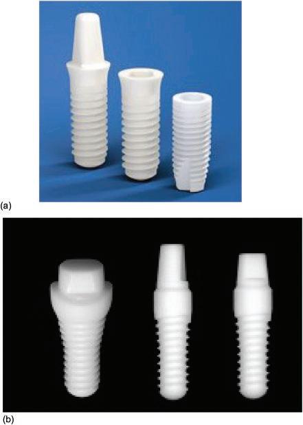
● Zirconium/Ceramic
Zirconium, a ceramic biocompatible biomaterial, has gained popularity as an implant material due to its aesthetics and was first tested during the 1990s as an implant material. The oxide form of zirconium has the capacity to create OI of a lesser quality and etching techniques have been used to improve the quality and extent of the OI. There are two zirconium implants available on the market. The ‘Ceraroot’ implant is an acidetched zirconium implant (Oral Iceberg) and Straumann Pure ZLA is also an acidetched ceramic implant (Figure 2.5a, 2.5b).
● Implant Design
The design of an implant facilitates the primary stability during the placement of the implant and includes the shape and thread configuration of the implant. Other parameters such as diameter and length are also important and influence primary stability indirectly.
– Implant shape
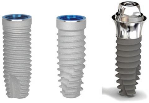
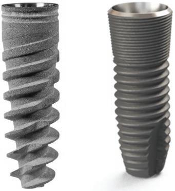
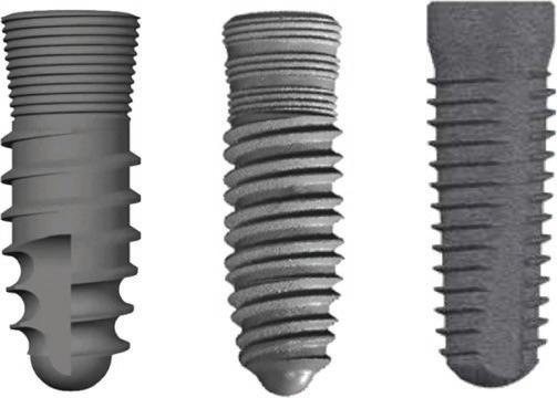
The implant shape ensures there is a good approximation between the bone and implant and facilitates force distribution to the bone. Three shapes, cylindrical, parallel sided and tapered, have been used with the cylindrical form implants manufactured as unthreaded implants inserted using a ‘press-fit’ action into the prepared site (Figure 2.6). These implants are no longer available due to compromised treatment outcomes. Parallel-sided and tapered/conical screw (also called threaded) form implants have been used with the former being more popular until recently where hybrid designs combining the features of both these shapes becoming more widely used (Figure 2.7). These hybrid implants are designed to increase the contact area between the implant and bone as well as relieving stress concentrations due to the different thread designs thereby improving the stability enabling reduced healing times (Figure 2.8). Most implant systems have introduced these ‘hybrid’ versions into their portfolio whilst still retaining the standard implant designs.
● Implant threads
The threads on a implant screw determine the degree to which the stability of the implant when placed is achieved and also facilitates the stress distribution to the bone. The degree of the stability achieved will be dependent on the profile and configuration of the threads called the thread depth and pitch (Figure 2.9). Screw form implants have threads that are
designed to engage into the alveolar bone thereby improving primary stability and the sharper the thread pitch the better the stability. This is a feature that has been introduced into most implants systems as the surgical provision of implants has moved towards immediate and delayed placement. Figure 2.10 shows the different thread configurations and shapes which influence the stress concentration on the bone. Most implant systems adopt a combination of thread designs incorporating microthreads coronally with a tapering thread profile, double and triple thread configurations and increased thread width, pitch and depth. The use of microthreads coronally is deemed to increase the contact between the coronal bone and implant thus reducing the risk of coronal bone loss whereas, changes in thread pitch and depth are aimed at providing faster penetration into the bone thereby reducing risk of trauma to the bone. These changes in design have enabled improved primary stability thus contributing to faster osseointegration and healing (Figure 2.11a, 2.11b).
Figure 2.5 a. b: Zirconium implants. a: Straumann. b: Ceraroot.
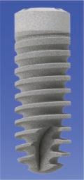
Figure 2.6 Cylindrical implants used with a press fit action for placement (IMZ implant with TPS-coated surface).
Figure 2.7 Parallel-sided and tapered implants.
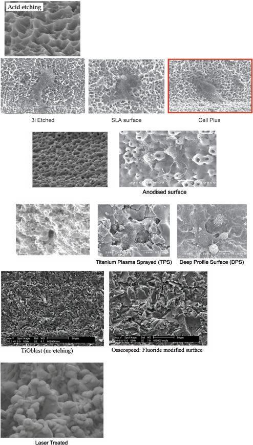
● Implant Surfaces
Surface topography of the implant influences the attraction of the osteoblasts to the implant surface with rough surfaces promoting osteogenesis by increasing the surface area over which the osteogenic cells can adhere and cellular activity takes place. The surface roughness can be described at the macroscopic (few mm to microns), microscopic (1–10microns) and nanometric (1–100 nm) level with the different levels having a profound effect on cellular healing accelerating the migration and proliferation of osteoblasts thereby improving the rate of osseointegration. Machined surface implants also
2.8 Hybrid-designed implant (parallel sided at the top; tapered apically).
Figure 2.9 a, b: Different implant thread configurations. a: The machined implant on the right was the first implant and the change in the shape and design as well as the thread pitch is evident as you go from left to right. b: Different thread shapes and pitch (macrodesign).
known as ‘turned’ surfaces were the first to be used and whilst at a macroscopic level, the surface looked smooth, under the microscope the processing of the implants still created some irregularities on the surface. The irregularities increase the surface area and different techniques have been used with these implants being largely superseded by implants with different surface topographies. The two main methods used to modify the implant surface are either by removing material on the surface or adding material on the surface. The former include acid etching, blasting with an abrasive material and treatment with
Figure
Figure 2.10 Shows the progressive change in an implant thread design: The implant on the left shows the change in the depth and pitch of the threads, and the implant on the right shows the microthreads at the top aimed at improving the contact with the coronal bone.
lasers whereas the latter includes hydroxyapatite coating and titanium plasma spraying, oxidation or anodization and deposition of nanoparticles by physical or chemical methods. Figure 2.12 shows the different surface topography and modifications associated with these methods. Plasma spraying and laser treatments are no longer used as the rate of integration was similar to that of the machined surfaces; however, laser treatment to promote gingival tissue adhesion, called the ‘laser lock’ has been used as a feature in one of the implant systems who state that this locking mechanism leads to an integration of the gingival tissue fibres to the surface. Table 2.2 shows the different generation of dental implants categorised according to the surface roughness.
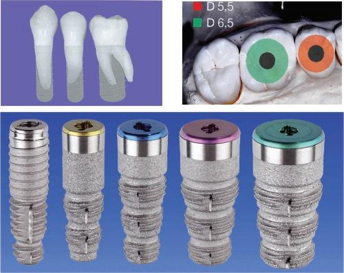
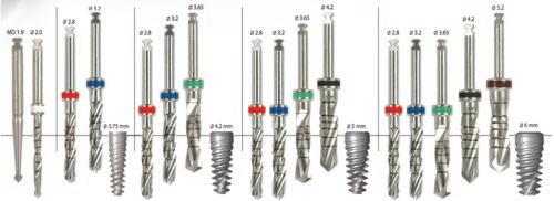
● Implant Diameter and Length
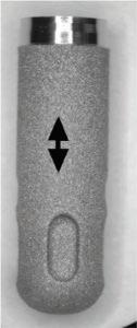
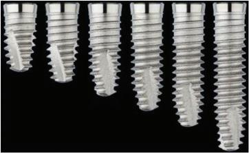
Implant diameters have changed over the years to enable improved profiles of the restorations. Early dental implants were available in one diameter (3.75 mm); however, as different types of teeth started being replaced with implants, the need for different diameters became evident to avoid ‘mushroom’ designs of the prosthesis. Implant diameters, today, fall into three groups, narrow (2.8–3.4 mm diameter), regular (3.75–4.1 mm) and wide (5.00–6 mm) with each group matching the type of tooth being replaced thus enabling the appropriate emergence profile of the restoration to maintain gingival tissue health (Figure 2.10) with each diameter group being colour coded enabling the correct components to be used. Implant lengths of up to 20 mm were advocated to achieve bicortical stabilisation where the border of the jaw bone and the alveolar crest was engaged to obtain stability. This concept has now evolved with the modified implant surfaces promoting healing with the average implant length being 11–13 mm. Whilst
previously, the shortest implant fixture advised was 7 mm, today the use of even shorter implant fixtures with lengths of 4–6 mm have become available as sites with compromised bone height are being considered for implant placement thus avoiding the need for advanced surgical interventions (Figure 2.13a, b). The use of the different diameters and lengths should be based on the outcome of the treatment plan agreed and finalised with the patient.
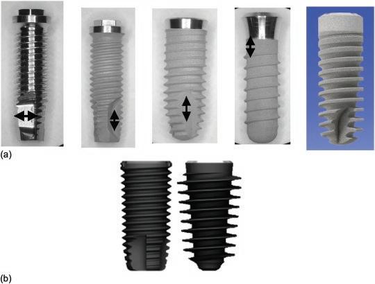
Figure 2.11a The progressive change in the implant shape and thread profiles.
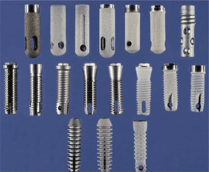
Figure 2.11b Range of different implants.
Figure 2.12 Electron Micrographs of the different surface topographies; acid etch, grit blasted, grit blasted and acid etched, anodised, plasma sprayed and laser treated. The SLA surface if both acid etched and grit blasted.
Table 2.2 Different Generations of Implant Systems with the Surface Modifications.
Generation Surface Treatment
Type of System
First None Blades, subperiosteal, ramus, transosteal
Implant System
Second Weak Machined (turned) Old Branemark (Nobelbiocare); Titanium plasma sprayed (TPS) Old Straumann
Coated Old IMZ
Third Strong response Moderately roughened surfaces
Sandblasted and etched Straumann SLA
Dentsply Frialit and Friadent
Etched Biomet 3i
Anodised Nobelbiocare TiUnite
Blasted
Laser ablation
Dentsply Astra TioBlast
Biohorizons Laser Lok
Fourth Optimal response Chemically active Straumann SLA Active Fluoride coated Dentsply Astra
Calcium phosphate coating Biomet 3i
Bioactive glasses and bisphosphonates
Experimental
Figure 2.13a Implant diameters to match the type of tooth being replaced giving a better emergence profile for the restoration.
● Environment-Related Factors
These factors influence the second phase of the osseointegration process during which the secondary stability of the implant is achieved. They include the status of the host bed, the surgical technique, healing period and loading conditions which determine the vascularity and the stability of the blood clot during the initial healing phase.
– Status of the host bed
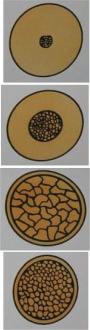
The host bed is the alveolar bone and the surrounding soft tissues into which the implant is placed. The health of these tissues is crucial in determining how well the implant will integrate into the bone. Once the implant is placed, good vascularity (bleeding into the site) is needed to facilitate blood clot formation and remodelling of the cells into bone forming cells. In sites where the blood supply is compromised or reduced, the rate of osseointegration will be compromised due to the lack of bone-forming cells. The vascularity of the bone is dependent on the bone quality which is assessed by the ratio of the cortical to the cancellous bone and is described as Type 1, which is dense with little cancellous bone and Type IV which is soft with predominantly cancellous bone (Figure 2.14). Type 1 bone has poor vascularity due to the reduced number of bone cells and specialised techniques using a ‘screw tap’ are needed during the site preparation to minimise the risk of bone necrosis caused by pressure and overheating during placement. Type 4 bone is like ‘quick sand’ and has good vascularity but poor density and thus modifications, such as using specialised tools, e.g. bone condensers, during site preparation are needed to ensure that primary stability of the implant is achieved. Any movement of the implant or the blood clot during the initial healing period will lead to fibrous tissue formation and loss of osseointegration. Smoking affects the vascularity of the soft tissues and the bone and thus has a negative outcome on healing and therefore the quality of osseointegration but has also been associated with a higher risk of complications.
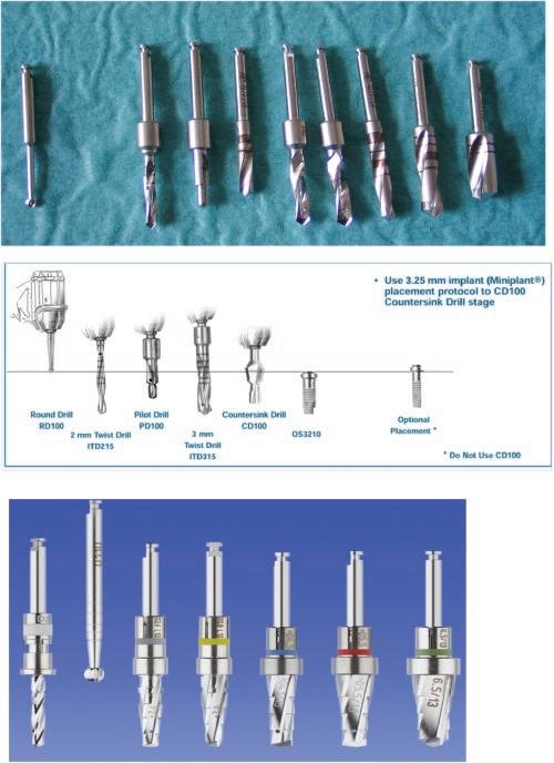
● Surgical Technique
The surgical technique is of paramount importance in determining predictability of osseointegration ensuring that the site is not overheated to more that 47oC during site preparation. This is achieved by the use of sharp drills, used sequentially at recommended speeds ranging from 800rpm to 1200 rpm depending on the implant system
Figure 2.13b Different implant lengths.