
Nanomaterials and Nanotechnology in Medicine Visakh P. M.
Visit to download the full and correct content document: https://ebookmass.com/product/nanomaterials-and-nanotechnology-in-medicine-visak h-p-m/
More products digital (pdf, epub, mobi) instant download maybe you interests ...
Nanotechnology in Plant Growth Promotion and Protection
Avinash P. Ingle (Ed.)
https://ebookmass.com/product/nanotechnology-in-plant-growthpromotion-and-protection-avinash-p-ingle-ed/

Nanotechnology in the Automotive Industry Ghulam Yasin
https://ebookmass.com/product/nanotechnology-in-the-automotiveindustry-ghulam-yasin/
Polyvinyl Alcohol-Based Biocomposites and Bionanocomposites 1st Edition Visakh P.M.
https://ebookmass.com/product/polyvinyl-alcohol-basedbiocomposites-and-bionanocomposites-1st-edition-visakh-p-m/
Nanotechnology in Fuel Cells Huaihe Song
https://ebookmass.com/product/nanotechnology-in-fuel-cellshuaihe-song/

Large Animal Internal Medicine, 6th Edition Bradford P. Smith
https://ebookmass.com/product/large-animal-internal-medicine-6thedition-bradford-p-smith/

Hazzard's Geriatric Medicine and Gerontology, 8th ed 8th Edition Kevin P. High
https://ebookmass.com/product/hazzards-geriatric-medicine-andgerontology-8th-ed-8th-edition-kevin-p-high/
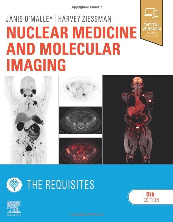
Nuclear Medicine and Molecular Imaging: the Requisites 5th Edition Janis P. O'Malley
https://ebookmass.com/product/nuclear-medicine-and-molecularimaging-the-requisites-5th-edition-janis-p-omalley/
Applications of Nanotechnology in Drug Discovery and Delivery Chukwuebuka Egbuna
https://ebookmass.com/product/applications-of-nanotechnology-indrug-discovery-and-delivery-chukwuebuka-egbuna/
Micro and Nanotechnology in Vaccine Development 1st Edition Mariusz Skwarczynski
https://ebookmass.com/product/micro-and-nanotechnology-invaccine-development-1st-edition-mariusz-skwarczynski/
Nanomaterials and Nanotechnology in Medicine
Nanomaterials and Nanotechnology in Medicine
Edited by
Dr. P.M. Visakh
Assistant Professor, Department of Physical Electronics
TUSUR University, Tomsk, Russia
This edition first published 2023
© 2023 John Wiley & Sons Ltd
All rights reserved. No part of this publication may be reproduced, stored in a retrieval system, or transmitted, in any form or by any means, electronic, mechanical, photocopying, recording or otherwise, except as permitted by law. Advice on how to obtain permission to reuse material from this title is available at http://www.wiley.com/go/permissions.
The right of P.M. Visakh to be identified as the author of the editorial material of this work has been asserted in accordance with law.
Registered Offices
John Wiley & Sons, Inc., 111 River Street, Hoboken, NJ 07030, USA
John Wiley & Sons Ltd, The Atrium, Southern Gate, Chichester, West Sussex, PO19 8SQ, UK
Editorial Office
The Atrium, Southern Gate, Chichester, West Sussex, PO19 8SQ, UK
For details of our global editorial offices, customer services, and more information about Wiley products visit us at www.wiley.com.
Wiley also publishes its books in a variety of electronic formats and by print-on-demand. Some content that appears in standard print versions of this book may not be available in other formats.
Limit of Liability/Disclaimer of Warranty
In view of ongoing research, equipment modifications, changes in governmental regulations, and the constant flow of information relating to the use of experimental reagents, equipment, and devices, the reader is urged to review and evaluate the information provided in the package insert or instructions for each chemical, piece of equipment, reagent, or device for, among other things, any changes in the instructions or indication of usage and for added warnings and precautions. While the publisher and authors have used their best efforts in preparing this book, they make no representations or warranties with respect to the accuracy or completeness of the contents of this work and specifically disclaim all warranties, including without limitation any implied warranties of merchantability or fitness for a particular purpose. No warranty may be created or extended by sales representatives, written sales materials or promotional statements for this work. This work is sold with the understanding that the publisher is not engaged in rendering professional services. The advice and strategies contained herein may not be suitable for your situation. You should consult with a professional where appropriate. Neither the publisher nor author shall be liable for any loss of profit or any other commercial damages, including but not limited to special, incidental, consequential, or other damages. The fact that an organization, website, or product is referred to in this work as a citation and/or potential source of further information does not mean that the publisher and authors endorse the information or services the organization, website, or product may provide or recommendations it may make. Further, readers should be aware that websites listed in this work may have changed or disappeared between when this work was written and when it is read. Neither the publisher nor authors shall be liable for any loss of profit or any other commercial damages, including but not limited to special, incidental, consequential, or other damages.
Library of Congress Cataloging-in-Publication Data
Names: P. M., Visakh, editor.
Title: Nanomaterials and nanotechnology in medicine / edited by Dr. P.M. Visakh, Assistant Professor, Department of Physical Electronics, TUSUR University, Tomsk, Russia.
Description: Hoboken, NJ : Wiley, 2022. | Includes bibliographical references and index.
Identifiers: LCCN 2022027360 (print) | LCCN 2022027361 (ebook) | ISBN 9781119558033 (hardback) | ISBN 9781119558040 (adobe pdf) | ISBN 9781119558095 (epub)
Subjects: LCSH: Nanomedicine. | Nanotechnology. | Nanostructured materials–Therapeutic use. | Medical technology.
Classification: LCC R857.N34 N25643 2022 (print) | LCC R857.N34 (ebook) | DDC 610.285–dc23/eng/20220802
LC record available at https://lccn.loc.gov/2022027360
LC ebook record available at https://lccn.loc.gov/2022027361
Cover Design: Wiley
Cover Image: © Volodymyr Horbovyy/Shutterstock
Set in 9.5/12.5pt STIXTwoText by Straive, Pondicherry, India
Contents
List of Contributors xi
Preface xvii
1 Nanomaterials and Nanotechnology in Medicine: Medical Applications: Challenges and Opportunities 1
P. M. Visakh
1.1 Nanoneurology 1
1.2 Nanomolecular Diagnostics 3
1.3 Nanopharmaceuticals 4
1.4 Role of Nanotechnology in Biological Therapies 6
1.5 Nanomaterials for Gene Therapy 8
1.6 Nanotools for the Treatment of Ocular Diseases 10
1.7 Nanotechnology Applications in Food and Nutrition Science 11
1.8 Rubber Nanocomposites for Biomedical Applications 13
References 15
2 Nanoneurology 27
Liron L. Israel and Eggehard Holler
2.1 Introduction and Recent Advances 27
2.2 Types of Nanomaterials 30
2.3 Nanomaterial Applications for Neurodegenerative Diseases 32
2.4 Nanomaterial Applications for Strokes 38
2.5 Nanomaterial Applications for Spinal Cord Injuries 41
2.6 Nanomaterial Applications for Brain Tumors 44
2.7 Adverse Effects of Nanomaterials 50
2.8 Regulatory Issues 53
2.9 Conclusions 54
References 54
3 Nanomolecular Diagnostics 65 Anila Fariq, Ayesha Selhaba, Anum Zulfiqar, and Azra Yasmin
3.1 Introduction 65
3.2 Nanodiagnostics 67
3.3 Nanoparticles for Molecular Diagnostics 68
3.4 Applications of Nanoparticles for Molecular Diagnostics 75
3.5 Comparison Between Nanomaterials and Other Materials in Molecular Diagnostics 79
3.6 Prospects of Nanodiagnostics 79
3.7 Regulatory Issues 80
3.8 Conclusion 81
References 82
4 Nanopharmaceuticals 87
David Quintanar-Guerrero, Gerardo Leyva-Gómez, Nancy Evelyn Magaña Vergara, and Néstor Mendoza Muñoz
4.1 Introduction 87
4.2 Liposomes in Nanopharmaceuticals 89
4.3 Polymeric Nanoparticles in Nanopharmaceuticals 93
4.4 Solid Lipid Nanoparticles in Nanopharmaceuticals 99
4.5 Dendrimers in Nanopharmaceuticals 101
4.6 Quantum Dots in Nanopharmaceuticals 104
4.7 Regulatory Issues 106
4.8 Conclusion 109
References 109
5 Role of Nanotechnology in Biological Therapies 115
Blanca Ocampo-García, Liliana Aranda Lara, Guillermina Ferro-Flores, Enrique Morales-Avila, and Keila Isaac-Olivé
5.1 Introduction 115
5.2 Biological Therapies 116
5.3 Nanoparticles in Biological Therapies 118
5.4 Application of Nanotechnology in Biological Therapies 131
5.5 Advantages and Disadvantages of Nanoparticles in Biological Therapies 136
5.6 Conclusion 137
References 137
6 Nanomaterials for Gene Therapy 153
V. Karthik, A. Vigneshwaran, D. Dharun Daniel Raj, T.G.N. Nagarjun, S. Poornima, R. Subbaiya, and M. Saravanan
6.1 Introduction and Recent Advances 153
6.2 Nanomaterials and their Physicochemical Properties 153
6.3 Methods of Characterizing the Physicochemical Properties of Nanomaterials 154
6.4 Target Organ Biocompatibility/Toxicity 155
6.5 Gene Delivery 158
6.6 Regulatory Issues 163
References 163
7 Nanotools for the Treatment of Ocular Diseases 169
Elisa J. Campos, António Campos, João Martins, and António Francisco Ambrósio
7.1 Introduction 169
7.2 Ocular Anatomy 170
7.3 Physiological Barriers in the Eye 171
7.4 Methods of Ocular Disease Treatment 173
7.5 Nanomedicine in Ocular Therapy 174
7.6 Closing Remarks 180 Conflict of Interest 181
References 181
8 Nanotechnology Applications in Food and Nutrition Science 185
Kobra S. Rizi, Majid Rezayi, Ehsan Aryan, Zahra Meshkat, and Majid G. Mobarhan
8.1 Introduction 185
8.2 Nanostructured Delivery Systems 186
8.3 Nanoparticles Based on Inorganic Materials 202
8.4 Metal Nanoparticles 204
8.5 Conclusion 205
References 206
9 Rubber Nanocomposites for Biomedical Applications 225 Jayalatha Gopalakrishnan
9.1 Introduction 225
9.2 Rubbers for Biomedical Applications 226
9.3 Rubber-based Nanocomposites 229
9.4 Conclusions 246
References 246
10 Nanomaterials and Nanotechnology in Medicine: Materials Development: Challenges and Opportunities 251 P. M Visakh
10.1 Nanomaterials and Scaffolds for Tissue Engineering and Regenerative Medicine 251
10.2 Nanorobotics in Nanomedicine 253
10.3 Nanosensors 255
10.4 Inorganic Nanoparticles for Drug-delivery Applications 257
10.5 Intelligent Nanomaterials for Medicine 259
10.6 Polymer-based Nanocomposites for Biomedical Applications 261
10.7 Toxicity of Nanomaterials 263
10.8 Multifunctional Nanomaterials for Medical Applications 264
10.9 Antimicrobial Applications of Nanoparticles 265 References 266
11 Nanomaterials and Scaffolds for Tissue Engineering and Regenerative Medicine 279
Saeid Kargozar, Simin Nazarnezhad, Farzad Kermani, and Francesco Baino
11.1 Introduction and Recent Advances 279
11.2 Tissue Engineering and Regenerative Medicine: General Concepts 280
11.3 Implantable Nanomaterials to Regenerate Living Tissues 282
11.4 Nanomaterials as Carriers for Therapeutic Agents 283
11.5 Nanofibrous Scaffolds 285
11.6 Nano-topography Techniques for Tissue-engineered Scaffolds 288
11.7 Regulatory Issues 290
11.8 Conclusion 290 References 291
12 Nanorobotics in Nanomedicine 303
Vaishali Y. Londhe and Rupali S. Bhadale
12.1 Introduction 303
12.2 What is Nanorobotics? 304
12.3 Nanorobotics in Nanomedicines 306
12.4 Nanorobots for Medical Imaging 307
12.5 Nanorobots for Targeted Drug Delivery 309
12.6 Enzymatic Nanolithography 313
12.7 Biomimetic Approach 314
12.8 Cell Biochips 315
12.9 Nanorobots for Precision Surgery 315
12.10 Nanorobots for Detoxification 317
12.11 Fabrication of Nanorobots 318
12.12 Toxicity 321
12.13 Administration and Retrieval 321
12.14 Clinical Presence of Nanorobots 322
12.15 Reproducibility and Standardization 322
12.16 Regulatory Issues 323
12.17 Conclusion 324 References 325
13 Nanosensors 333
Asit Behera, A.K. Sahoo, and S.S. Mohapatra
13.1 Introduction and Recent Advances 333
13.2 Classification of Nanosensors 336
13.3 Nanosensor Fabrication 339
13.4 Inorganic Nanosensors 343
13.5 Biopolymer-derived Nanosensors 346
13.6 Applications 348
13.7 Regulatory Issues 358
13.8 Conclusions 359 References 360
14 Inorganic Nanopar ticles for Drug-delivery Applications 367
Chinnu Sabu, V.K. Ameena Shirin, Renu Sankar, and K. Pramod
14.1 Introduction 367
14.2 Synthesis of Inorganic Nanoparticles 368
14.3 Properties of Inorganic Nanoparticles 371
14.4 Functionalization of Inorganic Nanoparticles 372
14.5 Quantum Dots for Drug Delivery 374
14.6 Drug Delivery by Mesoporous Silica Nanoparticles 376
14.7 Silver Nanoparticles for Drug Delivery 379
14.8 Gold Nanoparticles for Drug Delivery 383
14.9 Superparamagnetic Iron Oxide Nanoparticles for Drug Delivery 385
14.10 Hybrid Systems of Inorganic Nanoparticles 386
14.11 Prospects of Inorganic Nanoparticles 388
14.12 Conclusion 388
References 388
15 Intelligent Nanomaterials for Medicine 401 Ajit Behera and Ranjan K. Mohapatra
15.1 Introduction to Intelligent Nanomaterials 401
15.2 Design and Function of Intelligent Nanoparticles 403
15.3 Various Intelligent Materials for Medicines 405
15.4 Type of Stimuli in Intelligent Nanomaterials in Medicine 415
15.5 Clinical Applications of Intelligent Nanomaterials 416
15.6 Potential Risk Factors in Nanomaterial Application 419
15.7 Summary and Future Prospects 420
References 420
16 Polymer-based Nanocomposites for Biomedical Applications 427
Chander Amgoth, Kaxi Yu, Shuai Chen, Hongzhen Bai, and Guping Tang
16.1 Introduction and Recent Advances 427
16.2 Polymers for Biomedical Fields 428
16.3 Synthesis of Nanocomposites 432
16.4 Characterization Tools for Nanocomposites 432
16.5 Size, Shape, and Morphology of Nanocomposites 433
16.6 Polymer Nanocomposites for Various Applications 436
16.7 Nanocomposites for Molecular Diagnosis and Biopharmaceutics 437
16.8 Perspectives of Nanocomposites 441
16.9 Conclusion 442
References 442
17 Toxicity of Nanomaterials 447
Elham Abohamzeh, M. Sheikholeslami, and Ahmad Shafee
17.1 Introduction and Recent Advances 447
17.2 Biomedical Applications of Nanomaterials 449
17.3 Biodistribution, Mechanism, and Excretion of Nanomaterials 453
17.4 Toxicity of Nanomaterials 457
17.5 Physicochemical Properties and Toxicity of Nanomaterials 461
17.6 Regulatory Issues 464
17.7 Conclusion 466
References 466
18 Multifunctional Nanomaterials for Medical Applications 479
Jitha S Jayan, Ramya Rajan, Saritha Appukuttan, and Kuruvilla Joseph
18.1 Introduction and Recent Advances 479
18.2 Multifunctional Nanomaterials 481
18.3 Diagnostic Application 483
18.4 Therapeutic Application 485
18.5 External Stimuli-responsive Nanoparticles for Medicinal Applications 487
18.6 Regulatory Issues 498
18.7 Conclusion and Future Perspectives 498
References 499
19 Antimicrobial Applications of Nanopar ticles 517
Moataz A. Elsawy and Mohamed H. Mostafa
19.1 Introduction 517
19.2 Antimicrobial Properties of Nanoparticles 518
19.3 Antimicrobial Applications of Nanoparticles 525
19.4 Conclusions 539
References 540
Index 553
List of Contributors
Elham Abohamzeh
Department of Energy, Materials, and Energy Research Center (MERC) Karaj, Iran
Chander Amgoth Department of Chemistry Zhejiang University Hangzhou, China
António Francisco Ambrósio
University of Coimbra, Coimbra Institute for Clinical and Biomedical Research (iCBR), Faculty of Medicine Coimbra, Portugal and University of Coimbra Center for Innovative Biomedicine and Biotechnology (CIBB), Coimbra, Portugal and
Clinical Academic Center of Coimbra (CACC) Coimbra, Portugal and
Association for Innovation and Biomedical Research on Light and Image (AIBILI) Coimbra, Portugal
V.K. Ameena Shirin College of Pharmaceutical Sciences Government Medical College Kozhikode, Kerala, India
Saritha Appukuttan Department of Chemistry
Amrita Vishwa Vidyapeetham Kollam, Kerala, India
Liliana Aranda Lara Facultad de Medicina Universidad Autónoma del Estado de México
Toluca, Estado de México, Mexico
Ehsan Aryan Antimicrobial Resistance Research Center Department of Medical Bacteriology and Virology, Qaem University Hospital School of Medicine, Mashhad University of Medical Sciences Mashhad, Iran
Hongzhen Bai Department of Chemistry Zhejiang University Hangzhou, China
Francesco Baino Institute of Materials Physics and Engineering
Department of Applied Science and Technology, Politecnico di Torino Torino, Italy
Ajit Behera Department of Metallurgical & Materials Engineering National Institute of Technology Rourkela, Odisha, India
List of Contributors
Asit Behera
School of Mechanical Engineering, KIIT
Bhubaneswar, India
Rupali S. Bhadale
Shobhaben Pratapbhai Patel School of Pharmacy and Technology Management
SVKM’s NMIMS Mumbai, Maharashtra, India
Elisa J. Campos
University of Coimbra, Coimbra Institute for Clinical and Biomedical Research (iCBR), Faculty of Medicine Coimbra, Portugal and
University of Coimbra Center for Innovative Biomedicine and Biotechnology (CIBB) Coimbra, Portugal and
Clinical Academic Center of Coimbra (CACC), Coimbra, Portugal
António Campos
University of Coimbra, Coimbra Institute for Clinical and Biomedical Research (iCBR), Faculty of Medicine Coimbra, Portugal and University of Coimbra Center for Innovative Biomedicine and Biotechnology (CIBB), Coimbra, Portugal and
Clinical Academic Center of Coimbra (CACC) Coimbra, Portugal and
Department of Ophthalmology, Centro Hospitalar Leiria E.P.E., Leiria, Portugal and ciTechCare, Center for Innovative Care and Health Technology, Polytechnic Institute of Leiria, Leiria, Portugal
Shuai Chen
Department of Chemistry
Zhejiang University
Hangzhou, China
D. Dharun Daniel Raj
Department of Biotechnology
Maulana Abul Kalam Azad University of Technology
Haringhata, West Bengal, India
Moataz A. Elsawy
Polymer Laboratory, Petrochemical Department
Egyptian Petroleum Research Institute
Nasr City, Cairo, Egypt
Anila Fariq
Department of Biotechnology
University of Kotli Azad and Jammu Kashmir, Pakistan and
Department of Biotechnology
Fatima Jinnah Women University
Rawalpindi, Pakistan
Guillermina Ferro-Flores
Departamento de Materiales Radiactivos
Instituto Nacional de Investigaciones
Nucleares
Ocoyoacac, Estado de México Mexico
Jayalatha Gopalakrishnan
Department of Polymer Science and Rubber Technology
Cochin University of Science and Technology
Kochi, Kerala, India
Eggehard Holler
Nanomedicine Research Center in the Department of Neurosurgery
Cedars Sinai Medical Center
Los Angeles, CA, USA
Keila Isaac-Olivé
Facultad de Medicina
Universidad Autónoma del Estado de México
Toluca, Estado de México, Mexico
Liron L. Israel
Nanomedicine Research Center in the Department of Neurosurgery
Cedars Sinai Medical Center
Los Angeles, CA, USA
Jitha S. Jayan
Department of Chemistry
Amrita Vishwa Vidyapeetham
Kollam, Kerala, India
Kuruvilla Joseph
Department of Chemistry
Indian Institute of Space Science and Technology
Valiyamala, Kerala, India
Saeid Kargozar
Tissue Engineering Research Group (TERG), Department of Anatomy and Cell Biology
School of Medicine
Mashhad
University of Medical Sciences Mashhad, Iran
V. Karthik
Department of Biotechnology
K.S. Rangasamy College of Technology
Tiruchengode, Tamil Nadu, India
Farzad Kermani
Department of Materials Engineering
Faculty of Engineering
Ferdowsi University of Mashhad (FUM) Mashhad, Iran
Gerardo Leyva-Gómez
Facultad de Química
Universidad Nacional Autónoma de México
Ciudad de México, Mexico
Vaishali Y. Londhe
Shobhaben Pratapbhai Patel School of Pharmacy and Technology Management
SVKM’s NMIMS, Mumbai Maharashtra, India
List of Contributors
Nancy Evelyn Magaña Vergara
Facultad de Ciencias Químicas
Universidad de Colima Colima, Mexico
João Martins
University of Coimbra, Coimbra Institute for Clinical and Biomedical Research (iCBR), Faculty of Medicine Coimbra, Portugal and University of Coimbra Center for Innovative Biomedicine and Biotechnology (CIBB), Coimbra, Portugal and
Clinical Academic Center of Coimbra (CACC) Coimbra, Portugal and
University of Coimbra, Coimbra Institute for Biomedical Imaging and Translational Research (CIBIT), Institute for Nuclear Sciences Applied to Health (ICNAS) Coimbra, Portugal
Néstor Mendoza Muñoz Facultad de Ciencias Químicas, Universidad de Colima Colima, Mexico
Zahra Meshkat
Antimicrobial Resistance Research Center Department of Medical Bacteriology and Virology, Qaem University Hospital School of Medicine
Mashhad
University of Medical Sciences Mashhad, Iran
Majid G. Mobarhan
Metabolic Syndrome Research Center School of Medicine
Mashhad
University of Medical Sciences
Mashhad, Iran
List of Contributors
Mohamed H. Mostafa
Polymer Laboratory, Petrochemical
Department
Egyptian Petroleum Research Institute
Nasr City, Cairo, Egypt
S.S. Mohapatra
Department of Mechanical Engineering
NIT Rourkela, Odisha, India
Ranjan K. Mohapatra
Department of Chemistry
Government College of Enginnering
Keonjhar, Odisha, India
Enrique Morales-Avila
Facultad de Química
Universidad Autónoma del Estado de México
Toluca, Estado de México, Mexico
T.G.N. Nagarjun
Department of Food Technology
K.S. Rangasamy College of Technology Tiruchengode, Tamil Nadu, India
Simin Nazarnezhad
Tissue Engineering Research Group (TERG)
Department of Anatomy and Cell Biology
School of Medicine
Mashhad
University of Medical Sciences Mashhad, Iran
Blanca Ocampo-García
Departamento de Materiales Radiactivos
Instituto Nacional de Investigaciones
Nucleares
Ocoyoacac, Estado de México, Mexico
S. Poornima
Department of Biotechnology
K.S. Rangasamy College of Technology
Tiruchengode, Tamil Nadu, India
K. Pramod
College of Pharmaceutical Sciences
Government Medical College
Kozhikode, Kerala, India
David Quintanar-Guerrero
Facultad de Estudios Superiores Cuautitlán
Universidad Nacional Autónoma de México
Mexico, Mexico
Ramya Rajan
Department of Chemistry
Amrita Vishwa Vidyapeetham
Kollam, Kerala, India
Majid Rezayi
Medical Toxicology Research Center
Mashhad
University of Medical Sciences
Mashhad, Iran and
Department of Medical Biotechnology and Nanotechnology, School of Medicine
Mashhad University of Medical Sciences
Mashhad, Iran
Kobra S. Rizi
Antimicrobial Resistance Research Center
Department of Medical Bacteriology and Virology, Qaem University Hospital School of Medicine
Mashhad
University of Medical Sciences
Mashhad, Iran and
Department of Medical Biotechnology and Nanotechnology, School of Medicine
Mashhad
University of Medical Sciences
Mashhad, Iran
Chinnu Sabu
College of Pharmaceutical Sciences
Government Medical College
Kozhikode, Kerala, India
A.K. Sahoo
School of Mechanical Engineering
KIIT
Bhubaneswar, India
Renu Sankar
College of Pharmaceutical Sciences
Government Medical College
Kozhikode, Kerala, India
M. Saravanan
AMR and Nanotherapeutics Laboratory
Department of Pharmacology
Saveetha Dental College
Saveetha Institute of Medical and Technical Sciences
Chennai, India
Ayesha Selhaba
Department of Biotechnology
University of Kotli
Azad and Jammu Kashmir, Pakistan
Ahmad Shafee
Public Authority of Applied Education
& Training, College of Technological
Studie
Applied Science Department
Shuwaikh, Kuwait
M. Sheikholeslami
Department of Mechanical Engineering
Babol Noshirvani University of Technology
Babol, Iran
List of Contributors xv
and
Renewable energy systems and nanofluid applications in heat transfer Laboratory
Babol Noshirvani University of Technology
Babol, Iran
R. Subbaiya
Department of Biological Sciences
School of Mathematics and Natural Sciences
The Copperbelt University
Riverside, Kitwe, Zambia
Guping Tang
Department of Chemistry
Zhejiang University
Hangzhou, China
P. M. Visakh
Department of Physical Electronics
TUSUR University
Tomsk, Russia
A. Vigneshwaran
School of Biosciences and Technology
Vellore Institute of Technology
Vellore, Tamil Nadu, India
Azra Yasmin
Department of Biotechnology
Fatima Jinnah Women University
Rawalpindi, Pakistan
Kaxi Yu
Department of Chemistry
Zhejiang University
Hangzhou, China
Anum Zulfiqar
Department of Biotechnology
Fatima Jinnah Women University
Rawalpindi, Pakistan
Preface
This book, Nanomaterials and Nanotechnology in Medicine, summarizes recent research accomplishments in the area of nanomedicine. It discusses topics such as: challenges and opportunities, nanoneurology, nanomolecular diagnostics, nanopharmaceuticals, the role of nanotechnology in biological therapies, nanomaterials for gene therapy, nanomaterials in ophthalmology/ocular diseases, nanonutrition, rubber-based nanocomposites for biomedical applications etc. The book is intended to provide a valuable reference source for university and college faculties, professionals, post- doctoral research fellows, senior graduate students, researchers, and medical doctors. The various chapters in this book are written by prominent researchers from industry, hospitals, academia, and government and private research laboratories across the globe. It provides an up-to-date record of the major findings and observations in the field of nanomedicine.
Chapter 1 provides an overview of the main points of the various chapters by focusing on scope, state-of-the-art, applications, new challenges and opportunities of nanomedicine. The second chapter describes important applications of nanoneurology. In this chapter the author introduces the topic and discusses recent advances in nanomaterials and their application in: neurodegenerative diseases; nanomaterial application for stroke; nanomaterial application for spinal cord injury; nanomaterial application for brain tumor; the potential adverse effects of nanomaterials in nanoneurology; and regulatory issues.
Nanodiagnostics, nanoparticles for molecular diagnostics, the comparison between nanomaterials and other materials in molecular diagnostics, and the prospects of nanodiagnostics are described in Chapter 3. The fourth chapter focuses on nanopharmaceuticals. In this chapter, after the introduction and identification of recent advances, the authors discuss the following topics: liposomes in nanopharmaceuticals, polymeric nanoparticles in nanopharmaceuticals, solid lipid nanoparticles in nanopharmaceuticals, dendrimers in nanopharmaceuticals, and quantum dots in nanopharmaceuticals. The role of nanotechnology in biological therapies is the emphasis in the fifth chapter. In this chapter, the authors address different topics such as biological therapies, nanoparticles in biological therapies, application of nanotechnology in biological therapies, and the advantages and disadvantages of nanoparticles in biological therapies.
The sixth chapter’s focus is gene therapy and includes nanomaterials and their physicochemical properties, target organ biocompatibility/toxicity, and gene delivery. The following chapter concentrates on nanomaterials in ophthalmology and ocular diseases. In this chapter, the author explains ocular anatomy, physiological barriers in the eye, methods of ocular disease treatment, and the use of nanomedicine in ocular therapy. Nanonutrition, the focus of
Chapter 8, covers topics such as nanostructured delivery systems, nanoparticles based on inorganic materials, and metal nanoparticles. Rubber-based nanocomposites provide the subject content for Chapter 9 and includes discussion of biomedical applications for rubber-based nanoparticles including their applications and their use in nanocomposites.
Chapter 10 provides an overview of the materials development of nanoparticles and the challenges and opportunities encountered including the scope and state-of-the-art of nanomedicine. The next chapter, 11, describes the past, present, and future aspects of nanomedicine, that is, research and education in nanomedicine. In discussing tissue engineering and regenerative medicine the authors cover the general concepts, implantable nanomaterials to regenerate living tissues, nanomaterials as carriers for therapeutic agents, nanofibrous scaffolds, and nanotopography techniques for tissue-engineered scaffolds. The authors of Chapter 12 address a wide range of topics under the umbrella of nanorobotics. This includes nanorobots in: nanomedicine; medical imaging; targeted drug delivery; enzymatic nanolithography; a biomimetic approach; cell biochips; precision surgery; and detoxification. They further discuss the fabrication, toxicity, administration and retrieval of nanorobots as well as the clinical presence of nanorobots and their reproducibility and standardization.
Chapter 13’s focus is on nanosensors and their application where the authors discuss the classification and fabrication of nanosensors, and inorganic and biopolymer-derived nanosensors.
Inorganic nanoparticles for drug-delivery applications are addressed in Chapter 14. This chapter contains explanations and discussions of the synthesis of inorganic nanoparticles, as well as inorganic nanoparticles’ properties and functionalization, and drug delivery by: quantum dots; mesoporous silica nanoparticles; silver, gold, and iron oxide nanoparticles. Also contained in this chapter is a consideration of hybrid systems and the prospects for inorganic nanoparticle applications. Chapter 15 introduces intelligent nanomaterials, including: their design and function; the range for medicines; and the type of stimuli required in medicine as well as their clinical applications and potential risk factors in use.
Polymer-based nanocomposites, Chapter 16, addresses topics such as: polymers for biomedical fields, the synthesis of nanocomposites; characterization tools for nanocomposites; the size, shape, and morphology of nanocomposites, and polymer nanocomposites for various applications including use in molecular diagnosis, biopharmaceutics. Chapter 17 considers the effect of the toxicity and physiochemical properties of nanoparticle applications and includes discussion of the biodistribution, mechanism, and excretion of nanomaterials. The following chapter, Chapter 18, focuses on multifunctional nanoparticles in medical applications. The authors discuss diagnostic and therapeutic application, and external-stimuli responsive nanoparticles as part of their account. The authors in the final chapter provide information on and discussion of the antimicrobial properties and applications of nanoparticles.
Finally, I would like to express my sincere gratitude to all the contributors of this book, who provided excellent support in the successful completion of this venture. I am grateful to them for the commitment and the sincerity they have shown toward their contributions in the book. Without their enthusiasm and support, the compilation of book could have not been possible. I would like to thank all the reviewers who have taken their valuable time to make critical comments on each chapter. I also thank John Wiley & Sons for recognizing the demand for such a book, and for realizing the increasing importance of Nanomaterials and Nanotechnology in Medicine and for starting such a new project area.
Dr. Visakh P. M.
Nanomaterials and Nanotechnology in Medicine
Medical Applications: Challenges and Opportunities
P. M. Visakh
1.1 Nanoneurology
In principle, nanosystems with surface modifications can provide blood-brain barrier (BBB)- crossing, imaging, and therapeutic treatment especially in the case of surfacemodified, encapsulating nanosystems. They can also reduce off-target effects. Frequently, nanosystems are designed to combine ample targeting, imaging, and drug loads designed for high functional capacity. In many cases, disease pathology can cause the impairment of the BBB. For example, in brain tumors or metastasis, rapid angiogenesis in the growing tumor results in leaky vessels. The phenomenon that distinguishes tumor from healthy vessels is known as the enhanced permeability and retention effect (EPR) [1, 2].
The main medicinal applications for nanomaterials are imaging and therapy that require targeted delivery. Nanomaterials can be applied in separate or combined “theranostic” fashion since nanomaterials can often carry multiple functionalities. More frequently, targeting moieties are attached to a nanosystem for tracking the administered drug and its safety to unfold its pharmaceutical activity only at the intended site and avoid side (“off-target”) effects.
Therapeutic agents that can be delivered by nanotechnology or that are under investigation vary from low molecular weight molecules such as synthetic pharmaceuticals and peptides and high molecular weight proteins such as antibodies, enzymes, and, most challenging, nucleic acids. The mechanisms underlying treatments are versatile as well and may include immunotherapy, photodynamic therapy, thermotherapy, and gene delivery. Nanoparticles, which include micelles, dendrimers, and spheres, are often being studied for central nervous system (CNS) delivery. Micelles [3] are spherical structures of amphiphilic molecules that are organized in accordance with their hydrophobic tail forming the core and their hydrophilic head contacting the aqueous solvent. In contrast with polymers of linear platforms, dendrimers are repetitively branched nanomolecules.
Nanomaterials and Nanotechnology in Medicine, First Edition. Edited by P.M. Visakh. © 2023 John Wiley & Sons Ltd. Published 2023 by John Wiley & Sons Ltd.
Another type of nanomaterial includes carbon nanotubes (CNTs), consisting of a crosslinked carbon–carbon structure. Members of the fullerene structural family are allotropes of carbon with a cylindrical nanostructure, either single- or multi-walled involving entirely sp2-hybrid carbon atoms. Since they have unique properties, in term of strength as well as electrical (semiconductors), optical, and thermal properties, they are also studied for BBB delivery, as well as fullerenes of spherical structure. The biocompatibility is disputed. In recent years, cell-derived delivery systems have been studied for CNS delivery. One example is the “nanoghost” (NG) system [4, 5].
Using mostly but not exclusively mesenchymal stem cells [45–48], these derived vesicles are prepared by physical (e.g., sonication) and chemical treatment of cells to remove cytoplasm and nuclei leaving a small (~200 nm) membrane compartment that has inherited targeting capabilities from the “mother” cell. Current NGs are derived from cancer stem cells to treat tumors. Alzheimer’s disease (AD) and Parkinson’s disease (PD) are the most common types, and some other examples include Huntington’s disease, amyotrophic lateral sclerosis (ALS), prion disease, tauopathies, and several types of non-AD dementia, which is a collective term used to describe various symptoms of cognitive decline. Unlike other neurodegenerative diseases such as PD, where there are treatments available, the complexity of AD leads to the fact that to date, only the symptoms are treatable and there is no cure for the disease. As the age of the population is on the rise, so is the number of people who will be affected by the disease in older age [6]. Therefore, AD is one of the most studied neurodegenerative diseases.
Studies suggest that passive diffusion across the BBB may be reduced due to a thickened basement membrane and that the BBB paracellular route remains unaffected, potentially lowering the ability of nanomedicines to cross AD BBB; therefore, the extent of BBB disruption in AD remains controversial [7]. In another study, magnetic Fe3O4 nanoparticles coated with oleic acid molecules were used as a nanocarrier loaded with α-synuclein RNAi plasmid [8].
N-isopropylacrylamide derivative (NIPAm-AA) was photo-immobilized onto the oleic acid molecules, and short hairpin RNA (shRNA) was absorbed. The same method was used to absorb nerve growth factor (NGF) to NIPAm-AA to specifically promote neuronal uptake via NGF receptor-mediated endocytosis.
PD, is another neurodegenerative disease characterized by a progressive loss of dopaminergic neurons and an elevated level of intracytoplasmic Lewy bodies (abnormal aggregates of α-synuclein protein) in the remaining neurons located in the substantia nigra (SN) pars compacta, a region of the midbrain. This loss of neurons is accompanied by the death of astrocytes and a significant increase in the number of microglia in the SN. In order to study both the mechanism and drug delivery for PD, 1-methyl-4-phenyl-1,2,3,6-tetrahydropyridine (MPTP) is often used to facilitate PD in mice. In a recent study, graphene quantum dots (GQDs) were used to prevent α-synucleinopathy in PD by inhibiting fibrillization of α-syn and interacting directly with mature fibrils [9].
For that purpose, GQDs were synthesized by placing carbon fibers in a mixture of strong acid. Initial tests showed that in the absence of GQDs, α-syn monomers assembled into mature fibrils, while the GQDs predominantly inhibited α-syn fibrillization. GQDs also induced the dissociation of α-syn fibrils into short fragments. In a study, a functionalized nanogel-based nanovector was selectively internalized in activated mouse or human
1.2
iagnostics 3 astrocytes [10]. The nanogel was based on two main polymers, polyethylene glycol 8000 and linear polyethylenimine 2500. Rolipram, an anti-inflammatory drug, when administered by these nanovectors, limited the inflammatory response in A1 astrocytes, reducing iNOS and Lcn2, which in turn reverses the toxic effect of proinflammatory astrocytes on motor neurons in vitro, showing advantages over conventionally administered antiinflammatory therapy. Nanoscale immunoconjugates (NIC) treatment of mice bearing intracranial GL261 glioblastoma (GBM) results in an increase of CD8+ T cells, NK cells, and macrophages with a decrease of regulatory T cells (Tregs) in the brain tumor area. Survival of GBM-bearing mice treated with NIC combination is significantly longer compared to animals treated with single checkpoint inhibitor-bearing NICs or free a- CTLA-4 and a-PD-1. The same polymeric backbone was also used to deliver antisense oligo nucleotides (AONs) against extracellular matrix (ECM) laminins [11].
One example are iron oxide NPs, which may be used for heat therapy in brain tumor systems [12]. The increased temperature in the tumor can reach a certain temperature which can cause cell death, but also recruit the local immune system to fight the tumor or influence gene expression [13, 14]. However, not all iron oxide particles will reach the target. Some will be cleared mainly to the liver by macrophages via the reticuloendothelial system (RES).
1.2 Nanomolecular Diagnostics
Molecular diagnostics is defined as the detection of genomic variations for the diagnosis, prediction, and monitoring feedback of therapy. The integration of genomics, molecular biology techniques, and laboratory medicine has led to the foundation of molecular diagnostics. Together these elements provide accurate characterization of inherited diseases essential for an accurate diagnosis. High-throughput next-generation sequencing methods and genome-wide association studies provide instrumental discernments into the disease mechanisms and allow physicians to assess disease predilection, devise precise diagnostic methods, and personalize a cure [15]. In recent years, the advancement of molecular technologies has remarkably accelerated the growth of molecular diagnostics. Diagnostic molecular biology has been extensively used in immunology, hematology, microbiology, and clinical biochemistry [16].
Nanodiagnostics is defined as the use of nanotechnology for state-of-the-art clinical diagnostic purposes to meet the demands of enhanced sensitivity and prognosis of disease in clinical diagnostics. The increased need for accuracy can be met only through a diagnostically significant interaction between analyte molecules and signal-generating particles to detect a single analyte molecule. Nanotechnology has empowered one-to-one interaction between analytes and signal-generating particles in the size range of proteins (1–20 nm) and other biomolecules [17]. Nanotechnology features matter with dimensions between 1 and 100 nm. This field is playing a substantial role in molecular diagnostics by incorporating new methods based on the inimitable properties of nanometer-scale materials. They include biobarcodes, dendrimers, nanowires, nanocantilevers, nanopores, and nanofluidic devices [18].
Nanowires are categorized on the basis of metals used—that is, silver nanowires, gold nanowires, and magnetic nanowires. One of the nanowires of great interest is gold nanoparticles-decorated silicon nanowires (AuNPs@SiNWs). These nanowires are highly efficient near-infrared (NIR) hyperthermia agents for the destruction of cancerous cells. Three different types of cancerous cells are destructed in 3 mins when these cells are treated with AuNPs@SiNWs by producing efficient heat [19]. CNTs were first reported by Iijima and Ebbese and Ajayan isolated CNTs in large quantities [20]. CNTs are defined as tubular structures with a diameter typically measured in nanometers. The most promising feature of nanotubes is the target delivery of drug toward the tumor [21].
Quantum dots (QDs) are zero-dimensional semiconductor nanocrystals with very unique properties [22]. Carbon-based QDs that consist of graphite QDs and carbon QDs were first observed during the purification of single walled carbon nanotubes (SWCNTs) in 2004 and in 2006 from laser ablation of graphite and cement [23]. They are also known as carbon nanolights because of their good solubility and strong luminescence [24]. In the field of biomedicines, it has the potential application in biosensing, drug delivery, and bioimaging because of less toxicity and good biocompatibility [25]. Carbon QDs have low toxicity, and good biocompatibility provides promising features for fluorescent bioimaging ad multimodal bioimaging of cells and tissues. Carbon nanodots are also present in our daily-life foods such as coffee and breads, proving that they are more biocompatible than the inorganic nanodots. Due to high solubility in water, nontoxicity, excellent biocompatibility, good cell absorbency, multicolor emission, flexibility in surface modification, and high photostability, nanodots are also used as biosensors. Nanorobots, also known as computerized surgical nanorobots or nanobots, can function like a surgeon inside the body under the control of experts [26]. These nanobots are extensions of injectables which go into the body and gather information through images. Properties like chemical sensing; receiving information from, to, and outside the body; locomotion; and computation play very crucial roles in giving output. Picowatts are important for propelling a nanobot at a speed of 1 mm s−1 [27].
Nanocantilevers are being introduced in nanorobots. The main significant property of nanocantilevers is real-time detection. Simply put, they can be used to control objects at the single-molecule level. Nanocantilevers were fabricated in arrays and selected according to biomolecules. The nanocantilevers vary with the variation in biomolecules. Nanobased cardiovascular imaging is used for monitoring the live physiological system with high sensitivity and no pain. This live imaging is used for the understanding of pathological conditions and proper diagnosis. The majority of the nano-based cardiovascular imaging modules include thrombus imaging, theranostic imaging, stem cell imaging, and graft imaging [28].
1.3 Nanopharmaceuticals
The first conceptualization of nanopharmaceuticals could be fairly attributed to Paul Ehrlich when, at the beginning of the twentieth century, he introduced the concept of “magic bullets,” a term used to refer to “ideal therapeutic agents” that act specifically against a particular pathogen without causing damage to host cells. One of the specific applications of nanotechnology has been in the creation of advanced drug-delivery
systems, which have given rise to the term “nanopharmaceuticals.” In order to be classified as a “nanopharmaceutical,” Weissig et al. [29] suggest that the drug product has to meet two major criteria: (i) nanoengineering has to be employed during the manufacturing process, and (ii) the nanomaterial used has to be either essential for the therapeutic activity or has to confer additional and unique properties to the active drug entity.
There are two approaches used in the construction of nanopharmaceuticals: (i) the topdown approach, consisting of the step-wise size reduction of large pieces by mechanical or physicochemical methods to create smaller structures, and (ii) the bottom-up approach, involving the construction of nanometric structures through the self-assembly of atoms or molecules induced by physical or chemical forces.
During the last decade, the diversification of nanoplatforms has allowed other types of nanopharmaceuticals that are already in clinical trials or approved; we refer here to proteinbased, micelle-based, and inorganic NPs including pure metals (Au, Ag, etc.) and metal oxides. The majority of nanopharmaceuticals have been employed directly for therapeutic use. The majority of the drugs included among nanopharmaceuticals are pre-approved drugs; the discovery of novel drugs is not mandatory. Nanoencapsulation solely improves the pharmacokinetic and/or pharmacodynamic characteristics of the drug, increasing the efficacy of the treatments. Nanopharmaceuticals face a series of challenges, including ensuring low toxicity in patients as well as in the environment, the designing of manufacturing methods that represent a good cost–benefit ratio, and, finally, generating a regulatory framework that provides a rapid arrival on the market to products proven to be safe and effective. Nanopharmaceuticals must prove that they can be a revolutionary technological advance to improve the performance of already available or of novel therapeutic agents. Novel applications are now in the pipeline and will soon appear on the market; gene therapy, stimuli–response nanopharmaceutics, and theranostic agents are examples of the next generation of nanodrugs. Liposomes are a submicron and vesicular system for the transport and release of drugs; they are composed of a bilayer or multiple layers with a central aqueous compartment. The development of formulations with liposomes dates from a considerable time back: 1965 [30]. This period has made it possible for these to be one of the drug-delivery systems possessing a broad impact in the clinic.
Nanopharmaceuticals comprise a new generation of therapeutic-containing nanomaterials with unique properties and high potential that are capable of solving the constant and ongoing challenge for physicians in proposing novel therapeutic treatments to improve their patients’ quality of life.
One of the main advantages of using polymeric nanoparticles (PNPs) with respect to other nanosystems is the possibility of utilizing biocompatible and biodegradable polymers such as poly(alpha-hydroxy acids), poly(anhydrides), poly(ortho esters), poly(amino acids), etc., avoiding the requirement to remove the residual polymer within the patient’s body. PNPs show several advantages in relation to other colloidal systems (e.g. liposomes): (i) better stability in biological fluids and during storage, (ii) easy preparation and diversity in preparation techniques, (iii) reproducibility and easy large-scale manufacturing, and (iv) batch-to-batch reproducibility. Another recent, innovative proposal in nanodrug-delivery systems comprises polymeric lipid hybrid NPs or lipomers, which are nanocontainers composed of a polymeric core and lipid shell that imparts physicochemical stability and biocompatibility to the NPs. Lipomers have been reported as a blend of the positive attributes
and Nanotechnology in Medicine
of both liposomes and PNPs wherein their individual innate flaws are negated. These lipomers have demonstrated good results as drug-delivery systems to treat cancer and infectious diseases, as well as for theranostic purposes [31, 32]. The application of nanotechnology in nanopharmaceutical products is an emerging field that provides novel approaches to revolutionizing conventional medical treatments. These nanosystems are designed to modify the properties of drug molecules such as solubility, half-life, hall biocompatibility, and release characteristics. They can also increase the possibility of crossing the cell membranes and decreasing unwanted adverse effects [33].
Dendrimers offer several mechanisms of interactions with different drugs or biomolecules. The interior is well suited for host-guest interaction and for the incorporation of hydrophobic/hydrophilic molecules inside their empty cavities (nanoscale containers), which present around the core through hydrophobic interactions and hydrogen bonding. Thus, these can be used as dendritic boxes and unimolecular micelles [34].
In addition, terminal functionalities provide a platform for conjugation by covalent bonding (the prodrug approach) of the drug and targeting moieties. These peripheral functional groups can be employed to tailor-make the properties of dendrimers, enhancing their versatility. Dendrimers have also been employed to track the drug in specific sited by magnetic resonance imaging (MRI); for instance, Gonawala et al. developed water-insoluble Combretastatin conjugated to a water-soluble G3-succinamic acid Poly(AMidoAMine) (PAMAM) dendrimer, obtaining nanomolecules ranging in size from 3 to 5 nm to monitor cerebral blood flow across the blood-tumor barrier, observing that the intratumor blood vessels collapsed, leading to necrosis at the core of the tumor [35].
1.4 Role of Nanotechnology in Biological Therapies
Biological therapy is defined as a type of treatment which employs our own organic capabilities (biomolecules) as a source of drugs to treat various diseases. For example, the therapies based on biomolecules for the treatment of infections, cancer, immune system suppression or stimulation, as well as biosystems to directly or indirectly inhibit the proliferation of cancer cells. Biomolecule-coated CNTs (fullerenes) also release heat under radiofrequency fields [8–10] The thermal properties of nanotubes conjugated to different biomolecules could be useful in the therapy of deep-tissue tumors, where heat can induce necrosis or apoptosis, since radiofrequency easily penetrates the tissues [36]. AuNPs attach to target-specific biomolecules (peptides, antibodies, siRNA, aptamers, etc.), which can be luminescent and plasmonic [37].
Their luminescent properties are suitable for acquiring biomedical images, while the photothermal effect in plasmonic AuNPs produces localized heating under irradiation or exposition to laser light or to radiofrequency fields. The combination of polymeric and magnetic nanoparticles (MNPs) functionalized with biomolecules produces smart nanosystems with a response to pH changes, redox fluctuations, or thermal stimuli for drug release, as well as thermal therapy for metastatic cancer, if they are subjected to alternating magnetic fields (AMFs) [38–40]. Therefore, they can be used against tumors by merging hyperthermia with stimuli-dependent controlled drug delivery [41]. Biological therapies with humanized, chimeric, or human antibodies and in general fusion proteins modulate
1.4 oleoo Nanotechnologyin iological heraaies 7 the immunosystem response to treat diseases [42]. Antibodies and protein fusion have been developed for B or T cell–directed therapies. B cell–directed treatments are based on the depletion and inhibition of costimulation, survival, or differentiation of B cells, while T cell–directed therapies are based on T cell activation [43]. Due to the large variety of NPs, a simple classification is difficult. It is well known that literature lacks a standardized nomenclature [44]. For example, although organic NPs are made from organic matter, the classification excludes carbon-coated NPs. When designing NPs, size is the most important factor to be considered. For drug delivery, organic NPs are in the size range from 5 to 200 nm and include liposomes, PNPs, dendrimers, micelles, and nanocapsules. Liposomes, lipidbased NPs, and PNPs are the most studied nanosystems [45–47].
PNPs are submicron-sized colloidal particles, formed by biodegradable or nondegradable polymers [48, 49]. This type of nanoparticle has promising properties that make it useful as a delivery vehicle. The encapsulation is relatively easy, with high capacity and low toxicity [50]. It is well known that encapsulation of hydrophobic drugs improves their bioavailability and tolerability. Also, PNPs are functionalized with specific ligands to make them able to reach a specific receptor site, thus improving the tumor-targeting properties with practically no toxic effects. The main properties of polymeric nanosystems are biocompatibility, biodegradability, and the advantage to encapsulate a great number of therapeutic drugs (alone or combined). Exosomes are a bilayer membrane containing several types of phospholipids and proteins that are involved in several biological and pathological processes. They provide specialized cell-to-cell communications useful for drug delivery systems in targeting cells. Exosomes originate from endosomes and have a size between 40 and 100 nm. An exhaustive investigation has been carried out to use exosomes as drug delivery approaches [51, 52]. Therapeutic applications of functionalized AuNPs are widely used because of their multifunctional properties as well as their simple synthesis, controllable size, strength dispersity, easy ligand exchange, biocompatibility, and absence of toxicity. Due to the “gentle” chemical nature of gold, functionalization with target-specific ligands is relatively easy, mainly via gold-thiolate bonding
QDs are inorganic nanomaterials (e.g. CdSe, CdS, CdTe/CdS, ZnS, PbS, and InP) with optical properties for different bioapplications due to their emissions in the NIR region. Although QDs have been widely used for diagnosis, therapeutic and theranostic alternatives are possible. The conjugation of QDs to biomolecules such as antibodies has also been explored for biocompatible systems such as anti-VEGFR2- QD655 (QDs with emission at 655 nm) [53], bevacizumab- QD [54] and Anti-HER2- QD [55]. In addition to conventional targeting and delivery, QDs have also been used to deliver peptides and proteins (cellpenetrating peptide and His6-tagged beta-D-galactosidase: JB577- Qds- GALC) [56], nucleic acids (siRNA) [57], drugs, polymers [58], polysaccharides [59], etc. Conventional NPs are used for the transport of biomolecules such as proteins and drugs based on nucleic acids [60–68], interleukines [69–71], monoclonal antibodies [72–74], enzymes [75, 76], peptides [77, 78] and substances of natural origin [78–80]. The in vivo stability of these biological substances—in particular, proteins (of easy renal clearance and enzymatic degradation)—is significantly improved by being protected by the nanoparticle [81]. Cellular internalization is vital for the therapy to be effective. The primary role is played by the cell membrane [82, 83], although the size, shape, load, and stiffness of the nanoparticle also play a significant role. The main mechanism of internalization of nanoparticle-based
and Nanotechnology in Medicine
drug delivery systems (NDDS) is endocytosis (phagocytosis and pinocytosis); particles larger than 500 nm are internalized by phagocytosis, while smaller ones are internalized by pinocytosis (caltrin-independent or not) that may or not be mediated by receptors [84].
The type of endocytosis determines the degree of internalization. In caltrin-mediated endocytosis, the particle reaches the lysosomal compartment, but if mediated by caveolin, the particle does not reach the compartment. When the surface of the NPs is functionalized with ligands for targeted therapy, the fundamental mechanism of internalization is receptor-mediated endocytosis [85]. The changes that occur as the NPs penetrate the tumor tissue influence the release. Tumor tissue is characterized by low oxygen levels and low perfusion, which cause hypoxia. The pH is more acidic as a result of the production of carbon dioxide and lactic acid [86]; this difference in pH causes variation in the concentrations of several enzymes that contribute to particle degradation and drug release [87].
1.5 Nanomaterials for Gene Therapy
Nanotechnology has found increased applications in biomedical therapies due to their nanosize and the ability to mimic the native molecules of the biological system of the human body. Current understanding of the potential toxicity of nanomaterials is very limited because of their extremely small size. They could be retained longer within the biological system. Not only in therapies but also in day-to-day applications, nanotechnology could lead to exposure to NPs, which can get accumulated in the biological system by ingestion, inhalation, and penetration. In vivo studies on rodents and humans [88] focused on the clearance of nanomaterials from organs of the human body after exposure by inhalation. From this study it was evident that lungs (as the port for entry of the nanomaterial), the liver, and kidneys are the major organs affected by nanomaterials. If the nanomaterial enters the human body via ingestion or inhalation, they can cause tissue injury by accumulating in tissue. But mostly, when they enter the bloodstream, they can be translocated to different parts of the body via blood circulation [89]. In the case of lungs, airway and alveolar macrophages are the first line of defense against nanomaterials. But it was observed in case studies that any nanomaterial aggregates less than 100 nm were not cleared by phagocytosis [90, 91].
If the nanomaterial gets opsonized, then clearance can be assured by opsonin-dependent phagocytosis. As discussed in target organ toxicity, the liver is the major organ in which nanomaterials get deposited. When nanomaterials get deposited in the lysozyme of kupffer cells (KC) cells in the liver, they can activate lysozyme-associated degradation of nanomaterial. The nanomaterial when enters the acidic pH of the KC cells lot of hydrolase enzymes are generated, which degrades the nanomaterial, making nanotherapy unsuccessful [92]. It was observed that haemoxygenase-1, peroxidase and α-glucosidase facilitated in the degradation of nanomaterials internalized into liver cells. In order to carry out successful gene delivery, the delivery vector must be chosen carefully. Vectors are the agents that carry the genetic material intended to cure the disease and transport the material to the target cell or tissue. The success of gene delivery lies in decreasing the protein expressions that can cause harm to the host and down-regulation of genes that cause tumors and other side-effects [93].
Commonly used inorganic NPs for gene delivery are AuNPs, CNTs, fullerenes, MNPs, QDs, silica NPs, and calcium phosphate NPs. The AuNPs are used for the delivery of genes into mesenchymal stem cells. The AuNPs are cationic so that they can carry the genes to the target location without undergoing any changes. They also have antibacterial peptides that enhance the transfection efficiency and provide an antibacterial activity to the same [94]. The CNTs are used as a gene delivery vehicle because they have better biocompatibility, high length-todiameter ratio, and more surface area. The CNT surface can be modified so that it can deliver small interfering ribonucleic acid (siRNA), pDNA, and micro ribonucleic acid (miRNA) into the target mammalian cells. The CNTs can be single-walled or multi-walled [95]. They are employed to treat cancer, HIV, and diabetes [96]. The process of gene transfer using MNPs is called Magnetofection. The plasmids form complexes with the polymer-coated MNPs. The complex formation is called a Magnetoplex. Gene regulation is a kind of vast mechanism used by all prokaryotic and eukaryotic cells to control the production of specific gene products like proteins and RNAs. It increases the adaptability of the cells by express protein when it needed. In multicellular organisms, the regulation of gene expression in the same genome sequence leads to cell differentiation (creating different cell types) and embryo morphogenesis [97]. This helps to understand how the biological evolution performs at a molecular level.
The carbon-based nanomaterials with high efficiency have many concerns, mainly to avoid immune response while performing cellular metabolic regulations. With the help of nanocarriers we can avoid immune responses by coating the functional component that regulates gene expression over the surface of NPs so it can easily recognize target cells and aid intracellular entry [98]. AuNPs are a perfect example of a gene regulatory nanocarrier. These AuNPs are widely used in medical applications because of their remarkable properties such as high biocompatibility, low cytotoxicity, ability to easily bind with thiol derivatives by covalent bonding as well as without change its complex form tightly anchored with genetic speck [99]. In several studies involving photothermal therapy or chemotherapy, these gold nanocarriers were used as secondary material to increase the efficacy of gene regulation in cancer cells. During the gene regulation process in cancer cells, antisense sequences coated over the AuNPs which complementary to the target mRNAs are incorporated in the cancer cells that degrade mRNAs [100]. The fluorescence emitted by the nanocarrier was helps to monitor and confirm the mRNAs gets reduced.
During the process of protein synthesis from DNA, all the gene expression stages from transcription to post-translational modification were regulated. For example, the modification of histone in chromatin structure by DNA methylation, ncRNA, or DNA binding proteins regulates the gene expression as inheritable, commonly known as epigenetic regulation [101]. In DNA transcription, as a structural modification of DNA segment which wound around by tightly packed histone protein are leads to DNA supercoiling, these processes were temporarily modified by phosphorylation or permanently modified by methylation. Such modifications create a direct impact on the regulation of gene expressions [102, 103]. Though nanomedicine has more promising results in gene therapy, it also imparts a great challenge to public health in terms of regulatory issues. Many research studies state that not all nanomaterial is toxic for a human system in case of therapeutic applications of nanomaterials, but it is important to screen undesirable properties to aid in producing bio-nanomaterial at least with minimum risk. In the development of novel drugs, nanotechnology contributes especially in the area of cancer therapy
and the treatment of neurological disorders. Biological vectors like viruses are one of the ways that nanotechnology may facilitate gene therapy. Immunologically, since the nanomaterials are much less reactive than the vectors, the vectors allow for the delivery of the maximum quantity of genetic material.
1.6 Nanotools for the Treatment of Ocular Diseases
The treatment of ocular diseases is quite challenging because of the anatomy, physiology, and biochemistry of the eye. The development of new and effective therapeutic approaches for ocular diseases may be reached with the contribution of nanotechnology. Nanomedicine is already an established market. According to research by the Business Communications Company (BCC), the nanomedicine market is forecast to reach nearly $300 billion by 2022. Several nanomedicine tools offer already efficient solutions for ophthalmological issues, and many novel products are expected to appear in this field soon.
The precorneal tear film is continuously produced and drained by the lacrimal system; the eyelids play an important role in the distribution of the tear fluid as they spread it all over the anterior surface of the eye—that is, the cornea and the conjunctiva. The tear film is made of three layers (lipid, aqueous, and mucus), but there is no clear-cut barrier between these layers. The inner mucus layer is in direct contact with the epithelial cells of the cornea and conjunctiva and is mainly composed of water (95%) and high molecular weight (0.5–20 MDa) glycoproteins (mucins). Mucins reduce the surface tension between the aqueous layer and the cornea, thus allowing the aqueous layer to adhere to the epithelial cells of the cornea [104]. This central aqueous layer represents the bulk of the tear film. Topical administration is the most common and patient-friendly route for ocular disease treatment, particularly for eye diseases that affect the anterior segment. Nevertheless, it is known that less than 5% of administrated drug is able to reach the aqueous humor. The limited contact time with the ocular surface due to elimination mechanisms, such as tear turnover, nasolacrimal drainage, and systemic absorption in the precorneal area, and the simultaneous restricted entry into the eye due to the cornea barrier account for this ineffectiveness.
Intravitreal administration is the most used route for the treatment of posterior segment diseases, since the aforementioned elimination mechanisms and barriers are overcome. However, this approach is invasive, requiring an operating room setting and an experienced clinician. Furthermore, frequent injections are generally needed, which may cause pain, endophthalmitis, sterile inflammation, glaucoma, retinal detachment, and cataracts. Pharmacokinetic insights concerning intravitreal drug administration were deeply discussed elsewhere [105, 106].
Currently, the use of nanotechnology has been investigated for several ophthalmic applications in the treatment of eye diseases [107]. NPs have been receiving attention as one of the novel drug delivery systems that overcome the barriers of the eye, including the cornea, conjunctiva, and blood-retinal barrier (BRB) [108]. Also, the number of intravitreal injections is expected to decrease by using NPs [109], when compared to the use of intravitreal implants and intravitreal injections. NPs may have potential application for several medical purposes, such as gene therapy, drug delivery (treatment and/or prevention of diseases), and imaging [110, 111]. Furthermore, these systems are able to
1.7 Nanotechnology aalicationsin ood andNctrition cience 11 combine both therapy and diagnosis, delivering therapeutic drugs and diagnostic imaging agents simultaneously, and they are remarkably useful tools for theranostics [112]. As drug delivery systems, NPs can provide: (i) sustained delivery; (ii) targeted delivery to specific cells or tissues; (iii) improved delivery of water-insoluble and large molecules; and (iv) reduced side-effects, minimizing toxicological reactions. Nevertheless, toxicity of NPs on neuronal cells is still questionable. Size, dose, interaction time, chemical composition (core and coating), surface charge (zeta potential), and water solubility are factors that contribute to neuronal toxicity of NPs. Therefore, NPs must be thoroughly characterized before their application. The neuronal toxicity due to NPs is likely to occur via the formation of reactive oxygen species [113] and the alteration of gene expression patterns [114]. Microglial cells are able to phagocyte NPs, thus increasing the production of intracellular reactive oxygen species and reactive nitrogen species [115], as well secretion levels of proinflammatory cytokines such as tumor necrosis factor (TNF), IL-1β, and IL-6 [116].
There are various mechanisms responsible for the light-triggered release of therapeutic agents, including photocleavage, photoisomerization, and photo-cross-linking [117, 118]. Light-responsive NPs also have been used for the intracellular delivery of biomolecules, namely proteins/peptides, such as green fluorescent protein (GFP) and hemoglobin, and siRNA [119]. Light-responsive NPs for ophthalmic application are still in their early stages of development. Almutairi and collaborators described a light-sensitive degradable polymer containing a quinone-methide self-immolative moiety, which was triggered to degrade by UV irradiation through a light-sensitive group (4,5-dimethoxy-2-nitrobenzyl) along the backbone [120]. Later, for the first time, they reported the in vivo light-triggered drug release in the eye [121]. The drug-loaded NPs and pure NPs (without drug) can also be dispersed into contact lenses. Silver NPs, whose antimicrobial [122] and toxic [123] activity are well-known, have been incorporated into contact lenses. Rad and co-authors evaluated the antibacterial efficacy of silver NP-laden contact lenses [124].
Keratitis is described as an infectious ophthalmic inflammation, which can develop from bacteria, such as Pseudomonas Aeruginosa. The biofilm formation on the contact lens surface is assumed to be an example for hydrogel contact lens–induced infection. Silver can be loaded on the contact lens surface and released as an antimicrobial agent, thus reducing bacterial colonization. New drug delivery nanosystems need to be designed to improve the bioavailability of drugs, to allow better cellular targeting, to better release profile control, to lower side-effects, and to improve patient compliance. Despite the promising results already reported with NPs in preclinical studies, most of them still need further research. The cost of development and manufacture, the ability to scale up, the lack of knowledge about the impact of NPs on living systems, and the need for approval by the regulatory authorities have been hampering their entrance in the pharmaceutical market.
1.7 Nanotechnology Applications in Food and Nutrition Science
Nanotechnology maintains the covenant of improving the quality of human life and advancing knowledge in a magnitude of methods. The science that will determine the investigation of materials that are one-billionth of a meter in size (smaller than 100 nm)
1 Nanomaterials and Nanotechnology in Medicine
has grown remarkably over the past decades [125]. The development, usage, and manipulation of materials, devices, or systems at the nanometer scale were done in the field of nanotechnology [126]. Nanocochleates are a new drug delivery set based upon to be trapped of optimal drug molecules into the multilayered structure including solid-lipid bilayer in the form of sheets rolled up in spiral form [127]. Due to their high surfaceto-volume ratio and new physicochemical effects, nanomaterials can influence color, solubility, and thermodynamics of food substances and so improve and modify the texture, taste, and appearance of food [128, 129]. These new effects supply opportunities to rectify the sensory qualities of food such as taste, texture, and color.Nanomaterials also can be employed to improve preservation mechanisms for food. Recruiting nanosensors and nanopackaging materials provide fast, sensitive, and reliable detection of microbial contamination, harmful chemicals, and pesticides. On the other hand, nanoencapsulation systems can increase the quality of food processing by enabling the delivery of bioactive compounds for increasing the bioavailability in foods [130]. Maintenance of the bioactive ingredients in the food industry for protection against the non-standard process and environmental conditions and correctly delivered to the target organs and cells should be formulated under the nanoencapsulation technique [131].
Archaea (Archaeobacteria) are considered to be distinct from eubacteria and eukaryotes, and they include aerobic, anaerobic, thermophilic, extremely thermophilic, thermoacidophilic, and halophilic microorganisms. All lipid extracts from individual species of Archaea consist of polar ether lipids and from 5 to 20% neutral lipids. Biopolymer NPs were first designed using albumin and non-biodegradable synthetic polymers such as polyacrylamide and poly (methyl acrylate). The risks of chronic toxicity due to the intracellular and/ or tissue overloading of non-degradable polymers were soon considered as a major limitation for the systemic management of polyacrylamide and poly (methyl acrylate) NPs in humans. The chemical structure of liposomes includes phospholipids or synthetic amphiphiles merged with sterols, such as cholesterol, to impress membrane permeability. The thin-film hydration method (followed by extrusion) is the most widely used preparation for liposomes, in which lipid components with or without a drug are soluble in an organic solvent [132]. The effects of nanofibers can be specified as having a low unit length (diameter), high surface-area-to-volume ratio, high strength value, low basis weight, high porosity, and small pore size. These make them indispensable in numerous usages [133–136].
Examples of the most important applications of cellulosic nanofibers (a type of nanofiber) in food science are (i) immobilization of bioactive substances such as enzymes, vitamins, and antimicrobials; (ii) nutraceutical delivery systems and controlled release of materials; (iii) as biosensors; (iv) filtration; and (v) for reinforcing composites and in films [137]. Various polymers such as keratin, collagen, silk fibroin, cellulose, gelatin, poly(lactic acid) (PLA), poly(lactic-co-glycolic acid) (PLGA), poly(ethylene-co-vinyl acetate) (PEVA), and polysaccharides such as alginate and chitosan can be utilized in the production of nanofibers [138]. Routine methods for the formation of alginate hydrogel microparticles forfood use involves ionotropic gelation via manipulation of laminar flow, spray-drying, microsized liposome entrapment, emulsification, lyophilization, and homogenization. Alginate and chitosan layer-by-layer coating of nanoliposomes and electrospinning provided superior stability of vitamin B9 or vitamin C in an acidic environment, relating to behavior in the gastric environment and natural fruit juices [139–141]. A limitation of the usage of
PNPs in the food industry is that the host is exposed repeatedly to nano-engineered particles for long periods [142]. So must be used only food-grade components as particles for polymeric particle synthesis process planned for food utilization in the form of natural polymers, and biodegradable and biocompatible surfactants [143]. Furthermore, chitosan and many other natural polymers such as gelatin and sodium alginate have many advantages over conventional synthetic pathways [144].
Chitosan, alginate, pectin, cellulose, carrageenan, hyaluronate, gelatin, dextran, zein (a corn protein rich in prolamines), xanthan, albumin, milk proteins (casein), and whey proteins (βlactoglobulin and α-lactalbumin) as natural polymers are the most widely used biopolymers in this field [145]. Carbon-based nanomaterials have been used in the development of highperformance sensing devices for food safety inspections to produce, identify, and increase the sensing signals [146]. According to their spatial dimensions, carbon-based nanomaterials can be roughly divided into fullerenes (zero-dimensional), CNTs (one-dimensional), graphene (two-dimensional), graphene coil (multidimensional), etc. Activated carbon (AC) has been widely used as a sorbent for conventional wastewater treatment due to its large surface area and high potential to adsorb an extended spectrum of organic and inorganic contaminants [147–150]. The mono-targeted therapies or “smart drugs” have been designed over the past few years for the treatment of chronic diseases such as cardiovascular diseases, metabolic diseases, cancer, and neurological diseases. The main reason for these chronic disorders is a defect in signaling pathways. Targeting a single pathway among many of the pathways involved is not likely to be effective in the prevention and treatment of these diseases [151].
Several studies over the past five decades indicate that the herbal extract curcumin has antioxidant, anti-inflammatory, antiviral, antimicrobial, antihypertensive, and anticancer effects [152–155]. Curcumin shows poor solubility and low absorption rates. Therefore, its silver NPs were formed to increase solubility, absorption, andeffectiveness against tumors [156]. Another effect of curcumin NPs is their protective property against heavy metal–induced liver damage in humans [157]. Toxic results of the heavy-metal ions for example, arsenic (As), cadmium (Cd), chromium (Cr), copper (Cu), lead (Pb), and mercury (Hg)—are shown primarily as oxidative stress [158]. Nanotitanium particles as the whitest plastic packaging and white ink materials are manufactured with TiO2 for keep from ultraviolet light or for aesthetic applications [159]. These transparent nanoform particles are UV-resistant and UV-absorbant [160]. They can be used as UV-protected food packaging by incorporating them into plastic (e.g., co polyether-ester) [161–163]. They can be used in filler materials and plastic containers, bottles, and boxes [164]. Peiró et al. (2006) studied the behavior of photocatalytic oxygen consumption in the form of nanocrystalline TiO2 composite films. They exhibited high scavenging activity and fast oxygen reduction in the presence of ethanol due to the photocatalytic redox reaction [165].
1.8 Rubber Nanocomposites for Biomedical Applications
During the past few years, rubber nanocomposites have gained major attention in the area of biomedical applications. Since the 1940s, elastomers have been known to be used for a wide range of biomedical products. They are generally categorized into three: (i) commodity elastomers used in the biomedical field, (ii) medical grade elastomers ideal
for short-term physiological contact, and (iii) elastomers suitable for implants or longterm physiological contact. The prerequisites of rubbers/elastomers for biomedical applications include high purity; biocompatibility; desirable physical, chemical, and mechanical properties; ease of fabrication; sterilizability; and high stability. For the manufacture of biomedical elastomers, the catalysts selected for polymerization should be less toxic and the additives for modifying the polymer properties—such as fillers, plasticizers, accelerators, antioxidants, curatives, pigments, and stabilizers—should be nontoxic and non-leaching. It has been observed that the presence of residual monomers, oligomers, catalysts, and polymerization additives can lead to adverse biological reactions.
For applications such as catheters with balloon and wound dressing, the manufacturing process proceeds with the dispersion of silicone in solvents such as Freon or trichloroethane to be dipped or cast resulting in a uniformly smooth and flawless surface. Organosilicones, which contain low molecular weight dimethyl silioxane with silanol end groups, a tetraethoxy silane curing agent, a tin octoate catalyst, and X-ray contrasting substances, are effective for treating embolisms in kidney arteries. Silicone elastomer implants are widely used in plastic surgery that includes reconstruction of breasts, chin, nose, ear armature, etc. The polyurethanes have been used for many years for products including blood bags and dialysis tubing. The flexibility of these materials obviates the need for the addition of plasticizers that can leach during use. They also resist the possibility of shrinking, cracking, and kinking. In addition, they have resistance to hydrolysis and fungus.
The poly(ether-urethane -urea) segments with the trade names Biomer and Mitrathane have been used in cardiovascular applications. Polyurethane-ureas are processed via solution casting since heating of these polymers to a softening temperature may lead to degradation. The factors that decide the quality of the product depends on the concentration of polymer in solution, solvent characteristics, mold surface, humidity, etc. Biomer exhibits excellent blood compatibility and resilience and hence is notable as a coating for cannulas, catheters, left ventricular assist blood pumps, intra-aorta balloon pumps, etc. It has been reported that interactions of polyurethanes with body fluids can result in failure due to calcification, environmental stress cracking, and chain scission. The deposition of calcium hydroxyapatite on the implanted device is referred to as calcification. When in contact with body fluids, polyurethane can undergo environmental stress cracking under stress conditions. In addition, polyetherurethanes can develop cracks on surfaces that are in contact with tissues. This may be due to the auto- oxidative degradation process accelerated by the corrosion products of metallic components in the implanted device. Polyurethanes with varying degrees of hydrophobicity and hydrophilicity are excellent candidates for the replacement of articular cartilage, artificial joint prosthesis, and percutaneous implantable devices.
Styrenic thermoplastic elastomers have been used for medical tubing due to their cleanliness, chemical inertness, dimensional stability, ease of sterilization, and low gas and vapor transmission. It has been reported that tubing for peristaltic pumps made of styrenic thermoplastic elastomers (TPEs) lasts 30 times longer than silicone rubber. Some catheters and tubing made of polyvinyl alcohol (PVC) have been replaced with TPE to prevent plasticizer migration and extraction. TPEs are flexible at low temperatures so that they can replace natural rubber with no chemical irritants/proteins. Reconstructive surgery is mostly performed when there is a deformity or defect in human organs caused by congenital factors