
Visit to download the full and correct content document: https://ebookmass.com/product/endodontic-advances-and-evidence-based-clinical-gu idelines-1st-edition-hany-m-a-ahmed/
More products digital (pdf, epub, mobi) instant download maybe you interests ...

Communication Sciences and Disorders: A Clinical Evidence-Based Approach
https://ebookmass.com/product/communication-sciences-anddisorders-a-clinical-evidence-based-approach/
Evidence-Based Emergency Care: DIAGNOSTIC TESTING AND CLINICAL DECISION RULES 3rd Edition Jesse M. Pines
https://ebookmass.com/product/evidence-based-emergency-carediagnostic-testing-and-clinical-decision-rules-3rd-edition-jessem-pines/
Best Practices in School Neuropsychology: Guidelines for Effective Practice, Assessment, and Evidence-Based Intervention
https://ebookmass.com/product/best-practices-in-schoolneuropsychology-guidelines-for-effective-practice-assessment-andevidence-based-intervention/
Trigger Point Dry Needling: An Evidence and ClinicalBased Approach 1st Edition – Ebook PDF Version
https://ebookmass.com/product/trigger-point-dry-needling-anevidence-and-clinical-based-approach-1st-edition-ebook-pdfversion/
Evidence-Based Interventional Pain Medicine: According to Clinical Diagnoses
https://ebookmass.com/product/evidence-based-interventional-painmedicine-according-to-clinical-diagnoses/
978-1259824005 Principles of Athletic Training: A Guide to Evidence-Based Clinical Practice 16th Edition
https://ebookmass.com/product/978-1259824005-principles-ofathletic-training-a-guide-to-evidence-based-clinicalpractice-16th-edition/

Principles of Athletic Training: A Guide to Evidence Based Clinical Practice 16th Edition, (Ebook PDF)
https://ebookmass.com/product/principles-of-athletic-training-aguide-to-evidence-based-clinical-practice-16th-edition-ebook-pdf/
Oxford Clinical Guidelines: Newly Qualified Doctor 1st Edition David Fisher
https://ebookmass.com/product/oxford-clinical-guidelines-newlyqualified-doctor-1st-edition-david-fisher/
How to Implement Evidence-Based Healthcare 1st Edition
https://ebookmass.com/product/how-to-implement-evidence-basedhealthcare-1st-edition/
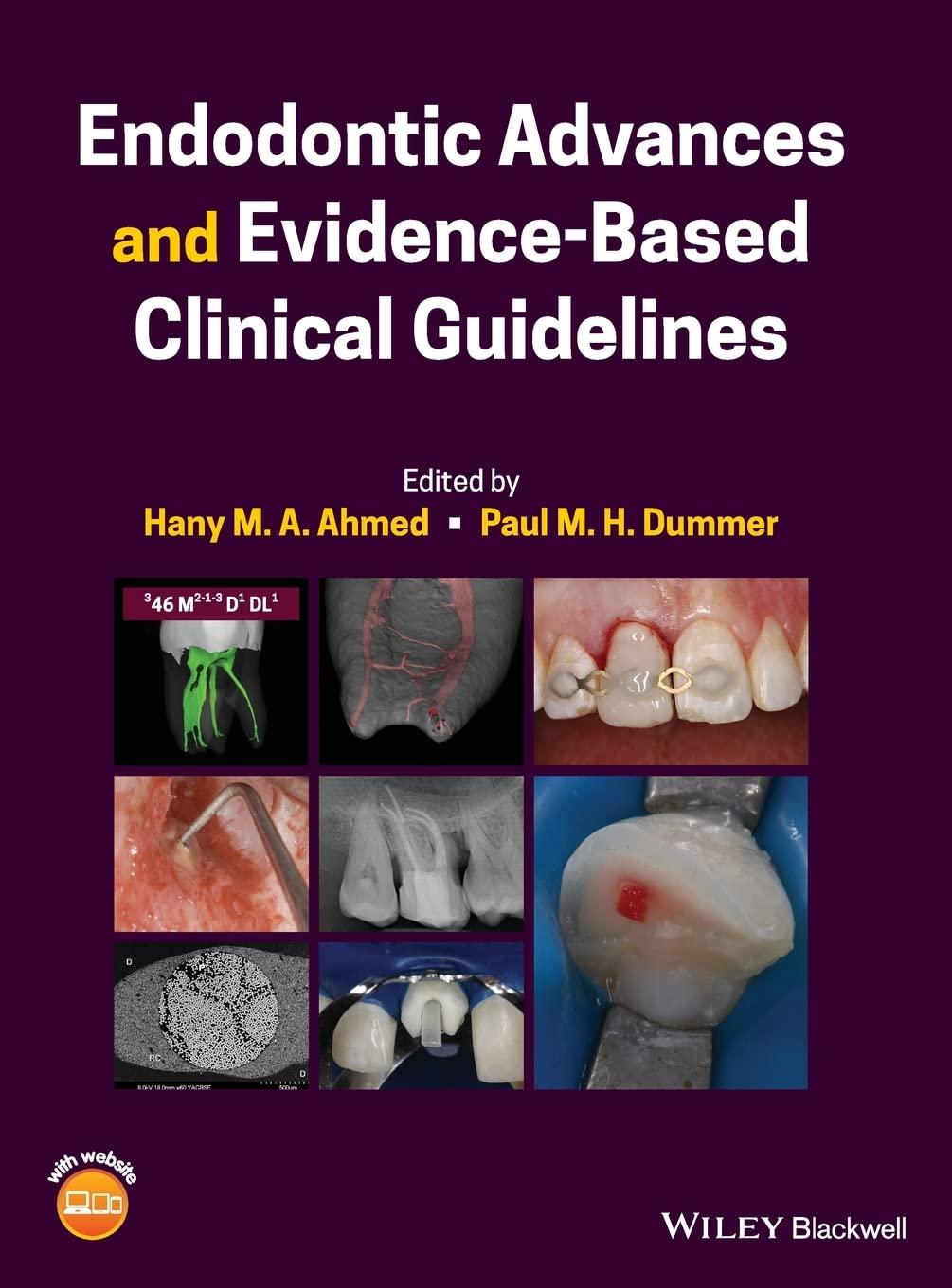
Endodontic Advances and Evidence-Based Clinical Guidelines
Endodontic Advances and Evidence-Based Clinical Guidelines
Edited by
Hany M. A. Ahmed
Department of Restorative Dentistry, Faculty of Dentistry, University of Malaya, Kuala Lumpur, Malaysia
Paul M. H. Dummer School of Dentistry, College of Biomedical and Life Sciences, Cardiff University, Cardiff, UK
Companion Website: www.wiley.com/go/ahmed/endodontics
This edition first published 2022 © 2022 John Wiley & Sons Ltd
All rights reserved. No part of this publication may be reproduced, stored in a retrieval system, or transmitted, in any form or by any means, electronic, mechanical, photocopying, recording or otherwise, except as permitted by law. Advice on how to obtain permission to reuse material from this title is available at http:// www.wiley.com/go/permissions.
The right of Hany M. A. Ahmed and Paul M. H. Dummer to be identified as the authors of the editorial material in this work has been asserted in accordance with law.
Registered Offices
John Wiley & Sons, Inc., 111 River Street, Hoboken, NJ 07030, USA
John Wiley & Sons Ltd, The Atrium, Southern Gate, Chichester, West Sussex, PO19 8SQ, UK
Editorial Office
The Atrium, Southern Gate, Chichester, West Sussex, PO19 8SQ, UK
For details of our global editorial offices, customer services, and more information about Wiley products visit us at www.wiley.com.
Wiley also publishes its books in a variety of electronic formats and by print-on-demand. Some content that appears in standard print versions of this book may not be available in other formats.
Limit of Liability/Disclaimer of Warranty
The contents of this work are intended to further general scientific research, understanding, and discussion only and are not intended and should not be relied upon as recommending or promoting scientific method, diagnosis, or treatment by physicians for any particular patient. In view of ongoing research, equipment modifications, changes in governmental regulations, and the constant flow of information relating to the use of medicines, equipment, and devices, the reader is urged to review and evaluate the information provided in the package insert or instructions for each medicine, equipment, or device for, among other things, any changes in the instructions or indication of usage and for added warnings and precautions. While the publisher and authors have used their best efforts in preparing this work, they make no representations or warranties with respect to the accuracy or completeness of the contents of this work and specifically disclaim all warranties, including without limitation any implied warranties of merchantability or fitness for a particular purpose. No warranty may be created or extended by sales representatives, written sales materials, or promotional statements for this work. The fact that an organization, website, or product is referred to in this work as a citation and/or potential source of further information does not mean that the publisher and authors endorse the information or services the organization, website, or product may provide or recommendations it may make. This work is sold with the understanding that the publisher is not engaged in rendering professional services. The advice and strategies contained herein may not be suitable for your situation. You should consult with a specialist where appropriate. Furthermore, readers should be aware that websites listed in this work may have changed or disappeared between when this work was written and when it is read. Neither the publisher nor authors shall be liable for any loss of profit or any other commercial damages, including but not limited to special, incidental, consequential, or other damages.
Library of Congress Cataloging-in-Publication Data
Names: Ahmed, Hany Mohamed Aly, author. | Dummer, Paul Michael Howell, author.
Title: Endodontic advances and evidence-based clinical guidelines / Hany Mohamed Aly Ahmed, Paul Michael Howell Dummer.
Description: Hoboken, NJ : John Wiley & Sons, 2022. | Includes bibliographical references and index.
Identifiers: LCCN 2021045254 (print) | LCCN 2021045255 (ebook) | ISBN 9781119553885 (paperback) | ISBN 9781119553793 (pdf) | ISBN 9781119553892 (epub) | ISBN 9781119553939 (obook)
Subjects: LCSH: Endodontics.
Classification: LCC RK351 .A36 2022 (print) | LCC RK351 (ebook) | DDC 617.6/342--dc23/eng/20211108
LC record available at https://lccn.loc.gov/2021045254
LC ebook record available at https://lccn.loc.gov/2021045255
Cover image: Courtesy of Hany Ahmed, Gabriel Krastl, Jorge Perdigão, Gianluca Plotino, and Silvio Taschieri
Cover design by Wiley
Set in 9.5/12.5pt STIXTwoText by Integra Software Service Pvt. Ltd, Pondicherry, India
Contents
Preface xviii
Acknowledgements xix
Editors’ Biography xx
List of Contributors xxi
About the Companion Website xxv
Part I: Advances in Knowledge 1
1 Tooth, Root, and Canal Anatomy 3
Hany M. A. Ahmed, Ali Keleş, Jorge N. R. Martins, and Paul M. H. Dummer
Summary 3
1.1 Introduction 3
1.2 Different Perspectives in Characterizing Root and Canal Morphology 4
1.2.1 Deficiencies of Current Classification Systems for Root Canal Morphology 5
1.2.2 Introduction to the New Coding System for Root and Canal Morphology 8
1.3 Advances in Apical Canal Morphology 17
1.3.1 Cemento-dentinal Junction (CDJ) 17
1.3.2 Apical Constriction (AC) 18
1.3.3 Major Apical Foramen (MAF) 19
1.3.4 Isthmus 22
1.3.5 Accessory Canals and Apical Deltas 25
1.3.6 Bifid Apex 26
1.3.7 The Importance of Apical Canal Anatomy in Apical Surgery 26
1.4 Root and Canal Morphology in Different Tooth Types 27
1.4.1 Maxillary Anterior Teeth 27
1.4.2 Maxillary First Premolar 28
1.4.3 Maxillary Second Premolar 29
1.4.4 Maxillary First Molar 31
1.4.5 Maxillary Second Molar 31
1.4.6 Mandibular Anterior Teeth 34
1.4.7 Mandibular First Premolar 36
1.4.8 Mandibular Second Premolar 36
1.4.9 Mandibular First Molar 36
1.4.10 Mandibular Second Molar 37 References 39
2 The Bioactive Properties of Dentine and Molecular Advances in Pulp Regeneration 51
Henry F. Duncan and Paul R. Cooper Summary 51
2.1 Introduction 51
2.2 Regenerative Endodontics 53
2.3 The Role of Dentine in Pulpar Repair and Regeneration 53
2.4 Infection, Inflammation, and Stem Cells Interaction in Pulp Regeneration 54
2.4.1 Immune Response 54
2.4.2 Inflammation and Regeneration 56
2.4.3 Opportunities for Clinical Translation 56
2.5 Regenerative Endodontic Procedures (REPs) 57
2.5.1 Cell-Homing 57
2.5.2 Cell-based Therapies 62
2.6 Conclusion 65 References 66
3 Microbial Biofilms in Root Canal Systems 74
Luis E. Chávez de Paz Summary 74
3.1 Introduction 74
3.2 General Characteristics of Microbial Biofilms 75
3.2.1 How do Bacteria Build Biofilms? 75
3.2.2 Formation of Biofilms in Root Canals 75
3.2.3 Planktonic Versus Biofilm Lifestyles 76
3.2.4 The Biofilm Phenotype 76
3.3 Ecological Factors Affecting Biofilms in Root Canals 77
3.3.1 The Inflammatory and Necrotic Environments 78
3.3.2 The Post-Treatment Environment 78
3.4 Survival of Biofilm Bacteria to Antimicrobials 79
3.5 Biofilm Resistance in Endodontics 80
3.6 Conclusion 81 References 81
4 Pulp, Root Canal, and Periradicular Conditions 85
Paul V. Abbott Summary 85
4.1 Introduction 85
4.2 What Causes Pulp, Root Canal, and Periradicular Conditions? 86
4.3 The Development and Progression of Pulp and Root Canal Conditions 88
4.4 The Development and Progression of Periradicular Conditions 90
4.5 Classifications of Conditions and/or Diseases 94
4.6 Classification and Description of Pulp and Root Canal Conditions 96
4.7 Classification and Description of Periradicular Conditions 104
4.8 Summary 115 References 115
5 Root Resorption 117
Abdulaziz Bakhsh, Shanon Patel, and Bhavin Bhuva Summary 117
5.1 Introduction 117
5.2 Histopathology of Root Resorption 119
5.3 Internal Root Resorption 119
5.3.1 Internal Inflammatory Root Resorption 119
5.3.2 Internal Replacement Resorption 120
5.4 External Root Resorption 121
5.4.1 External Inflammatory Resorption 121
5.4.2
External Replacement Resorption 122
5.4.3 External Surface Resorption 122
5.4.4 External Cervical Resorption 123
References 127
6
Minimally Invasive Endodontics 130
Prasanna Neelakantan, Antonis Chaniotis, and Avijit Banerjee
Summary 130
6.1 Introduction 130
6.2 Embracing the Concept of Minimally Invasive Endodontics 130
6.2.1 The Need for Patient-focused Approaches 131
6.2.2 Technological Advancements in Endodontics that Has Made Minimally Invasive Strategies Possible 132
6.2.3 What Does Minimum Intervention Root Canal Treatment Encompass? 133
6.3 Rationale for Minimally Invasive Root Canal Treatment 133
6.3.1 Failure of Root Canal Treatment: Microbial Causes 133
6.3.2 Failure of Root Canal Treatment: Structural Causes 134
6.4 Minimum Intervention in Endodontics: Prevention Is Better than Cure? 134
6.5 Minimally Invasive Management of the Deep Carious Lesion 134
6.6 Minimally Invasive Root Canal Treatment Procedures 136
6.7 Access Cavity Terminology 136
6.8 Minimally Invasive Root Canal Preparation 140
6.8.1 Goals of Root Canal Preparation 140
6.8.2 So, What Happens to These Untouched Walls? 141
6.8.3 Apical Preparation Sizes and Root Canal Preparation Tapers – How Much Is Enough? 143
6.8.4 Supplementary Irrigation Strategies in Minimally Prepared Root Canals 145
6.8.5 Is It Possible to Clean Root Canals with No Instrumentation at All? 146
6.9 Minimally Invasive Surgical Endodontics 146
6.10 Conclusion 147 References 147
7 Systemic Health and Endodontics 153 Juan J. Segura-Egea and Jenifer Martín-González Summary 153
7.1 From Focal Infection Theory to Endodontic Medicine 153
7.1.1 The Discredited Focal Infection Theory 154
7.1.2 Endodontic Medicine: Interrelation Between Systemic and Endodontic Pathosis 154
7.2 Pathways Linking Periapical Inflammatory Lesions to Systemic Health Status 156
7.2.1 The Spread of Endodontic Bacteria to Adjacent Tissues and Organs 156
7.2.2 Local Production of Soluble Regulatory Molecules that May Initiate or Sustain Inflammatory Events in Remote Tissues and Organs 157
7.2.3 Extrinsic or Intrinsic Pathological Mechanisms Resulting or Contributing to Both Local and Systemic Inflammation 158
7.3 Endodontic Implications of Systemic Diseases – Systemic Factors Affecting Periapical Repair 159
7.4 Diabetes and Endodontics 160
7.4.1 Scientific Evidence on the Association Between Diabetes and Endodontics 160
7.4.2 Biological Mechanisms Involved in the Association Between Diabetes and Endodontics 162
7.4.3 Endodontic Management of Diabetic Patients 164
7.5 Cardiovascular Disease and Endodontics 164
7.5.1 Scientific Evidence on the Association Between Cardiovascular Diseases and Endodontics 165
7.5.2 Mechanisms Involved in the Association Between Cardiovascular Diseases and Endodontics 166
7.5.3 Endodontic Management of Cardiovascular Patients 167
7.6 Relationship Amongst Other Systemic States and Endodontics 168
7.6.1 Smoking Habits 168
7.6.2 Digestive Diseases and Other Systemic Diseases 169 References 170
8 Technology Enhanced Education 178 Nor Nadia Zakaria, Muaiyed M. Buzayan, Donnie Adams, Hany M. A. Ahmed, and Paul M. H. Dummer Summary 178
8.1 Introduction 178
8.2 E-learning in Dentistry 179
8.3 Contemporary E-learning Models 179
8.4 E-learning During the COVID-19 Pandemic 181
8.5 Limitations of E-learning in Dental Education 181
8.6 Advances in Endodontology Education 182
8.6.1 3D Technology for Pre-clinical Training 182
8.6.2 Augmented and Virtual Reality 183
8.7 Digital Dentistry and Clinical Endodontics 185
8.7.1 Guided Endodontics 185
8.7.2 Surgical Endodontics 186
8.8 Conclusion 186 References 186
Par t II: Advances in Materials and Technology 191
9 Computed Tomography Imaging Devices and Techniques 193
Marco A. Versiani and Gianluca Gambarini
Summary 193
9.1 Digital Dentistry and Impact on Clinical Training and Education 193
9.1.1 3D Endodontic Rendering 193
9.1.2 3D Endodontic Software 195
9.1.3 Dynamic Navigation Systems (Software and Devices) 200
9.2 Advances in Micro- CT and Nano- CT Technologies and Their Impact on Clinical Training and Education 208
9.2.1 Fundamentals of Micro- CT and Nano- CT Imaging 209
9.2.2 Micro-CT Versus Nano- CT Technology 211
9.2.3 CT Technology in Dental Education and Training 211
9.2.4 Advances in Micro- CT and Nano- CT Applications 214
9.5 Conclusion 214 References 215
10 Advances in Working Length Determination 218
Mohammad H. Nekoofar Summary 218
10.1 Introduction 218
10.2 Morphology of the Root Canal Terminus 219
10.3 Determining the Root Canal Terminus 220
10.4 An Overview of Basic Electronics 222
10.4.1 Atom Structure 222
10.4.2 Ions and Electrolytes 222
10.4.3 Electrical Charge, Voltage, and Current 223
10.4.4 Resistance 223
10.4.5 Electric Circuits and the Human Body 224
10.4.6 Ohm’s Law 224
10.4.7 Direct Current and Alternating Current 224
10.4.8 Capacitor 225
10.4.9 Impedance and its Measurement 226
10.5 Electrical Features of Tooth Structure 226
10.6 Electronic Root Canal Length Measurement Devices (ERCLMDs) 227
10.6.1 Fundamental Assumption 227
10.6.2 Background 227
10.6.3 Resistance-based ERCLMDs 228
10.6.4 Low-frequency Oscillation ERCLMDs 229
10.6.5 High-frequency Devices (Capacitance-based Devices; ERCLMDs) 229
10.6.6 Capacitance and Resistance ERCLMDs (Look-up Tables) 230
10.6.7 Voltage Gradient ERCLMDs (Difference in Impedance with Three Nodes) 230
10.6.8 Two Frequencies: Impedance and Difference ERCLMDs 231
10.6.9 Two Frequencies: Impedance Ratio (Quotient) ERCLMDs 231
10.6.10 Multifrequency ERCLMDs 233
10.6.11 Root Canal Length Measurement Devices Integrated Into Rotary Endodontic Motors 233
10.6.12 Effect of ERCLMDs on Cardiac Devices 234
10.6.13 Application of ERCLMDs in the Primary Dentition 234
References 234
11 Advances in Materials and Techniques for Microbial Control 243
Ronald Ordinola-Zapata, Maria T. Arias-Moliz, Prasanna Neelakantan, Alejandro Perez-Ron, and Joseph T. Crepps
Summary 243
11.1 Introduction 243
11.2 Biofilms 244
11.3 Sodium Hypochlorite 245
11.4 Detoxification of the Root Canal System by Endodontic Procedures 247
11.5 Inactivation of Root Canal Irrigants 248
11.6 Etidronic Acid and the Continuous Chelation Concept 249
11.7 Cetrimide and Surfactants 251
11.8 Passive Ultrasonic Irrigation 252
11.9 Negative Apical Pressure 252
11.10 Photodynamic Antimicrobial Therapy 253
11.11 Laser-activated Irrigation 255
11.12 Multisonic Technique 256
11.13 Conclusion 257
References 258
12 Nickel-Titanium Metallurgy 268
Ya Shen and Markus Haapasalo
Summary 268
12.1 Introduction and Classification of Current NiTi Alloy Phases 268
12.2 Properties of Each Phase (Austenitic, Martinsitic, R-phase) 270
12.3 Surface Treatment of NiTi Alloys 272
12.4 Post-machining Heat Treatment of NiTi Alloys 273
12.5 Effects of Irrigants and Sterilisation Procedures on NiTi Alloys 275
12.6 Relevance of Current Studies 275
12.7 Conclusion 277 References 277
13 Rotary and Reciprocating Motions During Canal Preparation 283
Ove A. Peters and Ana Arias Summary 283
13.1 Goals and Limitations of Engine-driven Root Canal Preparation 283
13.2 Current Instrument Designs, Movements, and Manufacturing Methods 284
13.3 Clinical Recommendations for Rotary and Reciprocating Canal Preparation 287
13.3.1 Preparation for Treatment 287
13.3.2 Early Coronal Modification 288
13.3.3 Working Length and Patency 289
13.3.4 Glide Path Preparation 290
13.3.5 Canal Preparation 291
13.4 Physical Properties of Engine-driven Root Canal Instruments 292
13.4.1 Cutting Efficiency 292
13.4.2 Cyclic Fatigue Resistance 292
13.4.3 Torsional Performance 293
13.5 Surrogate and Clinical Parameters Affecting Outcomes 294
13.5.1 Geometry of Root Canals After Preparation 295
13.5.2 Induction of Dentinal Micro-crack Formation 297
13.5.3 Debris Extrusion and Postoperative Pain 298
13.5.4 Functionality in Retreatment 300
13.6 Clinical Experiences with Rotary and Reciprocating Root Canal Instruments 300 References 301
14 Hydraulic Calcium Silicate-based Endodontic Cements 311
Josette Camilleri Summary 311
14.1 Introduction 311
14.2 Material Properties 312
14.2.1 Cement Characteristics 313
14.2.2 Radiopacifier Characteristics 318
14.2.3 Admixtures, Additives, and Vehicles 318
14.3 Classification of Hydraulic Cements 320
14.4 Specific Uses and Material Properties 321
14.4.1 Application on the Coronal Pulp 321
14.4.2 Intraradicular Use 326
14.4.3 Extraradicular Use 330
14.5 Current Challenges and Conclusions 333 References 334
15 Nanomaterials in Endodontics 347
Anil Kishen and Annie Shrestha Summary 347
15.1 Introduction 347
15.2 Applications and Challenges 348
15.3 Nanomaterials in Endodontics 348
15.3.1 Application of nanomaterials for Endodontic Disinfection 350
15.3.2 Nanomaterials in Root Canal Fillings 353
15.3.3 Nanomaterials in Restorative Materials 354
15.3.4 Nanomaterials in Regenerative Endodontic Procedures 356
15.3.5 Nanomaterials as Bioactive Molecule Delivery Systems 357
15.3.6 Nanomaterials in Scaffolds 358
References 359
Par t III: Advances in Clinical Management 367
16 Vital Pulp Treatment 369
Henry F. Duncan and Lars Bjørndal
Summary 369
16.1 Introduction 369
16.2 Caries: Current Thinking and Radiographic Classification 370
16.3 Role of Pulp and Dentine in Repair 371
16.4 What Does Vital Pulp Treatment Encompass? 373
16.5 How Do We Classify and Diagnose Pulpal Disease? 374
16.6 How Do We Treat Pulpal Disease? Techniques to Avoid Pulpal Exposure 375
16.6.1 Indirect Pulp Capping 375
16.6.2 Selective Carious-tissue Removal in One Visit 376
16.6.3 Stepwise Excavation 376
16.6.4 When Should Pulp Exposure Be Avoided? 378
16.6.5 Follow-up 378
16.6.6 Outcome Analysis 378
16.7 How Do We Treat Pulpal Disease? Techniques When the Pulp Is Exposed 379
16.7.1 When Should We Expose the Pulp and How Much Tissue Should We Remove? 379
16.7.2 Direct Pulp Capping 380
16.7.3 Pulpotomy 381
16.7.4 Pulpectomy 383
16.7.5 Assessing Success 383
16.7.6 Future Opportunities and Therapies 384
16.7 Conclusion 385 References 385
17 Detection of Canal Orifices, Negotiation, and Management of Calcified and Curved Canals 393
Gianluca Plotino and Nicola M. Grande
Summary 393
17.1 Introduction 393
17.2 Detection of Canal Orifices 394
17.2.1 The Significance of Missed Anatomy on the Prognosis of Root Filled Teeth 394
17.2.2 Anatomical Landmarks for Detection of Root Canals 397
17.2.3 Clinical Detection of Canal Orifices 401
17.2.4 Magnification and Ultrasonics: The Perfect Tools for Detection of Canal Orifices 407
17.2.5 Radiographic Techniques for Detection of Root Canals 410
17.2.6 Guided Endodontics for Detection of Root Canals 411
17.3 Negotiation of Calcified and Curved Canals 413
17.3.1 Background 413
17.3.2 Negotiation, Glide Path, and Preflaring 414
17.3.3 Clinical Strategies for the Negotiation of Easily Scoutable Canals 415
17.3.4 Clinical Strategies for the Negotiation of Complex Canals 416
17.4 Shaping of Calcified and Curved Canals 421
17.4.1 Basic Principles 421
17.4.2 The Ideal Instruments for Shaping Calcified and Curved Canals 424
17.5 Conclusion 430
References 430
18 Management of Fractured Instruments 440
Yoshi Terauchi
Summary 440
18.1 Aetiology of Instrument Fracture 440
18.1.1 Factors Affecting Instrument Fracture 440
18.1.2 Incidence of Instrument Fracture 441
18.1.3 Mechanisms for Instrument Fracture 442
18.2 Diagnosis and Treatment Planning of Fractured Instruments 443
18.2.1 Factors Affecting the Success of Instrument Retrieval 443
18.2.2 Diagnostic Examination Using CBCT for Instrument Retrieval 443
18.2.3 Treatment Planning for Instrument Retrieval 446
18.3 Root Canal Preparation Techniques 449
18.3.1 Potential Accidents in Ultrasonic Activation 449
18.3.2 Refinement of the Damaged Ultrasonic Tip 450
18.3.3 Root Canal Preparation Techniques for Visible Instrument Retrieval 451
18.3.4 Root Canal Preparation for Nonvisible Instrument Retrieval 455
18.4 Instrument Retrieval Techniques 458
18.4.1 Type of fluid used in instrument removal attempts 458
18.4.2 Use of Ultrasonic Activation 460
18.4.3 Use of the Loop 462
18.4.4 Use of the XP-endo Shaper 463
18.4.5 Mechanical Techniques Other than Ultrasonics 465
18.4.6 Non-mechanical Techniques 467
18.5 Prognosis 467 References 469
19 Repair of Pulp Chamber and Root Perforations 475
Thomas Clauder Summary 475
19.1 Introduction 475
19.2 Occurrence and Diagnosis of Perforations During Root Canal Treatment 476
19.3 Diagnosis of Perforations 478
19.4 Classification of Perforations and Factors Affecting Prognosis 481
19.4.1 Time of Repair 482
19.4.2 Size of Perforation 482
19.4.3 Location of Perforation 483
19.4.4 MTA as a Perforation Repair Material 484
19.4.5 Alternative Materials for Perforation Repair in Specific Indications 485
19.5 Techniques and Considerations to Clinically Repair Perforations 486
19.5.1 Appropriate Material Selection 486
19.5.2 Use of a Matrix 487
19.6 Nonsurgical Management of Perforations 489
19.6.1 Crown, Pulpal Floor, and Furcation Areas 489
19.6.2 Middle One Third of the Root Canal 491
19.6.3 Apical One Third of the Root Canal 498
19.7 Surgical Management of Perforations 499
19.8 Clinical Outcomes 502
19.9 Conclusion 505 References 505
20 Removal of Root Canal Filling Materials 511
Tina Rödig and Michael Arnold Summary 511
20.1 Indications for Root Canal Retreatment 511
20.2 Objectives of Root Canal Retreatment Procedures 512
20.3 Removal of Crowns and Posts 513
20.3.1 Indications 513
20.3.2 Post Removal Techniques 514
20.3.3 Complications of Post Placement and Removal 516
20.3.4 Custom Cast Core Posts 516
20.3.5 Ceramic Posts 517
20.3.6 Removal of Fibre Posts (Tooth 26 Case with Video 3) 519
20.3.7 Prognostic Assessment of Post Removal 521
20.4 Methods for Removal of Gutta-percha 524
20.4.1 Hand Instruments 524
20.4.2 Softening of Gutta-percha 526
20.4.3 Engine-driven NiTi Instruments 535
20.4.4 Adjunctive Instruments and Techniques 537
20.5 Removal of Carrier-based Root Canal Filling Materials 539
20.6 Retrieval of Silver Cones 542
20.6.1 Need for Removal 543
20.6.2 Methods of Retrieval 544
20.6.3 Success of Silver Point Removal and Outcome 546
20.7 Removal of Calcium Silicate-based Cements 547
20.8 Removal of Calcium Silicate-based Sealers 548
20.9 Removal of Resorcinol-formaldehyde Resin Paste (Russian Red) 551 References 553
21 Restoration of Root-filled Teeth 565 Sanket Nagarkar, Nicole Theis-Mahon, Ronald Ordinola-Zapata, and Jorge Perdigão Summary 565
21.1 Introduction 565
21.2 Examination of Root-filled Teeth Before Selection of a Treatment Approach 566
21.2.1 Ferrule 566
21.2.2 Remaining Coronal Walls 566
21.2.3 Marginal Ridges 566
21.3 Evidence from Clinical Studies Regarding Factors Affecting the Prognosis of Root filled Teeth 567
21.3.1 Outcome Measures and Clinical Questions Addressed by Clinical Studiess 567
21.4 Decision-making for Restoration of Root filled Teeth 572
21.4.1 Root filled Teeth with Minimal Loss of Coronal Structure 572
21.4.2 Root filled Teeth with Significant Loss of Coronal Structure 572
21.5 Clinical Considerations for the Management of Root filled Teeth Using Posts 573
21.5.1 Relevance of Tooth Anatomy 573
21.5.2 Classification of Posts 575
21.5.3 Effect of Post Space Preparation and Post Placement on the Fracture Resistance of Root filled Teeth 577
21.5.4 Clinical Steps to Cement a Post 578
21.6 Importance of the Final Restoration 582
21.7 Conclusion 583 References 583
22 Classifications and Management of Endodontic-periodontal Lesions 591
Edoardo Foce, Hany M. A. Ahmed, and Ahmed A. R. Hashem Summary 591
22.1 Communication Pathways Between the Pulp and Periodontal Tissues 591
22.1.1 Endo-perio Lesions: A Terminological Controversy 592
22.1.2 Classifications of Endo-perio Lesions 594
22.1.3 Foce Classification System for Endo-perio Lesions 597
22.1.4 Ahmed Classification System for Endo-perio Lesions 598
22.2 Management and Prognosis of Endo-perio Lesions 601
22.2.1 Crown-down Plaque-induced Periodontal Lesions Without Pulpal Involvement 604
22.2.2 Crown-down Plaque-induced Periodontal Lesions With Pulpal Involvement 606
22.2.3 Down-crown Periodontal Lesions of Endodontic Origin 608
22.2.4 Combined Endo-perio Lesions 610
22.3 Conclusion 610 References 613
23 Management of Coronal Discolouration 617
Hany M. A. Ahmed, Gabriel Krastl, Brigitte Zimmerli, Mohamed Amer, and Peter Parashos Summary 617
23.1 Introduction 617
23.2 Aetiology 617
23.3 Prevention of Coronal Discolouration Related to Endodontic Procedures 620
23.4 Management Guidelines 621
23.4.1 History 621
23.4.2 Evaluation and Preparation 621
23.4.3 Selection of the Appropriate Treatment Approach 621
23.4.4 Types of Intracoronal Bleaching 625
23.5 Bleaching of Teeth with Calcified Pulp Chambers and Root Canals 627
23.6 Prognosis of Intracoronal Bleaching 628
23.6.1 Initial Results of Intracoronal Bleaching 628
23.6.2 Colour Stability 629
23.7 Complications After Intracoronal Bleaching 631
23.8 Other Treatment Options 632
23.8.1 Restoration of Teeth After Bleaching 632
23.9 Tooth Discolouration Following Regenerative Endodontic Procedures 633
23.10 Management of Tooth Discolouration Following Regenerative Endodontic Procedures 635 References 637
24 Surgical Endodontics 645
Silvio Taschieri and Stefano Corbella Summary 645
24.1 Introduction 645
24.2 Historical Perspective 645
24.3 Indications for Surgical Endodontics with Root-end Resection and Treatment Alternatives 646
24.4 Endodontic Microsurgery (EMS) Technique 647
24.4.1 Diagnosis 647
24.4.2 Anaesthesia 648
24.4.3 Mucoperiosteal Flap 648
24.4.4 Bone Access 650
24.4.5 Root-end Management 651
24.4.6 Root-end Filling Materials, Types, and Current Advances 653
24.4.7 Management of the Bone Cavity 653
24.5 Prognosis and Outcome Evaluation 654
24.6 Case Difficulty Classification for Surgical Endodontics 663
24.6.1 Patient Level 663
24.6.2 Tooth Level 664
24.7 Other Surgical Endodontics Procedures 665
24.7.1 Incision and Drainage 665
24.7.2 Exploratory Surgery 665
24.7.3 Periradicular Curettage and Biopsy 665
24.7.4 Root Resection 666
24.7.5 Tooth Resection 666
24.7.6 Extraction with Replantation 666 References 666
25 Alternatives to Root Canal Treatment: Tooth Autotransplantation 669 Monty Duggal and Hani Nazzal Summary 669
25.1 Introduction 669
25.2 Indications for Autotransplantation 671
25.3 Advantages and Disadvantages of Tooth Autotransplantation 671
25.4 The Role of Interdisciplinary Team Planning 672
25.5 Pretransplantation Bone Management 672
25.6 Case Selection 673
25.6.1 Availability of a Donor Tooth 674
25.6.2 Donor Tooth Assessment 675
25.6.3 Recipient Site Characteristics 675
25.7 Success and Survival of Tooth Autotransplantation 676
25.7.1 Factors Affecting Prognosis of Autotransplanted Teeth 676
25.8 Presurgical Preparations 676
25.9 Tooth Autotransplantation Surgical Technique and Considerations 677
25.10 Socket Assessment 679
25.11 Antibiotic Prophylaxis 681
25.12 Postoperative Instructions 681
25.12.1 Post-transplantation Pulpal and Periodontal Management 681
25.13 Interim Restorative Camouflage 681
25.14 Pulpal Management 682
25.15 Orthodontic Tooth Movement 682
25.16 Definitive Restoration 682
25.17 Conclusion 682
References 682
Par t IV: Evidence-based Clinical Guidelines 685
26 Endodontic Diagnosis 687 Pratik Kamalkant Shah and Bun San Chong Summary 687
26.1 Introduction 687
26.2 History Taking 688
26.2.1 Presenting Problem 688
26.2.2 Dental History 689
26.2.3 Medical History 689
26.2.4 Antibiotic Cover 690
26.2.5 Social History 690
26.3 Clinical Examination 690
26.3.1 Extraoral assessment 690
26.3.2 Intraoral assessment 690
26.3.3 Routine Tests 692
26.3.4 Special Tests 695
26.3.5 Radiography 704
26.4 Classification of Pulp and Periradicular Diseases 706
26.4.1 American Association of Endodontists Classification System 706
26.4.2 Limitations of the American Association of Endodontists
Classification System 709
26.4.3 Endolight Classification 710
26.5 Referred Pain 711
References 712
27 The Use of Cone-Beam Computed Tomography in Endodontics 719
Shanon Patel, Robert Kelly, and Tiago Pimentel Summary 719
27.1 Introduction 719
27.2 Detection of Apical Periodontitis 719
27.3 Root Canal Anatomy 720
27.4 Root Canal Retreatment 721
27.5 Endodontic Surgery 722
27.6 Dental Trauma 723
27.7 Diagnosis and Management of Root Resorption 724
27.8 Vertical Root Fractures 725
27.9 Limitations 726
27.10 Conclusion 727
References 727
28 Endodontic Emergencies and Systemic Antibiotics in Endodontics 734
Juan J. Segura-Egea and Jenifer Martín-González Summary 734
28.1 Endodontic Emergencies 734
28.1.1 Diagnosis and Treatment Planning in Endodontic Emergencies 735
28.1.2 Emergency Treatment of Symptomatic Reversible Pulpitis 735
28.1.3 Emergency Treatment of Symptomatic Irreversible Pulpitis 736
28.1.4 Emergency Treatment of Acute Periapical Abscess 736
28.1.5 Cracked Tooth 738
28.1.6 Traumatic Injuries of the Teeth 739
28.2 Systemic Antibiotics in Endodontics 739
28.2.1 Antibiotics as Antimicrobial Medicaments in Endodontic Infections 740
28.2.2 Indications for Systemic Antibiotics as Adjuvants in the Treatment of Endodontic Infections: European Society of Endodontology Position Statement 741
28.2.3 Indications for Antibiotic Prophylaxis in Endodontics: European Society of Endodontology Position Statement 743
28.2.4 Systemic Antibiotics for the Treatment of Traumatic Injuries of the Teeth 745
28.3 Conclusion 746
References 746
29 Revitalization Procedures 749
Kerstin M. Galler
Summary 749
29.1 Regeneration and Repair Processes in the Dental Pulp 749
29.2 Revitalization – Terminological Aspects 750
29.3 Position Statements of the ESE and AAE 751
29.4 Case Selection, Indications, and Contra-indications 751
29.5 Clinical Procedure 752
29.5.1 Disinfection 752
29.5.2 Provocation of Bleeding 754
29.6 Outcome 755
29.7 Future Perspectives 756
References 756
30 Management of Traumatic Dental Injuries in the Permanent Dentition 761 Gabriel Krastl, Roland Weiger, Andreas Filippi, Kurt A. Ebeleseder, and Kerstin M. Galler
Summary 761
30.1 Introduction and Epidemiological Data 761
30.2 Classification of Traumatic Dental Injuries 762
30.3 Diagnosis of Traumatic Dental Injuries 763
30.4 Enamel Cracks and Crown Fractures 764
30.4.1 Vital Pulp Treatment 765
30.4.2 Materials for Vital Pulp Treatment 766
30.4.3 Success Rates of Vital Pulp Treatment in Traumatised Teeth 767
30.4.4 Reattachment Restoration 767
30.4.5 Direct Resin Composite Restoration 769
30.4.6 Indirect Ceramic Restoration 770
30.5 Crown-root Fractures 770
30.5.1 Adhesive Fragment Reattachment 771
30.5.2 Two-step Direct Composite Restoration 771
30.5.3 Restorative Treatment of the Accessible Regions 772
30.5.4 Surgical Crown Lengthening 772
30.5.5 Extrusion 773
30.6 Splinting of Traumatised Teeth (Root Fractures and Luxation Injuries) 773
30.7 Root Fractures 775
30.8 Luxation Injuries (Concussion, Subluxation, Extrusion, Lateral Luxation) 777
30.9 Luxation Injuries (Intrusion) 779
30.10 Luxation Injuries (Avulsion) 781
30.10.1 Avulsed Teeth with Favourable Storage Conditions 781
30.10.2 Avulsed Teeth with Unfavourable Storage Conditions 782
30.11 Systemic Doxycycline Administration 783
30.12 Tetanus Prophylaxis 783
30.13 Conclusion 783
References 784
Index 794
Over the last few decades, there has been a substantial increase in the body of knowledge within the field of endodontology. This has been accompanied by a global increase in the awareness of clinicians, scientists, and the general public to the benefits of endodontic therapies and the ability of the dental profession to save teeth that in the past may have been extracted. We are delighted and honoured to contribute to the ever-increasing pool of knowledge in endodontology by presenting the first edition of our new book, Endodontic Advances and Evidence-Based Clinical Guidelines
The book is divided into four sections:
Advances in knowledge: This section focuses on the characterisation of root and canal anatomy using advanced diagnostic techniques, the bioactive properties of dentine and molecular advances in pulp regeneration, microbial biofilms in the root canal system, pulp and periodontal diseases, root resorption, minimally invasive endodontics, and technology-enhanced education.
Advances in materials and technology: This section covers advances in computed tomography imaging devices and techniques, working length determination, advances in materials and techniques for microbial control, and nickel titanium metallurgy together with recent automated motions and advances in calcium silicate-based cements.
Advances in clinical management: This section includes current trends for vital pulp therapies, an update for the detection, negotiation, and management of calcified and curved root canals, management of fractured instruments, repair of perforation defects, removal of root canal filling materials, restoration of root filled teeth, management of coronal discolouration, surgical endodontics, management of endo-perio lesions, and alternatives to root canal treatment.
Evidence-based clinical guidelines: This section includes guidelines for the use of cone-beam computed tomography in endodontics, endodontic emergencies and use of systemic antibiotics, regenerative endodontic procedures, and management of endodontic complications associated with traumatic injuries.
Recent advances in knowledge in the key areas of the specialty and their links to evidencebased clinical guidelines have allowed the latest scientific evidence to be integrated with treatment guidelines to address the needs of patients. This new and innovative format will enable undergraduate and postgraduate students, general dental practitioners, specialists, and clinical and non-clinical scientists to update their knowledge and engage with advanced treatment modalities that will have a significant, positive impact on patient management and treatment outcomes. Each chapter is accompanied by a large number of high-quality illustrations and clinical cases that will allow the reader to immediately understand current directions in research, the underlying concepts and new trends in education, and clinical techniques. The book is provided in print and eBook formats, and there is a companion website to enable the reader to access and browse the illustrations in a convenient manner.
Hany M. A. Ahmed
Paul M. H. Dummer
Acknowledgements
The editors would like to thank Wiley for supporting the concept of this new book and overseeing its production. Special thanks to Loan Nguyen, Susan Engelken, Erica Judisch, Tanya McMullin, Christy Michael and Amy Kopperude.
The editors gratefully acknowledge the contributing authors for sharing their valuable knowledge and experience.
We are also grateful to Muhammad Fairos Bin Jenal, Artist, Faculty of Dentistry, University of Malaya, for the drawings included in several chapters.
We would like to acknowledge and thank our families for their encouragement and continuous support along the way!

Editors’ Biography
Dr. Hany M. A. Ahmed, BDS, HDD, PhD, FICD, MDTFEd (RCSEd), FPFA, FADI
Dr. Ahmed graduated with a BDS (2002) from the Faculty of Dentistry, Ain Shams University, Egypt. In 2006, he obtained a Higher Dental Diploma degree in endodontics, followed by a PhD from the School of Dental Sciences, Universiti Sains Malaysia. He was awarded for his research including the IADR (SE Asian division) for the best laboratory research, in addition to the best publication award (2020), with a research group in Turkey, from the Journal of Endodontics
Dr. Ahmed has had work published in over 100 publications. In 2012, he introduced a new classification for endo-perio lesions, and in 2017, with experts in the field, he introduced a new system for classifying root and canal morphology, accessory canals, and dental anomalies, in addition to the PROUD-2020 reporting guidelines. He is an international consultant for research projects in several countries, and a key opinion leader for dental companies.
Currently, Dr. Ahmed is a senior lecturer in endodontics at the Faculty of Dentistry, University of Malaya (UM). He leads a number of grants related to root canal anatomy and endodontic bio-materials. In 2019, he was awarded the excellent service certificate from UM. Dr. Ahmed is also a registered specialist in endodontics with the Egyptian Dental Syndicate (2012-up to date).
Recently, Dr. Ahmed was awarded membership from the Faculty of Dental Trainers, Royal College of Surgeons (Edinburgh). He is a fellow of the International College of Dentists, Academy of Dentistry International, and Pierre Fauchard Academy.
Dr. Ahmed is a scientific reviewer and editorial board member for several journals. He is also the Deputy Editor-in-Chief of the European Endodontic Journal

Emeritus Professor Paul M. H. Dummer BDS, MScD, PhD, DDSc, FDS (RCSEd)
Professor Dummer graduated from the School of Dentistry, Welsh National School of Medicine, UK in 1973 with a bachelor’s degree in dental surgery and completed his MScD in 1980 and PhD in 1987 by research. He was awarded a senior doctorate in dental science (DDSc) in 2002 on the basis of his research record in endodontology. He has published over 300 original scientific articles in high-impact peer-reviewed journals and written several chapters in textbooks.
Professor Dummer was a professor of restorative dentistry at the School of Dentistry, Cardiff University, UK from 1995 to 2017 and vice dean between 2005 and 2011. He was dean for Education and Students in the College of Biomedical and Life Sciences, Cardiff University from 2011 to 2017, with overall responsibility for the quality of education in seven schools, including medicine, dentistry, pharmacy, nursing, optometry, psychology, and biosciences. He was director of the 3-year master’s programme in clinical dentistry (endodontology) at Cardiff from 2010 to 2017. He retired in 2017 and is now an emeritus professor at Cardiff University.
Paul Dummer was a clinical consultant in restorative dentistry with the Cardiff & Vale University Health Board (2000–2017) and a registered specialist in restorative dentistry and endodontics with the UK General Dental Council (2000–2017).
Paul Dummer was Editor-in-Chief of the International Endodontic Journal (1999–2021) and the president of the European Society of Endodontology (2020–2021), having been interim chief executive officer (2017–2019) and secretary (2009–2017).
List of Contributors
Paul V. Abbott
UWA Dental School
The University of Western Australia Perth, Australia
Donnie Adams
Department of Educational Management
Planning & Policy
Faculty of Education University of Malaya Kuala Lumpur, Malaysia
Hany M. A. Ahmed
Department of Restorative Dentistry
Faculty of Dentistry
University of Malaya Kuala Lumpur, Malaysia
Mohamed Amer
Melbourne Dental School
The University of Melbourne Victoria, Australia
Ana Arias
Department of Conservative and Prosthetic Dentistry
School of Dentistry
Complutense University Madrid, Spain
Maria T. Arias-Moliz
Department of Microbiology
School of Dentistry
University of Granada Granada, Spain
Michael Arnold
Private practice limited to Endodontics Dresden, Germany
Abdulaziz Bakhsh
Restorative Department
Endodontic Division
Umm Al-Qura University Faculty of Dentistry Makkah, Saudi Arabia
Avijit Banerjee
Conservative and MI Dentistry
Faculty of Dentistry, Oral and Craniofacial Sciences
King’s College London London, United Kingdom
Bhavin Bhuva
Department of Endodontics
Faculty of Dentistry, Oral & Craniofacial Sciences
King’s College London London, United Kingdom
Lars Bjørndal
Cariology and Endodontics
Faculty of Health and Medical Sciences
Department of Odontology
University of Copenhagen Copenhagen, Denmark
Muaiyed Mahmoud Ali Buzayan
Department of Restorative Dentistry Faculty of Dentistry
University of Malaya Kuala Lumpur, Malaysia
Josette Camilleri
School of Dentistry
Institute of Clinical Sciences
College of Medical and Dental Sciences
University of Birmingham Birmingham, United Kingdom
Antonis Chaniotis
Private practice limited to Microscopic
Endodontics
National and Kapodistrean University of Athens (NKUA)
Dental School, Department of Endodontics
Athens, Greece
Luis E. Chávez de Paz
Department of Endodontics
Karolinska Institute
Stockholm, Sweden
Bun San Chong
Institute of Dentistry
Faculty of Medicine & Dentistry
Queen Mary University of London
London, United Kingdom
Thomas Clauder
Private practice limited to Endodontics and Periodontics
Hamburg, Germany
Paul R. Cooper
Faculty of Dentistry
University of Otago
Dunedin, New Zealand
Stefano Corbella
Deptartment of Biomedical
Surgical and Dental Sciences - Università
degli Studi di Milano
Milan, Italy
IRCCS Ospedale Galeazzi - Sant’Ambrogio
Milan, Italy
Joseph T. Crepps
Division of Endodontics
University of Minnesota
School of Dentistry, Minneapolis Minnesota, United States of America
Monty Duggal
College of Dental Medicine
Qatar University
Doha, Qatar
Paul M. H. Dummer
School of Dentistry
College of Biomedical and Life Sciences
Cardiff University
Cardiff, United Kingdom
Henry F. Duncan
Division of Restorative Dentistry & Periodontology
Dublin Dental University Hospital
Trinity College Dublin
Dublin 2, Ireland
Kurt A. Ebeleseder
University Clinic of Dental Medicine and Oral Health
Medical University Graz
Graz, Austria
Andreas Filippi
Department of Oral Surgery
Center of Dental Traumatology
University Center for Dental
Medicine UZB
University of Basel
Basel, Switzerland
Edoardo Foce
Private practice
La Spezia, Italy
Kerstin M. Galler
Department of Conservative Dentistry and Periodontology
University Hospital Erlangen
Erlangen, Germany
Giamluca Gambarini
Dipartimento di Scienze
Odontostomatologiche
Corso di Laurea in Odontoiatria e Protesi Dentaria
Università degli studi di Roma
La Sapienza
Roma, Italy
Nicola M. Grande
Department of Operative Dentistry and Endodontics
School of Dentistry
Catholic University of Sacred Heart (UCSC)
Rome, Italy
Markus Haapasalo
Division of Endodontics
Faculty of Dentistry
University of British Columbia
Vancouver, Canada
Ahmed A. R. Hashem
Department of Endodontics
Faculty of Dentistry
Ain Shams University
Cairo, Egypt
Ali Keleş
Department of Endodontics Faculty of Dentistry
Ondokuz Mayis University Samsun, Turkey
Robert Kelly Private practice Bristol, United Kingdom
Anil Kishen Faculty of Dentistry
University of Toronto Toronto, Canada
Gabriel Krastl
Department of Conservative Dentistry and Periodontology
Center of Dental Traumatology
University Hospital of Würzburg Würzburg, Germany
Jenifer Martín-González Department of Stomatology School of Dentistry
University of Sevilla Sevilla, Spain
Jorge N. R. Martins School of Dentistry
University of Lisbon Lisbon, Portugal
Sanket Nagarkar Private practice at Park Dental Minneapolis, MN USA
University of Minnesota Minneapolis, MN United States of America
Hani Nazzal College of Dental Medicine
Qatar University Doha, Qatar
Prasanna Neelakantan Division of Restorative Dental Sciences
Faculty of Dentistry
The university of Hong Kong Hong Kong
Mohammad H. Nekoofar
Department of Endodontics
School of Dentistry
Tehran University of Medical Science (TUMS)
Tehran, Iran
Ronald Ordinola-Zapata Division of Endodontics
University of Minnesota
School of Dentistry
Minneapolis, Minnesota
United States of America
Peter Parashos
Melbourne Dental School
The University of Melbourne Victoria, Australia
Shanon Patel
Guy’s, King’s, St Thomas’ Dental Institute
King’s College London, Guy’s Tower London, United Kingdom
Alejandro Perez-Ron
Department of Dental Research Faculty of Dentistry
Iguaçu University (UNIG)
Nova Iguaçu
Rio de Janeiro, Brazil
Endochat Research Group Rio de Janeiro
Rio de Janeiro, Brazil
Department of Endodontics
University Rey Juan Carlos Madrid, Spain
Jorge Perdigao
Department of Restorative Sciences
University of Minnesota School of Dentistry
Minneapolis, Minnesota
United States of America
Ove A. Peters
School of Dentistry
The University of Queensland Brisbane, Australia
Tiago Pimentel
Faculty of Dentistry, Oral & Craniofacial Sciences
King’s College London London, United Kingdom
Gianluca Plotino
Private Practice
Rome, Italy
Tina Rödig
Department of Preventive Dentistry
Periodontology and Cariology
University Medical Center Göttingen
Göttingen, Germany
Juan J. Segura-Egea
Department of Endodontics
School of Dentistry
University of Sevilla Sevilla, Spain
Pratik Kamalkant Shah
Institute of Dentistry
Faculty of Medicine & Dentistry
Queen Mary University of London
London, United Kingdom
Ya Shen
Division of Endodontics
Faculty of Dentistry
University of British Columbia Vancouver, Canada
Annie Shrestha
Faculty of Dentistry
University of Toronto Toronto, Canada
Silvio Taschieri
Deptartment of Biomedical, Surgical and Dental SciencesUniversità degli Studi di Milano
Milan, Italy
IRCCS Ospedale Galeazzi - Sant’Ambrogio Milan, Italy
Yoshi Terauchi
Department of Endodontics
Faculty of Dentistry
Bahçeşehir University
İstanbul, Turkey
Henry M. Goldman School of Dental Medicine
Boston University
Boston, United States of America
Tokyo Medical and Dental University
Tokyo, Japan
Private practice limited to Endodontics
Tokyo, Japan
Nicole Theis-Mahon
Health Sciences Library
University of Minnesota
Minneapolis, United States of America
Marco A. Versiani
Dental Specialty Center
Brazilian Military Police Belo Horizonte, Brazil
Roland Weiger
Department of Periodontology, Endodontology and Cariology
Center of Dental Traumatology
University Center for Dental Medicine UZB
University of Basel
Basel, Switzerland
Nor Nadia Zakaria
Department of Department of Paediatric Dentistry & Orthodontics
Faculty of Dentistry
University of Malaya
Kuala Lumpur, Malaysia
Brigitte Zimmerli Zahnarztpraxis Braun & Zimmerli (private practice)
Burgdorf Switzerland
About the Companion Website
This book is accompanied by a companion website which includes resources created by the authors that you will find helpful.
http://www.wiley.com/go/ahmed/endodontics
The website includes the following material:
● Figures
● Videos
● Additional resources