Manual of Equine Anesthesia and Analgesia
Second Edition
Edited by
Tom Doherty, MVB, MSc, DACVAA
Retired from the College of Veterinary Medicine, University of Tennessee Knoxville, TN, USA
Alex Valverde, DVM, DVSc, DACVAA
Ontario Veterinary College, University of Guelph Guelph, Ontario, Canada
Rachel A. Reed, DVM, DACVAA
College of Veterinary Medicine, University of Georgia Athens, GA, USA
This second edition first published 2022 © 2022 John Wiley & Sons, Inc.
Edition History
Blackwell Publishers Ltd (1e, 2006)
All rights reserved. No part of this publication may be reproduced, stored in a retrieval system, or transmitted, in any form or by any means, electronic, mechanical, photocopying, recording or otherwise, except as permitted by law. Advice on how to obtain permission to reuse material from this title is available at http://www.wiley.com/go/permissions.
The right of Tom Doherty, Alex Valverde, and Rachel A. Reed to be identified as the authors of the editorial material in this work has been asserted in accordance with law.
Registered Office
John Wiley & Sons, Inc., 111 River Street, Hoboken, NJ 07030, USA
Editorial Office
111 River Street, Hoboken, NJ 07030, USA
For details of our global editorial offices, customer services, and more information about Wiley products visit us at www.wiley.com.
Wiley also publishes its books in a variety of electronic formats and by print-on-demand. Some content that appears in standard print versions of this book may not be available in other formats.
Limit of Liability/Disclaimer of Warranty
The contents of this work are intended to further general scientific research, understanding, and discussion only and are not intended and should not be relied upon as recommending or promoting scientific method, diagnosis, or treatment by physicians for any particular patient. In view of ongoing research, equipment modifications, changes in governmental regulations, and the constant flow of information relating to the use of medicines, equipment, and devices, the reader is urged to review and evaluate the information provided in the package insert or instructions for each medicine, equipment, or device for, among other things, any changes in the instructions or indication of usage and for added warnings and precautions. While the publisher and authors have used their best efforts in preparing this work, they make no representations or warranties with respect to the accuracy or completeness of the contents of this work and specifically disclaim all warranties, including without limitation any implied warranties of merchantability or fitness for a particular purpose. No warranty may be created or extended by sales representatives, written sales materials or promotional statements for this work. The fact that an organization, website, or product is referred to in this work as a citation and/or potential source of further information does not mean that the publisher and authors endorse the information or services the organization, website, or product may provide or recommendations it may make. This work is sold with the understanding that the publisher is not engaged in rendering professional services. The advice and strategies contained herein may not be suitable for your situation. You should consult with a specialist where appropriate. Further, readers should be aware that websites listed in this work may have changed or disappeared between when this work was written and when it is read. Neither the publisher nor authors shall be liable for any loss of profit or any other commercial damages, including but not limited to special, incidental, consequential, or other damages.
Library of Congress Cataloging-in-Publication Data
Names: Doherty, T. J. (Tom J.), editor. | Valverde, Alex, editor. | Reed, Rachel A., editor.
Title: Manual of equine anesthesia and analgesia / edited by Tom Doherty, Alex Valverde, Rachel A. Reed.
Description: Second edition. | Hoboken, NJ : Wiley-Blackwell, 2022. | Includes bibliographical references.
Identifiers: LCCN 2021034858 (print) | LCCN 2021034859 (ebook) | ISBN 9781119631286 (paperback) | ISBN 9781119631293 (Adobe PDF) | ISBN 9781119631323 (epub)
Subjects: MESH: Equidae–surgery | Horses–surgery | Anesthesia–veterinary | Analgesia–veterinary | Handbook
Classification: LCC SF951 (print) | LCC SF951 (ebook) | NLM SF 951 | DDC 636.10897–dc23
LC record available at https://lccn.loc.gov/2021034858
LC ebook record available at https://lccn.loc.gov/2021034859
Cover Design: Wiley
Cover Images: © Photo Credit/Tom Doherty, Alex Valverde, Greg Hirshoren, Usama Hagag
Set in 9.5/12.5pt STIXTwoText by Straive, Pondicherry, India
Contents
List of Contributors xi
Preface xv
Acknowledgments xvi
1 Preoperative Evaluation and Patient Preparation 1
The Risk of Equine Anesthesia 1
Tanya Duke-Novakovski
Patient Preparation 5
Tanya Duke-Novakovski
2 Serum Chemistry and Hematology 8
Carla Sommardahl
3 The Cardiovascular System 19
Physiology of the Cardiovascular System 19
Tamara Grubb
Evaluation of the Cardiovascular System 36
Daniel G. Kenney
4 The Respiratory System 50
Anatomy of the Equine Respiratory System 50
Robert Reed
Physiology of the Respiratory System 55
Carolyn Kerr
Evaluation of the Respiratory System 67
Tom Doherty
Airway Management 68
Tom Doherty
Tracheostomy 78
Tanner Snowden and Jim Schumacher
5 The Renal System 86
Natalie S. Chow
6 Neurophysiology and Neuroanesthesia 98
Tanya Duke-Novakovski
7 The Autonomic Nervous System 110
Christine Egger
8 Electrolyte and Fluid Therapy 119
Electrolytes 119
Rachel A. Reed
Fluid Therapy 125
Christopher K. Smith
9
Acid-Base Physiology 137
Traditional Approach 137
Alex Valverde and Tom Doherty
Physicochemical Approach 147
Diego E. Gomez and Alex Valverde
10 Hemostasis and Hemotherapy 155 Hemostasis 155
Kira L. Epstein
Hemotherapy 162
Kira L. Epstein
11 Thermoregulation 173
Chiara E. Hampton
12 Pharmacology of Drugs Used in Equine Anesthesia 184
Phenothiazines 184
Alicia Skelding
Butyrophenones 186
Alicia Skelding
α2-Adrenergic Agonists 188
Alicia Skelding
Opioids 196
Alicia Skelding
Trazadone 205
Alicia Skelding
Benzodiazepines 206
Alicia Skelding
Guaifenesin 209
Alicia Skelding
Ketamine 210
Alicia Skelding
Tiletamine and Zolazepam (TZ) 213
Alicia Skelding
Alfaxalone 214
Alicia Skelding
Propofol 216
Alicia Skelding
Barbiturates 218
Alicia Skelding
Intravenous lidocaine 219
Patricia Queiroz-Williams
Horse-related drug Regulations in Europe 222
Regula Bettschart-Wolfensberger and Simone K. Ringer
13
14
Inhalational Anesthetics 223
Rachel A. Reed
Local Anesthetics 232
Catherine M. Creighton and Leigh Lamont
15 Neuromuscular Blocking Agents in Horses 244
Manuel Martin-Flores and Daniel M. Sakai
16 Non-steroidal Anti-Inflammatory Drugs and Corticosteroids 250
Non-steroidal Anti-Inflammatory Drugs 250
Stephanie Kleine
Corticosteroids 259
Stephanie Kleine
17 Anesthetic Machines and Equipment 266
Rachel A. Reed
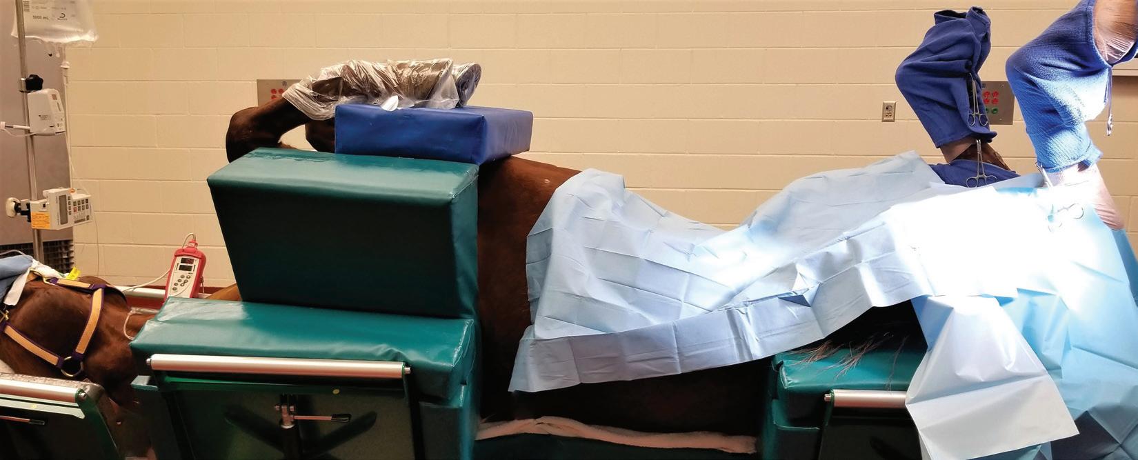
18 Positioning the Anesthetized Horse 287
Hui Chu Lin
19 Monitoring the Anesthetized Horse 294
Monitoring the Central Nervous System 294
Joanna C. Murrell
Cardiovascular Monitoring 300
Alanna N. Johnson
Respiratory Monitoring 310
Alanna N. Johnson
Anesthetic Agent Monitoring 323
Alanna N. Johnson
Monitoring Temperature 324
Chiara E. Hampton
Monitoring Neuromuscular Function 328
Manuel Martin-Flores and Daniel M. Sakai
20 Standing Sedation 333
Catherine M. Creighton
21 General Anesthesia Techniques 339
Regula Bettschart-Wolfensberger and Simone K. Ringer
22 Anesthesia of the Head and Neck 352
Anesthesia of the Head 352
Jim Schumacher, John Schumacher, and Ray Wilhite
Maxillary Nerve Block in Donkeys 358
Usama Hagag
Cervical Plexus Block 363
Luis Campoy
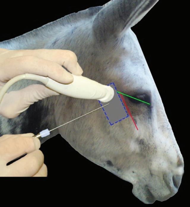
23 Anesthesia of the Eye 369
Daniel S. Ward
24 Anesthesia of the Limbs 376
Jim Schumacher, John Schumacher, and Ray Wilhite
25 Anesthesia of the Perineum and Testicle 396
Pudendal Nerve Block – Electrostimulation Technique 396
Kirsty Gallacher and Luiz Santos
Pudendal Nerve Block – Blind Approach 399
Jim Schumacher
Local Anesthesia for Castration 400
Philip D. Jones
26 Anesthesia of the Abdominal Wall 404
Thoracolumbar Paravertebral Block (TPVB) – Electrostimulation Technique 404
Luiz Santos and Kirsty Gallacher
Paravertebral Nerve Block – Blind Technique 406
Alex Valverde
Transversus Abdominis Plane Block 409
Alex Valverde and Flavio SA. Freitag
Caudal Intercostal Block for Abdominal Surgery (CIBAS) 413
Benjamin Gingold
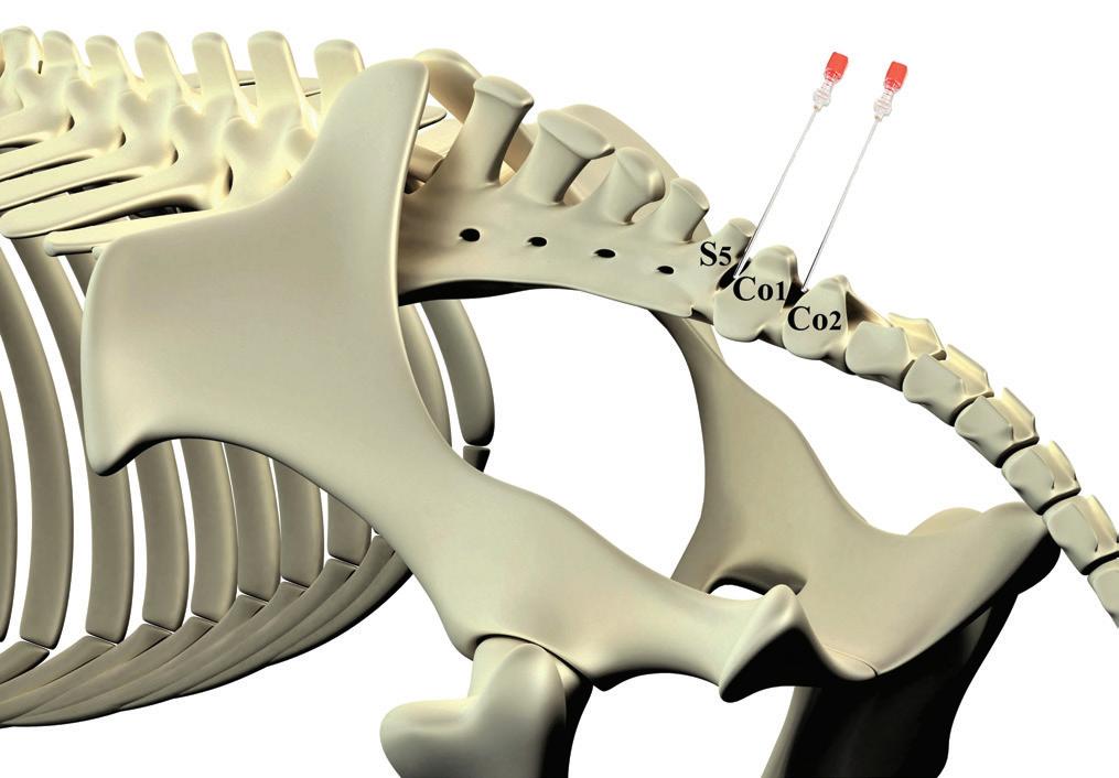
27 Epidural Analgesia and Anesthesia 416
Alex Valverde
28 Pathophysiology of Pain 425
Rachel A. Reed
29 Pain Recognition in Horses 431
Karina B. Gleerup, Casper Lindegaard, and Pia Haubro Andersen
30 Management of Pain 450
The Pharmacologic Approach to Pain Management 450
Rachel A. Reed
Rehabilitation Modalities for Acute and Chronic Pain in Horses 456
Tena L. Ursini
Equine Acupuncture 462
Neal Valk
31 Anesthesia of Foals 472
Tom Doherty and Alex Valverde
32 Anesthesia of Horses with Intestinal Emergencies (Colic) 494
Tom Doherty
33 Anesthesia of the Geriatric Horse 501
Reza M. Seddighi
34 Anesthesia and Pregnancy 511
Lydia Donaldson
35 Anesthesia for Equine Imaging 522
Carrie A. Davis
36 Anesthesia of Donkeys and Mules 543
Anatomic, Physiologic, and Behavioral Differences 543
Suzanne L. Burnham
Sedation and Anesthesia of Donkeys and Mules 548
Tom Doherty
Donkey Pain Assessment Scales 552
Machteld van Dierendonck and Thijs van Loon
37 Remote Capture of Equids 565
Nigel Caulkett
38 Complications 573
Intraoperative Hypotension 573
Christopher K. Smith and Tom Doherty
Intraoperative Hypertension 580
Tom Doherty
Hypoxia and Hypoxemia 582
Rachel A. Reed
Hypercarbia 588
Tom Doherty
Pulmonary Edema as a Consequence of Airway Obstruction 590
Tom Doherty
Endotoxemia 592
Tom Doherty
Postanesthetic Myopathy 594
Krista B. Mitchell
Neuropathy 599
Rachel A. Reed
Hyperkalemic Periodic Paralysis (HYPP) 604
Alanna N. Johnson and Rachel A. Reed
Malignant Hyperthermia 609
Alanna N. Johnson and Rachel A. Reed
Delayed Awakening and Recovery 611
Tom Doherty
Paraphimosis and Priaprism 614
Meggan Graves and Jim Schumacher
Anaphylactic and Anaphylactoid Reactions 621
Rachel A. Reed
Intra-carotid and Perivascular Injections 624
Rachel A. Reed
Equine Cardiopulmonary Resuscitation 627
Genevieve Bussieres
39 Recovery from Anesthesia 633
Bernd Driessen
40 Euthanasia 653
Ron Jones and Tom Doherty
Index 661
List of Contributors
Dr. Pia Haubro Andersen, DVM, PhD Department of Veterinary Clinical Sciences
Section of Medicine & Surgery
University of Copenhagen Denmark
Dr. Regula Bettschart-Wolfensberger, Dr. med. vet. PhD, DECVAA
Vetsuisse Faculty Department of Diagnostics and Clinical Services
Section of Anaesthesiology University of Zurich Zurich 8057
Switzerland
Dr. Suzanne L. Burnham, DVM Department of Veterinary Pathobiology College of Veterinary Medicine Texas A&M University College Station TX 77843-4474, USA
Dr. Genevieve Bussieres, DVM, MSc Department of Small Animal Clinical Sciences
The University of Tennessee College of Veterinary Medicine
Knoxville TN 37996-4545, USA
Dr. Luis Campoy, LV, Cert VA, DECVAA, MRCVS Department of Clinical Sciences
Cornell University College of Veterinary Medicine
Ithaca NY 14853, USA
Dr. Nigel Caulkett, DVM, MVSc, DACVAA Veterinary Clinical & Diagnostic Sciences University of Calgary, Calgary Alberta, Canada
Dr. Natalie S. Chow, DVM Department of Small Animal Clinical Sciences The University of Tennessee College of Veterinary Medicine Knoxville TN 37996-4545, USA
Dr. Catherine M. Creighton, DVM, MS, DACVAA Department of Companion Animals University of Prince Edward Island Charlottetown Prince Edward Island C1A 4P3, Canada
Dr. Carrie A. Davis, DVM, DACVAA Irving TX, USA
Dr. Machteld van Dierendonck, PhD Equine Behaviour Clinic Department of Equine Sciences Faculty of Veterinary Medicine Utrecht University, the Netherlands
Dr. Tom Doherty, MVB, MSc, DACVAA Knoxville TN, USA
Dr. Lydia Donaldson, DVM, PhD, DACVAA Middleburg VA 20118, USA
List of Contributors
Dr. Bernd Driessen, DVM, DrMedVet., DACVAA, DECVPT
University of Pennsylvania New Bolton Center Kennett Square PA 19348, USA
Dr. Tanya Duke-Novakovski, B VetMed, DACVAA Department of Veterinary Anesthesia
Radiology and Surgery
Western College of Veterinary Medicine University of Saskatchewan Saskatoon
Saskatchewan S7N 5B4, Canada
Dr. Christine Egger, DVM, MVSc, DACVAA Department of Veterinary Anesthesia, Radiology and Surgery
Western College of Veterinary Medicine University of Saskatchewan Saskatoon
Saskatchewan S7N 5B4, Canada
Dr. Kira L. Epstein, DVM, DACVS-LA, DACVECC
University of Georgia College of Veterinary Medicine
Athens GA 30602, USA
Dr. Flavio SA. Freitag, DVM, MSc Department of Clinical Studies Ontario Veterinary College The University of Guelph Ontario, Canada
Dr. Kirsty Gallacher School of Animal and Veterinary Sciences Roseworthy Campus
The University of Adelaide Adelaide 5371
Australia
Dr. Benjamin Gingold, DVM, DACVAA Turner ACT
Australia, 2612
Dr. Karina B. Gleerup, DVM, PhD Elleorevej 9 4000 Roskilde
Denmark
Dr. Diego E. Gomez, MV, MSc, MVSc, PhD, DACVIM Department of Clinical Studies Ontario Veterinary College The University of Guelph Ontario, Canada
Dr. Meggan Graves, DVM Department of Large Animal Clinical Sciences
The University of Tennessee College of Veterinary Medicine
Knoxville TN 37996-4545, USA
Dr. Tamara Grubb, DVM, PhD, DACVAA Uniontown WA 99179, USA
Dr. Usama Hagag, Dr. med. vet, PhD Faculty of Veterinary Medicine Department of Surgery, Anesthesiology and Radiology
Beni-Suef University
Beni-Suef 62511, Egypt
Dr. Chiara E. Hampton, DVM, MS, DACVAA School of Veterinary Medicine
Baton Rouge LA 70803, USA
Dr. Allana N. Johnson, DVM, DACVAA Department of Comparative, Diagnostic & Population Medicine University of Florida
Gainesville FL 32608, USA
Dr. Philip D. Jones, DVM, MS, DACVS-LA Department of Large Animal Clinical Sciences
The University of Tennessee College of Veterinary Medicine
Knoxville TN 37996-4545, USA
Dr. Ron Jones, MVSc, FRCVS, DVA, DrMedVet, DECVAA, DACVAA (Hon) Oxton, Prenton Merseyside CH43 5UF England
Dr. Daniel G. Kenney, VMD, DACVIM Health Sciences Centre Ontario Veterinary College
The University of Guelph Ontario, Canada
Dr. Carolyn Kerr, DVM, DVSc, PhD, DACVAA Department of Clinical Studies
Ontario Veterinary College
The University of Guelph Ontario, Canada
Dr. Stephanie Kleine, DVM, PhD, DACVAA Department of Small Animal Clinical Sciences
The University of Tennessee College of Veterinary Medicine
Knoxville TN 37996-4545, USA
Dr. Leigh Lamont, DVM, MS, DACVAA Department of Companion Animals University of Prince Edward Island Charlottetown Prince Edward Island Canada
Dr. Hui Chu Lin, DVM, MS, DACVAA Department of Clinical Sciences College of Veterinary Medicine
Auburn University AL 36849, USA
Dr. Casper Lindegaard, DVM, PhD, DECVS Department of Veterinary Clinical Sciences Section of Medicine & Surgery University of Copenhagen Denmark
Dr. Thijs van Loon, DVM, PhD, DACVAA Division of Anaesthesia and Intensive Care Department of Equine Sciences Faculty of Veterinary Medicine
Utrecht University, The Netherlands
Dr. Manuel Martin-Flores, MV, DACVAA Department of Clinical Sciences
Cornell University College of Veterinary Medicine
Ithaca NY 14853, USA
Dr. Krista B. Mitchell, DVM, DACVAA Department of Clinical Studies
Ontario Veterinary College
The University of Guelph Ontario, Canada
Dr. Joanna C. Murrell, BVSc, PhD, DECVAA
Highcroft Veterinary Referrals Whitchurch Bristol, UK
Dr. Patricia Queiroz-Williams, DVM, MS School of Veterinary Medicine
Baton Rouge LA 70803, USA
Dr. Rachel A. Reed, DVM, DACVAA University of Georgia College of Veterinary Medicine
Athens GA 30602, USA
Dr. Robert Reed, DVM, PhD Department of Biomedical and Diagnostic Sciences
The University of Tennessee College of Veterinary Medicine Knoxville TN 37996-4545, USA
Dr. Simone K. Ringer, PD, Dr. med. vet. PhD, DECVAA
Department of Clinical Diagnostics and Services
Section of Anaesthesiology
Vetsuisse Faculty University of Zurich
Zurich
Switzerland
List of Contributors
Dr. Daniel M. Sakai, MV, DACVAA University of Georgia College of Veterinary Medicine
Athens GA 30602, USA
Dr. Luiz Santos, DVM, MS, MANZCVS, DACVAA School of Animal and Veterinary Sciences
Roseworthy Campus
The University of Adelaide Adelaide 5371 Australia
Dr. Jim Schumacher, DVM, MS, DACVS Department of Large Animal Clinical Sciences
The University of Tennessee College of Veterinary Medicine Knoxville TN 37996-4545, USA
Dr. John Schumacher, DVM, MS, DACVIM Department of Clinical Sciences College of Veterinary Medicine
Auburn University AL 36849, USA
Dr. Reza M. Seddighi, DVM, PhD, DACVAA Department of Large Animal Clinical Sciences
The University of Tennessee College of Veterinary Medicine Knoxville TN 37996-4545, USA
Dr. Alicia Skelding, DVM, MSc, DVSc, DACVAA
Toronto Animal Health Partners Emergency and Specialty Hospital North York, Ontario Canada
Dr. Christopher K. Smith, DVM, DACVAA Department of Small Animal Clinical Sciences
The University of Tennessee College of Veterinary Medicine Knoxville TN 37996-4545, USA
Dr. Tanner Snowden, DVM
Oklahoma Equine Hospital
Washington OK 73093, USA
Dr. Carla Sommardahl, DVM, PhD, DACVIM Department of Large Animal Clinical Sciences
The University of Tennessee College of Veterinary Medicine
Knoxville TN 37996-4545, USA
Dr. Tena L. Ursini, DVM, DDACVSMR Department of Large Animal Clinical Sciences
The University of Tennessee College of Veterinary Medicine Knoxville TN 37996-4545, USA
Dr. Neal Valk, DVM, DACVS Department of Large Animal Clinical Sciences
The University of Tennessee College of Veterinary Medicine
Knoxville
TN 37996-4545, USA
Dr. Alex Valverde, DVM, DVSc, DACVAA Department of Clinical Studies
Ontario Veterinary College
The University of Guelph Guelph
Ontario NIG 2W1, Canada
Dr. Daniel S. Ward, DVM, PhD, DACVO Department of Small Animal Clinical Sciences
The University of Tennessee College of Veterinary Medicine
Knoxville
TN 37996-4545, USA
Dr. Ray Wilhite, MS, PhD Department of Anatomy, Physiology and Pharmacology College of Veterinary Medicine
Auburn University AL 36849, USA
Preface
The second edition of the Manual of Equine Anesthesia & Analgesia has been updated and rearranged. Most topics have been expanded and new chapters added. Chapters devoted to the sedation and anesthesia of horses undergoing imaging procedures, anesthesia of donkeys and mules, and recognition of pain in horses and donkeys are now included. Case examples of the pharmacologic control of pain are provided, and information on non-pharmacologic treatment of acute and chronic pain using acupuncture and physical rehabilitation techniques have also been added. In addition, a suggested reading list is included for the reader who wishes to seek further information on the topic.
This edition of the Manual of Equine Anesthesia & Analgesia strives to deliver relevant information on the physiologic and pharmacologic principles underpinning the anesthesia of equids, in addition to providing useful information on the clinical practice of anesthesia. The Manual has retained the same easily accessible format of the first edition, and we believe that it will be a useful guide to all those involved in the anesthesia of equids.
Tom Doherty
Alex Valverde
Rachel A. Reed
Acknowledgments
We wish to acknowledge our contributors for providing their expertise, and we greatly appreciate the time and effort they expended on this project. A special word of thanks to the staff at John Wiley & Sons, especially Ms. Erica Judisch, Ms. Susan Engelken, and Ms. Merryl Le Roux, for making this project possible.
1
Preoperative Evaluation and Patient Preparation
The Risk of Equine Anesthesia
Tanya Duke-Novakovski
● Risks of equine anesthesia have been linked with various conditions and situations and are reviewed in detail elsewhere and summarized in this chapter.
I Risk of equine anesthesia
● Anesthesia of the horse always involves an assessment of risk.
● Potential complications range from the less serious (e.g. skin wounds) to the more serious (e.g. long bone fractures, myopathies, and peripheral neuropathies), and to death in some cases.
● There is also risk of injury to personnel and safe handling should be practiced.
● The goal of the anesthetist is to minimize the adverse effects (ideally at minimum cost) by:
⚪ Identifying and defining the risk(s).
⚪ Selecting the best strategy for controlling or minimizing the risk(s).
● Data from single clinics have cited mortality rates of 0.24–1.8% in healthy horses.
● Data from multicenter studies cite the death rate for healthy horses undergoing anesthesia at around 0.9% (approximately 1 : 100).
● The overall death rate, when sick horses undergoing emergency colic surgery are included, has been reported to be 0.12% when fatalities were directly related to anesthesia in one study, and 1.9% in another study.
II Classification of physical status (see
Box 1.1)
● Classification of health status is generally based on the American Society of Anesthesiologists (ASA) system.
● This system uses information from the history, physical examination, and laboratory findings to place horses into one of five categories.
● The classification allows for standardization of physical status only.
● The ASA system does not classify risk, although increased risk of complication is associated with a high ASA status.
● These classifications are not always useful for horses: nevertheless, the system serves as a guide to case management.
Manual of Equine Anesthesia and Analgesia, Second Edition. Edited by Tom Doherty, Alex Valverde, and Rachel A. Reed. © 2022 John Wiley & Sons, Inc. Published 2022 by John Wiley & Sons, Inc.
Box 1.1 ASA Classification System
ASA 1 Healthy horse does not require intervention (e.g. castration).
ASA 2 Horse with mild systemic disease (e.g. mild anemia, mild recurrent airway obstruction) or localized injury (e.g. wound repair).
ASA 3 Horse with moderate systemic disease (e.g. stable colic, infected joint).
ASA 4 Horse with severe systemic disease (e.g. recent ruptured bladder, endotoxemia).
ASA 5 A moribund horse not expected to survive longer than 24 hours (e.g. unstable colic, ruptured bladder of several days duration).
E The letter E is added to status 2–5 under emergency conditions.
III Risk factors
A Age and physical status
⚪ The risk increases with age, and horses aged 12 years or older are at an increased risk of mortality.
⚪ Older horses may be more prone to fracture of a long bone in the recovery period, which could result in euthanasia.
⚪ Foals have an increased risk of fatality and this is probably associated with unfamiliarity with neonatal anesthesia, an immature cardiovascular system, and presence of systemic illness.
⚪ Pregnant mares have increased risk of mortality in the last trimester of pregnancy and this is probably associated with a need for emergency surgery. Otherwise, there is no difference between sexes.
⚪ Horses with a high ASA physical status have increased risk of mortality.
B Type of surgery and recovery
⚪ In otherwise healthy horses, the risk of mortality (euthanasia) following fracture repair is highest from repair failure or from fracture of another bone.
⚪ However, long periods of anesthesia typical of fracture repair have also been associated with increased mortality, and horses presented for fracture repair may be dehydrated and stressed. Emergency surgery (non-colic) carries a 4.25 times higher risk of mortality compared with elective surgery, and for emergency abdominal surgery the risk of fatality is 11.7%.
Colic surgery is associated with increased mortality because of a higher ASA physical status, emergency procedure with less time for stabilization, and use of dorsal recumbency possibly with episodes of hypotension.
Eye surgery, in one institution, resulted in longer recovery times and risk of complications and were associated with long anesthesia time compared to non-ophthalmic procedures. Fluconazole (microsomal P450 enzyme inhibitor) use was associated with increased risk of postoperative colic and longer recovery time. The use of the lowest effective volume of local anesthetic for a retrobulbar block was also recommended (10 ml/500 kg horse). Ophthalmic procedures have been associated with unsatisfactory recovery quality.
⚪ Assisted recovery with ropes can decrease the risk of fracture and dislocation, although the benefit of assisted-recovery is still debatable.
C Time of day
⚪ Performing anesthesia outside of normal working hours carries an increased risk for horses. This increase in risk is separate from the fact that most of these cases are emergency in nature.
⚪ Surgeries performed between midnight and 6 a.m. carry the highest risk of mortality. This may be due to the nature of the emergency, as well as to staff shortages and personnel fatigue.
D Body position
⚪ Dorsal recumbency was found to increase risk compared to either lateral recumbency, but most “colic” surgeries are performed with the horse in dorsal recumbency.
⚪ An increased risk of postanesthetic myelopathy has mostly been associated with draft breeds between 6 and 24 months of age and dorsal recumbency.
E Drug choice
⚪ Using total inhalational anesthesia regimen in foals (<12 months of age) without premedication carries the highest risk.
⚪ Halothane sensitizes the myocardium to circulating catecholamines. Fewer cardiac arrests occurred when isoflurane was substituted for halothane, although overall mortality did not differ between groups because limb fracture in recovery was prevalent for the isoflurane group.
⚪ Isoflurane and sevoflurane may be associated with unsatisfactory recovery, and sedation is often used following anesthesia to reduce the risk of excitable recovery.
⚪ Use of isoflurane and sevoflurane was linked with increased mortality, but this was because these drugs are more likely to be selected for sick horses.
⚪ Not using any premedication is associated with the highest risk, probably owing to increased circulating catecholamines from stress. It may be prudent to premedicate foals before induction of anesthesia, especially when using halothane.
⚪ Acepromazine lowers the risk of mortality when used alone as a premedicant, because it reduces the incidence of ventricular arrhythmias in the presence of halothane.
⚪ No particular injectable induction regimen is associated with greater risk when used with inhalational anesthesia.
⚪ Total intravenous anesthesia (TIVA) is associated with the lowest risk of all, but TIVA is often used for short procedures. TIVA has been associated with reduced stress response.
F Duration and management of anesthesia
⚪ Long periods of anesthesia (>2 hours) with volatile anesthetics are often associated with cardiovascular depression and poor tissue perfusion, leading to problems such as cardiac arrest or postanesthetic myopathy.
⚪ Intraoperative hypotension during anesthesia has clearly been associated with postanesthetic myopathy, and can still occur with short anesthetic periods. Direct arterial pressure monitoring should be used during lengthy anesthetic periods.
⚪ Postanesthetic myopathy may lead to bone fracture or dislocation.
⚪ Postanesthetic airway obstruction and pulmonary edema may be prevented by keeping the head in a normal position thereby reducing the risk of laryngeal nerve paralysis, and with good airway management in the recovery period.
Suggested Reading
Arndt, S., Hopster, K., Sill, V. et al. (2020). Comparison between head-tail-rope assisted and unassisted recoveries in healthy horses undergoing general anesthesia for surgeries. Vet. Surg. 49: 329–338.
Bidwell, L.A., Bramlage, L.R., and Rood, W.A. (2007). Equine perioperative fatalities associated with general anaesthesia. Vet. Anaesth. Analg. 34: 23–30.
Niimura del Barrio, M.C., David, F., Hughes, J.M.L. et al. (2018). A retrospective report (2003–2013) of the complications associated with the use of a one-man (head and tail) rope recovery system in horses following general anaesthesia. Ir. Vet. J. 71: 1–9.
Curto, E.M., Griffith, E.H., Posner, L.P. et al. (2018). Factors associated with postoperative complications in healthy horses after general anesthesia for ophthalmic versus non-ophthalmic procedures: 556 cases (2012–2014). J. Am. Vet. Med. Assoc. 252: 1113–1119.
Donaldson, L.J., Dunlop, C.S., Holland, M.S., and Burton, B.A. (2000). The recovery of horses from inhalant anesthesia: a comparison of halothane and isoflurane. Vet. Surg. 29: 92–101.
Dugdale, A.H.A. and Taylor, P.M. (2016). Equine anaesthesia-associated mortality: where are we now? Vet. Anaesth. Analg. 43: 242–255.
Dugdale, A.H.A., Obhrai, J., and Cripps, P.J. (2016). Twenty years later: a single-centre, repeat retrospective analysis of equine perioperative mortality and investigation of recovery quality. Vet. Anaesth. Analg. 43: 171–178.
Duke, T., Filsek, U., and Read, M.R. (2006). Clinical observations surrounding an increased incidence of post-anesthetic myopathy in halothane-anesthetized horses. Vet. Anaesth. Analg. 33: 122–127.
Grandy, J.L., Steffey, E.P., Hodgson, D.S., and Woliner, M.J. (1987). Arterial hypotension and the development of postanesthetic myopathy in halothane-anesthetized horses. Am. J. Vet. Res. 48: 192–197.
Johnston, G.M., Taylor, P.M., Holmes, M.A., and Wood, J.L.N. (1995). Confidential enquiry of perioperative equine fatalities (CEPEF-1): preliminary results. Equine. Vet. J. 27: 193–200.
Johnston, G.M., Eastment, J.K., Wood, J.L.N., and Taylor, P.M. (2002). The confidential enquiry into perioperative equine fatalities (CEPEF): mortality results of phases 1 and 2. Vet. Anaesth. Analg. 29: 159–170.
Johnston, G.M., Eastment, J.K., Taylor, P.M. et al. (2004). Is isoflurane safer than halothane in equine anaesthesia? Results from a prospective multicenter randomized controlled trial. Equine. Vet. J. 36: 64–71.
Leece, L., Corletto, F., and Brearley, J.C. (2008). A comparison of recovery times and characteristics with sevoflurane and isoflurane anaesthesia in horse undergoing magnetic resonance imaging. Vet. Anaesth. Analg. 35: 383–391.
Parviainen, A.K.J. and Trim, C.M. (2000). Complications associated with anaesthesia for ocular surgery: a retrospective study 1989-1996. Equine. Vet. J. 32: 555–559.
Ragle, C., Baetge, C., Yiannikouris, S. et al. (2011). Development of equine post anesthetic myelopathy: thirty cases (1979-2010). Equine. Vet. Educ. 23: 630–635.
Santos, M., Fuente, M., Garcia-Iturralde, P. et al. (2003). Effects of alpha2 adrenoceptor agonists during recovery from isoflurane anaesthesia in horses. Equine. Vet. J. 35: 170–175.
Senior, M. (2005). Post-anesthetic pulmonary oedema in horses: a review. Vet. Anaesth. Analg. 32: 193–200.
Young, S.S. and Taylor, P.M. (1990). Factors leading to serious anaesthetic related problems in equine anaesthesia. J. Ass. Vet. Anaesth. 17: 59. (abstr).
Young, S.S. and Taylor, P.M. (1993). Factors influencing the outcome of equine anaesthesia: a review of 1,314 cases. Equine. Vet. J. 25: 147–151.
Patient Preparation
Tanya Duke-Novakovski
I Preparation of the horse
A Evaluation
⚪ History and physical examination findings help with evaluation of health.
⚪ Many emergency cases, especially intestinal emergencies, are in cardiovascular shock and must be stabilized as much as possible prior to induction of anesthesia.
B Laboratory tests
⚪ In normal horses undergoing elective surgery, there is generally no value in performing extensive laboratory tests.
⚪ In emergency cases, performing laboratory tests such as electrolyte and metabolic acidbase status may be vital to the management of the case (e.g. a foal with uroabdomen).
C Physical examination
⚪ During the examination, attention should be directed to the neurological, cardiovascular, and respiratory systems.
⚪ Musculoskeletal problems, which may affect recovery, should be considered, and a plan should be made to assist recovery if deemed necessary.
D History
⚪ May reveal information that affects case management.
⚪ A recent history of coughing may indicate a viral infection of the airway, in which case elective surgeries should be postponed until one month following resolution of clinical signs.
⚪ Owners might report that the horse previously had a “bad” or “over” reaction to an anesthetic or sedative drug. These concerns should be investigated.
E Fasting
⚪ Fasting (~12 hours) was previously advised because of the potential benefits for lung function and the reduced risk of stomach rupture from trauma at induction or recovery.
⚪ Many equine hospitals do not fast horses prior to elective surgery. In one study, the PaO2 values during anesthesia were not significantly better in fasted versus non-fasted horses, and not fasting might reduce the incidence of postanesthetic colic due to changes in gastrointestinal motility.
⚪ However, it is generally the case that grain is removed.
⚪ Water should be made available up to the time of surgery.
F Medications
⚪ It is best to administer all ancillary drugs (e.g. antimicrobials, anti-inflammatories) prior to sedation. Sodium penicillin can reduce systolic arterial pressure by 8–15 mmHg in anesthetized horses. If antimicrobials are administered during the anesthetic event, this effect can be minimized by administering the drug slowly.
G Jugular catheter
⚪ An intravenous (IV) catheter should always be placed prior to anesthesia.
⚪ This reduces the likelihood of perivascular injection and provides ready access for further IV anesthetic or emergency drugs.
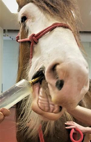
H Flushing the oral cavity
⚪ It is important to flush food debris from the oral cavity, especially if the airway is to be intubated (see Figure 1.1).
I Removal of shoes
⚪ Removal of shoes prevents damage to the horse and hospital flooring.
– However, removal of shoes is not popular with owners. An alternative is to apply bandage material or tape to improve grip and to cover metal points.
⚪ Certainly, loose shoes and nails should be removed.
⚪ Removal of shoes and metallic debris is necessary when an MRI is to be performed.
Figure 1.1 Rinsing of the mouth with water prior to induction of anesthesia using a large dosing syringe.
Suggested Reading
Bailey, P.A., Hague, B.A., Davis, M. et al. (2016). Incidence of post-anesthetic colic in non-fasted adult equine patients. Can. Vet. J. 57: 1263–1266.
Dobromylskyj, P., Taylor, P.M., Brearley, J.C. et al. (1996). Effect of pre-operative starvation on intraoperative arterial oxygen status in horses. J. Vet. Anaesth. 23: 75–77.
Hubbell, J.A.E., Muir, W.W., Robertson, J.T., and Sams, R.A. (1987). Cardiovascular effects of intravenous sodium penicillin, sodium cefazolin, and sodium citrate in awake and anesthetized horses. Vet. Surg. 16: 245–250.
Toews, A.R. and Campbell, J.R. (1997). Influence of preoperative complete blood cell counts on surgical outcomes in healthy horses: 102 cases (1986-1996). J. Am. Vet. Med. Assoc. 211: 887–888.
Serum Chemistry and Hematology
Carla Sommardahl
● As previously mentioned, there is little value in performing laboratory tests for healthy horses undergoing elective procedures.
● However, when indicated, the appropriate tests should, ideally, be performed before general anesthesia is induced. Nevertheless, this may not always be feasible in emergency situations.
I Complete blood count (CBC)
A Erythrocytes
○ Evaluation of erythrocyte numbers can begin with a determination of the packed cell volume (PCV) or hematocrit (HCT), which measure the percentage of the volume of whole blood that the red blood cells (RBCs) occupy.
○ Erythrocyte numbers are 6.9–10.7 x 10¹²/l, which represent a normal PCV of 35–45% and a normal hemoglobin of 12–15 g/dl.
○ A stained blood smear can be used to evaluate RBC morphology and the presence of infectious organisms on the RBCs.
○ Increased RBC numbers (erythrocytosis) is most commonly associated with hemoconcentration – this is called a relative erythrocytosis as there is no increase in RBC mass.
– Splenic contraction can cause a transient increase in circulating RBC mass.
○ In rare cases, there is an increase in numbers due to an increase in RBC production, absolute erythrocytosis
– Absolute erythrocytosis is further categorized as primary, as in polycythemia vera, and secondary, as results from chronic hypoxemia.
○ Decreased RBC numbers (anemia) can be caused by increased removal from the circulation by blood loss or destruction (hemolysis); or by decreased production by the bone marrow.
– Intravascular hemolysis is accompanied by a decrease in hemoglobin concentration; however, extravascular hemolysis is harder to confirm.
– Decreased production with iron deficiency secondary to chronic inflammation is a common cause of nonregenerative anemia in the horse, and is characterized by microcytic hypochromic RBCs and decreased RBC indices.
Manual of Equine Anesthesia and Analgesia, Second Edition. Edited by Tom Doherty, Alex Valverde, and Rachel A. Reed. © 2022 John Wiley & Sons, Inc. Published 2022 by John Wiley & Sons, Inc.
– Red cell distribution width (RDW) – a measure of RBC variation in size and volume - is used to detect RBC regeneration and is interpreted in conjunction with other values on the complete blood count (CBC).
B Leukocytes
○ Granulocytes (neutrophils, eosinophils, basophils, and mast cells), monocytes, macrophages, and lymphocytes are important for immune function, and changes in their numbers reflect a response to a disease process.
○ Total leukocyte count is 5.1–11.0 x 10⁹/l.
○ Neoplasia, bone marrow disease, or functional defects can cause a decrease or increase in leukocyte numbers.
Neutrophils
– Changes in numbers and morphology are important in evaluating the response to systemic inflammation, infection, and stress.
– However, the neutrophil count can be normal in the presence of inflammation or infection.
– Reference values include 2.8–7.7 x 10⁹/l segmented neutrophils and 0.0–0.2 × 10⁹/l bands.
– Neutrophilia is often associated with chronic bacterial infections.
– Neutropenia in adult horses is most often associated with endotoxemia (and other bacterial by-products).
– Neutropenia in foals is most commonly associated with sepsis and systemic inflammation.
Eosinophils
– Reference values are 0.0–0.7 × 10⁹/l.
– Eosinophilia is rare in horses. Conditions to rule out include:
▪ Parasitism with cyathastomes.
▪ Eosinophilic colitis, enteritis, or multisystemic disease.
Lymphocytes
– Reference values are 1.3–4.7 × 10⁹/l.
– Lymphopenia: Glucocorticoid release or administration, viral infection, old age, and immunodeficiency in foals (e.g. combined immunodeficiency disease in Arabian or Arabian crossbred foals).
– Lymphocytosis: epinephrine release or administration, exercise, equine herpesvirus 2 (foals), leukemia.
Monocytes and Basophils
– Reference values for monocytes are 0.1–0.8 × 10⁹/l.
– Monocytosis: Chronic inflammation, use of corticosteroids.
– Reference values for basophils are 0.0–0.1 × 10⁹/l.
C Platelets
○ The normal platelet count for horses is 75 000–300 000/μl.
○ Platelets function in hemostasis, inflammatory responses, immunity, tissue regeneration, and disease pathology.
Thrombocytosis (platelet count >400 000/μl)
Reactive or Secondary:
– Age < 3 years, intact males, pyrexia, infectious, or inflammatory conditions. – Platelet count: 400 000–850 000/μl.
Primary or Clonal:
– A rare chronic myeloproliferative disorder.
Platelet count >1 000 000/μl.
Thrombocytopenia (platelet count <75 000/μl)
Increased platelet destruction:
– Immune mediated.
– Causes include:
▪ Equine infectious anemia, anaplasma phagocytophilum.
▪ Drugs, toxins, snake bites.
Increased use:
– Hemorrhage.
– Disseminated intravascular coagulation.
Decreased production:
– Myelosuppressive drugs (e.g. phenylbutazone). – Bone marrow disease.
– Idiopathic.
II Blood chemistry interpretation
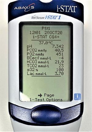
● Point of care (POC) analyzers have made the evaluation of chemistry, electrolyte, and blood gas values available rapidly as stall-side or immediate vicinity testing (see Figures 2.1 and 2.2).
Figure 2.1 Bench top point-of-care blood analyzer, Source: NOVA Biomedical.
Figure 2.2 Stall-side point-of-care blood analyzer, Source: Abbott point-of-care iSTAT system.
● Specific reference ranges from the laboratory or analyzer conducting the analysis should be utilized.
○ Reference ranges for foals should be used based on age.
○ Reference ranges for donkeys (asses, burros), mules, and hinnies should be used accordingly.
A Creatine kinase (CK)
○ Also called creatine phosphokinase (CPK).
○ The reference range for the horse is 108–430 U/l.
○ CK is a specific indicator of muscle damage.
– It is a “leakage” enzyme released secondary to myocyte damage or death.
○ Serum concentrations increase quickly following myocyte damage.
– In contrast to aspartate aminotransferase (AST) concentrations which increase more slowly and persist for longer (see below).
○ CK has a short half-life (hours); thus, serum concentrations decrease quickly after an episode.
○ Urine will be positive for blood on test strips if myoglobin is present.
– Myoglobin and free hemoglobin have peroxidase-like activity, which results in oxygen liberation from organic peroxide in the reagent strip, causing a color change in the strip.
– The urine may be “coffee” colored.
Mild increases in CK (1000–5000 U/l) may result from:
– Recumbency, trailer ride, recent exercise, or intramuscular injections.
– Consider a diagnosis of mild exertional rhabdomyolysis (ER) and/or polysaccharide storage myopathy (PSSM) if the above conditions are not applicable.
Moderate (~5000 U/l) to severe increases in CK (>10 000 U/l):
– Severe cases of ER may have associated CK values >100 000 U/l.
– These high values may be associated with a traumatic event, but may occur following general anesthesia.
Note: Because myoglobin may cause renal tubular damage, treatment may be indicated.
B Aspartate aminotransferase (AST)
○ AST is a “leakage” enzyme released secondary to myocyte or hepatocyte damage or death.
Small amounts are also present in myocardial cells.
○ Increased AST values in horses are most commonly associated with skeletal muscle damage.
– The reference range for the horse is 160–400 U/l.
○ Serum AST may also increase significantly in horses in early training.
○ AST values in foals may be greater than adult values for many months.
– This increase may be related to muscle growth.
○ Small amounts are present in RBCs.
– Thus, hemolysis can cause small increases in serum AST.
○ AST in serum increases more slowly (peak 6–12 hours) than does CK after muscle injury.
○ It has a long half-life (~seven days) in horses compared to other domestic species, and serum concentrations fall slowly (three to four days to return to normal) after an episode.
Note: AST activity will remain increased after CK activity has returned to normal.
○ AST values should be interpreted in association with CK and other liver enzymes (Sorbitol (Iditol) dehydrogenase [SDH], Gamma glutamyl transferase [GGT]).
○ Serum AST activity can also increase with in vivo or in vitro hemolysis.
C Gamma glutamyl transferase (GGT)
○ GGT resides in the cell membranes of all somatic cells, but its greatest activity is in biliary epithelial cells, pancreatic acinar cells, and renal tubular epithelial cells.
○ GGT is a cell surface glycoprotein and is a liver canalicular enzyme, and it is induced by dilation of the biliary tree.
○ It has multiple functions including the regulation of the anti-oxidant glutathione.
○ Increased GGT activity in serum is usually an indication of hepatobiliary toxicity, especially cholestasis.
○ Bile accumulation (cholestasis), biliary hyperplasia, and some drugs (e.g. phenobarbital, furosemide, phenytoin, and cimetidine) increase serum GGT activity.
○ GGT has a long half-life (approximately three days), thus serum concentrations fall slowly after an episode.
○ Chronic liver disease associated with hepatocyte destruction, fibrosis, and biliary hyperplasia are associated with severe increases in GGT.
○ Right dorsal displacement of the colon causes an increase in serum GGT activity by impeding the outflow of bile.
○ Proximal enteritis causes increases in AST and GGT activity secondary to ascending infection of the biliary tract and/or liver damage from systemic effects such as endotoxemia and hypovolemia.
○ Reference range: Adult horses 6–30 U/l.
○ The normal range for burros, donkeys and asses may be 2–3 times higher, and mules and hinnies may be two times higher than values for horses.