
Stoelting’s Anesthesia and Co-Existing Disease 6th Edition
Visit to download the full and correct content document: https://ebookmass.com/product/stoeltings-anesthesia-and-co-existing-disease-6th-edi tion/
More products digital (pdf, epub, mobi) instant download maybe you interests ...

Equine Anesthesia and Co-Existing Disease 1st Edition
Stuart Clark-Price
https://ebookmass.com/product/equine-anesthesia-and-co-existingdisease-1st-edition-stuart-clark-price/
Stoelting’s Anesthesia and Co Existing Disease E Book 7th Edition, (Ebook PDF)
https://ebookmass.com/product/stoeltings-anesthesia-and-coexisting-disease-e-book-7th-edition-ebook-pdf/
Stoelting's Anesthesia and Co-Existing Disease, 7e (May 2, 2017)_(0323401377)_(Elsevier).pdf Roberta L. Hines
https://ebookmass.com/product/stoeltings-anesthesia-and-coexisting-disease-7e-may-2-2017_0323401377_elsevier-pdf-roberta-lhines/
Anesthesia Secrets 6th Edition Edition Brian Keech
https://ebookmass.com/product/anesthesia-secrets-6th-editionedition-brian-keech/

Nurse Anesthesia 6th Edition – Ebook PDF Version
https://ebookmass.com/product/nurse-anesthesia-6th-edition-ebookpdf-version/
Anesthesia Secrets, 6th Edition Brian M. Keech
https://ebookmass.com/product/anesthesia-secrets-6th-editionbrian-m-keech/
Nurse Anesthesia 6th Edition Edition John J. Nagelhout
https://ebookmass.com/product/nurse-anesthesia-6th-editionedition-john-j-nagelhout/
Nurse Anesthesia E Book 6th Edition, (Ebook PDF)
https://ebookmass.com/product/nurse-anesthesia-e-book-6thedition-ebook-pdf/
Chestnut’s Obstetric Anesthesia: Principles and Practice 6th Edition Edition David Chestnut
https://ebookmass.com/product/chestnuts-obstetric-anesthesiaprinciples-and-practice-6th-edition-edition-david-chestnut/
(pseudonormalization of the T wave) during myocardial ischemia. These ECG changes are seen in 50% of patients. Variant angina, that is, angina that results from coronary vasospasm rather than occlusive coronary artery disease, is diagnosed by ST elevation during an episode of angina pectoris.
Exercise ECG is useful for detecting signs of myocardial ischemia and establishing their relationship to chest pain. The test also provides information about exercise capacity. The appearance of a new murmur of mitral regurgitation or a decrease in blood pressure during exercise adds to the diagnostic value of the test. Exercise testing is not always feasible, either because of the inability of a patient to exercise or the presence of conditions that interfere with interpretation of the exercise ECG (paced rhythm, left ventricular hypertrophy, digitalis administration, or preexcitation syndrome). Contraindications to exercise stress testing include severe aortic stenosis, severe hypertension, acute myocarditis, uncontrolled heart failure, and infective endocarditis.
The exercise ECG is most likely to indicate myocardial ischemia when there is at least 1 mm of horizontal or down-sloping ST-segment depression during or within 4 minutes after exercise. The greater the degree of ST-segment depression, the greater is the likelihood of significant coronary artery disease. When the ST-segment abnormality is associated with angina pectoris and occurs during the early stages of exercise and persists for several minutes after exercise, significant coronary artery disease is very likely. Exercise ECG is less accurate but more cost effective than imaging tests for detecting ischemic heart disease. A negative stress test result does not exclude the presence of coronary artery disease, but it makes the likelihood of three-vessel or left main coronary disease extremely low. Exercise ECG is less sensitive and specific in detecting ischemic heart disease than nuclear cardiology techniques (Table 1-3).
NUCLEAR CARDIOLOGY TECHNIQUES
Nuclear stress imaging is useful for assessing coronary perfusion. It has greater sensitivity than exercise testing for detection of ischemic heart disease. It can define vascular regions in which stress-induced coronary blood flow is limited and can estimate left ventricular systolic size and function. Tracers such as thallium and technetium can be detected over the myocardium by single-photon emission computed tomography (SPECT) techniques. A significant coronary obstructive lesion causes less blood flow and thus less tracer activity. Exercise perfusion imaging with simultaneous ECG testing is superior to exercise ECG alone (see Table 1-3). Exercise increases the difference in tracer activity between normal and underperfused regions, because coronary blood flow increases markedly with exercise except in those regions distal to a coronary artery obstruction. Imaging is carried out in two phases: the first is immediately after cessation of exercise to detect regional ischemia, and the second is 4 hours later to detect reversible ischemia. Areas of persistently absent uptake signify an old MI. The size of the perfusion abnormality is the most important indicator of the significance of the coronary artery disease detected.
TABLE
1-3 n Sensitivity and specificity of stress testing*
Data from Gibbons RJ, Abrams J, Chatterjee K, et al. ACC/AHA 2002 guideline update for the management of patients with chronic stable angina: a report of the American College of Cardiology/American Heart Association Task Force on Practice Guidelines. Circulation. 2003;107: 149–158. (Committee to Update the 1999 Guidelines for the Management of Patients with Chronic Stable Angina).
SPECT, Single-photon emission computed tomography.
*Without correction for referral bias.
†Weighted average pooled across individual trials.
Many patients who are at increased risk of coronary events cannot exercise because of peripheral vascular or musculoskeletal disease, deconditioning, dyspnea on exertion due to pulmonary disease, or prior stroke. Noninvasive imaging tests for the detection of ischemic heart disease are usually recommended when exercise ECG is not possible or interpretation of ST-segment changes would be difficult. Administration of atropine, infusion of dobutamine, or institution of artificial cardiac pacing produces a rapid heart rate to create cardiac stress. Alternatively, cardiac stress can be produced by administering a coronary vasodilator such as adenosine or dipyridamole. These drugs dilate normal coronary arteries but evoke minimal or no change in the diameter of atherosclerotic coronary arteries. After cardiac stress is induced by these interventions, radionuclide tracer scanning is performed to assess myocardial perfusion.
ECHOCARDIOGRAPHY
Echocardiographic wall motion analysis can be performed immediately after stressing the heart either pharmacologically or with exercise. New ventricular wall motion abnormalities induced by stress correspond to sites of myocardial ischemia, thereby localizing obstructive coronary lesions. In contrast, exercise ECG can indicate only the presence of ischemic heart disease and does not reliably predict the location of the obstructive coronary lesion. One can also visualize global wall motion under baseline conditions and under cardiac stress. Valvular function can be assessed as well. Limitations imposed by poor visualization have been improved by newer contrast-assisted technologies that have improved the accuracy of stress echocardiography.
STRESS CARDIAC MAGNETIC RESONANCE IMAGING
Pharmacologic stress imaging with cardiac magnetic resonance imaging compares favorably with other methods and is being used clinically in some centers, especially when other modalities cannot be used effectively.
ELECTRON BEAM COMPUTED TOMOGRAPHY
Calcium deposition occurs in atherosclerotic vessels. Coronary artery calcification can be detected by electron beam computed tomography. Although the sensitivity of electron beam computed tomography is high, it is not a very specific test and yields many false-positive results. Its routine use is not recommended.
CORONARY ANGIOGRAPHY
Coronary angiography provides the best information about the condition of the coronary arteries. It is indicated in patients with known or possible angina pectoris who have survived sudden cardiac death, those who continue to have angina pectoris despite maximal medical therapy, and those who are being considered for coronary revascularization, as well as for the definitive diagnosis of coronary disease for occupational reasons (e.g., in airline pilots). Coronary angiography is also useful for establishing the diagnosis of nonatherosclerotic coronary artery disease such as coronary artery spasm, Kawasaki's disease, radiation-induced vasculopathy, and primary coronary artery dissection. Among patients with chronic stable angina 25% will have significant single-, double-, or triple-vessel coronary artery disease, 5% to 10% will have left main coronary artery disease, and 15% will have no flow-limiting obstructions.
The important prognostic determinants in patients with coronary artery disease are the anatomic extent of the atherosclerotic disease, the state of left ventricular function (ejection fraction), and the stability of the coronary plaque. Left main coronary artery disease is the most dangerous anatomic lesion and is associated with an unfavorable prognosis when managed with medical therapy alone. Greater than 50% stenosis of the left main coronary artery is associated with a mortality rate of 15% per year.
Unfortunately, coronary angiography cannot predict which plaques are most likely to rupture and initiate acute coronary syndromes. Vulnerable plaques, that is, those most likely to rupture and form an occlusive thrombus, have a thin fibrous cap and a large lipid core containing a large number of macrophages. The presence of vulnerable plaque predicts a greater risk of MI regardless of the degree of coronary artery stenosis. Indeed, acute MI most often results from rupture of a plaque that had produced less than 50% stenosis of a coronary artery. Currently, there is no satisfactory test to measure the stability of plaques.
Treatment
Comprehensive management of ischemic heart disease has five aspects: (1) identification and treatment of diseases that can precipitate or worsen ischemia, (2) reduction of risk factors for coronary artery disease, (3) lifestyle modification, (4) pharmacologic management of angina, and (5) revascularization by coronary artery bypass grafting (CABG) or percutaneous coronary intervention (PCI) with or without placement of intracoronary stents. The goal of treatment of patients with
chronic stale angina is to achieve complete or almost complete elimination of anginal chest pain and a return to normal activities with minimal side effects.
TREATMENT OF ASSOCIATED DISEASES
Conditions that increase oxygen demand or decrease oxygen delivery may contribute to an exacerbation of previously stable angina or worsen existing angina. These conditions include fever, infection, anemia, tachycardia, thyrotoxicosis, heart failure, and cocaine use. Treatment of these conditions is critical to the management of stable ischemic heart disease.
REDUCTION OF RISK FACTORS AND LIFESTYLE MODIFICATION
The progression of atherosclerosis may be slowed by cessation of smoking; maintenance of an ideal body weight by consumption of a low-fat, low-cholesterol diet; regular aerobic exercise; and treatment of hypertension. Lowering the low-density lipoprotein (LDL) cholesterol level by diet and/or drugs such as statins is associated with a substantial decrease in the risk of death due to cardiac events. Drug treatment is appropriate when the LDL cholesterol level exceeds 130 mg/dL. The goal of treatment is a decrease in LDL to less than 100 mg/dL. Patients with ischemic heart disease may benefit from even lower LDL levels (<70 mg/dL), which can be achieved by a combination of diet and statin therapy. Hypertension increases the risk of coronary events as a result of direct vascular injury, left ventricular hypertrophy, and increased myocardial oxygen demand. Lowering the blood pressure from hypertensive levels to normal levels decreases the risk of MI, congestive heart failure, and stroke. In combination with lifestyle modifications, β-blockers, and calcium channel blockers are especially useful in managing hypertension in patients with angina pectoris. If left ventricular dysfunction accompanies hypertension, an angiotensin-converting enzyme (ACE) inhibitor or an angiotensin receptor blocker (ARB) is recommended.
MEDICAL TREATMENT OF MYOCARDIAL ISCHEMIA
Antiplatelet drugs, nitrates, β-blockers, calcium channel blockers, and ACE inhibitors are used in the medical treatment of angina pectoris.
Three classes of antiplatelet drugs are widely used in the management of ischemic heart disease: aspirin, thienopyridines (clopidogrel and prasugrel), and platelet glycoprotein IIb/IIIa inhibitors (eptifibatide, tirofiban, and abciximab).
A fourth class of antiplatelet drug, which affects platelet cyclic adenosine monophosphate (dipyridamole), is not widely used. A new class of short-acting, reversible platelet inhibitors (cangrelor and ticagrelor) is currently under development.
Aspirin inhibits the enzyme cyclooxygenase-1 (COX-1); this results in inhibition of thromboxane A2, which plays an important role in platelet aggregation. This inhibition of COX-1 is irreversible, lasts for the duration of platelet life span (around 7 days), and can be produced by low dosages of aspirin. Low-dose aspirin therapy (75 to 325 mg/day) decreases the risk of cardiac events in patients with stable or unstable angina
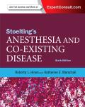
pectoris and is recommended for all patients with ischemic heart disease. Clopidogrel inhibits the adenosine diphosphate (ADP) receptor P2Y12 and inhibits platelet aggregation in response to ADP release from activated platelets (Figure 1-2).
Clopidogrel-induced inhibition of ADP receptors is irreversible and also lasts for the duration of the platelet's life span. Seven days after cessation of this drug 80% of platelets will have recovered normal aggregation function. Clopidogrel is a prodrug that is metabolized into an active compound in the liver. Due to genetic differences in the enzymes that metabolize clopidogrel to the active drug, significant variability in its activity has been observed. By some estimates, 10% to 20% of patients taking aspirin and clopidogrel demonstrate hyporesponsiveness (resistance) or hyperresponsiveness. Furthermore, some drugs, such as proton pump inhibitors, can affect the enzyme that metabolizes clopidogrel to its active compound and thereby can reduce the effectiveness of clopidogrel.
Clopidogrel can be used in patients who have a contraindication to or are intolerant of aspirin. Prasugrel also inhibits the ADP P2Y12 receptor irreversibly. However, the pharmacokinetics of prasugrel are more predictable. It is rapidly absorbed,
has a faster onset of action, and demonstrates less interindividual variability in platelet responses compared with clopidogrel. It also is more potent than clopidogrel, and a higher risk of bleeding has been associated with its use. Platelet glycoprotein IIb/IIIa receptor antagonists (abciximab, eptifibatide, tirofiban) inhibit platelet adhesion, activation, and aggregation. Short-term administration of antiplatelet drugs is particularly useful after placement of an intracoronary stent.
Organic nitrates decrease the frequency, duration, and severity of angina pectoris and increase the amount of exercise required to produce ST-segment depression. The antianginal effects of nitrates are greater when used in combination with β-blockers or calcium channel blockers. Nitrates dilate coronary arteries and collateral blood vessels and thereby improve coronary blood flow. Nitrates also decrease peripheral vascular resistance, which reduces left ventricular afterload and myocardial oxygen consumption. The venodilating effect of nitrates decreases venous return and hence left ventricular preload and myocardial oxygen consumption. They also have potential antithrombotic effects. Nitrates are contraindicated in the presence of hypertrophic cardiomyopathy or
Ticlopidine Clopidogrel Prasugrel
Cangrelor
FIGURE 1-2 Platelet activation mechanisms and sites of blockade of antiplatelet therapies. ↑, Increased; ↓, decreased; ADP, adenosine diphosphate; ASA, acetylsalicylic acid; ATP, adenosine triphosphate; cAMP, cyclic adenosine monophosphate; COX-1, cyclooxygenase-1; CYP450, cytochrome P450; GP, glycoprotein; GPVI, [glycoprotein VI]; P2X1, P2Y1, purinergic receptors; PAR, protease-activated receptor; TP, thromboxane receptor; TRA, [thrombin recepter agonist]; TxA2, thromboxane A2 (From Cannon CP, Braunwald E. Unstable angina and non-ST elevation myocardial infarction. In: Bonow RO, Mann DL, Zipes DP, et al, eds. Braunwald's Heart Disease. Philadelphia, PA: Saunders; 2012.)
severe aortic stenosis and should not be used within 24 hours of sildenafil, tadalafil, or vardenafil because this combination may produce severe hypotension. Administration of sublingual nitroglycerin by tablet or spray produces prompt relief of angina pectoris. The most common side effect of nitrate treatment is headache. Hypotension may occur after nitrate administration in hypovolemic patients. For long-term therapy, long-acting nitrate preparations (isosorbide, nitroglycerin ointment or patches) are equally effective. The therapeutic value of organic nitrates is compromised by the development of tolerance. To avoid nitrate tolerance, a daily 8- to 12-hour interval free of nitrate exposure is recommended.
β-Blockers are the principal drug treatment for patients with stable angina pectoris. They have antiischemic, antihypertensive, and antidysrhythmic properties. Long-term administration of β-blockers decreases the risk of death and myocardial reinfarction in patients who have had an MI, presumably by decreasing myocardial oxygen demand. This benefit is present even in patients in whom β-blockers were traditionally thought to be contraindicated, such as those with congestive heart failure, pulmonary disease, or advanced age. Drug-induced blockade of β1-adrenergic receptors (atenolol, metoprolol, acebutolol, bisoprolol) results in heart rate slowing and decreased myocardial contractility that are greater during activity than at rest. The result is a decrease in myocardial oxygen demand with a subsequent decrease in ischemic events during exertion. The decrease in heart rate also increases the length of diastole and thereby coronary perfusion time. β2-Adrenergic blockers (propranolol, nadolol) can increase the risk of bronchospasm in patients with reactive airway disease. Despite differences between β1 and β2 effects, all β-blockers seem to be equally effective in the treatment of angina pectoris. The most common side effects of β-blocker therapy are fatigue and insomnia. Heart failure may be intensified. β-Blockers are contraindicated in the presence of severe bradycardia, sick sinus syndrome, severe reactive airway disease, second- or third-degree atrioventricular heart block, and uncontrolled congestive heart failure. Diabetes mellitus is not a contraindication to β-blocker therapy, although these drugs may mask signs of hypoglycemia. Abrupt withdrawal of β-blockers after prolonged administration can worsen ischemia in patients with chronic stable angina.
Long-acting calcium channel blockers are comparable to β-blockers in relieving anginal pain. However, short-acting calcium channel blockers such as verapamil and diltiazem are not. Calcium channel blockers are uniquely effective in decreasing the frequency and severity of angina pectoris due to coronary artery spasm (Prinzmetal's or variant angina). They are not as effective as β-blockers in decreasing the incidence of myocardial reinfarction. The effectiveness of calcium channel blockers is due to their ability to decrease vascular smooth muscle tone, dilate coronary arteries, decrease myocardial contractility and oxygen consumption, and decrease systemic blood pressure. Many calcium channel blockers such as amlodipine, nicardipine, isradipine, felodipine, and longacting nifedipine are potent vasodilators and are useful in
treating both hypertension and angina. Common side effects of calcium channel blocker therapy are hypotension, peripheral edema, and headache. Calcium channel blockers are contraindicated in patients with severe congestive heart failure or severe aortic stenosis. They must be used cautiously if given in combination with β-blockers, because both classes of drugs have significant depressant effects on heart rate and myocardial contractility.
Excessive angiotensin II plays a significant role in the pathophysiology of cardiac disorders. It can lead to development of myocardial hypertrophy, interstitial myocardial fibrosis, increased coronary vasoconstriction, and endothelial dysfunction. Angiotensin II also promotes inflammatory responses and atheroma formation. ACE inhibitors are important not only in the treatment of heart failure but also in the treatment of hypertension and in cardiovascular protection. ACE inhibitors are recommended for patients with coronary artery disease, especially those with hypertension, left ventricular dysfunction, or diabetes. Angiotensin receptor blockers offer similar benefits. Contraindications to ACE inhibitor use include documented intolerance or allergy, hyperkalemia, bilateral renal artery stenosis, and renal failure.
REVASCULARIZATION
Revascularization by CABG or PCI with or without placement of intracoronary stents is indicated when optimal medical therapy fails to control angina pectoris. Revascularization is also indicated for specific anatomic lesions—in particular, left main coronary artery stenosis of more than 50% or the presence of a 70% or greater stenosis in an epicardial coronary artery. Revascularization is also indicated in patients with significant coronary artery disease with evidence of impaired left ventricular contractility (ejection fraction of <40%). The presence of hypokinetic or akinetic areas in the left ventricle connotes a poor prognosis. Extensive myocardial fibrosis from a prior MI is unlikely to be improved by revascularization. However, some patients with ischemic heart disease have chronically impaired myocardial function (hibernating myocardium) that demonstrates improvement in contractility following surgical revascularization. In patients with stable angina pectoris and one- or two-vessel coronary artery disease, a percutaneous intervention with or without stent placement, or surgical coronary artery bypass may be used for revascularization. CABG is preferred over PCI in patients with significant left main artery disease, those with three-vessel coronary artery obstruction, and patients with diabetes who have two- or three-vessel coronary artery disease. Operative mortality rates for CABG surgery currently range from 1.5% to 2%.
ACUTE CORONARY SYNDROME
Acute coronary syndrome represents a hypercoagulable state. Focal disruption of an atheromatous plaque triggers the coagulation cascade with subsequent generation of thrombin and partial or complete occlusion of the coronary artery by a thrombus. Imbalance of myocardial oxygen supply and
demand leads to ischemic chest pain. Patients who have ischemic chest pain can be categorized based on the findings of a 12-lead ECG. Patients with ST elevation at presentation are considered to have STEMI. Patients who have ST-segment depression or nonspecific changes on the ECG can be categorized based on the levels of cardiac-specific troponins or myocardial creatine kinase (CK-MB). Elevation of cardiac-specific biomarker levels in this situation indicates NSTEMI. If levels of cardiac-specific biomarkers are normal, then unstable angina is present (Figure 1-3). STEMI and UA/NSTEMI are treated differently and have different prognoses. Many more patients have UA/NSTEMI than have STEMI at presentation. The different kinds of MI often occur in different clinical situations.
ST Elevation Myocardial Infarction
Mortality rates from STEMI have declined steadily because of early therapeutic interventions such as angioplasty, thrombolysis and aspirin, heparin, and statin therapy. However, the mortality rate of acute MI remains significant. The short-term mortality rate of patients with STEMI who receive aggressive reperfusion therapy is about 6.5%. Data from the general medical community show a mortality rate of 15% to 20% (these patients have not received reperfusion therapy). Advanced age consistently emerges as one of the principal determinants of early mortality in patients with STEMI. Coronary angiography has documented that nearly all STEMIs are caused by thrombotic occlusion of a coronary artery.
The long-term prognosis after an acute MI is determined principally by the severity of residual left ventricular dysfunction, the presence and degree of residual ischemia, and the presence of malignant ventricular dysrhythmias. Most deaths that occur during the first year after hospital discharge take place within the first 3 months. Ventricular function can be substantially improved during the first few weeks after an acute MI, particularly in patients in whom early reperfusion was achieved. Therefore, measurement of ventricular function 2 to 3 months after an MI is a more accurate predictor of longterm prognosis than measurement of ventricular function during the acute phase of the infarction.
PATHOPHYSIOLOGY
Atherosclerosis is being increasingly recognized as an inflammatory disease. The presence of inflammatory cells in atherosclerotic plaques suggests that inflammation is important in the cascade of events leading to plaque rupture. Indeed, serum markers of inflammation, such as C-reactive protein and fibrinogen, are increased in those at greatest risk of developing coronary artery disease.
STEMI occurs when coronary blood flow decreases abruptly. This decrease in blood flow is attributable to acute thrombus formation at a site when an atherosclerotic plaque fissures, ruptures, or ulcerates. This creates a local environment that favors thrombogenesis. Typically, vulnerable plaques— that is, those with rich lipid cores and thin fibrous caps—are most prone to rupture. A platelet monolayer forms at the site
STABLE ANGINA
Ischemic type chest pain
New onset or change from baseline
Acute coronary syndrome
12-Lead ECG
No ST-segment elevation ST-segment elevation
Troponin/CK-MB negative
Troponin/CK-MB positive
NSTEMI
Unstable angina
Troponin/CK-MB positive
STEMI
Myocardial infarction
FIGURE 1-3 Terminology of acute coronary syndrome. CK-MB, Creatine kinase, myocardial-bound isoenzyme; ECG, electrocardiogram; NSTEMI, non–ST elevation myocardial infarction; STEMI, ST elevation myocardial infarction. (Adapted from Alpert JS, Thygesen K, Antman E, et al. Myocardial infarction redefined—a consensus document of the Joint European Society of Cardiology/American College of Cardiology Committee for the redefinition of myocardial infarction. J Am Coll Cardiol. 2000;36:959-969.)
of ruptured plaque, and various chemical mediators such as collagen, ADP, epinephrine, and serotonin stimulate platelet aggregation. The potent vasoconstrictor thromboxane A2 is released, which further compromises coronary blood flow. Glycoprotein IIb/IIIa receptors on the platelets are activated, which enhances the ability of platelets to interact with adhesive proteins and other platelets and causes growth and stabilization of the thrombus. Further activation of coagulation leads to strengthening of the clot by fibrin deposition. This makes the clot more resistant to thrombolysis. It is rather paradoxical that plaques that rupture and lead to acute coronary occlusion are rarely of a size that causes significant coronary obstruction. By contrast, flow-restrictive plaques that produce angina pectoris and stimulate development of collateral circulation are less likely to rupture. Rarely, STEMI develops as a result of acute coronary spasm or coronary artery embolism.
DIAGNOSIS
With recent advances in the techniques for detecting MI, the criteria for diagnosing an acute, evolving, or recent MI have been revised (Table 1-4). Diagnosis of acute MI requires the typical rise and subsequent fall in plasma levels of biochemical markers of myocardial necrosis in combination with at least one of the following: (1) ischemic symptoms, (2) development of pathologic Q waves on the ECG, (3) ECG changes
TABLE 1-4 n Revised definition of myocardial infarction
CRITERIA FOR ACUTE, EVOLVING, OR RECENT MYOCARDIAL INFARCTION
Either of the following criteria satisfies the diagnosis for acute, evolving, or recent myocardial infarction:
1. Typical rise and/or fall of biochemical markers of myocardial necrosis with at least one of the following:
a. Ischemic symptoms
b. Development of pathologic Q waves on the electrocardiogram (ECG)
c. ECG changes indicative of ischemia (ST-segment elevation or depression)
d. Imaging evidence of new loss of viable myocardium or new regional wall motion abnormality
2. Pathologic findings of an acute myocardial infarction
CRITERIA FOR HEALING OR HEALED MYOCARDIAL INFARCTION
Either of the following criteria satisfies the diagnosis for healing or healed myocardial infarction:
1. Development of new pathologic Q waves on serial ECGs. The patient may or may not remember previous symptoms. Levels of biochemical markers of myocardial necrosis may have normalized, depending on the length of time that has passed since the infarction developed.
2. Pathologic findings of a healed or healing infarction.
Adapted from Thygesen K, Alpert JS, White HD, et al. Universal definition of myocardial infarction. Circulation. 2007;116:2634-2653.
indicative of ischemia (ST-segment elevation or depression), and (4) imaging evidence of a new loss of viable myocardium or new regional wall motion abnormality.
Almost two thirds of patients describe new-onset angina pectoris or a change in their anginal pattern during the 30 days preceding an acute MI. The pain is often more severe than the previous angina pectoris and does not resolve with rest. Other potential causes of severe chest pain (pulmonary embolism, aortic dissection, spontaneous pneumothorax, pericarditis, cholecystitis) should be considered (see Table 1-2). About a quarter of patients, especially the elderly and those with diabetes, have no or only mild pain at the time of MI.
On physical examination, patients typically appear anxious, pale, and diaphoretic. Sinus tachycardia is usually present. Hypotension caused by left or right ventricular dysfunction or cardiac dysrhythmias may be present. Rales signal congestive heart failure due to left ventricular dysfunction. A cardiac murmur may indicate ischemic mitral regurgitation.
Laboratory Studies
Troponin is a cardiac-specific protein and biochemical marker for acute MI. An increase in the circulating concentration of troponin occurs early after myocardial injury. Levels of cardiac troponins (troponin T or I) increase within 3 hours after myocardial injury and remain elevated for 7 to 10 days (Table 1-5). Elevated troponin levels and the ECG are powerful predictors of adverse cardiac events in patients with anginal pain. Troponin is more specific than CK-MB for determining myocardial injury. The currently accepted definition of MI
TABLE 1-5 n Biomarkers for evaluation of patients with ST elevation myocardial infarction
Biomarker Range of time to initial elevation
FREQUENTLY USED IN CLINICAL PRACTICE
CK-MB†
T
INFREQUENTLY USED IN CLINICAL PRACTICE
Myoglobin 1-4 hr 6-7 hr 24 hr
CK-MB tissue isoform 2-6 hr 18 hr Unknown
CK-MM tissue isoform 1-6 hr 12 hr 38 hr
Modified from Antman EM, Anbe DT, Armstrong PW, et al. ACC/AHA guidelines for the management of patients with ST-elevation myocardial infarction. A report of the American College of Cardiology/American Heart Association Task Force on Practice Guidelines (Committee to Revise the 1999 Guidelines for the Management of Patients with Acute Myocardial Infarction). Circulation. 2004;110:e82-e292.
*Nonreperfused patients.
†Increased sensitivity can be achieved by sampling every 6 or 8 hr. ‡Multiple assays available for clinical use; the clinician should be familiar with the cutoff value used in his or her institution.
recommends assessing the magnitude of the infarction by measuring how much the cardiac biomarker level is elevated above the normal reference range (Figure 1-4).
Imaging Studies
Patients with typical ECG evidence of acute MI do not require evaluation with echocardiography. However, echocardiography is useful in patients with left bundle branch block or an abnormal ECG in whom the diagnosis of acute MI is uncertain and in patients with suspected aortic dissection. Echocardiography will demonstrate regional wall motion abnormalities in most patients with acute MI. The time required to perform myocardial perfusion imaging and the inability to differentiate between new and old MI limits the utility of radionuclide imaging in the early diagnosis of acute MI.
TREATMENT
Early treatment of acute MI reduces morbidity and mortality. Initial steps include evaluating hemodynamic stability, obtaining a 12-lead ECG, and administering oxygen to all patients suspected of having acute MI. Pain relief, usually provided by intravenous morphine and/or sublingual nitroglycerin, is necessary to reduce catecholamine release and the resultant increase in myocardial oxygen requirements. Aspirin (or clopidogrel for those intolerant of aspirin) is administered to decrease further thrombus formation. Prasugrel can be used as an alternative to clopidogrel. Platelet glycoprotein IIb/ IIIa inhibitors can be used if urgent surgical intervention is likely. β-Blockers relieve ischemic chest pain, infarct size, and
FIGURE 1-4 Rate and extent of rise of cardiac troponin and myocardial creatine kinase (CK-MB) levels after a typical acute myocardial infarction (AMI). Cardiac microinfarctions can raise the troponin levels without increasing the CK-MB levels. (From Antman EM. STsegment myocardial infarction: pathology, pathophysiology, and clinical features. In: Bonow RO, Mann DL, Zipes DP, et al, eds. Braunwald's Heart Disease. Philadelphia, PA: Saunders; 2012:Figure 54-14.)
life-threatening dysrhythmias. β-Blockers are administered to patients in hemodynamically stable condition who are not in heart failure, in a low cardiac output state, or at risk of cardiogenic shock. β-Blockers are not given to those with heart block. The primary goal in management of STEMI is to reestablish blood flow in the obstructed coronary artery as soon as possible. This can be achieved by reperfusion therapy or coronary angioplasty with or without placement of an intracoronary stent. The time to reperfusion therapy strongly influences the outcome of an acute STEMI.
Reperfusion Therapy
Thrombolytic therapy with streptokinase, tissue plasminogen activator, reteplase, or tenecteplase should be initiated within 30 to 60 minutes of hospital arrival, and within 12 hours of symptom onset. Thrombolytic therapy restores normal antegrade blood flow in the occluded coronary artery. Dissolution of the clot by thrombolytic therapy becomes much more difficult if therapy is delayed. The most feared complication of thrombolytic therapy is intracranial hemorrhage. This is most likely in elderly patients (>75 years of age) and in those with uncontrolled hypertension. Patients who have gastrointestinal bleeding or have recently undergone surgery are also at increased risk of bleeding complications.
Percutaneous Coronary Intervention
PCI may be preferable to thrombolytic therapy for restoring flow to an occluded coronary artery if appropriate resources are available. Ideally, angioplasty should be performed within 90 minutes of arrival at the health care facility and within 12 hours of symptom onset. It is the modality of choice in patients with a contraindication to thrombolytic therapy and those with severe heart failure and/or pulmonary edema. About 5% of patients who undergo immediate PCI require emergency cardiac surgery because of failed angioplasty or because the coronary artery anatomy precludes an intervention. The combined use of intracoronary stents and antiplatelet drugs (aspirin, clopidogrel or prasugrel, and a platelet glycoprotein IIb/IIIa inhibitor) during emergency PCI provides the maximum chance of achieving normal antegrade coronary blood flow, and this therapy decreases the need for a subsequent revascularization procedure.
Coronary Artery Bypass Graft Surgery
CABG can restore blood flow in an occluded coronary artery, but reperfusion can be achieved faster with thrombolytic therapy or coronary angioplasty. Emergency CABG
is usually reserved for patients in whom angiography reveals coronary anatomy that precludes PCI, patients with a failed angioplasty, and those with evidence of infarction-related ventricular septal rupture or mitral regurgitation. Patients with ST-segment elevation who develop cardiogenic shock, left bundle branch block, or a posterior wall MI within 36 hours of an acute STEMI are also candidates for early revascularization. Mortality from CABG is significant during the first 3 to 7 days after an acute MI.
Adjunctive Medical Therapy
Intravenous heparin therapy is commonly administered for 48 hours after thrombolytic therapy to decrease the risk of thrombus regeneration. A disadvantage of unfractionated heparin is the variability in the dose response due to its binding with plasma proteins other than antithrombin. Lowmolecular-weight heparin provides a more predictable pharmacologic effect, a long plasma half-life, and a more practical means of administration (subcutaneous), without the need to monitor the activated partial thromboplastin time. Thus, low-molecular-weight heparin is an excellent alternative to unfractionated heparin. Direct thrombin inhibitors such as bivalirudin can be used in patients with a history of heparininduced thrombocytopenia. Administration of β-blockers is associated with a significant decrease in early (in-hospital) and long-term mortality and myocardial reinfarction. Early administration of β-blockers can decrease infarct size by decreasing heart rate, blood pressure, and myocardial contractility. In the absence of specific contraindications, it is recommended that patients receive β-blockers as early as possible after an acute MI. β-Blocker therapy should be continued indefinitely.
All patients with a large anterior wall MI, clinical evidence of left ventricular failure, an ejection fraction of less than 40%, or diabetes should be treated with ACE inhibitors, or an angiotensin II receptor blocker if they are intolerant of ACE inhibitors.
In the absence of ventricular dysrhythmias, prophylactic administration of lidocaine or other antidysrhythmic drugs is not recommended. Calcium channel blockers should not be administered routinely but should be reserved for patients with persistent myocardial ischemia despite optimal use of aspirin, β-blockers, nitrates, and heparin. Glycemic control is part of the standard care of diabetic patients with an acute MI. Routine administration of magnesium is not recommended, but magnesium therapy is indicated in patients with torsade de pointes ventricular tachycardia. Statins have strong immune-modulating effects and should be started as soon as possible after MI, especially in patients receiving long-term statin therapy.
Unstable Angina/Non–ST Elevation Myocardial Infarction
UA/NSTEMI results from a reduction in myocardial oxygen supply. Typically, five pathophysiologic processes may contribute to the development of UA/NSTEMI: (1) rupture or erosion
of a coronary plaque that leads to nonocclusive thrombosis; (2) dynamic obstruction due to vasoconstriction (Prinzmetal's variant angina, cold, cocaine use); (3) worsening coronary luminal narrowing due to progressive atherosclerosis, in-stent restenosis, or narrowing of coronary artery bypass grafts; (4) inflammation (vasculitis); (5) myocardial ischemia due to increased oxygen demand (sepsis, fever, tachycardia, anemia). Most culprit arteries have less than 50% stenosis. Embolization of platelets and clot fragments into the coronary microvasculature leads to microcirculatory ischemia and infarction that can result in elevation of cardiac biomarker levels without elevation of the ST segments on a 12-lead ECG.
DIAGNOSIS
UA/NSTEMI has three principal presentations: angina at rest (usually lasting more than 20 minutes unless interrupted by antianginal medication), chronic angina pectoris that becomes more frequent and more easily provoked, and new-onset angina that is severe, prolonged, or disabling. UA/NSTEMI can also present with hemodynamic instability or congestive heart failure. Signs of congestive heart failure (S3 gallop, jugular venous distention, rales, peripheral edema) or ischemiainduced papillary muscle dysfunction causing acute mitral regurgitation may be evident. Fifty percent of patients with UA/NSTEMI have significant ECG abnormalities, including transient ST-segment elevation, ST depression, and T-wave inversion. Significant ST-segment depression in two or more contiguous leads and/or deep symmetrical T-wave inversion, especially in the setting of chest pain, is highly consistent with a diagnosis of myocardial ischemia and UA/NSTEMI. Elevated levels of cardiac biomarkers establish the diagnosis of acute MI. Approximately two thirds of patients who would have been classified as having unstable angina have now been found to show evidence of myocardial necrosis based on sensitive cardiac enzymes assays and should be classified as having NSTEMI.
TREATMENT
Management of UA/NSTEMI is directed at decreasing myocardial oxygen demand and limiting thrombus formation by inhibiting platelet activation and aggregation. Bed rest, supplemental oxygen, analgesia, and β-blocker therapy are indicated. Calcium channel blockers can also be used. Sublingual or intravenous nitroglycerin may improve myocardial oxygen supply. Aspirin, clopidogrel, or prasugrel and 48 hours of heparin therapy are strongly recommended to decrease further thrombus formation. Glycoprotein IIb/IIIa agents may be used as an alternative or in addition to other antiplatelet drugs in certain clinical situations. Thrombolytic therapy is not indicated in UA/NSTEMI and has been shown to increase mortality. Older age (>65 years), positive finding for cardiac biomarkers, rales, hypotension, tachycardia, and decreased left ventricular function (ejection fraction of <40%) are associated with increased mortality. Patients at high risk include the elderly, those with ischemic symptoms in the preceding 48 hours, those with prolonged chest pain (>20 minutes), those
with heart failure or hemodynamic instability, those with sustained ventricular dysrhythmias, those who had a PCI within the past 6 months or had prior CABG surgery, those with elevated troponin levels, and those with angina at low-level activity. These patients are considered for early invasive evaluation, which includes coronary angiography and revascularization by PCI or CABG, if needed. Patients with mild to moderate renal insufficiency (creatinine clearance of >30 mL/min) may also benefit from early invasive treatment. Patients at lower risk are treated medically and undergo stress testing at a later time. Coronary angiography is often considered for patients who demonstrate significant ischemia on stress testing.
COMPLICATIONS OF ACUTE MYOCARDIAL INFARCTION
Cardiac Dysrhythmias
Cardiac dysrhythmias, especially ventricular dysrhythmias, are a common cause of death during the early period following acute MI.
Ventricular fibrillation occurs in 3% to 5% of patients with acute MI, usually during the first 4 hours after the event. Rapid defibrillation with 200 to 300 J of energy is necessary when ventricular fibrillation occurs. Prophylactic lidocaine is not necessary if electrical defibrillation can be promptly accomplished. Amiodarone is regarded as one of the most effective antidysrhythmic drugs for control of ventricular tachydysrhythmias, especially after MI. Administration of β-blockers may decrease the early occurrence of ventricular fibrillation. Hypokalemia is a risk factor for ventricular fibrillation. Ventricular fibrillation is often fatal when it occurs in patients with co-existing hypotension and/or congestive heart failure.
Ventricular tachycardia is common in acute MI. Short periods of nonsustained ventricular tachycardia do not appear to predispose a patient to sustained ventricular tachycardia or ventricular fibrillation. Sustained or hemodynamically significant ventricular tachycardia must be treated promptly with electrical cardioversion. Asymptomatic ventricular tachycardia can be treated with intravenous lidocaine or amiodarone. Implantation of a cardioverter-defibrillator may be indicated in patients who experience recurrent ventricular tachycardia or ventricular fibrillation despite adequate revascularization.
Atrial fibrillation and atrial flutter are the most common atrial dysrhythmias seen with acute MI. They occur in about 20% of patients. Precipitating factors include hypoxia, acidosis, heart failure, pericarditis, and sinus node ischemia. Atrial fibrillation may also result from atrial ischemia or from an acute increase in left atrial pressure as a result of left ventricular dysfunction. The incidence of atrial fibrillation is decreased in patients who receive thrombolytic therapy. When atrial fibrillation is hemodynamically significant, cardioversion is necessary. If atrial fibrillation is well tolerated, β-blockers or calcium channel blockers are indicated to control the ventricular response.
Sinus bradycardia is common after acute MI, particularly in patients with inferior wall MI. This may reflect increased parasympathetic nervous system activity or acute ischemia of the sinus node or atrioventricular node. Treatment with atropine and/or a temporary cardiac pacemaker is needed only when there is hemodynamic compromise from the bradycardia. Second- or third-degree atrioventricular heart block occurs in about 20% of patients with inferior wall MI. Complete heart block requires temporary cardiac pacing.
Pericarditis
Acute pericarditis is a common complication that occurs 1 to 4 days after MI in 10% to 15% of patients. It may cause chest pain that can be confused with continuing or recurrent angina. However, in contrast to the pain of myocardial ischemia, the pain of pericarditis is pleuritic, gets worse with inspiration or lying down, and may be relieved by changes in posture. A pericardial friction rub can be heard but is often transient and positional. Diffuse ST-segment and T-wave changes may be present on the ECG. In the absence of a significant pericardial effusion, treatment of pericarditis is aimed at relieving the chest pain. Aspirin or indomethacin is recommended initially. Corticosteroids can relieve symptoms dramatically but are usually reserved for refractory cases, and it is recommended that steroid therapy be deferred for at least 4 weeks after an acute MI. Dressler's syndrome (post–MI syndrome) is a delayed form of pericarditis developing several weeks to months after an acute MI. It is thought to be immune mediated.
Mitral Regurgitation
Mitral regurgitation due to ischemic injury to the papillary muscles and/or the ventricular muscle to which the papillary muscles attach can occur after acute MI. Severe mitral regurgitation is rare and usually results from partial or complete rupture of a papillary muscle. Severe mitral regurgitation is 10 times more likely to occur after an inferior wall MI than after an anterior wall MI. Severe acute mitral regurgitation typically results in pulmonary edema and cardiogenic shock. Total papillary muscle rupture usually leads to death within 24 hours. Prompt surgical repair is required. Treatments that decrease left ventricular afterload and improve coronary perfusion, such as an intraaortic balloon pump or intravenous nitroprusside, can decrease the regurgitant volume and increase forward flow and cardiac output until surgery can be accomplished.
Ventricular Septal Rupture
Ventricular septal rupture is more likely after anterior wall rather than inferior wall MI. The characteristic holosystolic murmur of ventricular septal rupture may be difficult to distinguish from the murmur of severe mitral regurgitation. The diagnosis can be made by echocardiography. As soon as the diagnosis of ventricular septal rupture is made, intraaortic
balloon counterpulsation should be initiated. Emergency surgical repair is necessary when the ventricular defect is associated with hemodynamic compromise. The mortality rate associated with surgical repair of a post-MI ventricular septal defect is about 20%. It is better to wait at least a week before surgical repair is undertaken in patients in hemodynamically stable condition. If the defect is left untreated, mortality approaches 90%.
Congestive Heart Failure and Cardiogenic Shock
Acute MI is often complicated by some degree of left ventricular dysfunction. The term cardiogenic shock is restricted to an advanced form of acute heart failure in which the cardiac output is insufficient to maintain adequate perfusion of the brain, kidneys, and other vital organs. Hypotension and oliguria persist after relief of anginal pain, abatement of excess sympathetic nervous system activity, correction of hypovolemia, and treatment of dysrhythmias. Systolic blood pressure is low, and there may be associated pulmonary edema and arterial hypoxemia. Cardiogenic shock is usually a manifestation of infarction of more than 40% of the left ventricular myocardium. In the setting of an acute MI, the mortality of cardiogenic shock exceeds 50%.
Important in the management of cardiogenic shock is the diagnosis and prompt treatment of potentially reversible mechanical complications of MI. These include (1) rupture of the left ventricular free wall, septum, or papillary muscles; (2) cardiac tamponade; and (3) acute, severe mitral regurgitation. Echocardiography is extremely helpful in diagnosing and quantifying these pathologic conditions. Treatment of cardiogenic shock is dependent on blood pressure and peripheral perfusion. Norepinephrine, vasopressin, dopamine, or dobutamine may be administered in an attempt to improve blood pressure and cardiac output. If the blood pressure is adequate, nitroglycerin can be used to decrease left ventricular preload and afterload. Concomitant pulmonary edema may require the use of morphine, diuretics, and mechanical ventilation. Restoration of some coronary blood flow to the zone around the infarction by thrombolytic therapy, PCI, or surgical revascularization may be indicated. Circulatory assist devices can help sustain viable myocardium and support cardiac output until revascularization can be performed. Left ventricular assist devices improve cardiac output much more than intraaortic balloon counterpulsation, but intraaortic balloon pumps are much more widely available. The intraaortic balloon pump is programmed to the ECG so that it deflates just before systole and inflates during diastole. Inflation of the balloon during diastole increases diastolic blood pressure and thus improves coronary blood flow and myocardial oxygen delivery. Deflation of the balloon just before systole augments left ventricular ejection and decreases left ventricular afterload. Infusion of a combination of inotropic and vasodilator drugs may serve as a pharmacologic alternative to mechanical counterpulsation.
Myocardial Rupture
Myocardial rupture usually causes acute cardiac tamponade. This typically occurs within the first week after an MI and presents with sudden hemodynamic collapse or sudden death. In an extremely small percentage of cases, it is possible to have time for medical stabilization and emergency surgery.
Right Ventricular Infarction
Right ventricular infarction occurs in about one third of patients with acute inferior wall MI. Isolated right ventricular infarction is very unusual. The right ventricle has a more favorable oxygen supply/demand ratio than the left ventricle because of its smaller muscle mass and its improved oxygen delivery, which results from delivery of coronary blood flow during both systole and diastole. The clinical triad of hypotension, increased jugular venous pressure, and clear lung fields in a patient with an inferior wall MI is virtually pathognomonic for right ventricular infarction. Kussmaul's sign (distention of the jugular vein on inspiration) is often seen. Right ventricular dilation, right ventricular asynergy, and abnormal interventricular septal motion can be seen on echocardiography. Recognition of right ventricular infarction is important, because certain pharmacologic treatments for left ventricular failure may worsen right ventricular failure. In particular, administration of vasodilators and diuretics is very undesirable. Initial therapy for right ventricular failure consists of intravenous fluids. If hypotension persists, then inotropic support, with or without intraaortic balloon counterpulsation, may be necessary. Cardiogenic shock, although uncommon, is the most serious complication of right ventricular infarction. Improvement in right ventricular function generally occurs over time, which suggests reversal of "ischemic stunning" of the right ventricular myocardium. About one third of patients with right ventricular infarction develop atrial fibrillation. Heart block may occur in as many as 50% of these patients. Both of these situations may produce severe hemodynamic compromise. Third-degree atrioventricular heart block should be treated promptly with temporary atrioventricular sequential pacing, in recognition of the value of atrioventricular synchrony in maintaining ventricular filling in the ischemic, and therefore noncompliant, right ventricle.
Stroke
Infarction of the anterior wall and apex of the left ventricle results in thrombus formation there in as many as one third of patients. The risk of systemic embolization and the possibility of an ischemic stroke are very significant in these patients. Echocardiography is used to detect a left ventricular thrombus. The presence of such a thrombus is an indication for immediate anticoagulation with heparin followed by 6 months of anticoagulation with warfarin.
Thrombolytic therapy is associated with hemorrhagic stroke in 0.3% to 1% of patients. The stroke is usually evident within
the first 24 hours after treatment and is associated with a high mortality rate.
PERIOPERATIVE IMPLICATIONS OF PERCUTANEOUS CORONARY INTERVENTION
Percutaneous coronary angioplasty (PTCA) was introduced as an alternative to CABG to mechanically open stenosed coronary arteries. It was effective, but restenosis of the angioplasty site occurred in 15% to 60% of patients. To solve the problem of abrupt coronary closure after angioplasty, bare metal stents were introduced. However, coronary restenosis due to neointimal hyperplasia was observed in 10% to 30% of patients with bare metal stents. Stents coated with drugs (drug-eluting stents) were then introduced to reduce neointimal hyperplasia and subsequent stenosis. Today, at least three polymer-based drug-eluting stents are available: (1) the Cypher sirolimuseluting stent, (2) the Taxus paclitaxel-eluting stent, and (3) the Endeavor zotarolimus-eluting stent. The drugs in these stents prevent cell division and hence reduce neointimal hyperplasia. The two principal issues related to PCI with stent placement are thrombosis and an increased risk of bleeding due to dual antiplatelet therapy.
Percutaneous Coronary Intervention and Thrombosis
Mechanically opening a blood vessel by angiography causes vessel injury, especially destruction of the endothelium. This makes the area prone to thrombosis. It takes about 2 to 3 weeks for the vessel to reendothelialize after balloon angioplasty. After bare metal stent placement, reendothelialization can take up to 12 weeks, and a drug-eluting stent may not be completely endothelialized even after 1 year. Thus, thrombosis after angioplasty and stent placement is a major concern.
Stent thrombosis is categorized by the time interval between its occurrence and the PCI: acute (within 24 hours), subacute (between 2 and 30 days), late (between 30 days and a year), and very late (after a year). Early stent thrombosis is usually mechanical in origin and due to coronary artery dissection or underexpansion of the stent. In contrast, late stent thrombosis is typically related to stent malposition, abnormal reendothelialization, or hypersensitivity. Platelets play an important role in the pathophysiology of stent thrombosis, and use of antiplatelet drugs is critical in these patients until the stent becomes less prone to thrombosis. Platelets can be activated by many triggers, and there is significant redundancy and crosstalk between these pathways. Thus, multiple pathways must be blocked to achieve clinically effective platelet inhibition.
The discontinuation of antiplatelet therapy increases the risk of stent thrombosis. Dual antiplatelet therapy (aspirin with clopidogrel) is better in preventing stent thrombosis compared with aspirin alone. Clopidogrel discontinuation is the most significant independent predictor of stent thrombosis, with the probability of an event increased by more than
14 times after discontinuation. Patients with drug-eluting stents who stopped clopidogrel during the first month after PCI were 10 times more likely to have a fatal outcome during the next 11 months. Current recommendations for dual antiplatelet therapy are the following: it is needed for at least 2 weeks after balloon angioplasty without stenting, for at least 6 weeks after bare metal stent placement, and for at least 1 year after drug-eluting stent placement.
Other factors can predispose a patient to stent thrombosis, and these may be important in the perioperative period. Patients at risk for stent thrombosis include those with acute coronary syndrome, low ejection fraction, diabetes, renal impairment, advanced age, prior brachytherapy, and cancer. Factors related to coronary anatomy (length of the stents, placement of multiple stents, bifurcated lesions) may also predispose patients to stent thrombosis. Elective surgery and emergency surgery both increase the risk of stent thrombosis because of the prothrombotic state during the perioperative period.
Surgery and Risk of Stent Thrombosis
SURGERY AND BARE METAL STENTS
The frequency of major adverse cardiovascular events (death, MI, stent thrombosis, or the need for repeat revascularization) used to be 10.5% when noncardiac surgery was performed within 4 weeks of PCI. It decreased to 3.8% when surgery was performed between 31 and 90 days after PCI and to 2.8% when performed more than 90 days after PCI. The risk of death, MI, stent thrombosis, and urgent revascularization is increased by 5% to 30% if surgery is performed within 6 weeks of bare metal stent placement.
SURGERY AND DRUG-ELUTING STENTS
In the nonsurgical population, the chance of late stent thrombosis is higher after placement of a drug-eluting stent than after placement of a bare metal stent. This is attributed to the delayed endothelialization seen with drug-eluting stents. The incidence of major adverse cardiac events is quite significant if dual antiplatelet therapy is discontinued and noncardiac surgery is performed within 1 year of drug-eluting stent placement.
The risk of adverse events is higher in patients who undergo emergency surgery. In patients with bare metal stents, emergency surgery increases the adverse event rate threefold over elective surgery. For patients with drug-eluting stents, data indicate a 3.5-fold increase in adverse events.
Risk of Bleeding with Antiplatelet Agents
It is predictable that patients who are taking antiplatelet drugs will have a higher chance of bleeding, which can be of major concern in the perioperative period. The risk of spontaneous bleeding increases in patients who are receiving antiplatelet agents. It has been shown that continuing aspirin therapy increases the risk of bleeding by a factor of 1.5, but the severity of adverse events is not increased. The risk of bleeding in patients undergoing noncardiac surgery who are taking
clopidogrel has not been extensively studied. The addition of clopidogrel to aspirin increases the relative risk of bleeding by 50%. So far no increase in mortality has been noted except for intracranial surgery.
Bleeding versus Stent Thrombosis in the Perioperative Period
Discontinuing antiplatelet therapy causes a significant increase in coronary, cerebrovascular, and peripheral vascular events. However, in the perioperative patient, the risk of bleeding has to be weighed against the risk of thrombosis. In many situations the risk of coronary thrombosis is high and the consequence of thrombosis could be catastrophic; on the other hand, although the risk of bleeding is increased, bleeding could be manageable and does not contribute to significant morbidity and mortality. In such cases it may be prudent to continue antiplatelet therapy. However, some individuals are more prone to bleeding or need to undergo procedures in which bleeding can have severe consequences. These include neurosurgery, spinal cord decompression, aortic aneurysm surgery, and prostatectomy, among others. In such cases the risk of bleeding may outweigh the risk of thrombosis, so antiplatelet therapy should be stopped before these operations (at least 5 to 7 days before surgery for clopidogrel) and resumed as soon as feasible postoperatively. Some patients come for surgery receiving antiplatelet therapy for secondary prevention of cardiovascular events. These patients have no stents, so the risk of bleeding will outweigh the risk of cardiovascular events. Antiplatelet drugs can be temporarily withheld for high-risk surgery.
Management of Patients with Stents
Five factors should be considered when caring for a patient with a coronary stent: (1) timing of the operation after PCI, also called the PCI-to-surgery interval; (2) continuation of dual antiplatelet therapy; (3) perioperative monitoring strategies; (4) anesthetic technique; and (5) immediate availability of an interventional cardiologist.
PCI-TO-SURGERY INTERVAL
The risk of stent thrombosis is significant in the first month after stent placement and progressively decreases as the time from PCI to surgery increases. The longer one waits after stent placement the better it is. For patients with bare metal stents, waiting at least 6 weeks (preferably 90 days) before elective surgery is recommended. In patients with drug-eluting stents waiting at least 1 year before elective noncardiac surgery is recommended (Table 1-6).
CONTINUATION OF DUAL ANTIPLATELET THERAPY
Dual antiplatelet therapy should be continued for at least 6 weeks in patients with bare metal stents and 1 year in patients with drug-eluting stents. If dual antiplatelet therapy needs to be stopped, at least aspirin therapy should be continued. Aspirin should be stopped before elective surgery only
TABLE 1-6 n Recommended time intervals to wait for elective noncardiac surgery after coronary revascularization
Procedure Time to wait for elective surgery
Angioplasty without stenting 2-4 wk
Bare metal stent placement At least 6 wk; 12 wk preferable
Coronary artery bypass grafting At least 6 wk; 12 wk preferable
Drug-eluting stent placement At least 12 mo
when absolutely indicated. Although less than 6 weeks after bare metal stent placement and less than 1 year after drugeluting stent placement is considered a highly vulnerable period for stent thrombosis, stent thrombosis can happen at any time. Intraoperative and postoperative monitoring should be based on the risk of surgery, overall patient condition, and the interval between PCI and surgery. Patients who are in the vulnerable period should be monitored very closely, especially if antiplatelet therapy was discontinued for the surgery. In a bleeding patient, platelets can be administered to counteract the effects of antiplatelet drugs, but the effectiveness of the platelet infusions will depend on the timing of the last dose of clopidogrel. Platelet transfusions can be administered as soon as 4 hours after discontinuation of clopidogrel, but they will be most effective 24 hours after the last dose of clopidogrel.
PERIOPERATIVE MONITORING STRATEGIES
Practitioners should have a high index of suspicion for cardiac events and concentrate on monitoring for myocardial ischemia and infarction. Intraoperative continuous ECG monitoring with ST analysis is very helpful in monitoring for myocardial ischemia. Any angina in a patient with a stent should prompt evaluation to rule out acute MI, and an urgent cardiology evaluation should be sought.
ANESTHETIC TECHNIQUE
Use of neuraxial anesthetic techniques in patients who are receiving dual antiplatelet therapy is controversial. However, both the American Society of Regional Anesthesia and the European Society of Anaesthesiologists have adopted a conservative approach in this matter. Use of neuraxial blockade is not encouraged in patients who are receiving dual antiplatelet therapy. The risk of developing a spinal hematoma exists not only at the time of placement of the catheter, but also at the time of its removal. Recommended waiting times before placement or removal of an epidural catheter and administration of antiplatelet agents are given in Table 1-7.
IMMEDIATE AVAILABILITY OF AN INTERVENTIONAL CARDIOLOGIST
Although many MIs in the perioperative period are silent, any angina in a patient with a stent should prompt evaluation to rule out acute MI, and an urgent cardiology evaluation should
TABLE 1-7 n Recommended time intervals for withholding antiplatelet therapy before and after neuraxial puncture or catheter removal
Drug
Time before puncture/ catheter manipulation or removal
Time after puncture/ catheter manipulation or removal
Clopidogrel 7 days After catheter removal
Ticlopidine 10 days After catheter removal
Prasugrel 7-10 days 6 hr after catheter removal
Ticagrelor 5 days 6 hr after catheter removal
Data from recommendations of the European Society of Anaesthesiology.
be sought. There should ideally be immediate access to interventional cardiology services. Once the diagnosis of acute MI or acute stent thrombosis is made or considered, triage to interventional cardiology within 90 minutes is strongly recommended. Mortality increases substantially if reperfusion is delayed. Ambulatory surgical facilities, endoscopy suites, and other non–hospital-based operating locations without these resources on site should develop a relationship with interventional cardiologists that can facilitate rapid transfer if needed.
PERIOPERATIVE MYOCARDIAL INFARCTION
The incidence of perioperative cardiac injury is a cumulative result of the patient's preoperative medical condition, the specific surgical procedure, the expertise of the surgeon, the diagnostic criteria used to define MI, and the overall medical care at a particular institution. The risk of perioperative death due to cardiac causes is less than 1% in patients who do not have ischemic heart disease. The incidence of perioperative MI in patients who undergo elective high-risk vascular surgery is between 5% and 15%. The risk is even higher for emergency surgery. Patients who undergo urgent hip surgery have an incidence of perioperative MI of 5% to 7%, whereas fewer than 3% of patients who undergo elective total hip or knee arthroplasty have a perioperative MI. Perioperative MIs are associated with a 20% mortality.
Pathophysiology
Ischemia occurs early in the postoperative period and is associated with development of a perioperative MI. Contemporary studies indicate that most perioperative MIs occur in the first 24 to 48 hours after surgery. Many postoperative MIs are NSTEMIs and can be diagnosed by release of cardiac biomarkers and/or ECG changes. These MIs are usually preceded by tachycardia and ST depression and are often silent. Patients
with more severe coronary artery disease are at greater risk. These observations support the hypothesis that perioperative myocardial injury develops as a consequence of increased myocardial oxygen demand (increased blood pressure and heart rate) in the context of underlying compromised myocardial oxygen supply.
Another hypothesis suggests that perioperative MI is the result of sudden development of a thrombotic process associated with vulnerable plaque rupture. This hypothesis is based on postoperative autopsy studies and angiographic evidence of thrombi in coronary arteries that are not critically stenosed. Endothelial injury at the site of a plaque rupture triggers the cascade of platelet aggregation and release of mediators. Aggregation of platelets and activation of other inflammatory and noninflammatory mediators potentiates thrombus formation and leads to dynamic vasoconstriction distal to the thrombus. The combined effects of dynamic and physical blood vessel narrowing cause ischemia and/or infarction. In the postoperative period, changes in blood viscosity, catecholamine concentrations, cortisol levels, endogenous tissue plasminogen activator concentrations, and plasminogen activator inhibitor levels create a prothrombotic state. Changes in heart rate and blood pressure as a result of the stress response can increase the propensity for a plaque to fissure and develop endothelial damage. In combination, these factors can precipitate thrombus formation in an atherosclerotic coronary artery and lead to the development of an STEMI. Thus, two different pathophysiologic mechanisms can be responsible for perioperative MI. One could be related to acute coronary thrombosis, and the other could be the consequence of increased myocardial oxygen demand in the setting of compromised myocardial oxygen supply. These processes are not mutually exclusive. However, one process or the other usually predominates in a particular patient (Figure 1-5).
Diagnosis
In the perioperative period, ischemic episodes often are not associated with chest pain. In addition, many postoperative ECGs are nondiagnostic. Nonspecific ECG changes, newonset dysrhythmias, and noncardiac hemodynamic instability can further obscure the clinical picture of acute coronary syndrome in the perioperative period. Therefore, the diagnosis of perioperative MI may be quite difficult.
An acute increase in troponin levels should be considered to indicate MI in the perioperative setting. An increase in cardiac troponin level is a marker of myocardial injury, and there is a good correlation between the duration of myocardial ischemia and the increase in the level of cardiac-specific troponin. There is also a significant association between increased troponin levels and short- and long-term morbidity and mortality in surgical patients. This association exists for cardiac death, MI, myocardial ischemia, congestive heart failure, cardiac dysrhythmias, and stroke. Even relatively minor cardiovascular complications such as uncontrolled hypertension, palpitations, increased fatigue, and shortness of breath
FIGURE 1-5 Factors that can contribute to perioperative myocardial infarction. ↑, Increased.
Inflammatory response
Hypercoaguable state
Decreased hematocrit
are correlated with increased levels of cardiac-specific troponins. An increase in troponin level postoperatively, even in the absence of clear cardiovascular signs and symptoms, is an important finding that requires careful attention and referral to a cardiologist for further evaluation and management.
PREOPERATIVE ASSESSMENT OF PATIENTS WITH KNOWN OR SUSPECTED ISCHEMIC HEART DISEASE
History
The preoperative history taking is meant to elicit the severity, progression, and functional limitations imposed by ischemic heart disease. It should focus on determining the presence of major, intermediate, and minor clinical risk factors in a particular patient (Table 1-8). Myocardial ischemia, left ventricular dysfunction, and cardiac dysrhythmias are usually responsible for the signs and symptoms of ischemic heart disease. Symptoms such as angina and dyspnea may be absent at rest, which emphasizes the importance of evaluating the patient's response to various physical activities such as walking or climbing stairs. Limited exercise tolerance in the absence of significant lung disease is good evidence of decreased cardiac reserve. If a patient can climb two to three flights of stairs without symptoms, it is likely that cardiac reserve is adequate. Dyspnea after the onset of angina pectoris suggests the presence of acute left ventricular dysfunction caused by myocardial ischemia. In some patients myocardial ischemia does not evoke chest pain or discomfort. This silent myocardial ischemia usually occurs at a heart rate and blood pressure substantially lower than that present during exercise-induced ischemia. It is estimated
SURGERY
Neuroendocrine stress response
Plaque rupture
Thrombosis/ embolus
Decreased blood pressure
Vasoconstriction Hypoxia
Decreased oxygen delivery
Postoperative shivering
Increased oxygen demand
Perioperative myocardial injury/infarction
TABLE 1-8 n Clinical predictors of increased perioperative cardiovascular risk
MAJOR
Unstable coronary syndromes
Acute or recent MI with evidence of important ischemic risk based on clinical symptoms or noninvasive study
Unstable or severe angina
Decompensated heart failure
Significant dysrhythmias
High-grade atrioventricular block
Symptomatic ventricular dysrhythmias in the presence of underlying heart disease
Supraventricular dysrhythmias with uncontrolled ventricular rate
Severe valvular heart disease
INTERMEDIATE
Mild angina pectoris
Previous MI based on history or Q waves on ECG
Compensated or previous heart failure
Diabetes mellitus (particularly insulin dependent)
Renal insufficiency
MINOR
Advanced age (>70 years)
Abnormal ECG (left ventricular hypertrophy, left bundle branch block, ST-T abnormalities)
Rhythm other than sinus
Low functional capacity
History of stroke
Uncontrolled systemic hypertension
Adapted from Fleisher LA, Beckman JA, Brown KA, et al. ACC/AHA 2006 guideline update on perioperative cardiovascular evaluation for noncardiac surgery: focused update on perioperative beta-blocker therapy: a report of the American College of Cardiology/American Heart Association Task Force on Practice Guidelines. Circulation. 2006;113:2662-2674, with permission. ECG, Electrocardiogram; MI, myocardial infarction.
that nearly three quarters of ischemic episodes in patients with symptomatic ischemic heart disease are not associated with angina pectoris and 10% to 15% of acute MIs are silent. It is important to recognize the presence of incipient congestive heart failure preoperatively, because the added stresses of anesthesia, surgery, fluid replacement, and postoperative pain may result in overt congestive heart failure.
A history of MI is an important piece of information. It is common practice to delay elective surgery for some time (at least 30 days) following MI. Retrospective studies of large groups of adult patients have suggested that the incidence of myocardial reinfarction during the perioperative period is influenced by the time elapsed since the previous MI. Acute MI (1 to 7 days previously), recent MI (8 to 30 days previously), and unstable angina are associated with the highest risk of perioperative myocardial ischemia, MI, and cardiac death.
It is important to determine whether a patient has undergone cardiac revascularization with PCI and stent placement or CABG. Stent placement (drug-eluting or bare metal stent) is routinely followed by postprocedure antiplatelet therapy to prevent acute coronary thrombosis and maintain the long-term patency of the vessel. It is prudent to delay elective noncardiac surgery for 6 weeks after PCI with bare metal stent placement and as long as 12 months with drug-eluting stent placement. Ideally, elective noncardiac surgery should be delayed for 6 weeks after coronary bypass surgery (see Table 1-6).
The presence of aortic stenosis is associated with a two- to three-fold increase in the risk of perioperative cardiac morbidity and mortality. Patients with critical aortic stenosis have the highest risk of cardiac decompensation after noncardiac surgery. Mitral valve disease is associated with less risk of perioperative complications. The presence of prosthetic valves should be noted, since patients with these valves will require perioperative endocarditis prophylaxis and adjustment of their anticoagulation regimens.
The history taking should also elicit information relevant to co-existing noncardiac disease. For example, patients with ischemic heart disease are likely to have peripheral vascular disease. A history of syncope may reflect cerebrovascular disease, a seizure disorder, or cardiac dysrhythmias. Cough is often pulmonary rather than cardiac in origin. It may be difficult to differentiate dyspnea caused by cardiac dysfunction from that caused by chronic lung disease, although patients with ischemic heart disease more often complain of orthopnea and paroxysmal nocturnal dyspnea. Chronic obstructive pulmonary disease is likely in patients with a long history of cigarette smoking. Diabetes mellitus often co-exists with ischemic heart disease. Renal insufficiency (creatinine level of >2.0 mg/ dL) increases the risk of perioperative cardiac events.
Medical treatment for ischemic heart disease is designed to decrease myocardial oxygen requirements, improve coronary blood flow, stabilize plaque, prevent thrombosis, and remodel the injured myocardium. These goals are achieved by the use of β-blockers, nitrates, calcium entry blockers, statins, antiplatelet drugs, and ACE inhibitors. Effective β-blockade is suggested by a resting heart rate of 50 to 60 beats per minute.
Routine physical activity is expected to increase the heart rate by 10% to 20%. There is no evidence that β-blockers enhance the negative inotropic effects of volatile anesthetics. β-Blocker therapy should be continued throughout the perioperative period. Atropine or glycopyrrolate can be used to treat excessive bradycardia caused by β-blockers during the perioperative period. Isoproterenol is the specific pharmacologic antagonist for excessive β-blocker activity. The postoperative period is a time when inadvertent withdrawal of β-blocker therapy may occur and result in rebound hypertension and tachycardia.
Significant hypotension has been observed in patients receiving long-term treatment with ACE inhibitors who undergo general anesthesia. Many recommend withholding ACE inhibitors for 24 hours before surgery involving significant fluid shifts or blood loss. Hypotension attributable to ACE inhibitors is usually responsive to fluids or sympathomimetic drugs. If hypotension is refractory to these measures, treatment with vasopressin or one of its analogues may be required.
Antiplatelet drugs are an essential component in the pharmacotherapy of acute coronary syndrome and long-term management of ischemic heart disease. The use of dual antiplatelet therapy precludes neuraxial anesthesia and increases the risk of perioperative bleeding, which may require platelet transfusion in certain clinical situations.
Physical Examination
The physical examination of patients with ischemic heart disease often yields normal findings. Nevertheless, signs of right and left ventricular dysfunction must be sought. A carotid bruit may indicate cerebrovascular disease. Orthostatic hypotension may reflect attenuated autonomic nervous system activity because of treatment with antihypertensive drugs. Jugular venous distention and peripheral edema are signs of right ventricular dysfunction. Auscultation of the chest may reveal evidence of left ventricular dysfunction such as an S3 gallop or rales.
Specialized Preoperative Testing
Specialized preoperative cardiac testing includes ECG, echocardiography, radionuclide ventriculography, thallium scintigraphy, high-speed CT, MRI, and positron emission tomography scanning. Such testing is reserved for patients in whom the results are critical for guiding therapy during the perioperative period.
EXERCISE ELECTROCARDIOGRAPHY
Preoperative evaluation that includes tests that stimulate an increase in heart rate is appealing, because perioperative increases in myocardial oxygen consumption and the development of myocardial ischemia are often accompanied by tachycardia. Preoperative stress testing and/or the exercise tolerance of a patient can indicate the risk of perioperative myocardial
ischemia. Preoperative exercise stress testing is not usually indicated in patients with stable coronary artery disease and acceptable exercise tolerance.
ECHOCARDIOGRAPHY
Preoperative transthoracic or transesophageal echocardiography is useful for diagnosing left ventricular dysfunction and assessing valvular heart disease. Results of resting echocardiography do not contribute appreciably to the information provided by routine clinical and ECG data in predicting adverse outcomes. Echocardiographic wall motion analysis during infusion of dipyridamole or dobutamine or atropine (pharmacologic stress testing) is a good technique for evaluating ischemic heart disease, particularly in patients with no history of MI. Dobutamine stress echocardiography provides comparable, if not better, results than myocardial perfusion scintigraphy and provides additional information about valvular function.
THALLIUM SCINTIGRAPHY
Physical limitations, such as claudication or joint disease, may impair the ability of a patient to exercise. This limits the usefulness of exercise stress testing. Dipyridamole-thallium testing mimics the coronary vasodilator response associated with exercise. Like stress echocardiography, it is a useful test in patients with limited exercise capacity. Defects or "cold spots" on the nuclear scan denote areas of myocardial ischemia or infarction. The cost effectiveness of thallium scintigraphy is best when this test is restricted to patients who cannot exercise and whose risk of perioperative cardiac complications cannot be estimated based on other clinical factors.
RADIONUCLIDE VENTRICULOGRAPHY
Radionuclide ventriculography quantitates left and right ventricular systolic and diastolic function. The ejection fraction determined by radionuclide ventriculography does not provide information that can be used to predict the risk of perioperative myocardial ischemia, but an ejection fraction of less than 50% may predict an increased risk of postoperative congestive heart failure.
COMPUTED TOMOGRAPHY AND MAGNETIC RESONANCE IMAGING
High-speed CT can visualize coronary artery calcification. Intravenous administration of radiographic contrast media enhances the clarity of the images. MRI provides even greater image clarity and can delineate the proximal portions of the coronary arterial circulation. However, CT and MRI are more expensive and less mobile than other modalities of cardiac evaluation.
POSITRON EMISSION TOMOGRAPHY
Positron emission tomography is a highly sophisticated technique that demonstrates regional myocardial blood flow and metabolism. It can be used to delineate the extent of coronary artery disease and myocardial viability.
MANAGEMENT OF ANESTHESIA IN PATIENTS WITH KNOWN OR SUSPECTED ISCHEMIC HEART DISEASE UNDERGOING NONCARDIAC SURGERY
The preoperative management of patients with ischemic heart disease or risk factors for ischemic heart disease is geared toward the following goals: (1) determining the extent of ischemic heart disease and any previous interventions (CABG, PCI), (2) assessing the severity and stability of the disease, and (3) reviewing medical therapy and noting any drugs that can increase the risk of surgical bleeding or contraindicate use of a particular anesthetic technique. The first two goals are important in risk stratification.
Risk Stratification
For patients in stable condition undergoing elective major noncardiac surgery, six independent predictors of major cardiac complications have been identified and included in the Lee Revised Cardiac Risk Index (Table 1-9). These six predictors are high-risk surgery, history of ischemic heart disease, history of congestive heart failure, history of cerebrovascular disease, preoperative insulin-dependent diabetes mellitus, and preoperative serum creatinine level higher than 2.0 mg/dL. The higher the number of risk factors present, the greater the probability of perioperative cardiac complications such as cardiac death, cardiac arrest or ventricular fibrillation, complete heart block, acute MI, and pulmonary edema (Figure 1-6). These risk factors have been incorporated into the American
TABLE 1-9 n Cardiac risk factors in patients undergoing elective major noncardiac surgery
1. High-risk surgery
Abdominal aortic aneurysm
Peripheral vascular operation
Thoracotomy
Major abdominal operation
2. Ischemic heart disease
History of myocardial infarction
History of a positive finding on exercise testing
Current complaints of angina pectoris
Use of nitrate therapy
Presence of Q waves on electrocardiogram
3. Congestive heart failure
History of congestive heart failure
History of pulmonary edema
History of paroxysmal nocturnal dyspnea
Physical examination showing rales or S3 gallop
Chest radiograph showing pulmonary vascular redistribution
4. Cerebrovascular disease
History of stroke
History of transient ischemic attack
5. Insulin-dependent diabetes mellitus
6. Preoperative serum creatinine concentration >2 mg/dL
Adapted from Lee TH, Marcantonio ER, Mangione CM, et al. Derivation and prospective validation of a simple index for prediction of cardiac risk of major noncardiac surgery. Circulation. 1999;100:1043-1049, with permission.
FIGURE 1-6 Rates of major cardiac complications in patients in various Revised Cardiac Risk Index risk classes according to the type of surgery performed. Note that, by definition, patients undergoing abdominal aortic aneurysm (AAA), thoracic, and abdominal procedures are excluded from risk class I because these operations are all considered high-risk surgery. In all subsets, there was a statistically significant trend toward greater risk with higher risk class. (Adapted from Lee TH, Marcantonio ER, Mangione CM, et al. Derivation and prospective validation of a simple index for prediction of cardiac risk of major noncardiac surgery. Circulation. 1999;100:1043-1049.)
College of Cardiology/American Heart Association (ACC/AHA) guidelines for perioperative cardiovascular evaluation for noncardiac surgery. The principal theme of the guidelines is that preoperative intervention is rarely necessary simply to lower the risk of surgery. An intervention is indicated or not indicated irrespective of the need for surgery. Preoperative testing should be performed only if it the result is likely to influence perioperative management. Although no prospective, randomized study has been conducted to prove the efficacy of these guidelines, they offer a paradigm that has been widely adopted by clinicians.
The ACC/AHA guidelines provide a multistep algorithm for determining the need for preoperative cardiac evaluation. The first step assesses the urgency of the surgery. The need for emergency surgery takes precedence over the need for additional workup (Figure 1-7). Subsequent steps of the ACC/ AHA guidelines integrate risk stratification according to clinical risk factors, functional capacity, and surgery-specific risk factors. Clinical risk factors identified from the history, physical examination, and review of the ECG are grouped into three categories: (1) Major clinical risk factors (unstable coronary syndrome, decompensated heart failure, significant dysrhythmias, severe valvular heart disease) may require delay of elective surgery and cardiologic evaluation. Intensive preoperative management is necessary if surgery is urgent or emergent. (2) Moderate clinical risk factors (stable angina pectoris, previous MI identified by history or pathologic Q waves, compensated or previous heart failure, insulin-dependent diabetes mellitus, renal insufficiency) are well-validated markers of an enhanced risk of perioperative cardiac complications. (3) Minor clinical risk factors (hypertension, left bundle branch block, nonspecific ST-T wave changes, history of stroke) are recognized
Emergent or urgent surgery
Patient
Elective
FIGURE 1-7 Algorithm for preoperative assessment of patients with ischemic heart disease. Patients requiring urgent or emergent surgery are identified before proceeding to the operating room with medical management. In patients scheduled for elective surgery, the presence of major clinical risk factors or a change in medical condition may prompt further evaluation before surgery. CAD, Coronary artery disease.
markers of coronary artery disease that have not been proven to independently increase perioperative cardiac risk.
Functional capacity or exercise tolerance can be expressed in metabolic equivalent of the task (MET) units. The O2 consumption ( V O2) of a 70-kg, 40-year-old man in a resting state is 3.5 mL/kg/min or 1 MET. Perioperative cardiac risk is increased in patients with poor functional capacity, that is, those who are unable to meet a 4-MET demand during normal daily activities. These individuals may be able to perform
FIGURE 1-8 Algorithm for preoperative assessment of patients with ischemic heart disease scheduled for elective intermediateto high-risk surgery who are in stable clinical condition with moderate clinical risk factors. Determine whether previous coronary intervention was performed and assess the stability of the cardiac condition. If no change in cardiac condition has occurred, proceed with surgery with medical management. For patients with intracoronary stents, determine the date of insertion and location of the stent(s), the kind of stent(s), and the status of current antiplatelet therapy. Patients receiving antiplatelet therapy may require consultation with the cardiologist and the surgeon. BMS, Bare metal stent; CABG, coronary artery bypass graft; DES, drug-eluting stent; PCI, percutaneous coronary intervention.
No need for stress test Proceed to surgery
some activities, such as baking, slow ballroom dancing, golfing (riding in a cart), or walking at a speed of approximately 2 to 3 mph, but are unable to perform more strenuous activity without developing chest pain or significant shortness of breath. The ability to participate in activities requiring more than 4 METs indicates good functional capacity.
The surgery-specific risk of noncardiac surgical procedures is graded as high, intermediate, or low. High-risk surgery includes emergency major surgery, aortic and other major vascular surgery, peripheral vascular surgery, and prolonged surgery associated with large fluid shifts and/or blood loss. These operations are reported to carry a cardiac risk of more than 5%. Intermediate-risk surgery includes endovascular aortic surgery, carotid endarterectomy, head and neck surgery, intraperitoneal and intrathoracic surgery, orthopedic surgery, and prostate surgery. Such operations are reported to be associated with a cardiac risk of less than 5%. Low-risk procedures such as endoscopic surgery, superficial surgery, cataract surgery, breast surgery, and ambulatory surgery are reported to carry a less than 1% risk of perioperative cardiac events.
According to the most recent ACC/AHA guidelines, a select subgroup of patients should be considered for further cardiac evaluation preoperatively. Patients who have major clinical risk factors require cardiology consultation, workup, and optimization of care before elective surgery. If a patient previously underwent a revascularization procedure (CABG or PCI) and there has been no change in the patient's clinical status, the patient can proceed to surgery (Figure 1-8). It
High or intermediate-risk surgery and moderate to minor clinical risk factors
No need for stress test Proceed to surgery
Consult cardiology, balance the risk of thrombosis and/or bleeding
Stratify risk further (see Figure 1-9)
is recommended that patients who are scheduled to undergo elective high-risk surgery and have low exercise tolerance and three or more moderate clinical risk factors be sent for further cardiologic workup (Figure 1-9). Patients scheduled for intermediate-risk surgery with low exercise tolerance and three or more moderate clinical risk factors or those with low functional capacity and one or two clinical risk factors may be considered for further evaluation if it will affect perioperative management. Patients scheduled for elective low-risk surgery or those with minor clinical risk factors can proceed to surgery without further workup. Many patients who need further preoperative evaluation may not be candidates for exercise stress testing but can be referred for pharmacologic stress testing. Nuclear imaging can better detect myocardium at risk.
Preoperative coronary angiography is most suitable in a patient with a positive stress test result that suggests significant myocardium at risk. The aim of the angiographic study would be to identify significant coronary artery disease, that is, left main or severe multivessel coronary artery disease. Further management in such a patient would be dictated by the patient's clinical condition, the overall risk of an intervention, and available resources.
Management after Risk Stratification
The fundamental reason for risk stratification is to identify patients at increased risk so as to manage them with pharmacologic and other perioperative interventions that can lessen Elective
High or intermediate-risk surgery and moderate clinical risk factors
No prior revascularization
Stable CAD (medically optimized) or good exercise tolerance
Unable to assess CAD or decreased exercise tolerance
need for surgery. Optimal medical management can improve perioperative outcomes. Coronary intervention should be guided by the patient's cardiac condition and by the potential consequences of delaying surgery for recovery from the revascularization.
CORONARY ARTERY BYPASS GRAFTING
FIGURE 1-9 Algorithm for preoperative assessment of patients scheduled for intermediate- to high-risk surgery who have moderate clinical risk factors and poor exercise tolerance (or exercise tolerance cannot be established). Consider noninvasive stress testing to determine whether significant myocardium is at risk. If significant myocardium is at risk, consider coronary angiography. For patients with one or two clinical risk factors, consider noninvasive stress testing only if it will affect patient management; otherwise proceed to surgery with medical management. CAD, Coronary artery disease.
the risk and severity of perioperative cardiac events. Three therapeutic options are available before elective noncardiac surgery: (1) revascularization by surgery, (2) revascularization by PCI, and (3) optimal medical management.
In nonoperative settings, treatment strategies such as PCI (with or without stenting), CABG surgery, and medical therapy have proven efficacious in improving long-term morbidity and mortality. Patients with significant ischemic heart disease who come for noncardiac surgery are likely to be candidates for one or more of these therapies regardless of their
For CABG surgery to be beneficial before noncardiac surgery, the institutional risk of that particular noncardiac operation should be greater than the combined risk of coronary catheterization and coronary revascularization, plus the generally reported risk of that noncardiac operation. The indications for preoperative surgical coronary revascularization are the same as those in the nonoperative setting.
PERCUTANEOUS CORONARY INTERVENTION
It was thought that PCI before elective noncardiac surgery could improve perioperative outcomes. However, PCI, which is now often accompanied by stenting and dual antiplatelet therapy, poses its own unique set of problems that need to be considered in patients who are scheduled to undergo elective noncardiac surgery. There is no value in preoperative coronary intervention in a patient with stable ischemic heart disease.
PHARMACOLOGIC MANAGEMENT
The reason to formulate a risk stratification index is so that individuals at high risk will be identified and treated to reduce their risk of perioperative cardiac complications. In view of the serious limitations of current PCIs and lack of utility of CABG and PCI in patients with stable coronary artery disease, very few patients with stable coronary artery disease will undergo revascularization before surgery. Most patients with stable coronary artery disease and/or risk factors for coronary artery disease will be managed pharmacologically, as will patients with significant ischemic heart disease who come for emergent or urgent surgery.
Several pharmacologic agents have been used to reduce perioperative myocardial injury. These are drugs that have demonstrated pharmacologic efficacy in the management of coronary ischemia in the nonsurgical setting. Nitroglycerin may be helpful in the management of active perioperative ischemia. However, prophylactic use of nitroglycerin has not been shown to be efficacious in reducing perioperative morbidity and mortality.
Perioperative use of β-blockers has been shown to be efficacious in reducing perioperative cardiac morbidity and mortality in high-risk patients undergoing vascular surgery. However, recent trials have not shown efficacy of high-dose, acutely administered, perioperative β-blockers in reducing overall mortality in patients undergoing noncardiac surgery, but they did show better perioperative cardiac outcomes with β-blocker use. However a higher mortality and stroke rate has been noted with β-blocker use. Currently the only class I recommendation (action that should be taken) for perioperative administration of β-blockers is to continue their use in patients who are already receiving β-blockers. Elective surgery
TABLE 1-10 n Recommendations for perioperative β-blocker use
Already receiving β-blockers
Vascular surgery
High- or intermediaterisk surgery
Low-risk surgery
Major clinical risk factors or signs of ischemia on preoperative stress testing
Adapted from Fleisher LA, Beckman JA, Brown KA, et al. 2009 ACCF/AHA focused update on perioperative beta blockade incorporated into the ACC/AHA 2007 Guidelines on Perioperative Cardiovascular Evaluation and Care for Noncardiac Surgery: a report of the American College of Cardiology Foundation/American Heart Association Task Force on Practice Guidelines. Circulation. 2009;120(21):e169-e276.
++, Class I recommendation—β-blockers should be used; +, class IIa recommendation—β-blockers should probably be used; ±, class IIb recommendation—βblockers may be used.
*Insufficient data available.
Patients undergoing vascular surgery who have multiple risk factors or are found to have reversible ischemia on preoperative testing may benefit from perioperative administration of β-blockers. Although there are some differences between the European and the AHA guidelines, both agree that if β-blockers are used for prophylactic purposes in the perioperative period, they should be initiated at least a week before elective surgery, and acute administration of high-dose β-blockers in high-risk populations is not recommended (Table 1-10). Questions regarding the choice of β-blocker and the target heart rate are still unresolved. For ease of dosing and consistency of effect, longer-acting β-blockers such as atenolol or bisoprolol may be more efficacious in the perioperative period.
Patients with vascular disease should receive statin therapy for secondary prevention regardless of the need for noncardiac surgery. Clinical trials have demonstrated a beneficial effect of perioperative statin use. European guidelines recommend starting therapy 1 to 4 weeks before high-risk surgery. Discontinuation of statins in the perioperative period may cause a rebound effect that may be harmful. Thus, it is recommended that statins be continued perioperatively.
α2-Agonists, by virtue of their central action, have analgesic, sedative, and sympatholytic effects. Perioperative use of α2-agonists may be considered in patients who cannot tolerate β-blockers.
Controlling hyperglycemia in patients undergoing cardiac surgery and in patients in intensive care units has been associated with improved outcomes. The recent discoveries regarding the nonmetabolic effects of insulin and the harmful effects of hyperglycemia make it prudent to actively manage hyperglycemia with insulin. This is especially important in patients who are at high risk of cardiac injury. The goal is to keep the perioperative glucose level below 180 mg/dL. Because several pathophysiologic mechanisms can trigger a perioperative MI, it seems reasonable to think that multimodal therapy with β-blockers or α2-agonists, statins, and insulin may be more beneficial than treatment with any single drug (Figure 1-10).
Preoperative anxiety reduction can be achieved by both conversational and pharmacologic means. Patients are more likely to arrive in the operating room in a relaxed state if there has been a preoperative visit during which the anesthetic to be used was explained in detail and all questions and concerns were addressed. The goal of drug-induced sedation and anxiolysis is maximum sedation and/or amnesia without significant circulatory or ventilatory depression.
Intraoperative Management
The basic challenges during induction and maintenance of anesthesia in patients with ischemic heart disease are (1) to prevent myocardial ischemia by optimizing myocardial oxygen supply and reducing myocardial oxygen demand, (2) to monitor for ischemia, and (3) to treat ischemia if it develops. Intraoperative events associated with persistent tachycardia, systolic hypertension, sympathetic nervous system stimulation, arterial hypoxemia, or hypotension can adversely affect the patient with ischemic heart disease (Table 1-11). Perioperative myocardial injury is closely associated with heart rate in vascular surgery patients. A rapid heart rate increases myocardial oxygen requirements and decreases diastolic time for coronary blood flow and therefore oxygen delivery. The increased oxygen requirements produced by hypertension are offset to some degree by improved coronary perfusion. Hyperventilation must be avoided, because hypocapnia may cause coronary artery vasoconstriction. Maintenance of the balance between myocardial oxygen supply and demand is more important than which specific anesthetic technique or drugs are selected to produce anesthesia and muscle relaxation. Although isoflurane may decrease coronary vascular resistance, predisposing to coronary steal syndrome, there is no evidence that this drug increases the incidence of intraoperative myocardial ischemia.
It is important to avoid persistent and excessive changes in heart rate and blood pressure. A common recommendation is to keep the heart rate and blood pressure within 20% of the normal awake value for that patient. However, many episodes
Inflammatory response
Antiplatelets Anticoagulants
Decreased hematocrit Thrombosis/ embolus
FIGURE 1-10 Interventions that can modulate triggers of perioperative myocardial injury. ↑, Increased; ↓, decreased.
TABLE 1-11 n Intraoperative events that influence the balance between myocardial oxygen delivery and myocardial oxygen requirements
DECREASED OXYGEN DELIVERY
Decreased coronary blood flow
Tachycardia
Hypotension
Hypocapnia (coronary artery vasoconstriction)
Coronary artery spasm
Decreased oxygen content
Anemia
Arterial hypoxemia
Shift of the oxyhemoglobin dissociation curve to the left
INCREASED OXYGEN REQUIREMENTS
Sympathetic nervous system stimulation
Tachycardia
Hypertension
Increased myocardial contractility
Increased afterload
Increased preload
of intraoperative myocardial ischemia occur in the absence of hemodynamic changes. These episodes of myocardial ischemia may be due to regional decreases in myocardial perfusion and oxygenation. It is unlikely that this form of ischemia can be prevented by the anesthesiologist.
INDUCTION OF ANESTHESIA
Induction of anesthesia in patients with ischemic heart disease can be accomplished with an intravenous induction drug. Ketamine is not a likely choice, because the associated increase in heart rate and blood pressure transiently increases myocardial oxygen requirements. Tracheal intubation is facilitated by administration of succinylcholine or a nondepolarizing muscle relaxant.
Myocardial ischemia may accompany the sympathetic stimulation that results from direct laryngoscopy and endotracheal intubation. Keeping the duration of direct laryngoscopy short (≤15 seconds) is useful in minimizing the magnitude and duration of the circulatory changes associated with tracheal intubation. If the duration of direct laryngoscopy is not likely to be brief or if hypertension already exists, it is reasonable to consider administering drugs to minimize the sympathetic response. Laryngotracheal lidocaine, intravenous lidocaine, esmolol, fentanyl, and dexmedetomidine have all been shown to be useful for blunting the increase in heart rate evoked by tracheal intubation.
MAINTENANCE OF ANESTHESIA
In patients with normal left ventricular function, tachycardia and hypertension are likely to develop in response to intense stimulation, such as during direct laryngoscopy or painful surgical stimulation. Achieving controlled myocardial depression using a volatile anesthetic may be useful in such patients to minimize the increase in sympathetic nervous system activity.
Overall, volatile anesthetics may be beneficial in patients with ischemic heart disease because they decrease myocardial oxygen requirements and precondition the myocardium to tolerate ischemic events, or they may be detrimental because they lead to a decrease in blood pressure and an associated reduction in coronary perfusion pressure. The AHA guidelines state that it can be beneficial to use volatile anesthetic agents during noncardiac surgery for the maintenance of general anesthesia in patients in hemodynamically stable condition at risk for myocardial ischemia.
The use of nitrous oxide in patients with a history of coronary artery disease has been questioned since the early 1990s when animal and human studies showed an increase in pulmonary vascular resistance, diastolic dysfunction, and subsequent myocardial ischemia with its use. A large multicenter study is currently underway to better define the harmful effects of nitrous oxide.
Patients with severely impaired left ventricular function may not tolerate anesthesia-induced myocardial depression. Opioids may be selected as the principal anesthetic in these patients. The addition of nitrous oxide, a benzodiazepine, or a low-dose volatile anesthetic may be needed, because amnesia cannot be ensured with an opioid anesthetic. However, the addition of nitrous oxide or a volatile anesthetic may be associated with myocardial depression.
Regional anesthesia is an acceptable technique in patients with ischemic heart disease. However, the decrease in blood pressure associated with epidural or spinal anesthesia must be controlled. Prompt treatment of hypotension that exceeds 20% of the preblock blood pressure is necessary. Potential benefits of use of a regional anesthetic include excellent pain control, a decreased incidence of deep venous thrombosis in some patients, and the opportunity to continue the block into the postoperative period. However, the incidence of perioperative cardiac morbidity and mortality does not appear to be significantly different for general and regional anesthesia.
Hemodynamic goals of intraoperative therapy with β-blockers are unclear, and potential interactions with anesthetics that cause myocardial depression and vasodilation must be considered. It seems prudent to maintain the intraoperative heart rate at less than 80 beats per minute.
The choice of a nondepolarizing muscle relaxant in patients with ischemic heart disease is influenced by the impact these drugs can have on the balance between myocardial oxygen delivery and myocardial oxygen requirements. Muscle relaxants with minimal or no effect on heart rate and systemic blood pressure (vecuronium, rocuronium, cisatracurium) are attractive choices for patients with ischemic heart disease. The histamine release and resulting decrease in blood pressure caused by atracurium make it less desirable. Myocardial ischemia has been described in patients with ischemic heart disease given pancuronium, presumably because of the modest increase in heart rate and blood pressure produced by this drug. However, the circulatory changes produced by pancuronium may be useful in
offsetting the negative inotropic and chronotropic effects of some anesthetic drugs.
Reversal of neuromuscular blockade with an anticholinesterase-anticholinergic drug combination can be safely accomplished in patients with ischemic heart disease. Glycopyrrolate, which has much less chronotropic effect than atropine, is preferred in these patients.
MONITORING
Type of perioperative monitoring is influenced by the complexity of the operative procedure and the severity of the ischemic heart disease. The most important goal in selecting monitoring methods for patients with ischemic heart disease is to select those that allow early detection of myocardial ischemia. Most myocardial ischemia occurs in the absence of hemodynamic alterations, so one should be cautious in endorsing routine use of expensive or complex monitors to detect myocardial ischemia.
The simplest, most cost-effective method for detecting perioperative myocardial ischemia is electrocardiography. The diagnosis of myocardial ischemia focuses on changes in the ST segment, characterized as elevation or depression of at least 1 mm and T-wave inversions. However, other factors such as alterations in electrolytes can also produce such changes. The degree of ST-segment depression parallels the severity of myocardial ischemia. Because visual detection of ST-segment changes is unreliable, computerized ST-segment analysis has been incorporated into ECG monitors. Traditionally, monitoring of two leads (leads II and V5) has been the standard, but it appears that monitoring three leads (leads II, V4, and V5, or else V3, V4, and V5) improves the ability to detect ischemia. There is a correlation between the lead of the ECG that detects myocardial ischemia and the anatomic distribution of the diseased coronary artery (Table 1-12). For example, the V5 lead (fifth intercostal space in the anterior axillary line) reflects myocardial ischemia in the portion of the left ventricle supplied by the left anterior descending
TABLE 1-12 n Relationship of electrocardiogram (ECG) leads to areas of myocardial ischemia
ECG lead
Coronary artery responsible for ischemia Area of myocardium that may be involved
II, III, aVF Right coronary artery Right atrium Right ventricle
Sinoatrial node
Inferior aspect of left ventricle
Atrioventricular node
I, aVL Circumflex coronary artery
V3-V5 Left anterior descending coronary artery
Lateral aspect of left ventricle
Anterolateral aspect of left ventricle
coronary artery (Figure 1-11). Lead II is more likely to detect myocardial ischemia occurring in the distribution of the right coronary artery. Lead II is also very useful for analysis of cardiac rhythm disturbances.
Events other than myocardial ischemia that can cause STsegment abnormalities include cardiac dysrhythmias, cardiac conduction disturbances, digitalis therapy, electrolyte abnormalities, and hypothermia. However, in patients with known or suspected coronary artery disease, it is reasonable to assume that intraoperative ST-segment changes represent myocardial ischemia. The occurrence and duration of intraoperative ST-segment changes in high-risk patients are linked to an increased incidence of perioperative MI and adverse cardiac events. Interestingly, the overall incidence of myocardial ischemia is lower in the intraoperative period than that in the preoperative or postoperative period.
Intraoperative myocardial ischemia can manifest as an acute increase in pulmonary artery occlusion pressure due to changes in left ventricular compliance and left ventricular systolic performance. If myocardial ischemia is global or involves the papillary muscle, V waves may appear in the pulmonary artery occlusion pressure tracing. Nonischemic causes of increased pulmonary artery occlusion pressure include an acute increase in ventricular afterload, an increase in pulmonary venous resistance, and mitral regurgitation due to
nonischemic mechanisms. If only small regions of left ventricular myocardium become ischemic, overall ventricular compliance and pulmonary artery occlusion pressure will remain unchanged, so the pulmonary artery catheter is a relatively insensitive method of monitoring for myocardial ischemia. In addition, pulmonary artery occlusion pressure is measured only intermittently, and the pulmonary artery diastolic pressure is even less sensitive than the pulmonary artery occlusion pressure in detecting a change in left ventricular compliance. Pulmonary artery catheter measurements are more useful as a guide in the treatment of myocardial dysfunction. They can be used to guide fluid replacement, to measure cardiac output, and to calculate systemic vascular resistance and thereby evaluate the effectiveness of vasopressor, vasodilator, or inotropic therapy.
Indications for placing a pulmonary artery catheter are influenced by the information likely to be derived from it. Use of a pulmonary artery catheter has not been shown to be associated with improved cardiac outcomes. Nevertheless, the value and safety of pulmonary artery catheterization in selected patients are widely accepted. Central venous pressure and pulmonary artery occlusion pressure are correlated in patients with ischemic heart disease when the ejection fraction is more than 50%. However, if the ejection fraction is less than 50%, there is no longer a predictable correlation.
The development of new regional ventricular wall motion abnormalities is the accepted standard for the intraoperative diagnosis of myocardial ischemia. These regional wall motion abnormalities occur before ECG changes are seen. However, segmental wall motion abnormalities may also occur in response to events other than myocardial ischemia. The limitations of transesophageal echocardiography include its cost, the need for extensive training in interpreting the images, and the fact that the transducer cannot be inserted until after induction of anesthesia, so there is a critical period during which myocardial ischemia may develop in the absence of this monitoring.
INTRAOPERATIVE MANAGEMENT OF MYOCARDIAL ISCHEMIA
Treatment of myocardial ischemia should be instituted when there are 1-mm ST-segment changes on the ECG. Prompt pharmacologic treatment of changes in heart rate and/or blood pressure is indicated. Nitroglycerin is an appropriate choice when myocardial ischemia is associated with a normal or modestly elevated blood pressure. In this situation, the nitroglycerin-induced coronary vasodilation and decrease in preload facilitate improved subendocardial blood flow, but the nitroglycerin-induced decrease in afterload does not decrease systemic blood pressure to the point that coronary perfusion pressure is jeopardized. A persistent increase in heart rate in the setting of normal or high blood pressure can also be treated by administration of a β-blocker such as esmolol.
Hypotension is treated with sympathomimetic drugs to restore coronary perfusion pressure. In addition to administration of vasoconstrictor drugs, fluid infusion can be useful to help restore blood pressure. Regardless of the treatment, prompt restoration of blood pressure is necessary to maintain pressure-dependent flow through narrowed coronary arteries. In an unstable hemodynamic situation, circulatory support with inotropes or an intraaortic balloon pump may be necessary. It may also be necessary to plan for early postoperative cardiac catheterization.
Postoperative Management
Although significant advances have been made in researching and refining preoperative evaluation and risk management strategies, evidence-based strategies that can be adopted in the postoperative period to improve outcome have not yet been developed.
The goals of postoperative management are the same as those for intraoperative management: prevent ischemia, monitor for myocardial injury, and treat myocardial ischemia or infarction. Any situation that leads to prolonged and significant hemodynamic perturbations can stress the heart. Intraoperative hypothermia may predispose to shivering on awakening, leading to abrupt and dramatic increases in myocardial oxygen requirements. Pain, hypoxemia, hypercarbia, sepsis, and hemorrhage also lead to increased myocardial oxygen demand. The resulting oxygen supply/demand imbalance
in patients with ischemic heart disease can precipitate myocardial ischemia, infarction, or death. Although most adverse cardiac events occur within the first 48 hours postoperatively, delayed cardiac events can occur within the first 30 days and can be the result of secondary stresses. It is imperative that patients taking β-blockers continue to receive these drugs throughout the perioperative period.
Prevention of hypovolemia and hypotension is necessary postoperatively, and not only an adequate intravascular volume but also an adequate hemoglobin concentration must be maintained. Oxygen content and oxygen delivery depend significantly on the concentration of hemoglobin in blood. The degree of anemia that can be safely tolerated in patients with ischemic heart disease remains to be defined.
The timing of ventilatory weaning and tracheal extubation is another aspect of care that requires careful consideration. Early extubation is possible and desirable in many patients as long as they fulfill the criteria for extubation. However, patients with ischemic heart disease can become ischemic during emergence from anesthesia and/or weaning from mechanical ventilation. Any increase in heart rate and/or blood pressure must be managed diligently. Pharmacologic therapy with a β-blocker or a combined α- and β-blocker, such as labetalol, can be very helpful.
Continuous ECG monitoring is useful for detecting postoperative myocardial ischemia, which is often silent. Postoperative myocardial ischemia predicts adverse in-hospital and long-term cardiac events. It should be identified, evaluated, and managed, preferably in consultation with a cardiologist.
CARDIAC TRANSPLANTATION
Cardiac transplantation is most often performed in patients with end-stage heart failure due to an idiopathic or ischemic cardiomyopathy. Preoperatively, the ejection fraction is often less than 20%. Irreversible pulmonary hypertension is a contraindication to cardiac transplantation, and most centers do not consider candidates older than 65 years of age for this procedure. Active infection and recent pulmonary thromboembolism with pulmonary infarction are additional contraindications to heart transplantation.
Management of Anesthesia
Patients may come for cardiac transplantation with inotropic, vasodilator, or mechanical circulatory support. Most patients coming for cardiac transplantation will not be in a fasting state and should be considered as having a full stomach. They should be in hemodynamically stable condition before induction of anesthesia. Etomidate is preferred as an induction agent because it has little effect on hemodynamics. An opioid technique is often chosen for maintenance of anesthesia. Volatile anesthetics may produce undesirable degrees of myocardial depression and peripheral vasodilation. Nitrous oxide is rarely used because significant pulmonary hypertension is often present. In addition, there is concern about air
embolism because large blood vessels are opened during the surgical procedure. Nondepolarizing neuromuscular blocking drugs that do not cause histamine release are usually selected. The ability of pancuronium to increase heart rate and systemic blood pressure modestly may be desirable in some patients. Many patients undergoing cardiac transplantation have coagulation disturbances due to passive congestion of the liver as a result of chronic congestive heart failure.
The operative technique includes cardiopulmonary bypass and anastomosis of the aorta, pulmonary artery, and left and right atria. Immunosuppressive drugs are usually begun during the preoperative period. Intravascular catheters must be placed using strict aseptic technique. It is necessary to withdraw the central venous or pulmonary artery catheter into the superior vena cava when the native heart is removed. The catheter is then repositioned into the donor heart. These catheters are often inserted into the central circulation via the left internal jugular vein so that the right internal jugular vein is available as an access site when needed to perform endomyocardial biopsies during the postoperative period. Transesophageal echocardiography is used to monitor cardiac function intraoperatively.
After cessation of cardiopulmonary bypass, an inotropic drug may be needed briefly to maintain myocardial contractility and heart rate. Therapy to lower pulmonary vascular resistance may also be necessary and includes administration of a pulmonary vasodilator such as isoproterenol, a prostaglandin, nitric oxide, or a phosphodiesterase inhibitor. The denervated transplanted heart initially assumes an intrinsic heart rate of about 110 beats per minute, which reflects the absence of normal vagal tone. Stroke volume responds to an increase in preload by the Frank-Starling mechanism. These patients tolerate hypovolemia poorly. The transplanted heart does respond to direct-acting catecholamines, but drugs that act by indirect mechanisms, such as ephedrine, have a less intense effect. Vasopressin may be needed to treat severe hypotension unresponsive to catecholamines. The heart rate does not change in response to administration of anticholinergic or anticholinesterase drugs. About one quarter of patients develop bradycardia after transplantation that requires insertion of a permanent cardiac pacemaker.
Postoperative Complications
Cardiac transplant patients may require β-adrenergic stimulants for 3 to 4 days after transplantation. Early postoperative morbidity related to heart transplantation surgery usually involves sepsis and/or rejection. The most common early cause of death is opportunistic infection as a result of immunosuppressive therapy. Transvenous right ventricular endomyocardial biopsies are performed to provide early warning of asymptomatic allograft rejection. Congestive heart failure and development of dysrhythmias are late signs of rejection. Cyclosporine treatment can be associated with drug-induced hypertension that is often resistant to antihypertensive therapy. Nephrotoxicity is another complication of cyclosporine
and tacrolimus therapy. Long-term corticosteroid use may result in skeletal demineralization and glucose intolerance.
Late complications of cardiac transplantation include development of coronary artery disease in the allograft and an increased incidence of cancer. Diffuse obliterative coronary arteriopathy affects cardiac transplant recipients over time, and the ischemic sequelae of this form of coronary artery disease are the principal limitations to long-term survival. The arterial disease is restricted to the allograft and is present in about one half of cardiac transplant recipients after 5 years. The accelerated appearance of this coronary artery disease likely reflects a chronic rejection process in the vascular endothelium. This process is not unique to cardiac allografts and is thought to be analogous to the chronic immunologically mediated changes seen in other organ allografts (chronic rejection of the kidney, bronchiolitis obliterans in the lungs, vanishing bile duct syndrome in the liver). The clinical sequelae of this obliterative coronary artery disease include myocardial ischemia, left ventricular dysfunction, cardiac dysrhythmias, and sudden death. The prognosis for transplant recipients with angiographically established coronary artery disease is poor.
Any medical regimen involving long-term immunosuppression is associated with an increased incidence of cancer, especially lymphoproliferative and cutaneous cancers. Malignancy is responsible for a significant portion of the mortality of heart transplant patients. Most posttransplantation lymphoproliferative disease is related to infection with the Epstein-Barr virus.
Anesthetic Considerations in Heart Transplant Recipients
Heart transplant patients present unique anesthetic challenges because of the hemodynamic function of the transplanted denervated heart, the side effects of immunosuppressive therapy, the risk of infection, the potential for drug interactions given the complex drug regimens, and the potential for allograft rejection.
Allograft rejection results in progressive deterioration of cardiac function. The presence and degree of rejection should be noted preoperatively. The presence of infection must also be noted preoperatively, because infection is a significant cause of morbidity and mortality in these patients. Invasive monitoring requires the use of strict aseptic technique. When hepatic and renal function are normal, there is no contraindication to the use of any anesthetic drug.
The transplanted heart has no sympathetic, parasympathetic, or sensory innervation, and the loss of vagal tone results in a higher than normal resting heart rate. Two P waves are detectable on the ECG after heart transplantation. The native sinus node remains intact if a cuff of atrium is left in place to permit surgical anastomosis to the grafted heart. Because the native P wave cannot traverse the suture line, it has no influence on the chronotropic activity of the heart. Carotid sinus massage and Valsalva's maneuver have no effect on heart rate. There is no sympathetic response to direct laryngoscopy and
tracheal intubation, and the denervated heart has a blunted heart rate response to light anesthesia or intense pain. The transplanted heart is unable to increase its heart rate immediately in response to hypovolemia or hypotension but responds instead with an increase in stroke volume via the FrankStarling mechanism. The needed increase in cardiac output is dependent on venous return until the heart rate increases after several minutes in response to the effect of circulating catecholamines. Because α- and β-adrenergic receptors are intact on the transplanted heart, it will eventually respond to circulating catecholamines.
Cardiac dysrhythmias may occur in heart transplant patients, perhaps reflecting a lack of vagal innervation and/ or increased levels of circulating catecholamines. At rest, the heart rate reflects the intrinsic rate of depolarization of the donor sinoatrial node in the absence of any vagal tone. Firstdegree atrioventricular block (an increased PR interval) is common after cardiac transplantation. Some patients may require a cardiac pacemaker for treatment of bradydysrhythmias. A surgical transplantation technique that preserves the anatomic integrity of the right atrium by using anastomoses at the level of the superior and inferior vena cava rather than at the mid-atrial level results in better preservation of sinoatrial node and tricuspid valve function. Afferent denervation renders the cardiac transplant patient incapable of experiencing angina pectoris in response to myocardial ischemia.
RESPONSE TO DRUGS
Catecholamine responses are different in the transplanted heart because the intact sympathetic nerves required for normal uptake and metabolism of catecholamines are absent. The density of α and β receptors in the transplanted heart is unchanged, however, and responses to direct-acting sympathomimetic drugs are intact. Epinephrine, isoproterenol, and dobutamine have similar effects in normal and denervated hearts. Indirect-acting sympathomimetics such as ephedrine have a blunted effect in denervated hearts.
Vagolytic drugs such as atropine do not increase the heart rate. Pancuronium does not increase the heart rate, and neostigmine and other anticholinesterases do not slow the heart rate of denervated hearts.
PREOPERATIVE EVALUATION
At presentation heart transplant recipients may have ongoing rejection manifesting as myocardial dysfunction, accelerated coronary atherosclerosis, or dysrhythmias. All preoperative drug therapy must be continued, and proper functioning of a cardiac pacemaker must be confirmed. Cyclosporine-induced hypertension may require treatment with calcium channel–blocking drugs or ACE inhibitors. Cyclosporine-induced nephrotoxicity may present as an increased creatinine concentration. In such cases anesthetic drugs excreted mainly by renal clearance mechanisms should be avoided. Proper hydration is important and should be confirmed preoperatively, because heart transplant patients are preload dependent.
MANAGEMENT OF ANESTHESIA
Experience suggests that heart transplant recipients undergoing noncardiac surgery have monitoring and anesthetic requirements similar to those of other patients undergoing the same surgery. Intravascular volume must be maintained intraoperatively, because these patients are preload dependent and the denervated heart is unable to respond to sudden shifts in blood volume with an increase in heart rate. Invasive hemodynamic monitoring may be considered if the planned procedure is associated with large fluid shifts. Transesophageal echocardiography is an alternative to invasive hemodynamic monitoring in these patients. General anesthesia is usually selected because there may be an impaired response to the hypotension associated with spinal or epidural anesthesia. Anesthetic management includes avoidance of significant vasodilation and acute reductions in preload. Although volatile anesthetics may produce myocardial depression, they are usually well tolerated in heart transplant patients who do not have significant heart failure. Despite reports of cyclosporine-induced enhanced neuromuscular blockade, it does not appear that these patients require different dosing of muscle relaxants than normal patients. Careful attention must be paid to appropriate aseptic technique because of the increased susceptibility to infection.
KEY POINTS
n The exercise ECG is most likely to indicate myocardial ischemia when there is at least 1 mm of horizontal or downsloping ST-segment depression during or within 4 minutes after exercise. The greater the degree of ST-segment depression, the greater the likelihood of significant coronary disease. When the ST-segment abnormality is associated with angina pectoris and occurs during the early stages of exercise and persists for several minutes after exercise, significant coronary artery disease is very likely.
n Noninvasive imaging tests for the detection of ischemic heart disease are used when exercise ECG is not possible or interpretation of ST-segment changes would be difficult. Administration of atropine, infusion of dobutamine, institution of cardiac pacing, or administration of a coronary vasodilator such as adenosine or dipyridamole creates cardiac stress. After stress is induced, either echocardiography to assess myocardial function or radionuclide imaging to assess myocardial perfusion is performed.
n β-Blockers are the principal drug treatment for patients with angina pectoris. Long-term administration of β-blockers decreases the risk of death and myocardial reinfarction in patients who have had an MI, presumably by decreasing myocardial oxygen demand. This benefit is present even in patients in whom β-blockers were traditionally thought to be contraindicated, such as those with congestive heart failure, pulmonary disease, or advanced age.
n Patients with acute coronary syndrome can be categorized based on a 12-lead ECG. Patients with ST elevation at presentation are considered to have STEMI. Patients who have
ST-segment depression or nonspecific ECG changes can be classified based on the level of cardiac-specific troponins or CK-MB. Elevation of cardiac-specific biomarkers indicates NSTEMI. If levels of cardiac-specific biomarkers are normal, then unstable angina is present.
n STEMI occurs when coronary blood flow decreases abruptly. This decrease in blood flow is attributable to acute thrombus formation at a site where an atherosclerotic plaque fissures, ruptures, or ulcerates, which creates a local environment that favors thrombogenesis. Typically, vulnerable plaques—that is, those with rich lipid cores and thin fibrous caps—are most prone to rupture. Plaques that rupture are rarely of a size that causes significant coronary obstruction. By contrast, flow-restrictive plaques that produce angina pectoris and stimulate development of collateral circulation are less likely to rupture.
n The primary goal in the management of STEMI is reestablishment of blood flow in the obstructed coronary artery as soon as possible. This can be achieved by reperfusion therapy or coronary angioplasty with or without placement of an intracoronary stent. Thrombolytic therapy is associated with hemorrhagic stroke in 0.3% to 1% of patients.
n Administration of β-blockers after an acute MI is associated with a significant decrease in early (in-hospital) and longterm mortality and myocardial reinfarction. Early administration of β-blockers can decrease infarct size by decreasing heart rate, blood pressure, and myocardial contractility. In the absence of specific contraindications, it is recommended that all patients receive intravenous β-blockers as soon as possible after acute MI.
n NSTEMI and unstable angina result from a reduction in myocardial oxygen supply. Rupture or erosion of an atherosclerotic coronary plaque leads to thrombosis, inflammation, and vasoconstriction. Embolization of platelets and clot fragments into the coronary microvasculature leads to microcirculatory ischemia and infarction and results in elevation of cardiac biomarker levels.
n Infarction of the anterior wall and/or apex of the left ventricle can result in intracardiac thrombus formation in as many as one third of patients. Echocardiography can be used to detect this thrombus. The presence of a left ventricular thrombus is an indication for immediate anticoagulation with heparin followed by 6 months of anticoagulation with warfarin.
n Most postoperative MIs are NSTEMIs and can be diagnosed by ECG changes and/or release of cardiac biomarkers. Two different pathophysiologic mechanisms may be responsible for perioperative MI. One is related to acute coronary thrombosis, and the other is the consequence of increased myocardial oxygen demand in the setting of compromised myocardial oxygen supply.
n Acute MI (1 to 7 days previously), recent MI (8 to 30 days previously), and unstable angina are associated with the highest risk of perioperative myocardial ischemia, MI, and cardiac death.
n Coronary artery stent placement (drug-eluting or bare metal stent) is routinely followed by dual antiplatelet therapy to prevent acute coronary thrombosis and maintain long-term patency of the vessel. Elective noncardiac surgery should be delayed for 6 weeks after a PCI with bare metal stent placement and for at least 12 months after a PCI with drug-eluting stent placement to allow endothelialization of the stent and completion of dual antiplatelet therapy.
n The simplest, most cost-effective method for detecting perioperative myocardial ischemia is ECG. The diagnosis of myocardial ischemia focuses on changes in the ST segment characterized as elevation or depression of at least 1 mm. The degree of ST-segment depression parallels the severity of myocardial ischemia. T-wave inversion can also be associated with myocardial ischemia. Events other than myocardial ischemia that can cause ST-segment abnormalities include cardiac dysrhythmias, cardiac conduction disturbances, digitalis therapy, electrolyte abnormalities, and hypothermia.
n The transplanted heart has no sympathetic, parasympathetic, or sensory innervation, and the loss of vagal tone results in a higher than normal resting heart rate. Carotid sinus massage and Valsalva's maneuver have no effect on heart rate. There is no sympathetic response to direct laryngoscopy and tracheal intubation, and the denervated heart has a blunted heart rate response to light anesthesia or intense pain. The transplanted heart is unable to increase its heart rate immediately in response to hypovolemia or hypotension. It responds instead with an increase in stroke volume via the Frank-Starling mechanism. The needed increase in cardiac output is then dependent on venous return. After several minutes, the heart rate increases in response to the effect of circulating catecholamines. Because α- and β-adrenergic receptors are intact on the transplanted heart, it eventually responds to circulating catecholamines.
n One of the late complications of cardiac transplantation is the development of coronary artery disease in the allograft. Diffuse obliterative coronary arteriopathy affects cardiac transplant recipients over time, and the ischemic sequelae of this form of coronary disease are the principal limitations to long-term survival. The arterial disease is restricted to the allograft and is present in about one half of cardiac transplant recipients after 5 years. The accelerated appearance of this coronary artery disease likely reflects a chronic rejection process in the vascular endothelium.
RESOURCES
Anderson JL, Adams CD, Antman EM, et al. 2011 ACCF/AHA focused update incorporated into the ACC/AHA 2007 Guidelines for the Management of Patients with Unstable Angina/Non–ST-Elevation Myocardial Infarction: a report of the American College of Cardiology Foundation/ American Heart Association Task Force on Practice Guidelines. Circulation. 2011;123(18):e426-e579.
Antman EM. ST-segment myocardial infarction: pathology, pathophysiology, and clinical features. In: Bonow RO, Mann DL, Zipes DP, et al. eds. Braunwald's Heart Disease. Philadelphia, PA: Saunders; 2012.
Barash P, Akhtar S. Coronary stents: factors contributing to perioperative major adverse cardiovascular events. Br J Anaesth. 2010;105(suppl 1): i3-i15.
Cannon CP, Braunwald E. Unstable angina and non–ST elevation myocardial infarction. In: Bonow RO, Mann DL, Zipes DP, et al. eds. Braunwald's Heart Disease. Philadelphia, PA: Saunders; 2012.
Fleisher LA, Beckman JA, Brown KA, et al. 2009 ACCF/AHA focused update on perioperative beta blockade incorporated into the ACC/AHA 2007 Guidelines on Perioperative Cardiovascular Evaluation and Care for Noncardiac Surgery: a report of the American College of Cardiology Foundation/American Heart Association Task Force on Practice Guidelines. Circulation. 2009;120(21):e169-e276.
Gibbons RJ, Abrams J, Chatterjee K, et al. ACC/AHA 2002 guideline update for the management of patients with chronic stable angina—summary article: a report of the American College of Cardiology/American Heart Association Task Force on Practice Guidelines (Committee on the Management of Patients with Chronic Stable Angina). J Am Coll Cardiol 2003;41:159-168.
Kushner FG, Hand M, Smith Jr SC, et al. 2009 focused updates: ACC/AHA guidelines for the management of patients with ST-elevation myocardial infarction (updating the 2004 guideline and 2007 focused update) and ACC/AHA/SCAI guidelines on percutaneous coronary intervention (updating the 2005 guideline and 2007 focused update): a report of the American College of Cardiology Foundation/American Heart Association Task Force on Practice Guidelines. Circulation. 2009;120(22):2271-2306.
Opie L, Poole-Wilson P. Beta-blocking agents. In: Opie L, Gersh BJ, eds. Drugs for the Heart. Philadelphia, PA: Saunders; 2009.
Poldermans D, Bax JJ, Boersma E, et al. Guidelines for pre-operative cardiac risk assessment and perioperative cardiac management in non-cardiac surgery: the Task Force for Preoperative Cardiac Risk Assessment and Perioperative Cardiac Management in Non-cardiac Surgery of the European Society of Cardiology (ESC) and endorsed by the European Society of Anaesthesiology (ESA). Eur Heart J. 2009;30(22):2769-2812.
Thygesen K, Alpert JS, White HD, et al. Universal definition of myocardial infarction. Circulation. 2007;116:2634-2653.