Where can buy Essential cases in head and neck oncology 1st edition babak givi ebook with cheap pric
Essential Cases in Head and Neck Oncology 1st Edition Babak Givi
Visit to download the full and correct content document: https://ebookmass.com/product/essential-cases-in-head-and-neck-oncology-1st-editio n-babak-givi/
More products digital (pdf, epub, mobi) instant download maybe you interests ...
Oral, Head and Neck Oncology and Reconstructive Surgery
No part of this publication may be reproduced, stored in a retrieval system, or transmitted, in any form or by any means, electronic, mechanical, photocopying, recording or otherwise, except as permitted by law. Advice on how to obtain permission to reuse material from this title is available at http://www.wiley.com/go/permissions
The right of Michael G. Moore, Arnaud F. Bewley, Babak Givi and the American Head and Neck Society to be identified as the authors of the editorial material in this work has been asserted in accordance with law.
Registered Office(s)
John Wiley & Sons, Inc., 111 River Street, Hoboken, NJ 07030, USA
John Wiley & Sons Ltd, The Atrium, Southern Gate, Chichester, West Sussex, PO19 8SQ, UK
Editorial Office
9600 Garsington Road, Oxford, OX4 2DQ, UK
For details of our global editorial offices, customer services, and more information about Wiley products visit us at www.wiley.com.
Wiley also publishes its books in a variety of electronic formats and by print-on-demand. Some content that appears in standard print versions of this book may not be available in other formats.
Limit of Liability/Disclaimer of Warranty
The contents of this work are intended to further general scientific research, understanding, and discussion only and are not intended and should not be relied upon as recommending or promoting scientific method, diagnosis, or treatment by physicians for any particular patient. In view of ongoing research, equipment modifications, changes in governmental regulations, and the constant flow of information relating to the use of medicines, equipment, and devices, the reader is urged to review and evaluate the information provided in the package insert or instructions for each medicine, equipment, or device for, among other things, any changes in the instructions or indication of usage and for added warnings and precautions. While the publisher and authors have used their best efforts in preparing this work, they make no representations or warranties with respect to the accuracy or completeness of the contents of this work and specifically disclaim all warranties, including without limitation any implied warranties of merchantability or fitness for a particular purpose. No warranty may be created or extended by sales representatives, written sales materials or promotional statements for this work. The fact that an organization, website, or product is referred to in this work as a citation and/or potential source of further information does not mean that the publisher and authors endorse the information or services the organization, website, or product may provide or recommendations it may make. This work is sold with the understanding that the publisher is not engaged in rendering professional services. The advice and strategies contained herein may not be suitable for your situation. You should consult with a specialist where appropriate. Further, readers should be aware that websites listed in this work may have changed or disappeared between when this work was written and when it is read. Neither the publisher nor authors shall be liable for any loss of profit or any other commercial damages, including but not limited to special, incidental, consequential, or other damages.
Library of Congress Cataloging-in-Publication Data
Names: Moore, Michael G. (Michael Geoffrey), 1976-editor. | Bewley, Arnaud F., editor. | Givi, Babak, editor. | American Head and Neck Society, issuing body.
Title: Essential Cases in Head and Neck Oncology / Edited by Michael G. Moore, Arnaud F. Bewley, Babak Givi
Description: First edition. | Hoboken, NJ : Wiley, 2022. | Includes bibliographical references and index.
Identifiers: LCCN 2021034618 (print) | LCCN 2021034619 (ebook) | ISBN 9781119775942 (paperback) | ISBN 9781119775959 (adobe pdf) | ISBN 9781119775966 (epub)
Subjects: MESH: Head and Neck Neoplasms–surgery | Otorhinolaryngologic
Set in 10/12pt STIX Two Text by Straive, Pondicherry, India
LIST OF AUTHORS x
Section 1 Oral Cavity 1
Section Editor Chase Heaton
Case 1 1
Babak Givi
Case 2 3
Babak Givi
Case 3 6
Michael G. Moore
Case 4 8
Alok Pathak
Case 5 10
Arnaud Bewley
Case 6 14
Ian Ganly
Section 2 Oropharynx 21
Section Editor Liana Puscas
Case 7 21
Raymond Chai
Case 8 23
Jason I. Kass and Glenn J. Hanna
Case 9 26
Aru Panwar
Case 10 29
Daniel Sharbel and Kenneth Byrd
Case 11 31
Daniel Pinheiro
Section 3 Nasopharynx 37
Section Editor Chad Zender
Case 12 37
Levi Ledgerwood
Case 13 39
Jesse Ryan and Alice Tang
Case 14 42
Bharat Yarlagadda
Case 15 46
Brian Cervenka
Section 4 Laryngeal Cancer 51
Section Editor Bharat Yarlagadda
Case 16 51
Bharat Yarlagadda
Case 17 54
Bharat Yarlagadda
Case 18 58
Chase Heaton
Case 19 61
Laureano A. Giraldez-Rodriguez
Case 20 63
Rizwan Aslam
Case 21 66
Bharat Yarlagadda
Section 5 Hypopharynx 73
Section Editor Tanya Fancy
Case 22 73
Rizwan Aslam
Case 23 75
Chad Zender
Case 24 78
Bharat Yarlagadda and Chase Heaton
Section 6 Thyroid 83
Section Editor Rusha Patel
Case 25 83
Chad Zender
Case 26 85
Bharat Yarlagadda
Case 27 88
Luiz P. Kowalski
Case 28 90
Arnaud Bewley and Michael G. Moore
Case 29 93
Antoine Eskander
Case 30 95
Dustin A. Silverman, Peter J. Kneuertz, Fadi A. Nabhan, and Stephen Kang
Section 7 Parathyroid 103
Section Editor Liana Puscas
Case 31 103
Raymond Chai
Case 32 106
Tanya Fancy
Case 33 108
Liana Puscas
Section 8 Paraganglioma 111
Section Editor Kenneth Byrd
Case 34 111
Thomas J. Ow
Case 35 114
Camilo Reyes and J. Kenneth Byrd
Case 36 117
Michael G. Moore
Case 37 119
Charles Yates and Michael G. Moore
Section 9 Neck 125
Section Editor Jason Kass
Case 38 125
Chase Heaton
Case 39 127
Tanya Fancy
Case 40 129
Michael G. Moore
Case 41 132
Chase Heaton
Case 42 134
Avinash Mantravadi
Section 10 Trachea 141
Section Editor Stephen Kang
Case 43 141
David Neskey
Case 44 143
Basit Jawad and Rizwan Aslam
Case 45 146
Yash Patil
Section 11 Skull Base 151
Section Editor Paul O’Neill
Case 46 151
Kenneth Byrd
Case 47 155
Zoukaa Sargi
Case 48 158
Carl H. Snyderman
Section 12 Cutaneous Malignancies 165
Section Editor Charley Coffey
Case 49 165
Arnaud Bewley
Case 50 167
Vasu Divi
Case 51 169
David Neskey
Case 52 171
Bharat Yarlagadda
Case 53 173
Rizwan Aslam
Case 54 175
Lucy Shi and Stephen Kang
Section 13 Salivary 179
Section Editor Antoine Eskander
Case 55 179
Michael G. Moore
Case 56 181
Chris Rassekh
Case 57 183
Michael G. Moore
Case 58 185
Michael G. Moore
Case 59 186
Jessica Yesensky
Case 60 189
Michael G. Moore
Section 14 Reconstruction 195
Section Editor Rizwan Aslam
Case 61 195
Avinash Mantravadi
Case 62 198
Rizwan Aslam and Yash Patil
Case 63 200
Chase Heaton
Case 64 202
Rusha Patel
Case 65 204
Jesse Ryan
Section 15 Ethics 211
Section Editor Andrew Shuman
Case 66 211
Catherine T. Haring and Andrew G. Shuman
Case 67 213
Catherine T. Haring and Andrew G. Shuman
Case 68 215
Lulia A. Kana, Kevin J. Kovatch, and Andrew G. Shuman
Case 69 217
Lulia A. Kana, Kevin J. Kovatch, and Andrew G. Shuman
List of Authors
Rizwan Aslam, DO, MScEd, MBA, FACS
Associate Professor
Department of Otolaryngology
Tulane University
New Orleans, LA, USA
Arnaud F. Bewley, MD
Associate Professor
Department of Otolaryngology-Head and Neck Surgery
Chief of Head and Neck Surgery University of California Davis, Sacramento, CA, USA
Kenneth Byrd, MD
Assistant Professor
Department of Otolaryngology-Head and Neck Surgery
Augusta University Augusta, GA, USA
Brian Cervenka, MD
Assistant Professor
Department of Otolaryngology-Head and Neck Surgery University of Cincinnati Cincinnati, OH, USA
Raymond Chai, MD
Associate Professor Department of Otolaryngology
Icahn School of Medicine
Mount Sinai, NY, USA
Charley Coffey, MD
Associate Professor
Department of Otolaryngology-Head and Neck Surgery University of California San Diego San Diego, CA, USA
Vasu Divi, MD
Associate Professor
Department of Otolaryngology-Head and Neck Surgery
Stanford University Stanford, CA, USA
Antoine Eskander, MD
Assistant Professor
Department of Otolaryngology-Head & Neck Surgery University of Toronto Toronto, ON, Canada
Tanya Fancy, MD
Associate Professor
Department of Otolaryngology-Head & Neck Surgery
West Virginia University Morgantown, WV, USA
Ian Ganly, MD
Attending Surgeon, Head and Neck Service
Memorial Sloan Kettering Cancer Center
Professor of Otolaryngology – Head and Neck Surgery
Weill Medical College
Cornell University New York, NY, USA
Laureano A. Giraldez-Rodriguez, MD
Director, Voice and Swallowing Center of Puerto Rico
Adjunct Faculty Department of Otolaryngology –
Head and
Neck Surgery, University of Puerto Rico School of Medicine
San Jaun, PR
Babak Givi, MD, FACS
Associate Professor of Otolaryngology-Head & Neck Surgery
Section Chief, Otolaryngology, Manhattan VA Medical Center
Department of Otolaryngology-Head & Neck Surgery
NYU Langone Health
New York, NY, USA
Glenn J. Hanna, MD
Assistant Professor
Harvard Medical School
Dana-Farber Cancer Institute Boston, MA, USA
Catherine T. Haring, MD
Head & Neck Fellow
Department of Otolaryngology-Head and Neck Surgery
The Ohio State University Wexner Medical Center Columbus, OH, USA
Chase Heaton, MD
Associate Professor
Department of Otolaryngology-Head & Neck Surgery
University of California San Francisco, CA, USA
Basit Jawad, MD
Clinical Instructor
Department of Otolaryngology-Head & Neck Surgery
Tulane University
New Orleans, LA, USA
Lulia A. Kana
Medical Student
University of Michigan
Ann Arbor, MI, USA
Stephen Kang, MD
Associate Professor, Fellowship Director, Head and Neck Oncology and Microvascular Reconstruction
Department of Otolaryngology – Head and Neck Surgery the Ohio State University Wexner Medical Center the James Cancer Hospital and Solove Research Institute Columbus, OH, USA
Jason I. Kass, MD, PhD
Reliant Medical Group Head and Neck Surgeon Worcester, MA, USA
Peter J. Kneuertz, MD
Assistant Professor Division of Thoracic Surgery the Ohio State University Wexner Medical Center the James Cancer Hospital and Solove Research Institute Columbus, OH, USA
Kevin J. Kovatch, MD
Head & Neck Fellow
Department of Otolaryngology/Head and Neck Surgery
Vanderbilt University Medical Center Nashville, TN, USA
Luiz Paulo Kowalski, MD
Full Professor and Chairman, Director
Department of Head and Neck Surgery and Otorhinolaryngology
University of São Paulo Medical School
São Paulo, Brazil
Levi Ledgerwood, MD
Head & Neck Surgeon
South Sacramento Kaiser Permanente Medical Center Sacramento, CA, USA
Avinash Mantravadi, MD
Associate Professor Director, Head and Neck Surgical Oncology
Department of Otolaryngology - Head and Neck Surgery
Indiana University School of Medicine Indianapolis, IN, USA
Michael G. Moore, MD, FACS
Arilla Spence DeVault Professor Vice Chair of Academic Affairs
Department of Otolaryngology - Head and Neck Surgery
Indiana University School of Medicine
Medical Director, IUH Joe & Shelly Schwarz Cancer Center
Indianapolis, IN, USA
Fadi A. Nabhan, MD
Associate Professor Endocrinology and Metabolism
Department of Medicine
Ohio State University College of Medicine Columbus, OH, USA
David Neskey, MD
Director of Translational Research
Sarah Cannon Research Institute Charleston, SC, USA
Paul O’Neill, MB, FRCSI, MMSc, MD, MBA, ORL-HNS Professor
Otolaryngology-Head and Neck Surgery
Royal College of Surgeons – Ireland Dublin, Ireland
Thomas J. Ow, MD, MS
Associate Professor
Department of Otolaryngology-Head and Neck Surgery/Pathology
Albert Einstein College of Medicine Bronx, NY, USA
Aru Panwar, MD
Assistant Professor Department of Otolaryngology-Head and Neck Surgery University of Nebraska Lincoln, NE, USA
Rusha Patel, MD
Associate Professor Department of Otolaryngology-Head and Neck Surgery
The University of Oklahoma Oklahoma City, OK, USA
Yash Patil, MD
Associate Professor
Program Director Department of Otolaryngology-Head and Neck Surgery University of Cincinnati Cincinnati, OH, USA
Alok Pathak, MS, PhD
Professor, Director Department of Surgery, Surgical Oncology Research University of Manitoba Winnipeg, MB, Canada
Daniel Pinheiro, MD, Phd
Department of Otolaryngology-Head and Neck Surgery Cancer Center for the Ozarks, Mercy Hospital Springfield, MO, USA
Liana Puscas, MD, MHS
Associate Professor Department of Head and Neck Surgery & Communication Sciences
Duke University Durham, NC, USA
Chris Rassekh, MD, FACS
Professor
Department of Otolaryngology-Head and Neck Surgery University of Pennsylvania Philadelphia, PA, USA
Camilo Reyes, MD
Assistant Professor
Department of Otolaryngology-Head and Neck Surgery
Medical college of Georgia
Augusta University Augusta, GA, USA
Jesse Ryan, MD
Associate Professor Department of Otolaryngology and Communication Sciences
SUNY Upstate Medical University Syracuse, NY, USA
Zoukaa Sargi, MD, MPH Professor of Otolaryngology and Neurosurgery Residency Program Director Department of Otolaryngology
University of Miami Miller School of Medicine Miami, FL, USA
Daniel Sharbel, MD Department of Otolaryngology
Augusta University Augusta, GA, USA
Lucy Shi, MD
Department of Otolaryngology-Head and Neck Surgery
Ohio State University Wexner Medical Center Columbus, OH, USA
Andrew G. Shuman, MD, FACS, HEC- C Associate Professor, CBSSM Department of Otolaryngology - Head and Neck Surgery University of Michigan Ann Arbor, MI, USA
Dustin A. Silverman, MD
Head & Neck Fellow Department of Otolaryngology University of California – Davis Davis, CA, USA
Carl H. Snyderman, MD, MBA
Professor, Departments of Otolaryngology and Neurological Surgery
University of Pittsburgh School of Medicine
Co-Director, Center for Cranial Base Surgery
University of Pittsburgh Medical Center Pittsburgh, PA, USA
Bharat Yarlagadda, MD Division of Otolaryngology – Head and Neck Surgery
Lahey Hospital and Medical Center
Assistant Professor Department of Otolaryngology – Head and Neck Surgery
Boston University School of Medicine Boston, MA, USA
Charles Yates, MD
Associate Professor
Department of Otolaryngology – Head and Neck Surgery University of Indiana Bloomington, IN, USA
Jessica Yesensky, MD
Assistant Professor Department of Otolaryngology – Head and Neck Surgery University of Indiana Bloomington, IN, USA
Chad Zender, MD, FACS
Associate Chief Medical Officer, UC Health Center of Excellence Leader, Head and Neck, UC Cancer Institute
Professor Department of Otolaryngology – Head & Neck Surgery
University of Cincinnati College of Medicine Cincinnati, OH, USA
SECTION 1
Oral Cavity Chase Heaton
CASE 1
Babak Givi
History of Present Illness
A 53-year-old man presents with a 1-month history of tongue soreness and pain. He has not noticed any change in voice, difficulty swallowing, or a neck mass. However, the tongue pain is persistent and has not gone away with over-the-counter medications. His past medical history includes type II diabetes, controlled with oral agents, and hypertension. He does not smoke and has no history of tobacco or alcohol abuse. No other past medical history was identified.
Physical Examination
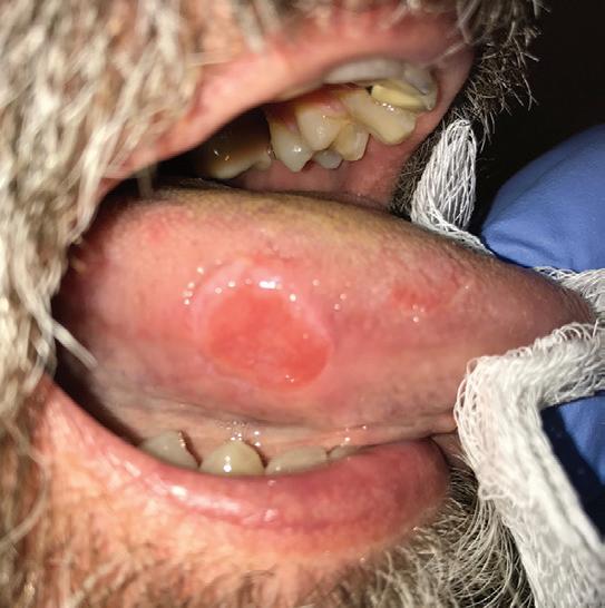
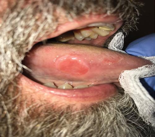
No palpable neck mass was identified. Oral cavity exam shows a slightly raised lesion on the right lateral tongue, which is soft and tender to palpation, measuring 1 cm in diameter (see Figure 1.1). There is no mass noted deep to the lesion. The rest of the exam, including fiberoptic laryngoscopy, is within normal limits.
Management
Question: What would you recommend next?
Answer: Tissue sampling with a punch or incisional biopsy of the lesion, preferably from the corner of the lesion.
Question: The biopsy shows squamous cell carcinoma (SCC), moderately differentiated, with a depth of invasion of at least 4 mm (punch biopsy specimen with tumor transected at the base). What would you recommend next in the workup?
Answer: Imaging of the neck is usually recommended to assess lymph node involvement. A computed tomography
(CT) scan with contrast or an ultrasound of the neck (which can be performed in the clinic) are both reasonable first options. The risk of distant metastases in early (T1 and T2) oral cavity SCC is extremely low. Therefore, extensive metastatic workup is not necessary. While positron emission tomography (PET)/ CT has become more common, the evidence for its added benefit does not exist. Obtaining a chest CT to rule out lung metastases is considered adequate.
Question: A CT scan of the head and neck does not show any evidence of regional metastases. How would you clinically stage this disease?
Answer: Based on the AJCC staging manual, 8th Edition, the clinical stage is cT1N0Mx, stage I.
Question: What treatment would you recommend?
Answer: Early stage tongue cancer treatment is wide local excision of the primary tumor and addressing the regional lymph nodes. If the risk of regional lymph node metastases is presumed to be higher than 20%, an elective, selective neck dissection should be performed. Depth of invasion is a prognostic marker for the presence of occult nodal metastases in the cN0 neck. With a depth of invasion >3 mm, it is believed that the risk of occult nodal metastases is >20%, and therefore an elective neck dissection should be performed. In this scenario, the recommended treatment is wide local excision of the primary tumor with 1 cm margins and elective neck dissection (ipsilateral levels I–III, i.e., supraomohyoid neck dissection). Alternatively, sentinel node biopsy could be offered if adequate expertise in the treating facility exists.
Question: Patient undergoes sentinel node mapping followed by wide local excision and sentinel node biopsy. The tongue defect is repaired with biologic dressing
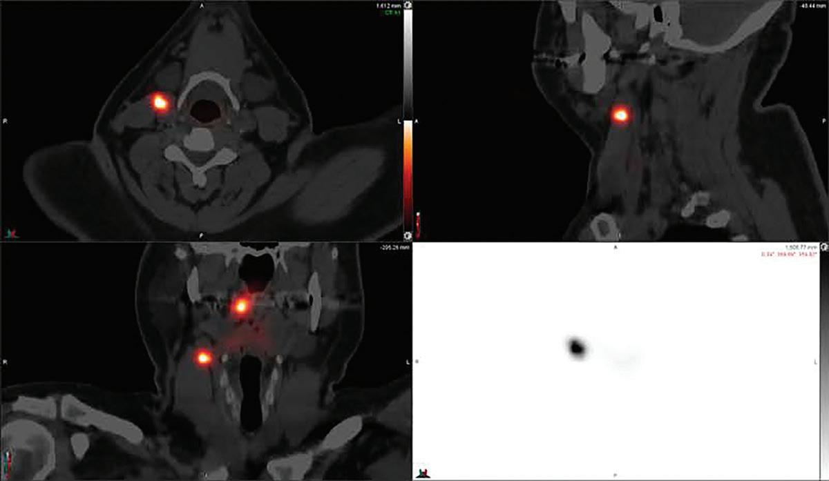
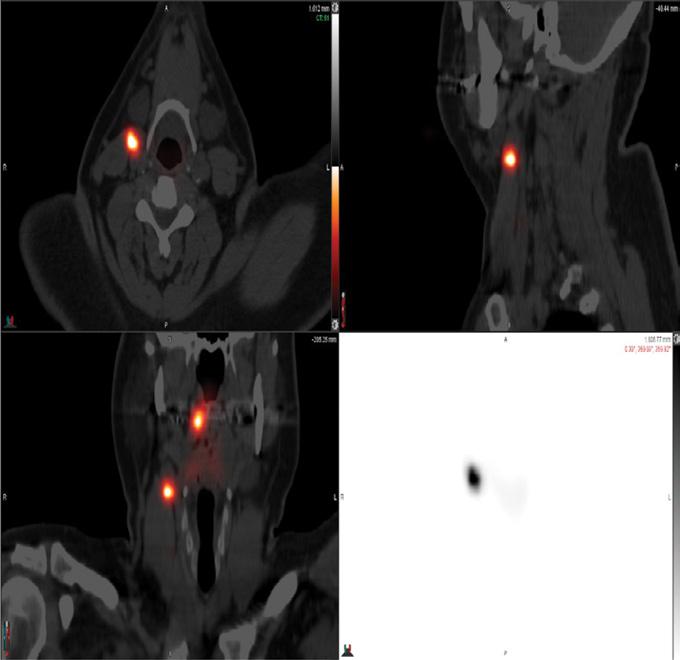
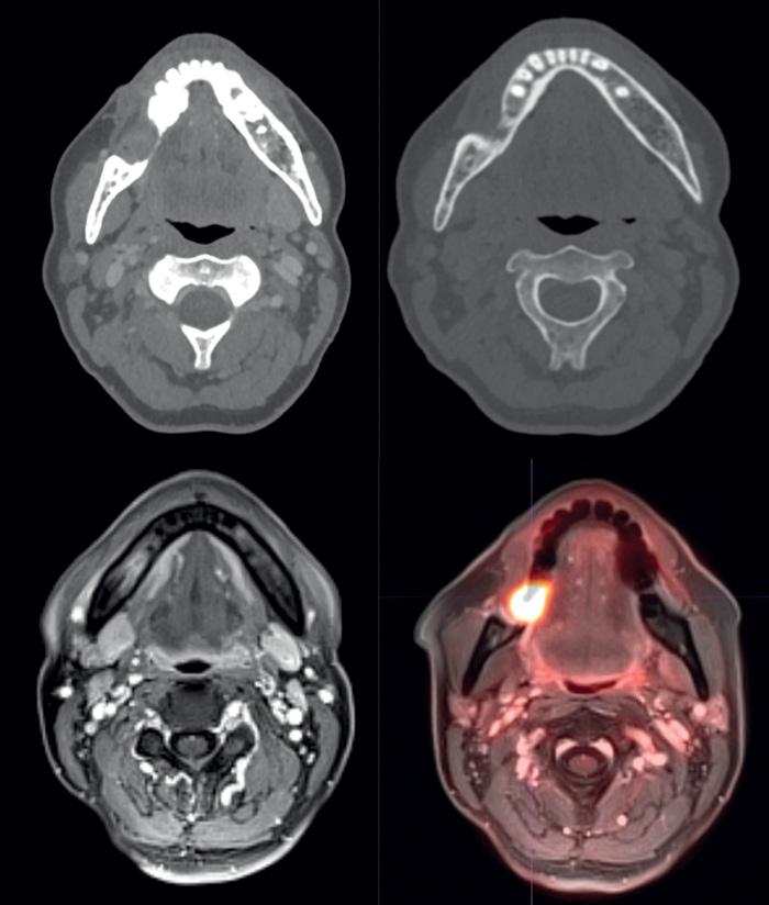
and secondary intention closure. On lymphoscintigraphy, the sentinel node is located in an ipsilateral level II lymph node (Figure 1.2). Excisional biopsy and frozen section assessment shows metastatic SCC in the level II node. How would you proceed?
Answer: If the sentinel node is positive, completion lymphadenectomy (selective neck dissection, level I–IV) is recommended.
The patient recovers well from the operation. The final pathology report shows a 1.5 cm moderately differentiated SCC with a depth of invasion of 7 mm. All margins are free of tumor, with the closest margin being 8 mm from tumor. No lymphovascular or perineural invasion is identified. One out of 30 lymph nodes is positive for metastatic SCC without extranodal extension, measuring 1.9 cm (sentinel node).
Question: Based on these pathologic findings, what is the appropriate stage for this patient?
Answer: According to AJCC 8th Edition, tumors of the oral cavity with a depth of invasion of more than 5 mm are
FIGURE 1.1 This photo demonstrates the patient’s right lateral tongue ulceration.
FIGURE 1.2 This image shows the patient’s fused CT- lymphoscintigraphy image. Note the uptake at the injection site and a right level II lymph node.
CASE 1 (continued
)
considered T2, even if the diameter is less than 2 cm. Therefore, the pathologic stage is pT2N1M0, stage III.
Question: What adjuvant treatment regimen, if any, would you recommend to this patient?
Answer: Since the disease is stage III, consideration of adjuvant treatment is warranted. Radiotherapy should be considered after discussion of the case at a multidisciplinary tumor board. The benefit of radiotherapy is not as clear in N1 disease; however, limited data exist that shows tumors with a depth of invasion of greater than 4 mm are at increased risk of regional failure without adjuvant therapy. Since there is no evidence of primary site positive margins or extranodal extension, there is no indication for adjuvant chemotherapy.
Key Points
• Oral tongue SCC is the most common malignancy of the oral cavity. The most important risk factors are tobacco, alcohol, poor dentition, diets low in fruits and vegetables, and Fanconi anemia.
• The risk of occult metastases in early stage oral cavity cancers is usually upward of 20%. Level I clinical trial evidence exists in the survival benefit of elective neck dissection in early stage tongue cancer and clinically negative cervical nodes when the depth of invasion is >3 mm. Currently, imaging techniques are not sensitive enough to identify occult metastases, and a negative CT or PET scan does not rule out microscopic metastases.
• Sentinel node biopsy in oral cavity cancers has been studied and shown to be reliable enough to identify the majority of occult metastases. Sentinel node sensitivity is reported
CASE 2
History of Present Illness
A 64-year-old woman presents with a nonhealing ulcer of the right mandibular alveolus for the past month after extraction of the second molar. The lesion is not painful and does not bleed. She has a more than 40 pack-year history of smoking and drinking two alcoholic drinks a day for the past 30 years. She does not report any history of other medical problems or prior malignancy.
Question: The patient completes a course of adjuvant radiotherapy. What is your recommended regimen for follow-up and clinical surveillance?
Answer: Based on National Comprehensive Cancer Network (NCCN) guidelines, baseline imaging at 12 weeks after completion of adjuvant treatment should be obtained, followed by physical examination every 1–3 months in the first year posttreatment and then 4–6 months in the second year. In years 3–5, a physical exam every 4–8 months is recommended and annually after 5 years. Annual thyroid-stimulating hormone (TSH) testing is recommended since the neck has received radiotherapy. Dental, nutrition, and ongoing depression evaluation are also recommended.
as 86% with a negative predictive value of 95% based on the European Organization for Research and Treatment of Cancer.
• Recommended primary treatment of oral cavity cancers is primarily surgical. Wide local excision with a 1 cm margin and lymph node dissection (selective node dissection in clinically negative neck) is the current recommendation.
• Depth of invasion is an important prognostic factor. A depth of invasion of more than 3 mm is associated with an increased risk of lymph node metastases.
• The current indications for adjuvant radiotherapy are (i) close or positive margins, (ii) nodal involvement, (iii) perineural invasion, and (iv) advanced stage tumor (T3–4).
• Concurrent chemotherapy with platinum-based agents is only recommended in positive margins or extranodal extension.
Physical Examination
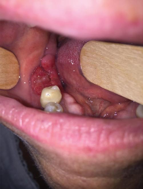
No palpable neck mass is identified. Oral cavity exam shows an ulcerative lesion limited to the occlusal surface of the right mandibular alveolus measuring 2 × 1 cm. The lesion is not tender to touch, does not extend to the buccal mucosa or floor of the mouth (see Figure 2.1). The rest of the exam, including flexible laryngoscopy, is within normal limits.
(Continued)
Babak Givi
Management
Question: What would you recommend next?
Answer: Since the lesion has been present for more than 2 weeks and a significant history of tobacco and alcohol abuse exists, tissue diagnosis via incisional or punch biopsy is warranted.
Question: The biopsy is performed and shows invasive, moderately differentiated keratinizing SCC. How would you stage this disease?
Answer: The clinical stage of the disease at this point is T2N0M0, stage II. However, the alveolar lesions can invade the mandible early in the process. Therefore, imaging of the mandible to better delineate osseous involvement is indicated.
Question: How would you determine the involvement of the mandible?
Answer: No imaging modality can offer 100% sensitivity and specificity. CT scanning, especially cone-beam CT, has been
utilized as a modality with high sensitivity. Magnetic resonance imaging (MRI) could be useful in determining the involvement of the bone marrow. In the absence of any single definitive modality, attention to the overall clinical exam and imaging findings could guide the clinician to determine the extent of the disease. For oral cavity lesions with suspected mandibular involvement, most surgeons would start with a CT scan with contrast of the head and neck to evaluate the primary site lesion and regional lymphatics.
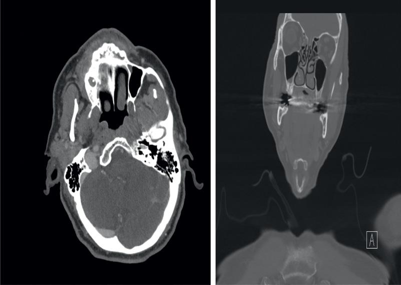
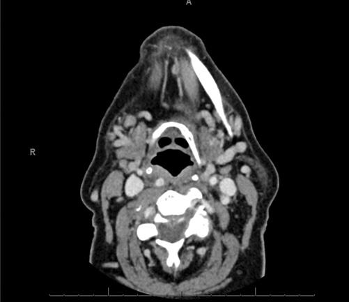
A PET/MRI and neck CT with IV contrast are obtained. The CT of the neck shows significant thinning of the lingual cortex of the mandible at the site of the lesion and the absence of the buccal cortex superiorly. The inferior alveolar canal appears intact (see Figure 2.2). The PET/MRI shows intense activity at the primary lesion without clear evidence of lymph node metastases. On MRI, there is an abnormal marrow signal at the location of the lesion, suspicious for marrow invasion. There is no evidence of involvement of the mandibular canal.
Question: Based on imaging findings, what is the clinical stage of the disease?
Answer: Since there is radiographic evidence of osseous invasion, the disease is upstaged to T4aN0M0, stage IVa.
Question: What is the recommended course of treatment?
Answer: Oral cavity SCC is primarily treated with surgery, followed by risk-adjusted adjuvant treatment. In this case, the recommended treatment is composite resection of the lesion with segmental mandibulectomy, selective neck dissection (levels I–III), and reconstruction with microvascular free tissue transfer, such as fibula free flap, to provide the best functional outcome.
The patient undergoes segmental mandibulectomy and selective neck dissection with fibula free flap reconstruction. The final pathology shows a 2 × 1 cm ulcerative SCC of the gingiva with invasion of the mandible. All margins are free of tumor (>5 mm). There is no perineural invasion. There is a microscopic focus of carcinoma present in 1 out of 32 lymph nodes (level Ib node, 2.3 cm) with no evidence of extracapsular extension.
Question: What is the pathologic stage?
Answer: Based on mandibular invasion and nodal involvement, the pathologic stage is pT4aN1M0, stage IVa.
Question: What adjuvant treatment regimen would you recommend to this patient?
Answer: In spite of negative margins and no perineural invasion, advanced T stage and involvement of regional nodes warrant adjuvant radiotherapy. There is no indication for chemotherapy (positive margins and extranodal extension). Therefore, adjuvant radiotherapy is adequate.
FIGURE 2.1 This photo demonstrates the ulcerative mucosal lesion of the right mandible gingiva.
FIGURE 2.2 These axial images are from the patient’s CT (a, b) as well as the T1-weighted MRI (c) and fused PET/ MRI (d). Note the lesion of the right mandible with associated cortical erosion and hypermetabolism.
Key Points
• Alveolus SCC is less common than tongue or floor of mouth cancers.
• Achieving negative margin resection (>5 mm) is an extremely important objective in the treatment of alveolar cancers.
• To achieve negative margin resection, most alveolar tumors will require resection of the underlying bone. Determining the extent of the mandibulectomy required to achieve negative margins can be challenging.
• It is accepted that if there is no bone erosion on CT or MRI, and there is enough height of the bone, marginal mandibulectomy can achieve negative margin resection. The remaining mandible should have at least a height of 1 cm.
• If there is bone erosion on CT imaging or MRI shows changes in the bone marrow signal, segmental mandibulectomy is recommended.
• There is no level I data on the best method to determine bone involvement. Cone beam CT is considered a very sensitive method.
• In good risk candidates, osseous reconstruction with fibula, scapula, or iliac crest free flap is recommended.
• Osteocutaneous radial forearm or vascularized rib graft has been described and used in patients who are not good candidates for fibula or scapula. However, the harvested bone is not thick enough to host implants.
CASE 3
Michael G. Moore
History of Present Illness
A 68-year-old white male presents with a chief complaint of a painful sore around his right lateral maxillary teeth. He states he initially thought it was related to a dental infection, but after having a tooth pulled, the area has continued to enlarge.
Question: What are the other important points in history taking?
Answer:
• Presence of other adjacent loose teeth.
• Facial numbness.
• Difficulty in opening the mouth.
• Dysphagia, odynophagia.
• Voice changes.
• Presence of neck mass.
For maxillary lesions, it is always important to determine the extent of the disease. Signs such as loose teeth, difficulty in opening the mouth (trismus), and facial numbness (perineural invasion) could provide critical clinical clues to the extent of disease and aggressive behavior.
Question: What additional aspects of the history and risk factors should be investigated?
Answer:
• Tobacco or alcohol use.
• Any history of head and neck cancers.
• Past medical history for significant diseases: peripheral vascular disease, diabetes, autoimmune diseases, chronic kidney disease, coagulation disorders, to name a few. This patient has a history of 10 pack-year smoking but quit 25 years ago. He drinks alcohol socially with no history of excessive drinking. There is no history of significant diseases or malignancy.
Physical Examination
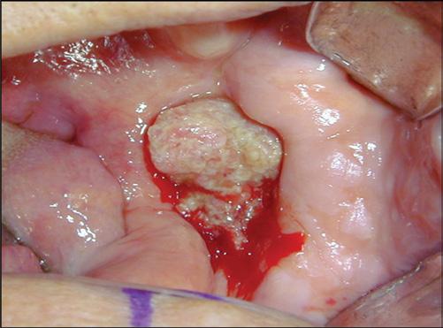
Oral cavity examination shows a 3 × 2 cm ulcerative lesion on the buccal aspects of teeth #2 to #4 with slight extension onto the palatal aspect of these teeth. No obvious loose teeth, but there is fleshy tissue at the site of the previously extracted tooth #3. His upper teeth are otherwise intact.
Neck exam revealed a 1.5 cm, firm, mobile, slightly tender right level 1b neck mass. No other neck masses were noted. No trismus or paresthesias noted. The rest of the examination is within the normal limits. Cranial nerves II–XII are intact.
Management
Question: What would you recommend next?
Biopsy of the lesion is the first step and is recommended even before imaging.
An office biopsy is performed and shows a moderately differentiated invasive SCC.
Question: What is the clinical stage at this point?
Answer: T2N1M0, stage III, based on a larger than 2 cm lesion and one palpable node. However, in alveolar lesions, it is important to evaluate the involvement of the bone, and these lesions could be upstaged to T4. Therefore, it is better to obtain imaging before assigning a clinical stage.
Question: What imaging modality would be an appropriate next step in the evaluation and management of this patient?
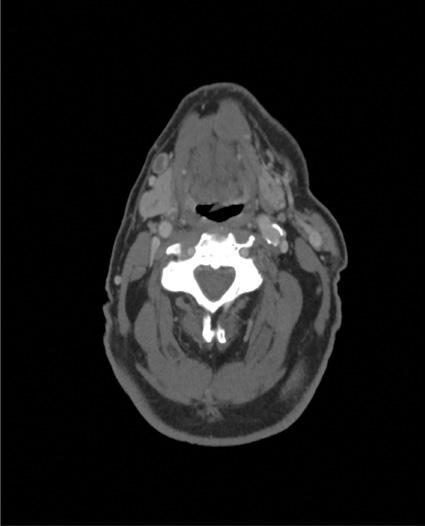
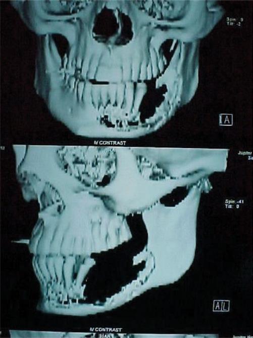
Answer: A CT of the neck and chest with IV contrast is an excellent next step as this will allow for evaluation of the extent of the primary lesion and to look for bone erosion as well as for any pathologic lymphadenopathy. CT of the chest helps to complete the staging. Alternatively, a PET/CT would be reasonable, but it may not allow for as good of resolution of the primary lesion to evaluate for bone involvement. Given that there is no evidence of neural deficits or concern for orbital or infratemporal fossa extension on physical exam, an MRI is probably not necessary (see Figures 3.1 and 3.2).
Chest CT demonstrated no evidence of metastatic disease. Oromaxillofacial prosthodontics is consulted to assist with a dental impression and the production of an obturator if maxillectomy is considered.
Question: Based on your current assessment, what would be this patient’s clinical stage?
Answer: This patient has oral cavity cancer. The primary lesion is staged based on size and depth of invasion. Here, depth is not known. Size puts it in the T2 category. Of note, minor bone erosion or maxillary involvement through a tooth socket alone does not upstage it to T4a. The patient has multiple ipsilateral pathologic nodes, none of which are larger than 6 cm in size, making him cN2b. Therefore, the stage is T2N2bM0: stage IVa.
Question: Given the above information, what would be the most appropriate management approach for this patient?
Answer: This patient has stage IVa oral cavity cancer. The optimal treatment strategy in patients who are amenable to surgery is for upfront surgical resection of the primary lesion with a concomitant neck dissection. Given the N2b neck dissection, he would benefit from adjuvant radiation therapy (RT) or chemoradiation therapy, depending on the final margin status and the presence or absence of extracapsular spread.
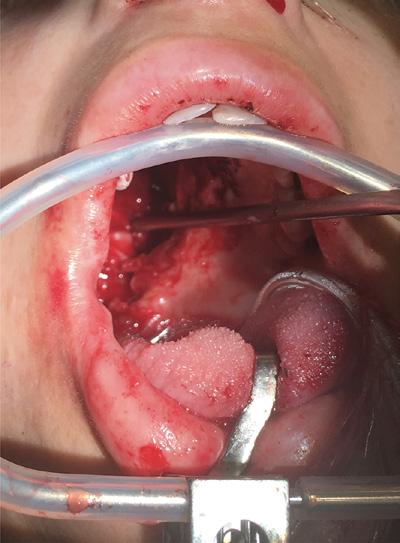
Question: Figure 3.3 shows an intraoperative photo of the oral cavity defect after surgical resection of the primary tumor. What would you recommend for the reconstruction of the defects? What are the factors that should be considered in the reconstruction of the maxillary defect?
FIGURE 3.1 Axial (a) and coronal (b) cut of the primary lesion. Note there is minor bone erosion – no obvious extension into the pterygoid plates or muscles.
FIGURE 3.2 Axial cut of the neck portion of the CT demonstrating the pathologic node felt on physical examination. There was one additional node with evidence of central necrosis in right level 1b. Both lymph nodes were less than 3 cm in size.
Intraoperative photo of the right oral cavity defect after surgical resection of the primary tumor.
FIGURE 3.3
CASE 3 (continued)
Answer: In a patient undergoing maxillectomy, it is critical to consider the best way to rehabilitate the defect. Limited defects, especially those with no communication to the sinonasal cavity, may require no reconstruction or simple adjacent tissue closure. For more extensive defects entering the sinonasal cavity, it is necessary to address this defect to allow for functional speech and swallowing. In this patient, an excellent option would be rehabilitation with a maxillofacial prosthesis. To do this, the patient must have adequate mouth opening and manual dexterity to allow for placement and removal. Moreover, retention is significantly improved when the ipsilateral canine tooth is maintained to allow for an adequate fulcrum for stability. Other contraindications to obturator use are resection of the orbital floor (unless a separate orbital floor repair is also performed) or resection of the anterior (premaxilla) or lateral (zygoma) projecting elements. Resection of the pterygoid plates does not significantly impact whether or not appropriate obturation can be achieved.
The patient’s final pathology demonstrates a 3.2 cm primary tumor, resected closest margin is 4 mm, posteriorly.
Key Points
• Evaluation of oral cavity cancer starts with a biopsy and typically a CT scan of the face and neck with IV contrast to assess the extent of the primary lesion and to evaluate for regional lymphadenopathy. An MRI may be indicated if there is concern for significant perineural invasion, deep tongue invasion, or extension near the orbit, skull base, or parapharyngeal space. The use of PET/CT should be in patients with stage III or IV disease.
• Management of tumors of the maxillary alveolus typically involves upfront surgery with removal of the primary tumor with clear surgical margins. A neck dissection should be performed for pathologic lymphadenopathy. For cN0 patients, elective neck dissection should be considered for advanced (T3 or T4) primary tumors as it may improve cancer control. more recent evidence suggests elective neck dissection in T2 tumors or consideration of sentinel node biopsy to determine the need for neck dissection.
CASE 4
Alok Pathak
History of Present Illness
A 75-year old man with a 35 pack-year smoking history presents for evaluation of a sore in the left side of his mouth. He quit tobacco 30 years ago but continues to drink two to three alcoholic drinks every day. He had extractions of tooth #17
There was only minor bone erosion seen. The neck dissection specimen showed 5 out of 58 lymph nodes positive. The largest lymph node was 2.7 cm, and there was evidence of extracapsular spread.
Question: What would be the recommended next step in this patient’s management?
Answer: This patient has pT2N3bM0, stage IVb disease based on the 8th Edition of the AJCC staging system. The presence of a close margin at the primary site would indicate the need to consider adjuvant radiation therapy. However, the presence of extracapsular spread of cervical lymph nodes (or the presence of a positive margin at the primary that cannot be re-excised) is an indication for adjuvant chemoradiation therapy as this has been shown to improve overall survival and disease-free survival. Since the posterior margin was close but clear, reexcision would not be necessary. The patient will benefit from concurrent adjuvant chemoradiotherapy with platinumbased agents.
• Adjuvant radiation therapy should be considered for advanced primary tumors, the presence of lymph node metastases, perineural invasion, lymphovascular invasion, or close surgical margins.
• Adjuvant chemoradiotherapy should be recommended in instances of positive surgical margins and/or the presence of extracapsular spread in cervical lymph nodes.
• In patients where a maxillectomy is considered, options for reconstruction include the use of a maxillofacial prosthesis or the use of a regional or free flap. If a maxillary obturator is planned, it is essential to have the patient see a maxillofacial prosthodontist soon after diagnosis to allow for a prosthesis to be made prior to the day of the resection.
• In instances when there is resection of the orbital floor or orbital exenteration, or when there will be inadequate remaining dentition to retain an obturator, reconstruction with free tissue transfer should be considered in suitable candidates.
and #18 3 months ago. However, the extraction site has not healed since then.
Physical Examination
Oral cavity examination demonstrates a 2.5 cm proliferative mass over the left retromolar trigone adherent to the underlying mandible with loss of sensation over the left chin (see
4 (continued)
Figure 4.1). There is also a palpable 2 cm mobile left submandibular lymph node.
Question: What is the most likely clinical diagnosis?
Answer: In view of progressive proliferative growth, with loss of sensation over the distribution of mental nerve with the risk factors of past tobacco use and current alcohol, the most likely diagnosis is SCC. In the presence of a gingival growth, tooth extraction should be avoided as it provides easy access to the mandible through the tooth socket.
Management
Question: What is the best way to achieve preoperative tissue diagnosis?
Answer: Punch/incisional biopsy. Since the lesion is easily accessible in the oral cavity, transoral incisional biopsy in the clinic is the most appropriate and expeditious way to get a tissue diagnosis. Fine needle aspiration of the lymph node may also be performed. However, a negative cytology from the lymph node does not rule out malignancy. Any consideration of an open biopsy of the lymph node should be avoided as it can complicate further neck treatment.
Question: What would be the most appropriate next step in the evaluation of this patient?
Answer: Considering the extent of the symptoms, imaging is recommended. CT scan of the head and neck with contrast is the most appropriate first step. Mental paresthesia is an indicator of the involvement of the inferior alveolar nerve in the mandibular canal. CT scan with contrast is the most appropriate imaging modality to assess the extent of mandibular
invasion and cervical lymphadenopathy. If proximal extension of the tumor along the mandibular nerve is a concern on CT scan, an MRI could be obtained. For advanced oral cavity cancer, distant imaging is recommended, in the form of a CT chest with contrast or PET/CT.
Contrast-enhanced CT of the neck shows a 4.5 cm heterogeneous tumor centered in the left retromolar trigone with underlying osseous destruction through the inferior alveolar canal. The erosive component of the tumor crosses the midline at the mandibular symphysis (see Figure 4.2). There are multiple enlarged and round lymph nodes in the left neck, the largest measuring 3.1 cm in left level 1b.
Chest CT does not demonstrate any distant metastatic disease.
The incisional biopsy came back as moderately differentiated SCC.
Question: What is the clinical tumor stage?
Answer: cT4aN2bM0, stage IVa. Mandibular involvement upstages the T stage to T4a. With multiple ipsilateral nodes <6 cm, the N stage is N2b.
FIGURE 4.1 This intraoral photograph shows the lesion of the patient’s left retromolar trigone extending on the left mandible body.
FIGURE 4.2 CT scan shows an extensive and destructive process of the left mandibular body.
CASE 4 (continued)
Question: What course of treatment would you recommend?
Answer: Treatment of advanced oral cavity SCC requires extirpation of the primary tumor with negative margins and addressing the lymphatics of the neck. Segmental mandibulectomy is required to obtain negative primary site surgical margins. The mandibular defect will require free tissue transfer reconstruction. The ipsilateral involved neck will require a neck dissection, level I–IV (skip metastases to level V in oral cavity carcinoma are uncommon). As the lesion crosses midline anteriorly, it is appropriate to consider or perform a contralateral selective (level I–III) neck dissection.
Question: What are the potential functional deficits after surgical treatment?
Answer: While rare, injuries to the spinal accessory nerve (CN XI) resulting in shoulder weakness, and marginal mandibular branch of the facial nerve (CN VII) resulting in lower lip depressor weakness, can occur but are not an expected outcome in experienced settings. Left chin numbness is to be expected after a segmental mandibulectomy as the inferior alveolar nerve is transected during the extirpation. Injury to the hypoglossal nerve (CN XII) is also rare and would be considered a complication. Treatment of alveolar cancer is not expected to result in significant dysphagia or aspiration postoperatively. However, when resection extends
Key Points
• Evaluation of oral cavity cancer starts with a biopsy and typically a CT scan of the face and neck with IV contrast to assess the extent of the primary lesion and to evaluate for regional lymphadenopathy. An MRI may be indicated if there is concern for significant perineural invasion, deep tongue invasion, or extension near the orbit, skull base, or parapharyngeal space. The use of PET/CT should be in patients with stage III or IV disease.
• Management of tumors of the oral cavity typically involves upfront surgery with removal of the primary tumor with
CASE 5
Arnaud Bewley
History of Present Illness
A 62-year-old white female is seen in the office with a 3-month history of a gradually enlarging lower lip mass. She has not had any previous biopsies, imaging, or treatment.
across the midline anteriorly, resulting in detachment of the genial muscular attachments, some loss of function can be observed.
Final pathology shows a 4.2 cm tumor centered in the left retromolar trigone with mandibular invasion, extensive perineural invasion, and a deep positive margin. In the right neck dissection specimen, 0/8 lymph nodes are involved with the tumor. In the left neck dissection specimen, 6/32 lymph nodes are involved with the tumor. There is a 3.1 cm level 1b lymph node that shows extranodal extension into the submandibular gland.
Question: Based on the final pathology, what is the pathologic stage and your recommended adjuvant treatment regimen?
Answer: pT4aN3bM0, stage IVb. Mandibular involvement results in pT4a staging. Any node with extranodal extension (ENE) results in an upstaging to N3b. There are many indications for radiation, including positive margins, perineural invasion (PNI), and multiple nodes involved. There are two indications for chemotherapy: positive surgical margins at the primary site and extranodal extension. In this case, considering that a large resection was performed and the defect is reconstructed with fibula flap, the expectation of re-resection is not realistic. Therefore, continuing with concurrent adjuvant chemoradiation with platinum-based agents is appropriate.
clear surgical margins. A neck dissection should be performed for pathologic lymphadenopathy. Contralateral selective neck dissections should be performed if the tumor crosses the midline.
• Adjuvant radiation therapy should be considered for advanced primary tumors, the presence of lymph node metastases, perineural invasion, lymphovascular invasion, or close surgical margins.
• Adjuvant chemoradiation therapy should be recommended in instances of positive surgical margins and/or the presence of extracapsular spread in cervical lymph nodes.
Her past medical history includes hypercholesterolemia, hypertension, appendectomy, and tonsillectomy. She takes Lisinopril and atorvastatin. She has a history of 30 pack-year smoking but quit 10 years ago. She drinks a glass of wine with dinner every night.
CASE 5 (continued)
Question: What are the other important points in history?
Answer:
• Pain and tenderness. This is an important question, as pain that is out of proportion with exam can be suggestive of perineural invasion.
• The presence of neck masses is an important finding, as advanced lower lip cancers have a high propensity for spread to the regional lymphatics.
• Voice change. It is important to screen for other head and neck cancer primaries in patients who are high risk.
• Weight loss, which is often associated with the duration and severity of symptoms.
The lesion is exquisitely painful, particularly along the right lower lip. She has not experienced any voice changes or weight loss and has not noticed any neck masses.
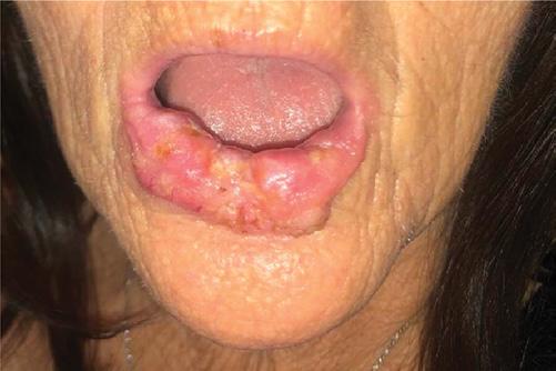
Physical Examination
An image of the patient’s lower lip lesion is shown in Figure 5.1. The tumor is very tender to palpation and extends from the right commissure to about 3 mm from the left commissure. Intraorally, it is free from the fixed gingiva by about 1 cm. There is subcutaneous extension palpable at the right lateral aspect of the tumor. There is no palpable lymphadenopathy. The remainder of her head and neck examination is unremarkable.
Management
Question: What would you recommend next?
Answer: Punch biopsy of the lower lip lesion. Pathologic diagnosis is imperative and is the most appropriate next step
in management. This would most easily be obtained with a punch biopsy at the time of her initial visit.
Question: What other tests or studies would you consider, if any?
Answer: A CT of the neck with IV contrast is an important next step in evaluating regional adenopathy. MRI of the neck could be considered, given the acute tenderness noted along the right lower lip. An MRI would be superior to a CT in evaluating for enhancement of the right mental nerve. An MRI would also allow for evaluation of the regional nodes, though less cost-effective than a CT scan. PET/CT scan could be considered. Also, a potential method of evaluating the regional nodal basin while also ruling out distant metastatic disease. For early stage disease (T1/T2) without clinical or radiographic evidence of regional disease, a PET/CT is usually unnecessary. If a PET/CT is considered, it is recommended that CT is performed with contrast and with adequate detail to delineate the neck anatomy.
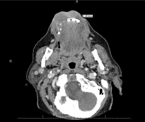
A punch biopsy is performed and demonstrates SCC. A contrast-enhanced CT scan is obtained with representative images shown below. No abnormal lymph nodes are reported. The tumor is measured around 5 cm on the CT scan (see Figure 5.2).
Question: Based on the patient’s examination and radiographic findings, how would you stage this disease?
Answer: T3N0M0, stage III. Cancers of the lip mucosa continue to be staged as cancers of the oral cavity, while cancers of the external vermillion lip are now staged as cutaneous carcinomas, per AJCC 8th Edition. However, in advanced tumors, this can be a difficult distinction to make when tumors involve both the mucosal and external vermillion surface. In these cases, staging should be based on the tumor’s historical origin when this can be deduced. In this case, the patient reports the tumor originated on the inner aspect of the lip, and this is corroborated by the greater degree of extension noted on the mucosal surface. The tumor is greater than 4 cm in diameter, therefore, it meets the criteria for a T3 primary. It would also likely meet the 1 cm depth of invasion criteria for T3 tumor. There is no clinical or radiographic evidence of regional metastatic spread; therefore, the patient should be staged as a T3N0M0, stage III.
Question: What is the appropriate treatment for this patient?
Answer: Primary surgical therapy is generally considered the standard of care for lip cancers as with oral cavity cancers. While the resection of lip cancer is relatively straightforward, the complex functional and aesthetic roles of the lip present major reconstructive challenges.
Due to the challenges of achieving acceptable functional and aesthetic outcomes with surgery for extensive lip
FIGURE 5.1 The patient’s lower lip mass. The tumor extends from the right Commissure to about 3mm from the left Commissure.
FIGURE 5.2 These representative axial cuts for the patient’s neck CT with IV contrast demonstrate a large ill-defined soft tissue tumor of the lower lip (a) with rounded level Ia and (b) Ib lymph nodes without obvious necrosis.
cancer, radiotherapy is considered an acceptable alternative. In particular, extensive lower lip cancers that extend over a significant proportion of the surface of the lip are particularly amenable to this approach.
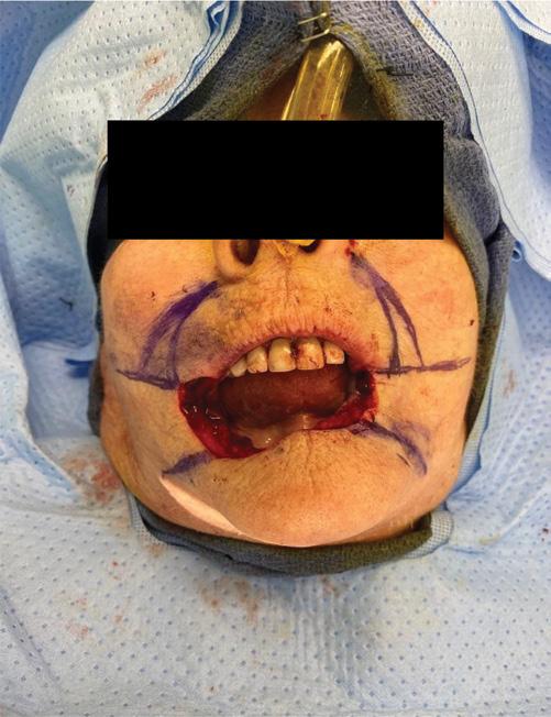
This intraoperative photo shows the patient’s total lower lip defect.
In this patient’s case, given the thickness of the tumor and soft tissue extension, she elected for definitive surgical resection, yielding the defect pictured below (see Figure 5.3).
Final pathology report showed SCC, 4.1 cm in greatest dimension, with a depth of invasion of 13 mm. Perineural invasion is present, but no lymphovascular invasion. The closest margin is 3 mm.
Question: How would you reconstruct the primary defect depicted in Figure 5.3?
Answer: While a Karapandzic flap is an excellent option for large lower lip defects, it relies entirely on the existing lip. As a result, some amount of preserved lower lip is needed to prevent severe microstomia. With a total lower lip defect, a Karapandzic flap would result in an unacceptable degree of microstomia.
Abbe/Estlander flaps are ideal for small lateral defects of the lip where sufficient lip can be recruited from the uninvolved
(a)
(b)
FIGURE 5.3
CASE 5 (continued)
lip to achieve a functional reconstruction. This defect is clearly too extensive to be amenable to these types of flaps.
Bernard/Webster flap could be used as an option. Total lower lip defects require the recruitment of additional tissue to recreate the lip, thereby minimizing the degree of microstomia. The Bernard and Webster flaps recruit tissue from the
cheek and buccal mucosa to reconstruct the lower lip and are therefore ideal for total or subtotal lip defects. The reconstructed tissue is not contractile but maintains sensation with good skin match.
The radial forearm free flap is another method of creating a new lip and is best for mitigating microstomia. This is often performed with a palmaris longus tendon sling to aid with oral competence. Disadvantages are lack of contractility, lack of sensation, and poor skin match.
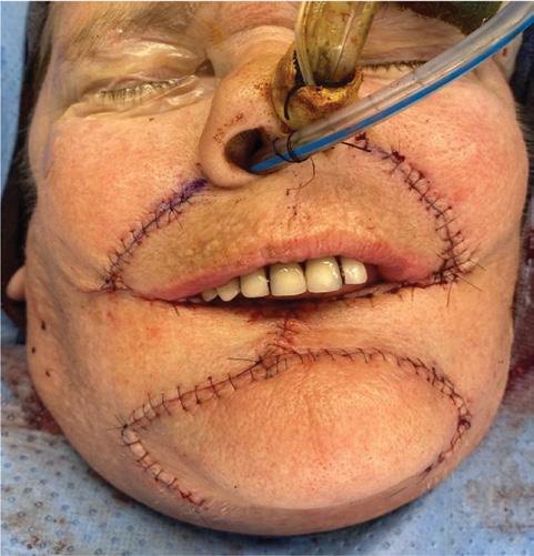
This patient was reconstructed with a Bernard-Webster bilateral advancement flap as pictured below (see Figure 5.4).
Question: How would you manage the patient’s regional lymph node basin?
Answer: Bilateral supra-omohyoid neck dissection is an acceptable way to address the high risk for occult regional disease in locally advanced lower lip cancer. This is a particularly appealing approach in patients who may be able to avoid adjuvant RT. Given the tumor involvement of the bilateral lower lip, any elective nodal dissection should address both sides of the neck.
Sentinel lymph node biopsy (SLNB) has been shown to be feasible and effective in patients who may be at high risk of metastases based on tumor size and depth. However, given the extensive nature of this primary tumor, the specificity of a sentinel node identification may be lower as four-quadrant injection of the radiotracer would likely trace to a variety of nodes. This would, therefore, not be an ideal case for SLNB. In general, sentinel node biopsy is recommended in T1, T2 tumors. Given the locally advanced nature of this patient’s tumor (T3) and the presence of PNI, she would benefit from adjuvant radiation therapy to the tumor bed. The regional lymphatics could likewise be irradiated to an adjuvant dose without the need for elective neck dissection.
Key Points
• Cancers of the lip mucosa continue to be staged as cancers of the oral cavity, while cancers of the external vermillion lip are now staged as cutaneous carcinomas.
• Primary radiotherapy can offer equivalent oncologic outcomes to surgical resection for early stage tumors.
• Extensive, superficial lower lip cancers are good candidates for primary radiotherapy as they avoid the potential high morbidity of surgical resection.
• Total lower lip defects require the recruitment of additional tissue to recreate the lip, thereby minimizing the degree of microstomia.
• The Bernard and Webster flaps recruit tissue from the cheek and buccal mucosa to reconstruct the lower lip and are therefore ideal for total or subtotal lip defects.
• SLNB has been shown to be feasible and effective in patients with lip tumors who may be at high risk of metastases based on tumor size and depth.
• Due to extensive lymphatic drainage from the upper lip and commissure, tumors of these subsites have a higher incidence of lymph node metastases at the time of diagnosis.
CASE 6
Ian Ganly
Presentation
A 45-year-old man presents with a soft tissue swelling of the left hard palate and soft palate. He denies pain. The patient had previously been seen by his dentist, who initiated a course of antibiotics with no effect. The patient was referred for further investigation. Examination showed a diffuse swelling of the hard palate as shown in Figure 6.1.
Question: What are the differential diagnoses for this mass?
Answer: Hard palate tumors are rare. The differential includes ameloblastoma, odontogenic keratocyst, and lymphoma, among others. However, the most common pathologies are minor salivary gland tumors.
Question: What is the most common minor salivary tumor in the oral cavity?
Answer: The most common minor salivary tumor of the oral cavity is mucoepidermoid carcinoma, followed by adenoid cystic carcinoma.
Question: What would you recommend next? What further investigations should be done for this patient?
Answer: Tissue diagnosis is the next step. Fine needle aspirate will most likely determine if this is a malignant versus benign tumor. However, core needle biopsy is often needed due to the heterogeneity in salivary gland pathology to establish the exact pathological diagnosis.
A contrast CT scan is a very reasonable first imaging study to determine the extent of bone invasion of the hard palate and upper alveolus and will determine if any neural foramens are widened in keeping with perineural invasion. An MRI scan might be necessary and is used more often to determine the extent of soft tissue invasion and to determine if any perineural invasion is present. MRI is also helpful in distinguishing between different salivary tumors.
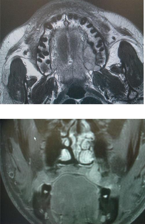
A core biopsy was done and reported as adenoid cystic carcinoma. A CT scan and MRI scan were done. The results of the MRI scan are shown in Figure 6.2.
FIGURE 6.1 This transoral photograph shows mild submucosal fullness of the left aspect of the hard palate.
(a)
(b)
FIGURE 6.2 An axial T1-weighted MRI image without contrast (a) and a coronal T1-weighted MRI with contrast (b) show an enhancing submucosal lesion of the left hard palate.
CASE 6 (continued)
Question: What are the pertinent findings in MRI on your review?
Answer: The MRI shows widening of the palatine canal, which is most likely due to the perineural invasion of the greater palatine nerve.
Question: What is the recommended management for this patient?
Answer: Adenoid cystic carcinoma is best managed by wide surgical resection followed by adjuvant radiation unless the tumor is considered unresectable. In this particular case, the extent of perineural invasion was limited to the greater palatine nerve with no extension to the skull base or cavernous sinus. The tumor was, therefore, deemed to be resectable. Due to the presence of perineural invasion and high incidence of positive margins in adenoid cystic cancer, adjuvant radiotherapy is recommended in the majority of cases to improve local control.
Question: What is the extent of surgery? What approach would you recommend?
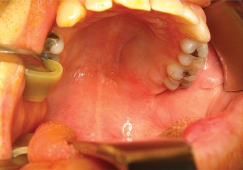
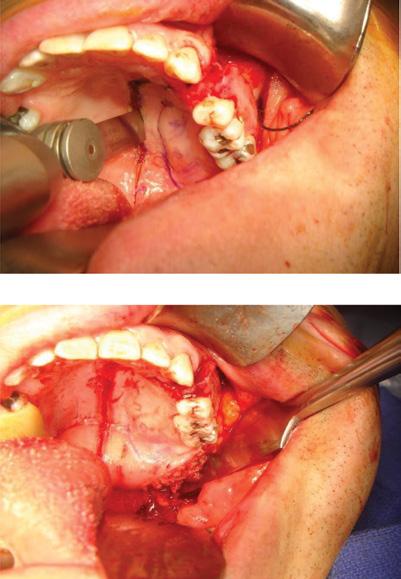
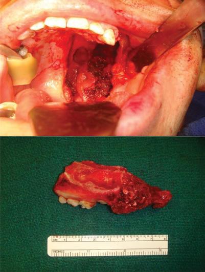
Answer: Based on the extent of the tumor on imaging and exam, peroral left infrastructure maxillectomy with resection of the hard palate, upper alveolus incorporating the palatine canal, and pterygoid plates appears to be a reasonable approach. The steps involved in the surgery are summarized in Figure 6.3.
An infrastructure maxillectomy is started by completing mucosal cuts to allow for at least a 1 cm soft tissue margin around the visible and palpable tumor. The next steps in the maxillectomy are accomplished by performing bone cuts. This is first done through the use of an oscillating saw. Due to the risk for bleeding, the posterior cut is often saved for last and is accomplished by the use of a curved osteotome followed by heavy curved scissors.
Question: What would you recommend for rehabilitation or reconstruction of this defect?
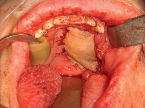
Answer: This patient could be rehabilitated with a dental obturator. The obturator allows for inspection of the resection site as well as for dental rehabilitation. The main disadvantage is the inability to eat without the prosthesis and nasal speech. An alternative method is with a free radial forearm flap, which allows for eating without a prosthesis and normal speech. However, a dental plate with teeth is still required for dental rehabilitation. In this particular case, a free radial forearm flap was used with donor vessels arising from the facial artery (see Figure 6.4).
Final pathology showed adenoid cystic carcinoma, predominantly tubular, 3.5 cm, no bone invasion, but with gross perineural invasion into the greater palatine nerve. All margins are free of tumor.
FIGURE 6.3 These intraoperative images show the surgical approach transorally and the resulting maxillectomy defect as well as the associated specimen.
CASE 6 (continued)
FIGURE 6.4 This intraoperative photo shows the palate defect after reconstruction using a radial forearm free flap. An alternative approach would have been a maxillary obturator given the preservation of the ipsilateral canine tooth and adequate contralateral maxillary dentition.
Question: What would you recommend next?
Answer: Although the tumor is completely excised, the risk of local failure is still significant in adenoid cystic carcinoma. Adjuvant radiotherapy is recommended. Since there are no adverse features (such as lymph node involvement, positive margins), adjuvant chemotherapy or consideration of clinical trials to add chemotherapy to radiotherapy are not appropriate in this case.
Question: What is the expected outcome for this patient?
Answer: Patients with minor salivary cancer tend to have a very good overall survival of 80% and disease- specific survival of 80–90%. The main predictors of outcome are stage III/IV disease and high- grade pathology. Female patients have a superior survival compared to male patients. Distant failure is more common and is the main cause of death.
This patient was treated with adjuvant radiation and remains disease-free at 3 years post-treatment.
Key Points
• The oral cavity is the most common location for the development of minor salivary gland tumors due to the high concentration of minor salivary gland tissue in this location.
• Adenoid cystic carcinoma is the most common minor salivary gland cancer.
• For minor salivary gland cancers of the palate, evaluation includes a detailed history, physical exam, a thorough cranial nerve exam, and a biopsy, often with a fine needle aspiration.
• For further assessment of palate salivary cancers, both CT and MRI are typically employed to assess for bone erosion and perineural invasion, respectively.
• Due to the close association of the mucosa of the hard palate and the underlying bone, infrastructure maxillectomy is often required.
• When considering a patient for an infrastructure maxillectomy, it is essential to consider the expected defect and discuss options for rehabilitation. If the use of a maxillary obturator is considered, a preoperative evaluation by a maxillofacial prosthodontist is crucial to get a surgical obturator created.
• To successfully retain an obturator, it is necessary to have adequate dentition to stabilize the prosthesis. Retention of the ipsilateral canine tooth typically allows for excellent obturator stability.
• In patients where a maxillary obturator is not appropriate or desired, more definitive reconstruction with a regional or free flap is needed.