An
Introduction to Genetic Analysis 11th Edition, (Ebook PDF)
Visit to download the full and correct content document: https://ebookmass.com/product/an-introduction-to-genetic-analysis-11th-edition-eboo k-pdf/
More products digital (pdf, epub, mobi) instant download maybe you interests ...
Humanity: An Introduction to Cultural Anthropology 11th Edition – Ebook PDF Version
https://ebookmass.com/product/humanity-an-introduction-tocultural-anthropology-11th-edition-ebook-pdf-version/
Electronic Media: An Introduction 11th Edition, (Ebook PDF)
https://ebookmass.com/product/electronic-media-anintroduction-11th-edition-ebook-pdf/
An Introduction to Language 11th Edition Victoria Fromkin
https://ebookmass.com/product/an-introduction-to-language-11thedition-victoria-fromkin/
American Public Policy: An Introduction 11th Edition, (Ebook PDF)
https://ebookmass.com/product/american-public-policy-anintroduction-11th-edition-ebook-pdf/
An Introduction to Statistical Methods and Data Analysis 7th Edition, (Ebook PDF)
https://ebookmass.com/product/an-introduction-to-statisticalmethods-and-data-analysis-7th-edition-ebook-pdf/
The Cultural Landscape: An Introduction to Human Geography (11th Edition – Ebook PDF Version) – Ebook PDF Version
https://ebookmass.com/product/the-cultural-landscape-anintroduction-to-human-geography-11th-edition-ebook-pdf-versionebook-pdf-version/
Scientific Farm Animal Production: An Introduction (11th Edition ) 11th Edition
https://ebookmass.com/product/scientific-farm-animal-productionan-introduction-11th-edition-11th-edition/
An Introduction to Correspondence Analysis 1st Edition
Eric J. Beh
https://ebookmass.com/product/an-introduction-to-correspondenceanalysis-1st-edition-eric-j-beh/
Exceptional Children: An Introduction to Special Education (Whatu2019s New in Special Education) 11th Edition, (Ebook PDF)
https://ebookmass.com/product/exceptional-children-anintroduction-to-special-education-whats-new-in-specialeducation-11th-edition-ebook-pdf/
3
3.1
5 The Genetics of Bacteria and
3.3
3.5
4 Mapping Eukaryote Chromosomes
4.1
Interaction
6.2
PArt II From DnA to PhenotyPe
7
7.1
9 Proteins and Their Synthesis
versus nonoverlapping codes
number of letters in the codon
use of suppressors to demonstrate a triplet code
degeneracy of the genetic code
7.2
7.3
7.4
7.5
10 Gene Isolation and Manipulation 351
7.7
Using Molecular Probes to Find and Analyze a Specific Clone of Interest
10.5
analysis of the lac promoter
characterization of the Lac repressor and the lac operator
13
Genetic Control
13.3 Defining the Entire Toolkit 482 the anteroposterior and dorsoventral axes 483
Expression of toolkit genes 484
13.4 Spatial Regulation of Gene Expression in Development 487
Maternal gradients and gene activation 488 drawing stripes: integration of gap-protein inputs 489 Making segments different: integration of Hox inputs 491
13.5 Post-transcriptional Regulation of Gene Expression in Development 494 rnA splicing and sex determination in Drosophila 494 regulation of mrnA translation and cell lineage in C. elegans 496 translational control in the early embryo 496 mirnA control of developmental timing in C. elegans and other species 499
13.6 From Flies to Fingers, Feathers, and Floor Plates: The Many Roles of Individual Toolkit Genes 500
13.7 Development and Disease 501 Polydactyly 501 Holoprosencephaly 502 cancer as a developmental disease 502
14 Genomes and Genomics 507
14.1 The Genomics Revolution 510
14.2 Obtaining the Sequence of a Genome 511 turning sequence reads into an assembled sequence 511
Whole-genome sequencing 513 traditional WGS 513 next-generation whole-genome shotgun sequencing 514
Whole-genome-sequence assembly 517
14.3 Bioinformatics: Meaning from Genomic Sequence 519 the nature of the information content of dnA 519 deducing the protein-encoding genes from genomic sequence 520
14.4 The Structure of the Human Genome 524 noncoding functional elements in the genome 525
14.5 The Comparative Genomics of Humans with Other Species 527
Phylogenetic inference 527 of mice and humans 530 comparative genomics of chimpanzees and humans 532
14.6 Comparative Genomics and Human Medicine 532 the exome and personalized genomics 533 comparative genomics of nonpathogenic and pathogenic E. coli 534
14.7
15 The Dynamic Genome: Transposable Elements
15.1 Discovery of Transposable Elements in Maize 549
Mcclintock’s experiments: the Ds element 549
Autonomous and nonautonomous elements 550 transposable elements: only in maize? 552
15.2 Transposable Elements in Prokaryotes 553 Bacterial insertion sequences 553 Prokaryotic transposons 554 Mechanism of transposition 556
15.3 Transposable Elements in Eukaryotes 558 class 1: retrotransposons 558 class 2: dnA transposons 562 utility of dnA transposons for gene discovery 564
15.4 The Dynamic Genome: More Transposable Elements Than Ever Imagined 566 Large genomes are largely transposable elements 567 transposable elements in the human genome 568 the grasses: Ltr-retrotransposons thrive in large genomes 569 Safe havens 569
15.5 Regulation of Transposable Element Movement by the Host 571 Genome surveillance in animals and bacteria 573
16 Mutation, Repair, and Recombination
16.1 The Phenotypic Consequences of DNA Mutations 583 types of point mutation 583 the molecular consequences of point mutations in a coding region 584 the molecular consequences of point mutations in a noncoding region 586
16.2 The Molecular Basis of Spontaneous Mutations 586 Luria and delbrück fluctuation test 586 Mechanisms of spontaneous mutations 588
Spontaneous mutations in humans: trinucleotiderepeat diseases 591
16.3 The Molecular Basis of Induced Mutations
16.4 Biological Repair Mechanisms
16.5
17 Large-Scale Chromosomal Changes 617
17.1
18 Population Genetics
18.2
19 The Inheritance of Complex Traits
20 Evolution of Genes and Traits
20.1 Evolution by Natural Selection 764
20.2 Natural Selection in Action: An Exemplary Case 766 the selective advantage of HbS 768 the molecular origins of HbS 770
20.3 Molecular Evolution: The Neutral Theory 771 the development of the neutral theory 771 the rate of neutral substitutions 772 the signature of purifying selection on dnA 772
20.4 Cumulative Selection and Multistep Paths to Functional Change 774 Multistep pathways in evolution 774 the signature of positive selection on dnA sequences 778
20.5 Morphological Evolution 779
Adaptive changes in a pigment-regulating protein 779
20.6 The Origin of New Genes and Protein Functions
This page intentionally left blank
Enduring Features
Coverage of model organisms
The eleventh edition retains the enhanced coverage of model systems in formats that are practical and flexible for both students and instructors.
• Chapter 1 introduces some key genetic model organisms and highlights some of the successes achieved through their use.
• Model Organism boxes presented in context where appropriate provide additional information about the organism in nature and its use experimentally.
• A Brief Guide to Model Organisms, at the back of the book, provides quick access to essential, practical information about the uses of specific model organisms in research studies.
• An Index to Model Organisms, on the endpapers at the back of the book, provides chapter-by-chapter page references to discussions of specific organisms in the text, enabling instructors and students to easily find and assemble comparative information across organisms.
Problem sets
No matter how clear the exposition, deep understanding requires the student to personally engage with the material. Hence our efforts to encourage student problem solving. Building on its focus on genetic analysis, the eleventh edition provides students with opportunities to practice problem-solving skills—both in the text and online through the following features.
• Versatile Problem Sets. Problems span the full range of degrees of difficulty. They are categorized according to level of difficulty—basic or challenging.
• Working with the Figures. An innovative set of problems included at the back of each chapter asks students pointed questions about figures in the chapter. These questions encourage students to think about the figures and help them to assess their understanding of key concepts.
• Solved Problems. Found at the end of each chapter, these worked examples illustrate how geneticists apply principles to experimental data.
• Unpacking the Problems. A genetics problem draws on a complex matrix of concepts and information. “Unpacking the Problem” helps students learn to approach problem solving strategically, one step at a time, concept on concept.
• NEW Multiple-choice versions of the end-of-chapter problems are available on our online LaunchPad for quick gradable quizzing and easily gradable homework assignments. The Unpacking the Problem tutorials from the text have been converted to in-depth online tutorials and expanded to help students learn to solve problems and think like a geneticist. New videos demonstrate how to solve selected difficult problems.
How genetics is practiced today
A feature called “What Geneticists Are Doing Today” suggests how genetic techniques are being used today to answer specific biological questions, such as “What is the link between telomere shortening and aging?” or “How can we find missing components in a specific biological pathway?”
Media and Supplements
The LaunchPad is a dynamic, fully integrated learning environment that brings together all the teaching and learning resources in one place. It features the fully interactive e-Book, end-of-chapter practice problems now assignable as homework, animations, and tutorials to help students with difficult-to-visualize concepts. This learning system also includes easy-to-use, powerful assessment tracking and grading tools, a personalized calendar, an announcement center, and communication tools all in one place to help you manage your course. Some examples:
• Hundreds of self-graded end-of-chapter problems allow students to practice their problem-solving skills. Most of the open-ended end-of-chapter questions have been carefully rewritten to create high-quality, analytical multiple-choice versions for assigning.
• Animations help students visualize genetics.
• Unpacking the Problem tutorials from the text have been converted and expanded to help students learn to solve problems and think like a geneticist. These in-depth online tutorials guide students toward the solution, offering guidance as needed via hints and detailed feedback.
• NEW Problem-solving videos walk students through solving difficult problems from the text.
Teaching resources for instructors
Electronic teaching resources are available online at the LaunchPad, at http://www.whfreeman.com/launchpad/iga11e
Includes all the electronic resources listed below for teachers. Contact your W. H. Freeman sales representative to learn how to log on as an instructor.
e-Book
The e-Book fully integrates the text and its interactive media in a format that features a variety of helpful study tools (full-text, Google-style searching; note taking; bookmarking; highlighting; and more). Available as a stand-alone item or on the LaunchPad.
Clicker Questions
Jump-start discussions, illuminate important points, and promote better conceptual understanding during lectures.
Layered PowerPoint Presentations
Illuminate challenging topics for students by deconstructing intricate genetic concepts, sequences, and processes step-by-step in a visual format.
All Images from the Text
More than 500 illustrations can be downloaded as JPEGs and PowerPoint slides. Use high-resolution images with enlarged labels to project clearly for lecture hall presentations. Additionally, these JPEG and PowerPoint files are available without labels for easy customization in PowerPoint.
67 Continuous-Play Animations
A comprehensive set of animations, updated and expanded for the eleventh edition, covers everything from basic molecular genetic events and lab techniques to analyzing crosses and genetic pathways. The complete list of animations appears on page xix.
Assessment Bank
This resource brings together a wide selection of genetics problems for use in testing, homework assignments, or in-class activities. Searchable by topic and provided in MS Word format, as well as in LaunchPad and Diploma, the assessment bank offers a high level of flexibility.
Student Solutions Manual (ISBN: 1-4641-8794-0)
The Student Solutions Manual contains complete worked-out solutions to all the problems in the textbook, including the “Unpacking the Problem” exercises. Available on the LaunchPad and the Instructor’s Web site as easy-to-print Word files.
Understanding Genetics: Strategies for Teachers and Learners in Universities and High Schools (ISBN: 0-7167-5216-6)
Written by Anthony Griffiths and Jolie-Mayer Smith, this collection of articles focuses on problem solving and describes methods for helping students improve their ability to process and integrate new information.
Resources for students
at http://www.whfreeman.com/launchpad/iga11e
LaunchPad 6-month Access Card (ISBN: 1-4641-8793-2)
The LaunchPad contains the following resources for students:
• Self-Graded End-of-Chapter Problems: To allow students to practice their problem-solving skills, most of the open-ended end-of-chapter questions have been carefully rewritten to create high-quality, analytical multiplechoice versions for assigning.
• Online Practice Tests: Students can test their understanding and receive immediate feedback by answering online questions that cover the core concepts in each chapter. Questions are page referenced to the text for easy review of the material.
• Animations: A comprehensive set of animations, updated and expanded for the eleventh edition, covers everything from basic molecular genetic events and lab techniques to analyzing crosses and genetic pathways. The complete list of animations appears on the facing page.
• Interactive “Unpacking the Problem”: An exercise from the problem set for many chapters is available online in interactive form. As with the text version, each Web-based “Unpacking the Problem” uses a series of questions to step students through the thought processes needed to solve a problem. The online version offers immediate feedback to students as they work through the problems as well as convenient tracking and grading functions. Authored by Craig Berezowsky, University of British Columbia.
• NEW Problem-Solving Videos: Twenty-five problem-solving videos walk students through solving difficult problems from the text.
Student Solutions Manual (ISBN: 1-4641-8794-0)
The Solutions Manual contains complete worked-out solutions to all the problems in the textbook, including the “Unpacking the Problem” exercises. Used in conjunction with the text, this manual is one of the best ways to develop a fuller appreciation of genetic principles.
Other genomic and bioinformatic resources for students:
Text Appendix A, Genetic Nomenclature, lists model organisms and their nomenclature.
Text Appendix B, Bioinformatic Resources for Genetics and Genomics, builds on the theme of introducing students to the latest genetic research tools by providing students with some valuable starting points for exploring the rapidly expanding universe of online resources for genetics and genomics.
Animations
Sixty-seven animations are fully integrated with the content and figures in the text chapters. These animations are available on the LaunchPad and the Book Companion site.
CHAPTER 1
A Basic Plant Cross (Figure 1-3)
The Central Dogma (Figure 1-10)
CHAPTER 2
Mitosis (Chapter Appendix 2-1)
Meiosis (Chapter Appendix 2-2)
X-Linked Inheritance in Flies (Figure 2-17)
CHAPTER 3
Punnett Square and Branch Diagram Methods for Predicting the Outcomes of Crosses (Figure 3-4)
Meiotic Recombination Between Unlinked Genes by Independent Assortment (Figures 3-8 and 3-13)
Analyzing a Cross: A Solved Problem (Solved Problem 2)
CHAPTER 4
Crossing Over Produces New Allelic Combinations (Figures 4-2 and 4-3)
Meiotic Recombination Between Linked Genes by Crossing Over (Figure 4-7)
A Molecular Model of Crossing Over (Figure 4-21)
A Mechanism of Crossing Over: A Heteroduplex Model (Figure 4-21)
A Mechanism of Crossing Over: Genetic Consequences of the Heteroduplex Model
Mapping a Three-Point Cross: A Solved Problem (Solved Problem 2)
CHAPTER 5
Bacterial Conjugation and Mapping by Recombination (Figures 5-11 and 5-17)
CHAPTER 6
Interactions Between Alleles at the Molecular Level, RR: Wild-Type
Interactions Between Alleles at the Molecular Level, rr: Homozygous Recessive, Null Mutation
Interactions Between Alleles at the Molecular Level, r ′r ′: Homozygous Recessive, Leaky Mutation
Interactions Between Alleles at the Molecular Level, Rr: Heterozygous, Complete Dominance
Screening and Selecting for Mutations
A Model for Synthetic Lethality (Figure 6-20)
CHAPTER 7
DNA Replication: The Nucleotide Polymerization Process (Figure 7-15)
DNA Replication: Coordination of Leading and Lagging Strand Synthesis (Figure 7-20)
DNA Replication: Replication of a Chromosome (Figure 7-23)
CHAPTER 8
Transcription in Prokaryotes (Figures 8-7 to 8-10)
Transcription in Eukaryotes (Figures 8-12 and 8-13)
Mechanism of RNA Splicing (Figures 8-16 and 8-17)
CHAPTER 9
Peptide-Bond Formation (Figure 9-2)
tRNA Charging (Figure 9-7)
Translation (Figure 9-14 to 9-16)
Nonsense Suppression at the Molecular Level: The rod ns Nonsense Mutation (Figure 9-18)
Nonsense Suppression at the Molecular Level: The tRNA Nonsense Suppressor (Figure 9-18)
Nonsense Suppression at the Molecular Level: Nonsense Suppression of the rod ns Allele (Figure 9-18)
CHAPTER 10
Polymerase Chain Reaction (Figure 10-3)
Plasmid Cloning (Figure 10-9)
Finding Specific Cloned Genes by Functional Complementation: Functional Complementation of the Gal Yeast Strain and Recovery of the Wild-Type GAL gene
Finding Specific Cloned Genes by Functional Complementation: Making a Library of Wild-Type Yeast DNA
Finding Specific Cloned Genes by Functional Complementation: Using the Cloned GAL Gene as a Probe for GAL mRNA
SDS Gel Electrophoresis and Immunoblotting
Dideoxy Sequencing of DNA (Figure 10-17)
Creating a Transgenic Mouse (Figures 10-29 and 10-30)
CHAPTER 11
Regulation of the Lactose System in E. coli: Assaying Lactose Presence/Absence Through the Lac Repressor (Figure 11-6)
Regulation of the Lactose System in E. coli: OC lac Operator Mutations (Figure 11-8)
Regulation of the Lactose System in E. coli: I Lac Repressor Mutations (Figure 11-9)
Regulation of the Lactose System in E. coli: IS Lac Superrepressor Mutations (Figure 11-10)
CHAPTER 12
Three-Dimensional Structure of Nuclear Chromosomes (Figure 12-11)
Gal4 Binding and Activation (Figures 12-6 through 12-9)
Chromatin Remodeling (Figures 12-13 and 12-14)
CHAPTER 13
Drosophila Embryonic Development
Sex Determination in Flies (Figure 13-23)
CHAPTER 14
DNA Microarrays: Using an Oligonucleotide Array to Analyze Patterns of Gene Expression (Figure 14-20)
DNA Microarrays: Synthesizing an Oligonucleotide Array
Yeast Two-Hybrid Systems (Figure 14-21)
CHAPTER 15
Replicative Transposition (Figure 15-9)
Life Cycle of a Retrovirus (Figure 15-11)
The Ty1 Mechanism of Retrotransposition (Figures 15-13 and 15-14)
CHAPTER 16
Replication Slippage Creates Insertion or Deletion Mutations (Figure 16-8)
UV-Induced Photodimers and Excision Repair (Figure 16-19)
Base-Excision Repair, Nucleotide Excision Repair, and Mismatch Repair (Figures 16-20, 16-22, and 16-23)
CHAPTER 17
Autotetraploid Meiosis (Figure 17-6)
Meiotic Nondisjunction at Meiosis I (Figure 17-12)
Meiotic Nondisjunction at Meiosis II (Figure 17-12)
Chromosome Rearrangements: Paracentric Inversion, Formation of Paracentric Inversions (Figure 17-27)
Chromosome Rearrangements: Paracentric Inversion, Meiotic Behavior of Paracentric Inversions (Figure 17-28)
Chromosome Rearrangements: Reciprocal Translocation, Formation of Reciprocal Translocations (Figure 17-30)
Chromosome Rearrangements: Reciprocal Translocation, Meiotic Behavior of Reciprocal Translocations (Figure 17-30)
Chromosome Rearrangements: Reciprocal Translocation, Pseudolinkage of Genes by Reciprocal Translocations (Figure 17-32)
Acknowledgments
We extend our thanks and gratitude to our colleagues who reviewed this edition and whose insights and advice were most helpful:
Anna Allen, Howard University
Melissa Antonio, California Baptist University
Dave Bachoon, Georgia College & State University
Brianne Barker, Drew University
Lina Begdache, Binghamton University
Edward Berger, Dartmouth College
Aimee Bernard, University of Colorado Denver
Jaime Blair, Franklin & Marshall College
Jay Brewster, Pepperdine University
Doug Broadfield, Florida Atlantic University
Mirjana Brockett, Georgia Institute of Technology
Judy Brusslan, California State University, Long Beach
Gerald Buldak, Loyola University Chicago
Aaron Cassill, University of Texas at San Antonio
Helen Chamberlin, Ohio State University
Henry Chang, Purdue University
Randolph Christensen, Coe College
Mary Clancy, University of New Orleans
Craig Coleman, Brigham Young University
Matthew Collier, Wittenberg University
Shannon Compton, University of Massachusetts–Amherst
Diane Cook, Louisburg College
Victoria Corbin, University of Kansas
Claudette Davis, George Mason University
Ann Marie Davison, Kwantlen Polytechnic University
Elizabeth De Stasio, Lawrence University
Matt Dean, University of Southern California
Michael Dohm, Chaminade University
Robert Dotson, Tulane University
Chunguang Du, Montclair State University
Erastus Dudley, Huntingdon College
Edward Eivers, California State University, Los Angeles
Robert Farrell, Penn State University
David Foltz, Louisiana State University
Wayne Forrester, Indiana University
Rachael French, San Jose State University
Shirlean Goodwin, University of Memphis
Topher Gee, UNC Charlotte
John Graham, Berry College
Theresa Grana, University of Mary Washington
Janet Guedon, Duquesne University
Patrick Gulick, Concordia University
Richard Heineman, Kutztown University
Anna Hicks, Memorial University
Susan Hoffman, Miami University
Stanton Hoegerman, College of William and Mary
Margaret Hollingsworth, University at Buffalo
Nancy Huang, Colorado College
Jeffrey Hughes, Millikin University
Varuni Jamburuthugoda, Fordham University
Pablo Jenik, Franklin & Marshall College
Aaron Johnson, University of Colorado School of Medicine
Anil Kapoor, University of La Verne
Jim Karagiannis, University of Western Ontario
Kathleen Karrer, Marquette University
Jessica Kaufman, Endicott College
Darrell Killian, Colorado College
Dennis Kraichely, Cabrini College
Anuj Kumar, University of Michigan
Janice Lai, Austin Community College
Evan Lau, West Liberty University
Min-Ken Liao, Furman University
Sarah Lijegren, University of Mississippi
Renyi Liu, University of California, Riverside
Diego Loayza, Hunter College
James Lodolce, Loyola University Chicago
Joshua Loomis, Nova Southeastern University
Amy Lyndaker, Ithaca College
Jessica Malisch, Claremont McKenna College
Patrick Martin, North Carolina A&T State University
Presley Martin, Hamline University
Dmitri Maslov, University of California, Riverside
Maria Julia Massimelli, Claremont McKenna College
Endre Mathe, Vasile Goldis Western University of Arad
Herman Mays, University of Cincinnati
Thomas McGuire, Penn State Abington
Mark Meade, Jacksonville State University
Ulrich Melcher, Oklahoma State University
Philip Meneely, Haverford College
Ron Michaelis, Rutgers University
Chris Mignone, Berry College
Sarah Mordan-McCombs, Franklin College of Indiana
Ann Murkowski, North Seattle Community College
Saraswathy Nair, University of Texas at Brownsville
Sang-Chul Nam, Texas A&M International University
Scot Nelson, University of Hawaii at Manoa
Brian Nichols, University of Illinois at Chicago
Todd Nickle, Mount Royal University
Juliet Noor, Duke University
Mohamed Noor, Duke University
Daniel Odom, California State University, Northridge
Kirk Olsen, East Los Angeles College
Kavita Oommen, Georgia State University
Maria Orive, University of Kansas
Laurie Pacarynuk, University of Lethbridge
Patricia Phelps, Austin Community College
Martin Poenie, University of Texas at Austin
Jennifer Powell, Gettysburg College
Robyn Puffenbarger, Bridgewater College
Jason Rauceo, John Jay College (CUNY)
Eugenia Ribiero-Hurley, Fordham University
Ronda Rolfes, Georgetown University
Edmund Rucker, University of Kentucky
Jeffrey Sands, Lehigh University
Monica Sauer, University of Toronto at Scarborough, UTSC
Ken Saville, Albion College
Pratibha Saxena, University of Texas at Austin
Jon Schnorr, Pacific University
Malcolm Schug, University of North Carolina at Greensboro
Deborah Schulman, Lake Erie College
Allan Showalter, Ohio University
Elaine Sia, University of Rochester
Robert Smith, Nova Southeastern University
Joyce Stamm, University of Evansville
Tara Stoulig, Southeastern Louisiana University
Julie Torruellas Garcia, Nova Southeastern University
Virginia Vandergon, California State University, Northridge
Charles Vigue, University of New Haven
Susan Walsh, Rollins College
Michael Watters, Valparaiso University
Roger Wartell, Georgia Institute of Technology
Matthew White, Ohio University
Dwayne Wise, Mississippi State University
Andrew Wood, Southern Illinois University
Mary Alice Yund, UC Berkeley Extension
Malcom Zellars, Georgia State University
Deborah Zies, University of Mary Washington
Tony Griffiths would like to acknowledge the pedagogical insights of David Suzuki, who was a co-author of the early editions of this book, and whose teaching in the media is now an inspiration to the general public around the world. Great credit is also due to Jolie Mayer-Smith and Barbara Moon, who introduced Tony to the power of the constructivist approach applied to teaching genetics. Sean Carroll would like to thank Leanne Olds for help with the artwork for Chapters 11, 12, 13, 14, and 20. John Doebley would like to thank his University of Wisconsin colleagues Bill Engels, Carter Denniston, and Jim Crow, who shaped his approach to teaching genetics.
The authors also thank the team at W. H. Freeman for their hard work and patience. In particular we thank our developmental and supplements editor, Erica Champion; senior acquisitions editor Lauren Schultz; senior project editor Jane O’Neill; and copy editor Teresa Wilson. We also thank Susan Wein, production supervisor; Diana Blume, art director; Vicki Tomaselli, cover and text designer; Sheridan Sellers, page layout; Janice Donnola, illustration coordinator; Jennifer MacMillan, permissions manager; Amanda Dunning, executive media editor; and Alexandra Garrett, editorial assistant. Finally, we especially appreciate the marketing and sales efforts of John Britch, executive marketing manager, and the entire sales force.
This page intentionally left blank
The Genetics Revolution
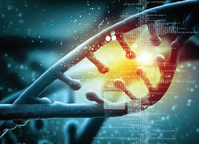
DNA (deoxyribonucleic acid) is the molecule that encodes genetic information. The strings of four different chemical bases in DNA store genetic information in much the same way that strings of 0’s and 1’s store information in computer code. [ Sergey Nivens/Shutterstock.]
O ut L ine
1.1 the birth of genetics
1.2 after cracking the code
1.3 genetics today
Learning Outc O mes
After completing this chapter, you will be able to
• Describe the way in which modern genetics developed.
• List the main cellular constituents involved in gene expression and action.
• Give some examples of how genetics has influenced modern medicine, agriculture, and evolution.
Genetics is a form of information science. Geneticists seek to understand the rules that govern the transmission of genetic information at three levels—from parent to offspring within families, from DNA to gene action within and between cells, and over many generations within populations of organisms. These three foci of genetics are known as transmission genetics, moleculardevelopmental genetics, and population-evolutionary genetics. The three parts of this text examine these three foci of genetics.
The science of genetics was born just over 100 years ago. Since that time, genetics has profoundly changed our understanding of life, from the level of the individual cell to that of a population of organisms evolving over millions of years. In 1900, William Bateson, a prominent British biologist, wrote presciently that an “exact determination of the laws of heredity will probably work more change in man’s outlook on the world, and in his power over nature, than any other advance in natural knowledge that can be foreseen.” Throughout this text, you will see the realization of Bateson’s prediction. Genetics has driven a revolution in both the biological sciences and society in general.
In this first chapter, we will look back briefly at the history of genetics, and in doing so, we will review some of the basic concepts of genetics that were discovered over the last 100 years. After that, we will look at a few examples of how genetic analysis is being applied to critical problems in biology, agriculture, and human health today. You will see how contemporary research in genetics integrates concepts discovered decades ago with recent technological advances. You will see that genetics today is a dynamic field of investigation in which new discoveries are continually advancing our understanding of the biological world.
1.1 the Birth of genetics
Throughout recorded history, people around the world have understood that “like begets like.” Children resemble their parents, the seed from a tree bearing flavorful fruit will in turn grow into a tree laden with flavorful fruit, and even members of wolf packs show familial resemblances (Figure 1-1). Although people were confident in these observations, they were left to wonder as to the underlying mechanism. The Native American Hopi tribe of the Southwestern United States understood that if they planted a red kernel of maize in their fields, it would grow into a plant that also gave red kernels. The same was true for blue, white, or yellow kernels. So they thought of the kernel as a message to the gods in the Earth about the type of maize the Hopi farmers hoped to harvest. Upon receiving this message, the gods would faithfully return them a plant that produced kernels of the desired color.
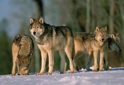
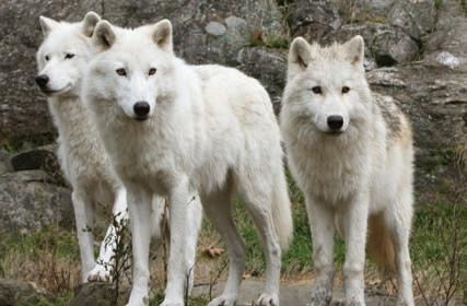
In the 1800s in Europe, horticulturalists, animal breeders, and biologists also sought to explain the resemblance between parents and offspring. A commonly held view at that time was the blending theory of inheritance, or the belief that inheritance worked like the mixing of fluids such as paints. Red and white paints, when mixed, give pink; and so a child of one tall parent and one short parent could be expected to grow to a middling height. While blending theory seemed to work at times, it was also clear that there were exceptions, such as tall children born to parents of average height. Blending theory also provided no mechanism by which the “heredity fluids” it imagined, once mixed, could be separated—the red and white paints cannot be reconstituted from the pink. Thus, the long-term expectation of blending theory over many generations of intermating among individuals is that all members of the population will come to express the same average value of a trait. Clearly, this is not how nature works. Human populations have people with a range of
FIGURE 1-1 Family groups in the gray wolf show familial resemblances for coat colors and patterning.
[ (Top) altrendo nature/Getty Images; (bottom) Bev McConnell/ Getty Images.]
Like begets like
heights, from short to tall, and we have not all narrowed in on a single average height despite the many generations that human populations have dwelled on Earth.
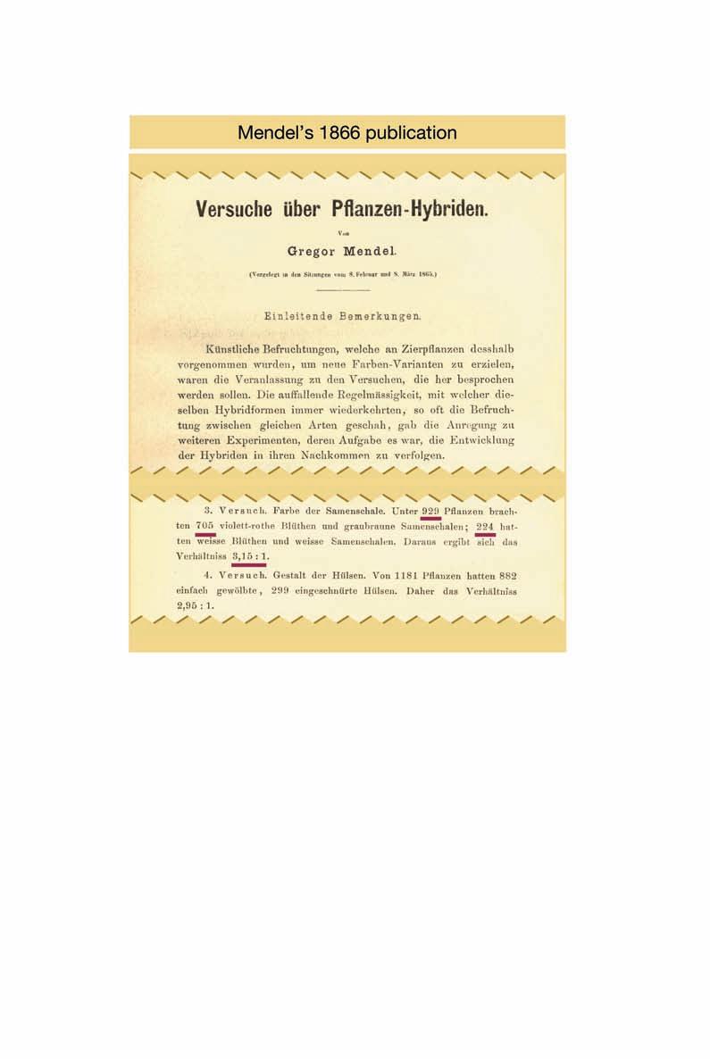
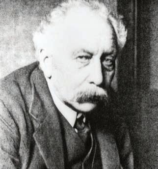
Gregor Mendel—A monk in the garden
While the merits and failings of blending theory were being debated, Gregor Mendel, an Austrian monk, was working to understand the rules that govern the transmission of traits from parent to offspring after hybridization among different varieties of pea plants (Figure 1-2). The setting for his work was the monastery garden in the town of Brünn, Austria (Brno, Czech Republic, today). From 1856 to 1863, Mendel cross-pollinated or intermated different varieties of the pea plant. One of his experiments involved crossing a pea variety with purple flowers to one with white flowers (Figure 1-3). Mendel recorded that the first hybrid generation
One of Mendel’s experiments
Parents
]
First-generation hybrid
Second-generation hybrids
3 purple : 1 white
FIGURE 1-3 The mating scheme for Mendel’s experiment involving the crossing of purple- and white-flowered varieties of pea plants. The purple and white circles signify the gene variants for purple vs. white flower color. Gametes carry one gene copy; the plants each carry two gene copies. The “×” signifies a cross-pollination between the purple- and white-flowered plants.
FIGURE 1-2 Gregor Mendel was an Austrian monk who discovered the laws of inheritance. [ James King-Holmes/Science Source.
04/15/14
05/01/14
Media Group
of offspring from this cross all had purple flowers, just like one of the parents. There was no blending. Then, Mendel selfpollinated the first-generation hybrid plants and grew a second generation of offspring. Among the progeny, he saw plants with purple flowers as well as plants with white flowers. Of the 929 plants, he recorded 705 with purple flowers and 224 with white flowers (Figure 1-4). He observed that there were roughly 3 purple-flowered plants for every 1 whiteflowered plant.
How did Mendel explain his results? Clearly, blending theory would not work since that theory predicts a uniform group of first-generation hybrid plants with light purple flowers. So Mendel proposed that the factors that control traits act like rather than fluids and that these particles do not blend together but are passed intact from one generation to the next. Today, Mendel’s particles are known as genes.
Mendel proposed that each individual pea plant has two copies of the gene controlling flower color in each of the cells of the plant body (somatic cells). However, when the plant forms gametes (eggs and sperm), only one copy of the gene enters into these reproductive cells (see Figure 1-3). Then, when egg and sperm unite to start a new individual, once again there will be two copies of the flower color gene in each cell of the plant body.
Mendel had some further insights. He proposed that the gene for flower color comes in two gene variants, or alleles one that conditions purple flowers and one that conditions white flowers. He proposed that the purple allele of the flower color gene is dominant to the white allele such that a plant with one purple allele and one white allele would have purple flowers. Only plants with two white alleles would have white flowers (see Figure 1-3). Mendel’s two conclusions, (1) that genes behaved like particles that do not blend together and (2) that one allele is dominant to the other, enabled him to explain the lack of blending in the first-generation hybrids and the reappearance of white-flowered plants in the second-generation hybrids with a 1 ratio of purple- to white-flowered plants. This revolutionary advance in our understanding of inheritance will be fully discussed in Chapter 2.
How did Mendel get it right when so many others before him were wrong? Mendel chose a good organism and good traits to study. The traits he studied were all controlled by single genes. Traits that are controlled by several genes, as many traits are, would not have allowed him to discover the laws of inheritance so easily. Mendel was also a careful observer, and he kept detailed records of each of his experiments. Finally, Mendel was a creative thinker capable of reasoning well beyond the ideas of his times.
Key Con C ept Gregor Mendel demonstrated that genes behave like particles and not fluids Introduction to Genetic Analysis, 11e Figure 01.04 #104
Mendel’s particulate theory of inheritance was published in 1866 in the Proceedings of the Natural History Society of Brünn (see Figure 1-4). At that time, his work was noticed and read by some other biologists, but its implications and importance went unappreciated for over 30 years. Unlike Charles Darwin, whose discovery of the theory of evolution by natural selection made him worldrenowned virtually overnight, when Mendel died in 1884, he was more or less unknown in the world of science. As biochemist Erwin Chargaff put it, “There are people who seem to be born in a vanishing cap. Mendel was one of them.”
Mendel rediscovered
As the legend goes, when the British biologist William Bateson (Figure 1-5) boarded a train bound for a conference in London in 1900, he had no idea how profoundly his world would change during the brief journey. Bateson carried with him a copy of Mendel’s 1866 paper on the hybridization of plant varieties. Bateson had recently learned that biologists in Germany, the Netherlands, and Austria had each independently reproduced Mendel’s 3 : 1 ratio, and they each cited Mendel’s original work. This trio had rediscovered Mendel’s laws of inheritance. Bateson needed to read Mendel’s paper. By the time he stepped off the train, Bateson had a new mission in life. He understood that the mystery of inheritance had been solved. He soon became a relentless apostle of Mendel’s laws of inheritance. A few years later in 1905, Bateson coined the term genetics—the study of inheritance. The genetics revolution had begun.
When Mendel’s laws of inheritance were rediscovered in 1900, a flood of new thinking and ideas was unleashed. Mendelism became the organizing principle for much of biology. There were many new questions to be asked about inheritance. Table 1-1 summarizes the chronology of seminal discoveries made over the coming decades and the chapters of this text that cover each of these topics. Let’s look briefly at a few of the questions and their answers that transformed the biological sciences.
Where in the cell are Mendel’s genes? The answer came in 1910, when Thomas H. Morgan at Columbia University in New York demonstrated that Mendel’s genes are located on chromosomes—he proved the chromosome theory of inheritance. The idea was not new. Walter Sutton, who was raised on a farm in Kansas and later served as a surgeon for the U.S. army during WWI had proposed the chromosome theory of inheritance in 1903. Theodor Boveri, a German biologist, independently proposed it at the same time. It was a compelling hypothesis, but there were no experimental data to support it. This changed in 1910, when Morgan proved the chromosome theory of inheritance using Mendelian genetics and the fruit fly as his experimental organism. In Chapter 4, you will retrace Morgan’s experiments that proved genes are on chromosomes.
Can Mendelian genes explain the inheritance of continuously variable traits like human height? While 3 : 1 segregation ratios could be directly observed for simple traits like flower color, many traits show a continuous range of values in secondgeneration hybrids without simple ratios like 3 : 1. In 1918, Ronald Fisher, the British statistician and geneticist, resolved how Mendelian genes explained the inheritance of continuously variable traits like height in people (Figure 1-6). Fisher’s core idea
continuous variation for height
William Bateson gave genetics its name
FIGURE 1-5 William Bateson, the British zoologist and evolutionist who introduced the term genetics for the study of inheritance and promoted Mendel’s work. [ SPL/Science Source.]
FIGURE 1-6 Students at the Connecticut Agriculture College in 1914 show a range of heights. Ronald Fisher proposed that continuously variable traits like human height are controlled by multiple Mendelian genes. [ A. F. Blakeslee, “Corn and Men,” Journal of Heredity 5, 11, 1914, 511–518.]
TABLE 1-1
Key events in the history of genetics
1865 Gregor Mendel showed that traits are controlled by discrete factors now known as genes. 2, 3
1869 Friedrich Miescher isolated DNA from the nuclei of white blood cells.
1903 Walter Sutton and Theodor Boveri hypothesized that chromosomes are the hereditary elements. 4
1905 William Bateson introduced the term “genetics” for the study of inheritance. 2
1908 G. H. Hardy and Wilhelm Weinberg proposed the Hardy–Weinberg law, the foundation for population genetics.
1910 Thomas H. Morgan demonstrated that genes are located on chromosomes.
1913 Alfred Sturtevant made a genetic linkage map of the Drosophila X chromosome, the first genetic map. 4
1918 Ronald Fisher proposed that multiple Mendelian factors can explain continuous variation for traits, founding the field of quantitative genetics. 19
1931 Harriet Creighton and Barbara McClintock showed that crossing over is the cause of recombination. 4, 16
1941 Edward Tatum and George Beadle proposed the one-gene—one-polypeptide hypothesis. 6
1944 Oswald Avery, Colin MacLeod, and Maclyn McCarty provided compelling evidence that DNA is the genetic material in bacterial cells. 7
1946 Joshua Lederberg and Edward Tatum discovered bacterial conjugation. 5
1948 Barbara McClintock discovered mobile elements (transposons) that move from one place to another in the genome. 15
1950 Erwin Chargaff showed DNA composition follows some simple rules for the relative amounts of A, C, G, and T. 7
1952 Alfred Hershey and Martha Chase proved that DNA is the molecule that encodes genetic information. 7
1953 James Watson and Francis Crick determined that DNA forms a double helix. 7
1958 Matthew Meselson and Franklin Stahl demonstrated the semiconservative nature of DNA replication. 7
1958 Jérôme Lejeune discovered that Down syndrome resulted from an extra copy of the 21st chromosome. 17
1961 François Jacob and Jacques Monod proposed that enzyme levels in cells are controlled by feedback mechanisms. 11
1961–1967 Marshall Nirenberg, Har Gobind Khorana, Sydney Brenner, and Francis Crick "cracked" the genetic code. 9
1968 Motoo Kimura proposed the neutral theory of molecular evolution. 18, 20
1977 Fred Sanger, Walter Gilbert, and Allan Maxam invented methods for determining the nucleotide sequences of DNA molecules.
1980 Christiane Nüsslein-Volhard and Eric F. Wieschaus defined the complex of genes that regulate body plan development in Drosophila.
1989 Francis Collins and Lap-Chee Tsui discovered the gene causing cystic fibrosis. 4, 10
1993 Victor Ambrose and colleagues described the first microRNA. 13
1995 First genome sequence of a living organism (Haemophilus influenzae) published.
1996 First genome sequence of a eukaryote (Saccharomyces cerevisiae) published.
1998 First genome sequence of an animal (Caenorhabditis elegans) published.
2000 First genome sequence of a plant (Arabidopsis thaliana) published.
2001 The sequence of the human genome first published.
2006 Andrew Fire and Craig Mello win the Nobel prize for their discovery of gene silencing by double-stranded RNA. 8
2012 John Gurdon and Shinya Yamanaka win the Nobel prize for their discovery that just four regulatory genes can convert adult cells into stem cells. 8, 12
The one-gene–one-enzyme model
was that continuous traits are each controlled by multiple Mendelian genes. Fisher’s insight is known as the multifactorial hypothesis. In Chapter 19, we will dissect the mathematical model and experimental evidence for Fisher’s hypothesis.
How do genes function inside cells in a way that enables them to control different states for a trait like flower color? In 1941, Edward Tatum and George Beadle proposed that genes encode enzymes. Using bread mold (Neurospora crassa) as their experimental organism, they demonstrated that genes encode the enzymes that perform metabolic functions within cells (Figure 1-7). In the case of the pea plant, there is a gene that encodes an enzyme required to make the purple pigment in the cells of a flower. Tatum and Beadle’s breakthrough became known as the one-gene–oneenzyme hypothesis. You’ll see how they developed this hypothesis in Chapter 6.
What is the physical nature of the gene? Are genes composed of protein, nucleic acid, or some other substance? In 1944, Oswald Avery, Colin MacLeod, and Maclyn McCarty offered the first compelling experimental evidence that genes are made of deoxyribonucleic acid (DNA). They showed that DNA extracted from a virulent strain of bacteria carried the necessary genetic information to transform a nonvirulent strain into a virulent one. You’ll learn exactly how they demonstrated this in Chapter 7.
How can DNA molecules store information? In the 1950s, there was something of a race among several groups of geneticists and chemists to answer this question. In 1953, James Watson and Francis Crick working at Cambridge University in England won that race. They determined that the molecular structure of DNA was in the form of a double helix—two strands of DNA wound side-by-side in a spiral. Their structure of the double helix is like a twisted ladder (Figure 1-8). The sides of the ladder are made of sugar and phosphate groups. The rungs of the ladder are made of four bases: adenine (A), thymine (T), guanine (G), and cytosine (C). The bases face the center, and each base is hydrogen bonded to the base facing it in the opposite strand. Adenine in one strand is always paired with thymine in the other by a double hydrogen bond, whereas guanine is always paired with cytosine by a triple hydrogen bond. The bonding specificity is based on the complementary shapes and charges of the bases. The sequence of A, T, G, and C represents the coded information carried by the DNA molecule. You will learn in Chapter 7 how this was all worked out.
Introduction to Genetic Analysis, 11e Figure 01.07 #123
04/03/14
Dragonfly Media Group
How are genes regulated? Cells need mechanisms to turn genes on or off in specific cell and tissue types and at specific times during development. In 1961, François Jacob and Jacques Monod made a conceptual breakthrough on this question. Working on the genes necessary to metabolize the sugar lactose in the bacterium Escherichia coli, they demonstrated that genes have regulatory elements that regulate gene expression—that is, whether a gene is turned on or off (Figure 1-9). The regulatory elements are specific DNA sequences to which a regulatory protein binds and acts as either an activator or repressor of the expression of the gene. In Chapter 11, you will explore the logic behind the experiments of Jacob and Monod with E. coli, and in Chapter 12, you will explore the details of gene regulation in eukaryotes.
FIGURE 1-7 The one-gene–oneenzyme model proposed that genes encode enzymes that carry out biochemical functions within cells. Tatum and Beadle proposed this model based on the study of the synthesis of arginine (an amino acid) in the bread mold Neurospora crassa.
FIGURE 1-8 (a) The double-helical structure of DNA, showing the sugar–phosphate backbone in blue and paired bases in brown. (b) A flattened representation of DNA showing how A always pairs with T and G with C. Each row of dots between the bases represents a hydrogen bond.
How is the information stored in DNA decoded to synthesize proteins? While the discovery of the double-helical structure of DNA was a watershed for biology, many details were still unknown. Precisely how information was encoded into DNA and how it was decoded to form the enzymes that Tatum and Beadle had shown to be the workhorses of gene action remained unknown. Over the years 1961 through 1967, teams of molecular geneticists and chemists working in several countries answered these questions when they “cracked the genetic code.” What this means is that they deduced how a string of DNA nucleotides, each with one of four different bases (A, T, C, or G), encodes the set of 20 different amino acids that are the building blocks of proteins. They also discovered that there is a messenger molecule made of ribonucleic acid (RNA) that carries information in the DNA in the nucleus to the cytoplasm where proteins are synthesized. By 1967, the basic flowchart for information transmission in cells was known. This flowchart is called the central dogma of molecular biology.
Key Con C ept The rediscovery of Mendel’s laws launched a new era in which geneticists resolved many fundamental questions about the nature of the gene and the flow of genetic information within cells. During this era, geneticists learned that genes reside on chromosomes and are made of DNA. Genes encode proteins that conduct the basic enzymatic work within cells.
the structure of Dna
(a) (b)
Genes have regulatory and coding regions
Regulatory protein
GGGCCC
Regulatory element
RNA polymerase complex
Site where the RNA polymerase complex binds
Protein coding sequence
The central dogma of molecular biology
FIGURE 1-9 The structure of a protein-coding gene showing a regulatory DNA element (GGGCCC) to which a regulatory protein binds, the promoter region where the RNA polymerase complex binds to initiate transcription, and a protein-coding region
In 1958, Francis Crick introduced the phrase “central dogma” to represent the flow of genetic information within cells from DNA to RNA to protein, and he drew a simple diagram to summarize these relationships (Figure 1-10a). Curiously, Crick chose the word dogma thinking that it meant “hypothesis,” which was his intention, unaware that its actual meaning is “a belief that is to be accepted without doubt.” Despite this awkward beginning, the phrase had an undeniable power and it has survived.
Figure 1-10b captures much of what was learned about the biochemistry of inheritance from 1905 until 1967. Let’s review the wealth of knowledge that this simple figure captures. At the left, you see DNA and a circular arrow representing DNA replication, the process by which a copy of the DNA is produced. This process enables each of the two daughter cells that result from cell division to have a
Replication
Direction of transcription Transcription
Information transfer among biological molecules
Introduction to Genetic Analysis, 11e
Figure 01.09 #125
04/03/14
05/01/14
Dragonfly Media Group
(a) (b)
Replication (DNA synthesis)
Transcription (RNA synthesis)
FIGURE 1-10 (a) One version of Francis Crick’s sketch of the central dogma, showing information flow between biological molecules. The circular arrow represents DNA replication, the central straight arrow represents the transcription of DNA into RNA, and the right arrow the translation of RNA into protein. (b) More detailed sketch showing how the two strands of the DNA double helix are independently replicated, how the two strands are disassociated for transcription, and how the messenger RNA (mRNA) is translated into protein at the ribosome.
(protein synthesis)