Surgical Critical Care and Emergency Surgery
Clinical Questions and Answers
Third Edition
Edited by Forrest “Dell” Moore,
MD, FACS
Associate Professor of Surgery
TCU & UNTHSC School of Medicine
Vice Chief of Surgery
Associate Trauma Medical Director
John Peter Smith Health Forth Worth, TX, USA
Peter M. Rhee, MD, MPH, FACS, FCCM, DMCC
Professor of Surgery at New York Medical College
USUHS, and Morehouse College of Medicine
Chief of Acute Care Surgery and Trauma
Vice Chair of Surgery
Carlos J. Rodriguez, DO
Associate Professor of Surgery
TCU & UNTHSC School of Medicine
Director, Emergency General Surgery
Director, Surgical Research
John Peter Smith Health Fort Worth, TX, USA
This third edition first published 2022
© 2022 John Wiley & Sons Ltd
Edition History
John Wiley & Sons Ltd (1e, 2012; 2e, 2018)
All rights reserved. No part of this publication may be reproduced, stored in a retrieval system, or transmitted, in any form or by any means, electronic, mechanical, photocopying, recording or otherwise, except as permitted by law. Advice on how to obtain permission to reuse material from this title is available at http://www.wiley.com/go/permissions.
The right of Forrest “Dell” Moore, Peter M. Rhee, and Carlos J. Rodriguez to be identified as the authors of the editorial material in this work has been asserted in accordance with law.
Registered Offices
John Wiley & Sons, Inc., 111 River Street, Hoboken, NJ 07030, USA
John Wiley & Sons Ltd, The Atrium, Southern Gate, Chichester, West Sussex, PO19 8SQ, UK
Editorial Office
9600 Garsington Road, Oxford, OX4 2DQ, UK
For details of our global editorial offices, customer services, and more information about Wiley products visit us at www.wiley.com.
Wiley also publishes its books in a variety of electronic formats and by print-on-demand. Some content that appears in standard print versions of this book may not be available in other formats.
Limit of Liability/Disclaimer of Warranty
The contents of this work are intended to further general scientific research, understanding, and discussion only and are not intended and should not be relied upon as recommending or promoting scientific method, diagnosis, or treatment by physicians for any particular patient. In view of ongoing research, equipment modifications, changes in governmental regulations, and the constant flow of information relating to the use of medicines, equipment, and devices, the reader is urged to review and evaluate the information provided in the package insert or instructions for each medicine, equipment, or device for, among other things, any changes in the instructions or indication of usage and for added warnings and precautions. While the publisher and authors have used their best efforts in preparing this work, they make no representations or warranties with respect to the accuracy or completeness of the contents of this work and specifically disclaim all warranties, including without limitation any implied warranties of merchantability or fitness for a particular purpose. No warranty may be created or extended by sales representatives, written sales materials or promotional statements for this work. The fact that an organization, website, or product is referred to in this work as a citation and/or potential source of further information does not mean that the publisher and authors endorse the information or services the organization, website, or product may provide or recommendations it may make. This work is sold with the understanding that the publisher is not engaged in rendering professional services. The advice and strategies contained herein may not be suitable for your situation. You should consult with a specialist where appropriate. Further, readers should be aware that websites listed in this work may have changed or disappeared between when this work was written and when it is read. Neither the publisher nor authors shall be liable for any loss of profit or any other commercial damages, including but not limited to special, incidental, consequential, or other damages.
Library of Congress Cataloging-in-Publication Data
Names: Moore, Forrest “Dell”. editor. | Rhee, Peter M., 1961- editor. | Rodriguez, Carlos J., editor.
Title: Surgical critical care and emergency surgery : clinical questions and answers / edited by Forrest “Dell”. Moore, Peter M. Rhee, Carlos J. Rodriguez.
Description: Third edition. | Hoboken, NJ : Wiley-Blackwell, 2022. | Includes bliographical references and index.
Identifiers: LCCN 2021029654 (print) | LCCN 2021029655 (ebook) | ISBN 9781119756750 (paperback) | ISBN 9781119756767 (adobe pdf) | ISBN 9781119756774 (epub)
Subjects: MESH: Critical Care–methods https://id.nlm.nih.gov/mesh/D003422Q000379 | Surgical Procedures, Operative–methods | Critical Illness–therapy | Emergencies | Emergency Treatment–methods | Wounds and Injuries–surgery | Examination
Questions
Classification: LCC RD93 (print) | LCC RD93 (ebook) | NLM WO 18.2 | DDC 617/.026–dc23
LC record available at https://lccn.loc.gov/2021029654
LC ebook record available at https://lccn.loc.gov/2021029655
Cover Design: Wiley
Cover Image: Courtesy of Peter Rhee
Set in 10/12pt Warnock Pro by Straive, Pondicherry, India
Contents
List of Contributors ix
About the Companion Website xii
Part One Surgical Critical Care 1
1 Respiratory and Cardiovascular Physiology 3
Anne Warner, MD, Harsh Desai, MD, and Frederick Giberson, MD
2 Cardiopulmonary Resuscitation, Oxygen Delivery and Shock 11
Kevin W. Cahill, MD, Harsh Desai, MD, and Luis Cardenas, DO, PhD
3 ECMO 19
Mauer Biscotti III, MD, Matthew A. Goldshore, MD, PhD, MPH, and Jeremy W. Cannon, MD, SM
4 Arrhythmias, Acute Coronary Syndromes, and Hypertensive Emergencies 33
Ryan Malcom, MD
5 Sepsis and the Inflammatory Response to Injury 45
Ilya Shnaydman, MD and Matthew Bronstein, MD
6 Hemodynamic and Respiratory Monitoring 55
Jared Sheppard, MD, Christopher S. Nelson, MD, and Stephen L. Barnes, MD
7 Airway and Perioperative Management 67
Jared Sheppard, MD, Jeffrey P. Coughenour, MD, and Stephen L. Barnes, MD
8 Acute Respiratory Failure and Mechanical Ventilation 77
Adrian A. Maung, MD and Lewis J. Kaplan, MD
9 Infectious Disease 85
Rathnayaka M. K. Gunasingha, MD, Patrick Benoit, DO, and Matthew J. Bradley, MD
10 Pharmacology and Antibiotics 99
Michelle Strong, MD, PhD and Elaine Cleveland, MD
11 Transfusion, Hemostasis, and Coagulation 113
Lindsey Karavites, MD and Kazuhide Matsushima, MD
12 Analgesia and Anesthesia 123
Toni Manougian, MD, MBA and Bardiya Zangbar, MD
13 Delirium, Alcohol Withdrawal and Psychiatric Disorders 137
Thomas Muse, MD and Rondi Gelbard, MD
14 Acid-Base, Fluids, and Electrolytes 145
Joshua Dilday, DO, Catherine Cameron, MD, and Christopher Bell, MD
15 Metabolic Illness and Endocrinopathies 155
Andrew J. Young, MD and Therese M. Duane, MD
16 Hypothermia and Hyperthermia 165
Drew Farmer, MD and Shariq Raza, MD
17 Acute Kidney Injury 173
Cassandra Q. White, MD and Terence O’Keeffe, MB ChB
18 Liver Failure 181
Bellal Joseph, MD and Omar Obaid, MD
19 Nutrition Support in Critically Ill Patients 189
Ida Molavi, MD and Jorge Con, MD
20 Neurocritical Care 203
Kevin W. Cahill, MD and Frederick Giberson, MD
21 Venous Thromboembolism 213
Brett M. Chapman, MD and Herb A. Phelan, MD, MSCS
22 Transplantation, Immunology, and Cell Biology 225
Jarrett Santorelli, MD and Leslie Kobayashi, MD
23 Obstetric Critical Care 237
Gary Lombardo, MD
24 Pediatric Critical Care 251
Juan P. Gurria, MD and J. Craig Egan, MD
25 Envenomation, Poisoning, and Toxicology 261
Michelle Strong, MD, PhD and Elaine Cleveland, MD
26 Common Procedures in the ICU 273
Fariha Sheikh, MD and Adam D. Fox, DO, DPM
27 Diagnostic Imaging, Ultrasound and Interventional Radiology 281
Hang Ho, MD and Terence O’Keeffe, MB ChB Part Two Emergency Surgery 293
28 Neurotrauma 295
Bellal Joseph, MD and Raul Reina Limon, MD
29 Blunt and Penetrating Neck Trauma 307
Eric Raschke, DO and Leslie Kobayashi, MD
30 Cardiothoracic and Thoracic Vascular Injury 319
Charles J. Fox, MD and Annalise Penikis, MD
31 Abdominal and Abdominal Vascular Injury 333
Melike Harfouche, MD and Joseph DuBose, MD
32
Orthopedic and Hand Trauma 349
Brett D. Crist, MD and Gregory J. Della Rocca, MD, PhD
33 Peripheral Vascular Trauma 359
Yousef Abuhakmeh, DO and Jonathan Swisher, MD
34 Urologic Trauma and Disorders 369
Daniel Roubik, MD and Luke Hofmann, DO
35 Care of the Pregnant Trauma Patient 379
Navdeep Samra, MD and Jaideep Sandhu, MBBS, MPH
36 Esophagus, Stomach, and Duodenum 391
Collin Stewart, MD and Andrew Tang, MD
37 Small Intestine, Appendix, and Colorectal 403
Elise Sienicki, MD, Vishal Bansal, MD, and Jay J. Doucet, MD, MSc
38 Gallbladder and Pancreas 415
Kirstie Jarrett, MD and Andrew Tang, MD
39 Liver and Spleen 429
Narong Kulvatunyou, MD and Peter M. Rhee, MD
40 Incarcerated Hernias and Abdominal Wall Reconstruction 443
Michael C. Smith, MD and Richard S. Miller, MD
41 Necrotizing Soft Tissue Infections and Other Soft Tissue Infections 451
MAJ Jacob Swann, MD and Joseph DuBose, MD
42 Obesity and Bariatric Surgery 459
Thomas A. O’Hara, DO and Gregory S. Peirce, MD
43 Burns, Inhalational Injury, and Lightning Injury 471
MAJ Jacob Swann, MD and William Mohr, III, MD
44 Gynecologic Surgery 481
Joshua Klein, DO
45 Cardiovascular and Thoracic Surgery 493
Kristine Tolentino Parra, MD, Theodore Pratt, MD, and Matthew J. Martin, MD
46 Pediatric Surgery 505
Brandt Sisson, MD, Matthew J. Martin, MD, and Romeo Ignacio, MD
47 Geriatrics 523
Douglas James, MD and Kartik Prabhakaran, MD
48 Statistics 537
Alan Cook, MD, MS
49 Ethics, End- of-Life, and Organ Retrieval 549
Lewis J. Kaplan, MD
Index 559
List of Contributors
Yousef Abuhakmeh, DO
MAJ, MC US Army
Banner University Medical Center
University of Arizona College of Medicine Tucson, AZ, USA
Vishal Bansal, MD
Scripps Mercy Hospital San Diego, CA, USA
Stephen L. Barnes, MD
Division of Acute Care Surgery, Department of Surgery University of Missouri Columbia, MO, USA
Elise Becker, MD Naval Medical Center San Diego, CA, USA
Christopher Bell, MD
William Beaumont Army Medical Center El Paso, TX, USA
Patrick Benoit, DO
Walter Reed National Military Medical Center Bethesda, MD, USA
Mauer Biscotti III, MD
Division of General Surgery, Department of Surgery
San Antonio Military Medical Center San Antonio, TX, USA
Matthew J. Bradley, MD
Uniformed Services University of the Health Sciences Program Director General Surgery Residency
Walter Reed National Military Medical Center Bethesda, MD, USA
Matthew Bronstein, MD
Division of Trauma and Acute Care Surgery
New York Medical College
Westchester Medical Center Valhalla, NY, USA
Kevin W. Cahill, MD
Christiana Care Health Care System Newark, DE, USA
Catherine Cameron, MD Landstuhl Regional Medical Center Landstuhl, Germany
Jeremy W. Cannon, MD, SM
Division of Traumatology, Surgical Critical Care & Emergency Surgery, Perelman School of Medicine at the University of Pennsylvania Philadelphia, PA, USA
Department of Surgery, F. Edward Hébert School of Medicine, Uniformed Services University of the Health Sciences Bethesda, MD, USA
Luis Cardenas, DO, PhD Department of Surgery Christiana Care Health Care System Newark, DE, USA
Brett M. Chapman, MD
LSUHSC-New Orleans New Orleans, LA, USA
Elaine Cleveland, MD
William Beaumont Army Medical Center El Paso, TX, USA
Jorge Con, MD
Division of Trauma and Acute Care Surgery
New York Medical College Westchester Medical Center Valhalla, NY, USA
Alan Cook, MD, MS University of Texas at Tyler Tyler, TX, USA
J. Craig Egan, MD
Phoenix Children’s Hospital Phoenix, AZ, USA
Brett D. Crist, MD
Department of Orthopaedic Surgery University of Missouri Columbia, MO, USA
Jeffrey P. Coughenour, MD
Division of Acute Care Surgery Department of Surgery University of Missouri Columbia, MO, USA
Gregory J. Della Rocca, MD, PhD Department of Orthopaedic Surgery University of Missouri Columbia, MO, USA
Harsh K. Desai, MD Department of Surgery Christiana Care Health Care System Newark, DE, USA
Joshua Dilday, DO
Division of Acute Care Surgery
University of Southern California LAC + USC Medical Center Los Angeles, CA, USA
Jay J. Doucet, MD, MSc Department of Surgery University of California San Diego San Diego, CA, USA
Therese M. Duane, MD
TCU & UNTHSC School of Medicine Department of Surgery Texas Health Resources Fort Worth, TX, USA
Joseph DuBose, MD Department of Surgery Dell School of Medicine University of Texas Austin Austin, TX, USA
Drew Farmer, MD
Trauma Surgery, Surgical Critical Care & Emergency Surgery
Perelman School of Medicine University of Pennsylvania Philadelphia, PA, USA
Adam D. Fox, DO
Division of Trauma and Critical Care Surgery
Rutgers New Jersey Medical School University Hospital Newark, NJ, USA
Charles J. Fox, MD
R Adams Cowley Shock Trauma Center Division of Vascular Surgery
University of Maryland School of Medicine Baltimore, MD, USA
Rondi Gelbard, MD
Division of Trauma and Acute Care Surgery
Department of Surgery
University of Alabama at Birmingham Birmingham, AL, USA
Frederick Giberson, MD Department of Surgery
Christiana Care Health Care System Newark DE, USA
Matthew A. Goldshore, MD, PhD, MPH Department of Surgery
Perelman School of Medicine at the University of Pennsylvania Philadelphia, PA, USA
Rathnayaka M. K. Gunasingha, MD
Walter Reed National Military Medical Center Bethesda, MD, USA
Juan P. Gurria, MD
Phoenix Children’s Hospital Phoenix, AZ, USA
Melike Harfouche MD
Division of Trauma and Acute Care Surgery
University of Maryland – Shock Trauma Center Baltimore, MD, USA
Hang Ho, MD
Augusta University Medical Center Augusta, GA, USA
Luke Hofmann, DO
Brooke Army Medical Center San Antonio, TX, USA
F. Edward Hebert School of Medicine Uniformed
Services University Bethesda, MD, USA
Romeo Ignacio, MD
Division of Pediatric Surgery
Rady Children’s Hospital San Diego, CA, USA
MAJ Jacob Swann, MD Regions Hospital Saint Paul, MN, USA
Douglas James, MD
Section of Trauma and Acute Care Surgery
Westchester Medical Center Valhalla, NY, USA
Kirstie Jarrett, MD
Banner University Medical Center Tucson, AZ, USA
Bellal Joseph, MD
Division of Trauma, Surgical Critical Care, Burns and Acute Care Surgery
University of Arizona College of Medicine
Banner University Medical Center Tucson, AZ, USA
Lewis J. Kaplan, MD, FCCM, FCCP
Division of Trauma, Surgical Critical Care and Emergency Surgery, Department of Surgery, Perelman School of Medicine, University of Pennsylvania Philadelphia, PA, USA
Corporal Michael J. Crescenz VA Medical Center Philadelphia, PA, USA
Lindsey Karavites, MD
Division of Acute Care Surgery
University of Southern California LAC+USC Medical Center Los Angeles, CA, USA
Joshua Klein, DO
Department of Surgery, Trauma & Acute Care Surgeon Westchester Medical Center, Division of Trauma & Acute Care Surgery
New York Medical College, Valhalla, NY, USA
Leslie Kobayashi, MD
Division of Trauma, Acute Care Surgery
Surgical Critical Care and Burns
University of California San Diego San Diego, CA, USA
Narong Kulvatunyou, MD
Department of Surgery
University of Arizona School of Medicine
Banner University Medical Center Tucson, AZ, USA
Raul Reina Limon, MD
Division of Trauma, Critical Care, Burns, and Emergency Surgery
Department of Surgery
University of Arizona Tucson, AZ, USA
Gary Lombardo, MD
Division of Trauma and Acute Care Surgery
New York Medical College Westchester Medical Center Valhalla, NY, USA
Ryan Malcom, MD
Division of Trauma and Acute Care Surgery
New York Medical College Westchester Medical Center Valhalla, NY, USA
Toni Manougian MD, MBA
Department of Critical Care Anesthesiology
New York Medical College
Westchester Medical Center Valhalla, NY, USA
Matthew J. Martin, MD
Trauma and Acute Care Surgery Service
Scripps Mercy Hospital San Diego, CA, USA
Kazuhide Matsushima, MD
Division of Acute Care Surgery
University of Southern California LAC+USC Medical Center Los Angeles, CA, USA
Adrian A. Maung, MD , FCCM
Yale School of Medicine New Haven, CT, USA
Richard S. Miller, MD Department of Surgery
TCU & UNTHSC School of Medicine
John Peter Smith Health Fort Worth, TX, USA
William Mohr III, MD
Regions Hospital Saint Paul, MN, USA
Ida Molavi, MD
Department of Trauma and Acute Care Surgery
Louisiana State University Health Shreveport, LA, USA
Thomas Muse, MD
Department of Trauma and Acute Care Surgery Department of Surgery University of Alabama at Birmingham Birmingham, AL, USA
Christopher S. Nelson, MD
Division of Acute Care Surgery Department of Surgery University of Missouri Columbia, MO, USA
Omar Obaid, MD Division of Trauma, Critical Care Burns, and Emergency Surgery Department of Surgery University of Arizona Tucson, AZ, USA
Thomas A. O’Hara, DO
Dwight D. Eisenhower Army Medical Center Fort Gordon, GA, USA
Terence O’Keeffe, MB ChB
Augusta University Medical Center Augusta, GA, USA
Kristine Tolentino Parra, MD Naval Medical Center San Diego, CA, USA
Gregory S. Peirce, MD Womack Army Medical Center Fort Bragg, NC, USA
Annalise Penikis, MD University of Maryland Medical Center Baltimore, MD, USA
Herb A. Phelan, MD, MSCS
Department of Surgery, LSU School of Medicine New Orleans, LA, USA
Kartik Prabhakaran, MD
New York Medical College Westchester Medical Center Valhalla, NY, USA
Theodore Pratt, MD Naval Medical Center San Diego, CA, USA
Eric Raschke, DO Madigan Army Medical Center Tacoma, WA, USA
Shariq Raza, MD
Trauma Surgery, Surgical Critical Care & Emergency Surgery
Perelman School of Medicine University of Pennsylvania Philadelphia, PA, USA
Peter M. Rhee, MD
Division of Trauma and Acute Care Surgery New York Medical College Westchester Medical Center Valhalla, NY, USA
Daniel Roubik, MD
Brooke Army Medical Center San Antonio, TX, USA
Navdeep Samra, MD
LSU Health Shreveport, LA, USA
Jaideep Sandhu, MBBS, MPH
City of Hope National Medical Center Duarte, CA, USA
Jarrett Santorelli, MD
Division of Trauma, Acute Care Surgery, Surgical Critical Care and Burns
University of California San Diego San Diego, CA, USA
Fariha Sheikh, MD
Division of Trauma and Critical Care Surgery
Rutgers New Jersey Medical School University Hospital Newark, NJ, USA
Jared Sheppard, MD
Division of Acute Care Surgery, Department of Surgery University of Missouri Columbia, MO, USA
Ilya Shnaydman, MD
Division of Trauma and Acute Care Surgery
New York Medical College Westchester Medical Center Valhalla, NY, USA
Elise Sienicki, MD
Naval Medical Center, San Diego, CA, USA
Brandt Sisson, MD
Naval Medical Center San Diego, CA, USA
Michael C. Smith, MD
Division of Trauma and Surgical Critical Care
Vanderbilt University Medical Center Nashville, TN, USA
Collin Stewart, MD
Banner University Medical Center University of Arizona College of Medicine Tucson, AZ, USA
Michelle Strong, MD, PhD Trauma and Acute Care Surgeon Austin, TX, USA
Jonathan Swisher, MD LTC, MC US Army
William Beaumont Army Medical Center El Paso, TX, USA
Andrew Tang, MD University of Arizona College of Medicine Banner University Medical Center Tucson, AZ, USA
Anne Warner, MD Department of Surgery Christiana Care Health Care System Newark, DE, USA
Cassandra Q. White, MD Department of Surgery Augusta University Augusta, GA, USA
Andrew J. Young, MD Division of Trauma, Critical Care and Burn
The Ohio State University Columbus, OH, USA
Bardiya Zangbar, MD Division of Trauma and Acute Care Surgery
New York Medical College Westchester Medical Center Valhalla, NY, USA
About the Companion Website
This book is accompanied by a companion website
www.wiley.com/go/surgicalcriticalcare3e
The website features:
● Interactive multiple choice questions
Part One
Surgical Critical Care
Respiratory and Cardiovascular Physiology
Anne Warner, MD, Harsh Desai, MD, and Frederick Giberson, MD
Department of Surgery, Christiana Care Health Care System, Newark, DE, USA
1 In a patient who develops ARDS, the addition of PEEP in optimizing ventilatory support has which of the following effects?
A Maximal alveolar recruitment with inspiration.
B Decreasing mean airway pressure.
C Decreased right ventricular afterload.
D Improvement of functional residual capacity (FRC).
E Increasing left ventricular afterload.
The use of positive end-expiratory pressure (PEEP) as part of the ARDS ventilatory strategy has been shown to improve the functional residual capacity (FRC) above the closing pressure of alveoli, thereby preventing alveolar collapse. PEEP maximizes alveolar recruitment at end expiration, not inspiration. The addition of PEEP increases inflation pressure, thereby increasing peak alveolar pressure and ultimately mean airway pressure. Increased PEEP increases pulmonary vascular resistance impeding right vascular stroke volume and thereby left ventricular filling. It also decreases the transmural pressure – the pressure needed to be overcome in order to eject stroke volume – thereby decreasing left ventricular afterload.
Answer: D
Briel M, Meade M, Mercat A, et al. Higher vs lower positive end-expiratory pressure in patients with acute lung injury and acute respiratory distress syndrome. JAMA. 2010; 303 (9): 865–873. Schmitt JM, Viellard-Baron A, Augarde R, et al. Positive end-expiratory pressure titration in acute respiratory distress syndrome patients: impact on right ventricular outflow impedance evaluated by pulmonary artery Doppler flow velocity measurements. Crit Care Med. 2001; 29: 1154–1158.
2 Which of the following is NOT a component of the inflammatory cascade leading to lung injury in ARDS?
A Injury to type I and type II epithelial cells within the alveoli.
B Capillary endothelial dysregulation resulting in recruitment of neutrophils.
C Sequestration of predominantly lymphocytes within the pulmonary microcirculation.
D Release of cytoplasmic granules from neutrophil degranulation.
E Exudation of protein-rich fluid into the distal airspaces.
The inflammatory cascade in ARDS is thought to be initiated by activation of circulating neutrophils by the release of IL-1 and TNF by macrophages and monocytes. Endothelial dysregulation attracts and retains neutrophils with subsequent sequestration within the pulmonary microcirculation. This occurs through adhesion of neutrophils to endothelial cells and neutrophil stiffening. Neutrophils then move into lung parenchyma and degranulate propagating injury to the type I and II epithelial cells within the alveoli allowing for exudation of protein-rich fluid, erythrocytes, and platelets into the distal airspaces.
Answer: C
Abraham E. Neutrophils and acute lung injury. Crit Care Med. 2003; 31(supp): S195–S199.
3 A 27-year-old man is undergoing exploratory laparotomy after presenting with a gunshot wound to the left flank. He is currently hemodynamically stable. The operative team has concern for possible ureteral
Surgical Critical Care and Emergency Surgery: Clinical Questions and Answers, Third Edition. Edited by Forrest “Dell” Moore, Peter M. Rhee, and Carlos J. Rodriguez. © 2022 John Wiley & Sons Ltd. Published 2022 by John Wiley & Sons Ltd. Companion website: www.wiley.com/go/surgicalcriticalcare3e
injury and asks that methylene blue be administered for identification of possible urine leak. Shortly after administration, the patient desaturates to SpO2 of 82% with remaining hemodynamics remaining appropriate. What is the management for the etiology of this patient’s desaturation event?
A Perform a left tube thoracostomy.
B Immediate bronchoscopy.
C Abort the procedure.
D Manual bag mask ventilation.
E Watch and wait without immediate intervention.
The multiple uses of methylene blue have been established including use in methemoglobinemia treatment as well as potential use in vasoplegic syndrome. In the operating room, methylene blue is often used to evaluate renal function and for potential leak in urologic procedures. However, one of the adverse effects of methylene blue is to decrease pulse oximetry readings.
Pulse oximeters are made up of a side containing two light emitting diodes that emit at 660nm and 940nm detecting deoxygenated and oxygenated hemoglobin, respectively. The light is captured after passing through the arteries in the finger by a probe on the other side of the oximeter. This is then passed through and alternating current amplifier to block nonpulsatile wave forms from veins. The ratio of oxygenated to total hemoglobin is used to calculate SpO2. When administered, methylene blue transiently decreases the detected oxygenated hemoglobin as the methemoglobin fraction, usually a small percentage of total circulating hemoglobin, increases until processed out through the renal system. Therefore, for this patient, aborting the procedure is not necessary. The desaturation is transient and not caused by mucus plugging, which may require bronchoscopy, pneumothorax, which would require tube thoracostomy, or significant atelectasis, which may require bag mask ventilation.
Answer: E
Clifton J and Leikin JB. Methylene blue. Am J Ther. 2003; 10(4): 289–291.
Rong LQ, Mauer E, Mustapich TL, et al. Characterization of the rapid drop in pulse oximetry reading after intraoperative administration of methylene blue in open thoracoabdominal aortic repairs. Anesth Analg. 2019; 129(5): 142–145.
4 A 65-year-old woman is in the post-anesthesia care unit following elective inguinal hernia surgery. Shortly after arriving, she is noted to have increasing shortness of breath and wheezing requiring administration of a nebulized beta agonist. The patient has a known
history of COPD. Which of the following pulmonary function test patterns would be expected in a patient with COPD?
A FEV1 decreased; FVC decreased/normal; FEV1/ FVC ratio decreased.
B FEV1 increased; FVC decreased; FEV1/FVC ratio increased.
C FEV1 decreased/normal; FVC decreased; FEV1/ FVC ratio normal.
D FEV1 increased; FVC increased; FEV1/FVC ratio increased.
E FEV1 decreased; FVC decreased; FEV1/FVC ratio decreased.
Pulmonary function testing is often used in preoperative evaluation, particularly prior to thoracic procedures. These can be used, in addition to history and exam, to identify obstructive versus restrictive lung processes. Three of the important measures are the forced vital capacity (FVC) – the total volume forcefully expired after maximal inspiratory effort; forced expiratory volume in 1 second (FEV1) – the volume of air forcefully expired after maximal inspiratory effort in 1 second; the FEV1/FVC ratio. In evaluating spirometry results, first step is to interpret the FEV1/FVC ratio. If less than the lower limit of normal, an obstructive pattern is suspected. If greater than lower limit of normal, the FVC is evaluated and if less than lower limit of normal, a restrictive process is considered. Obstructive diseases include COPD, asthma, and emphysema while restrictive lung diseases include neuromuscular disorders and interstitial lung diseases.
Answer: A
Barreiro TJ and Perillo I. An approach to interpreting spirometry. Am Fam Physician. 2004; 69(5): 1107–1115. Pellegrino R, Viegi G, Brurasco V, et al. Interpretative strategies for lung function tests. Eur Respir J. 2005; 26:948–968.
5 You are caring for a patient in your SICU who is post total abdominal colectomy and end ileostomy. Ileostomy output has been in excess of 1.5L daily with concomitant acute kidney injury noted on basic metabolic panel with continued required resuscitation. Which of the following represents the primary relationships between alveolar pressure (PA), pulmonary arterial pressure (Pa), and pulmonary venous pressure (P v) within the lung in a state of hypovolemia?
A Pa > Pv > PA and PA > Pv > Pa
B PA > Pv > Pa and Pa > PA > Pv
C Pa > PA > Pv and PA > Pa > Pv
D PA > Pa > Pv and Pv > Pa > PA E Pa > PA > Pv and Pa > Pv > PA
The relationship between alveolar pressure, pulmonary arterial pressure, and pulmonary venous pressure represents the West zones of the lung. Zone 1, not seen in normal physiology, signifies alveolar dead space secondary to increased alveolar pressure causing arterial collapse (PA > Pa > P v). Zone 2 represents pulsatile perfusion (Pa > PA > P v) typically the upper portions of lung in a typical, upright person. Zone 3 represents the bulk of healthy lung tissue with continuous blood flow without
extrinsic compression (Pa > P v > PA). In a hypovolemic individual, as in this patient, decreased circulating volume converts Zone 3 tissue to Zone 1 and 2, increasing dead space.
Answer: C
West JB and Dollery CT. Distribution of blood flow and the pressure-flow relations of the whole lung. J Appl Physiol. 1965; 20(2): 175–183.
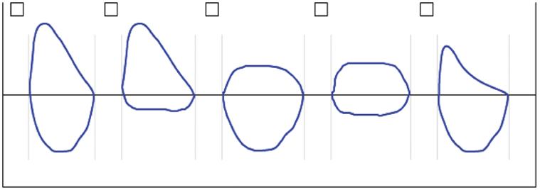
For questions 7–10, use the following figure to match the clinical scenario to the appropriate flow volume loop:
6 A 42-year-old man presents to the ICU following intubation for COPD exacerbation.
7 An 18-year-old woman diagnosed on bronchoscopy with intratracheal lipoma.
8 A recovered COVID-19 patient who develops tracheal stenosis following a 2 week intubation.
9 A 75-year-old male who undergoes emergent intubation following development of angioedema found to have R vocal cord paralysis.
Flow volume loops involve plotting inspiratory and expiratory flow on the Y-axis with volume on the X-axis, ideally during maximally forced inspiratory and expiratory effort. Flow volume loops are component of the information presented on mechanical ventilators as well and can aid in the diagnosis of airway obstruction. The normal loop is seen in loop A above representing a complete inspiratory and expiratory breath. Loop B demonstrates variable extrathoracic obstruction with a flattening of the inspiratory component. This is due to a combination of atmospheric extraluminal pressure and negative intraluminal pressure exacerbating extrathoracic obstruction as in vocal cord dysfunction and mobile tumors such as lipoma.
Intrathoracic variable obstruction, such as with bronchogenic cysts or intrathoracic tracheomalacia, is demonstrated by flattening of the expiratory component, as seen in loop C. Pleural pressure becomes positive relative to airway pressure exacerbating obstruction during expiration. Loop D demonstrates fixed airway obstruction, as with tracheal stenosis, causing flattening of both components of the loop. Finally, loop E demonstrates lower airway obstruction as seen in COPD and asthma. A scooped-out appearance to the loop comes from premature airway closure as heterogeneity of flow in expiration, i.e., areas with higher elastic recoil and lower airway resistance empty faster than diseased areas.
Answers: 6-E, 7-B, 8-C, 9-B
Loutfi SA and Stoller JK. Flow-volume loops. UpToDate. Retrieved November 16, 2020 from https://www.uptodate. com/contents/flow-volume-loops?search=flow%20 volume%20loops&source=search_result&selected Title=1~59&usage_type=default&display_rank=1 Pellegrino R, Viegi G, Brusasco V, et al. Interpretative strategies for lung function tests. Eur Respir J. 2005; 26(5): 948–968.
10 A 72-year-old woman is admitted to the trauma ICU after presentation following high-speed MVC.
A pulmonary artery catheter is placed given the patient’s refractory hypotension. Which of the following is consistent with cardiogenic shock?
Though used infrequently within the surgical ICU setting, the Swan-Ganz catheter is a useful adjunct in the diagnosis of undifferentiated shock. Normal values obtained, as in option A, show a pulmonary capillary wedge pressure (PCWP) 8–12 mmHg, cardiac output (CO) 5–7 L/min, systemic vascular resistance (SVR) 900–1300dyne-sec/cm5, and mixed venous oxygen (MVO2) approximately 65%. Option B indicates severe hypovolemic shock with decreased PCWP, decreased CO, increased SVR, and decreased MVO2. Option C indicates cardiogenic shock with increased PCWP, decreased CO, increased SVR, and decreased MVO2 Option D indicates distributive shock with normal PCWP, increased CO, decreased SVR, and increased MVO2. Option E indicates obstructive shock with normal PCWP, normal CO, increased SVR, and increased MVO2.
Answer: C
Cecconi M, De Backer D, Antonelli M, et al. Consensus on circulatory shock and hemodynamic monitoring. Task force of the European Society of Intensive Care Medicine. Intensive Care Med. 2014; 40: 1795–1815.
11 A 73-year-old female with past medical history of significant peripheral vascular disease, hypertension, and diabetes is admitted to the ICU with significant hypotension following a myocardial infarction in PACU after undergoing EVAR of a 6 cm AAA. STAT echocardiogram shows right-sided heart failure. SwanGanz catheter is placed with PCWP of 10 mmHg. What is the next appropriate intervention?
A Inotrope initiation.
B Vasopressor initiation.
C Placement of intra-aortic balloon pump.
D Volume resuscitation.
E Diuretic therapy.
The initial treatment of choice following acute right heart failure following MI is fluid resuscitation until PCWP > 15 mmHg is reached. Following this, initiation of inotropes, such as dobutamine, is done. Diuretic
therapy may play a role in normotensive individuals. Vasopressors may be used in hypotensive patients with the goal of increasing systemic vascular resistance without increasing pulmonary vascular resistance. Fluid resuscitation should be adequate before continuing to increase vasopressor use. The intra-aortic balloon pump is used in left heart failure, not right heart failure.
Answer: D
Ventetuolo CE and Klinger JR. Management of acute right ventricular failure in the intensive care unit. Ann Am Thorac Soc. 2014; 11(5): 811–822.
12 An 83-year-old woman with past medical history of significant peripheral vascular disease, ESRD on peritoneal dialysis admitted following below knee amputation for acute limb ischemia. You are called to bedside for patient’s mean arterial pressure of 55 mmHg. You note the systolic pressure is appropriate, but diastolic pressure remains low. Which of the following is part of the pathophysiology of diastolic heart failure?
A Adaptive myocyte remodeling.
B Volume overload of the ventricle.
C Cell loss secondary to increased oxygen demand.
D Impaired ventricular wall relaxation.
E Change of ventricle from elliptical to globular.
Diastolic heart failure stems from incomplete relaxation of the ventricle. Three pathophysiologic pathways include impaired ventricular wall relaxation, as left atrial pressure exceeds left ventricular pressure causing pulmonary edema; increased stiffness of the ventricle secondary to increased wall thickness and decreased internal diameter often seen with poorly controlled hypertension; excess collagen deposition as myofibrils are laid in parallel secondary to ischemia, as seen with MI, impairing contractility. The pathophysiology of systolic failure involves adaptive myocyte remodeling, as occurs with CAD, changing ventricular shape resulting in an increasingly overloaded ventricle with decreasing contractility resulting in cell loss due to increased oxygen demand and eventual change of the ventricle from elliptical to globular.
Answer: D
Zile MR, Baicu CF and Gaasch WH. Diastolic heart failure – abnormalities in active relaxation and passive stiffness of the left ventricle. NEJM. 2004; 350: 1953–1959.
13 You are utilizing central venous pressure monitoring to guide resuscitation of a patient with a 60% TBSA
burn injury in your ICU. Which of the following components of the CVP waveform represents isovolumic contraction?
A c wave
B x descent
C a wave
D y descent
E v wave a c x v y
CVP Waveform component Mechanical event
a wave Atrial contraction
c wave Isovolumic contraction
v wave Systolic filling of the atrium
x descent Atrial relaxation, systolic collapse
y descent Early diastolic filling, diastolic collapse
Answer: A
Magder S. Central venous pressure: a useful but not so simple measurement. Crit Care Med. 2006; 34: 2224–2227.
14 Which component of the cardiac cycle is represented by the answer given above?
A Early diastole
B Early systole
C End diastole
D Mid-systole
E End systole
The a wave occurs from right atrial contraction increasing venous pressure. Right ventricular contraction displaces the tricuspid valve into the right atrium, represented by the c wave. With the emptying of right ventricle, the right atrium relaxes and begins to fill, represented by the x descent. The v wave demonstrates the filled right atrium with increased atrial pressure. Finally, the y descent shows right ventricular filling as the tricuspid valve opens.
CVP Waveform component
a wave
Cardiac cycle event
End diastole
c wave Early systole
v wave Late systole
x descent Mid-systole
y descent Early diastole
Answer: B
Magder S. Central venous pressure: a useful but not so simple measurement. Crit Care Med. 2006; 34: 2224–2227.
15 Which of the following is NOT a physiologic effect of minimally invasive left ventricular assist device?
A Decreased left ventricular end diastolic pressure.
B Decreased left ventricular wall tension.
C Increased diastolic pressure.
D Increased mean arterial pressure.
E Increased pulmonary capillary pressure.
A minimally invasive left ventricular assist device is a miniature axial pump that allows blood to be aspirated from the left ventricle into the cannula component of the pump and expelled above the aortic valve into the ascending aorta. The device has been used for support in high-risk percutaneous coronary intervention as well as cardiogenic shock. The device works to unload the left ventricle reducing left ventricular end diastolic pressure and wall tension. This allows for decreased oxygen demand. Furthermore, it increased mean arterial pressure, diastolic pressure, and cardiac output, thereby improving both systemic and coronary blood flow. Finally, it decreases pulmonary capillary pressure and thereby right ventricular afterload.
Answer: E
Burzotta F, Trani C, Doshi S, et al. Impella ventricular support in clinical practice: collaborative viewpoint from a European expert user group. Int J Cardiol. 2015; 201: 684–691.
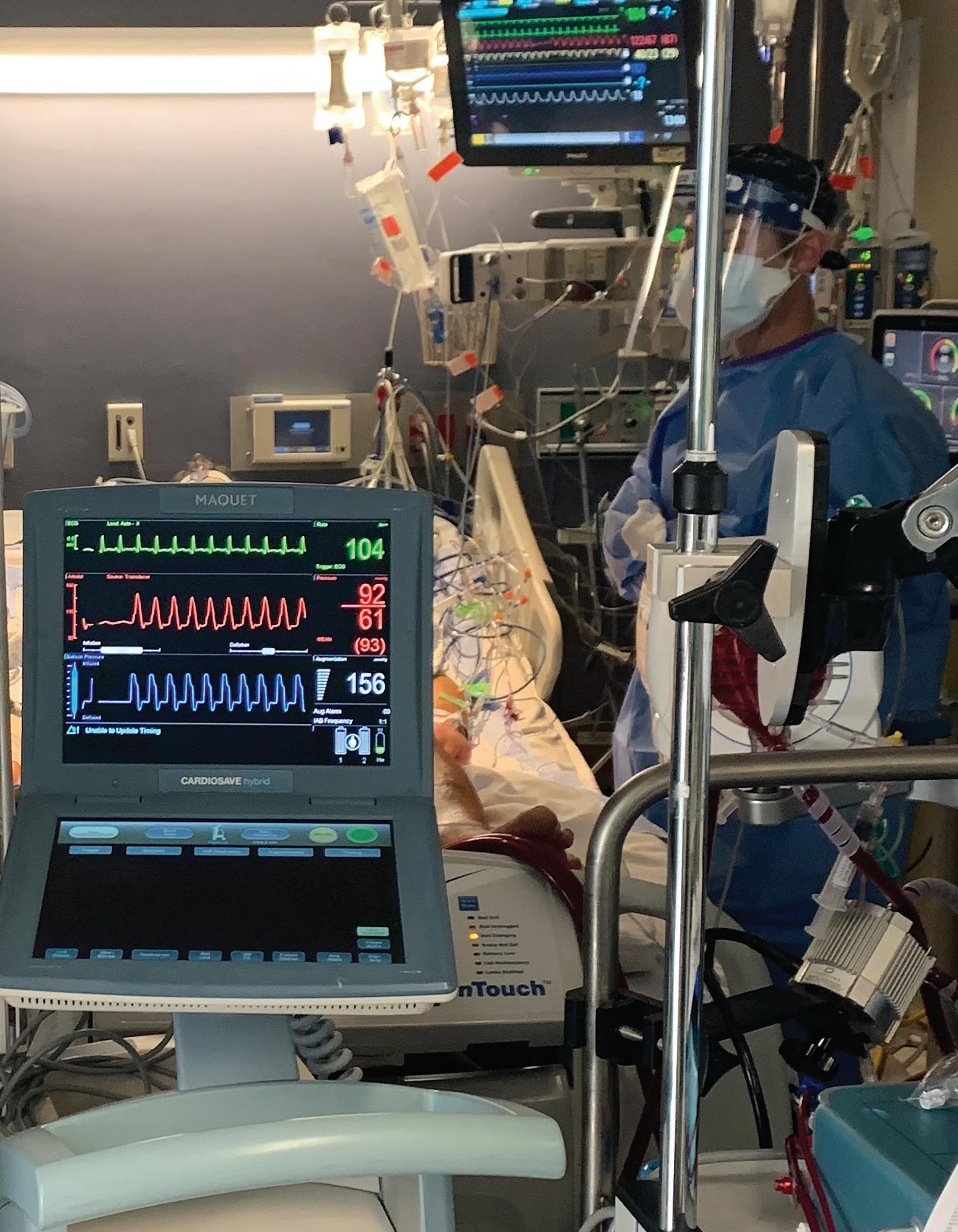
16 Which of the following is a physiologic impact of intra- aortic balloon pumps during systole?
A Increased systolic blood pressure.
B Decreased pre-systolic aortic pressure.
C Increase in the isometric phase of left ventricular contraction.
D Increased left ventricular wall tension.
E Decreased left ventricular ejection fraction.
The intra-aortic balloon pump follows the principle of counterpulsation i.e. inflation during diastole with deflation during systole. The physiologic impacts during the systolic phase include a decrease in aortic systolic pressure as well pre-systolic (end-diastolic) aortic pressure both of which contribute to decreased afterload by 10% and 30%, respectively; decrease in the isometric phase of left ventricular contraction, thereby reducing myocardial oxygen consumption; decreased left ventricular wall
tension by 20%; increased left ventricular ejection fraction by up to 30%.
Answer: B
Parissis H, Graham V, Lampridis S, et al. IABP: historyevolution-pathophysiology-indications: what we need to know. J Cardiothorac Surg. 2016; 11(1): 122.
17 Which of the following is an expected cardiovascular change during pregnancy?
A Decreased heart rate.
B Decreased cardiac output.
C Increased peripheral vascular resistance.
D Decreased ventricular distension.
E Decreased systemic vascular resistance.
Pregnancy results in increased heart rate, increased cardiac output, decreased peripheral vascular resistance, increased ventricular distension, and decreased systemic vascular resistance.
Answer: E
Hill CC and Pickinpaugh J. Physiologic changes in pregnancy. Surg Clin N Am. 2008; 88: 391–401.
18 Which of the following is a mechanism by which vasodilators improve cardiac function in acute decompensated heart failure?
A Increased ventricular preload.
B Decreased stroke volume.
C Increased ventricular afterload.
D Increased cardiac output.
E Increased ventricular filling pressure.
The pathophysiology of acute heart failure involves increased myocardial oxygen demand with increased ventricular filling pressures, low cardiac output, and increased systemic vascular resistance. Nitroprusside and nitroglycerin remain two of the most potent vasodilators used in therapy. Nitrogylcerin is a venodilator working to decrease preload, decrease afterload, and myocardial oxygen demand. Nitroprusside is an arterial and venous dilator decreasing preload, afterload, myocardial oxygen demand as well as increasing stroke volume and cardiac output.
Answer: D
Carlson MD and Eckman PM. Review of vasodilators in acute decompensated heart failure: the old and new. J Card Fail 2013; 19(7): 478–493.
19 Which of the following is an expected effect of increased intrapleural pressure from positive pressure ventilation?
A Increased venous return.
B Increased aortic pressure.
C Baroreceptor dampening.
D Increased systemic vascular resistance.
E Increased preload.
With positive pressure ventilation, increased intrapleural pressure results in initially increased aortic pressure causing compensatory reduction in systemic vascular resistance and left ventricular afterload by activated baroreceptors, thereby increasing cardiac output. Positive pressure additionally decreases venous return and, therefore, preload.
Answer: B
Alviar CL, Miller PE, McAreavey D, et al. Positive pressure ventilation in the cardiac intensive care unit. J Am Coll Cardiol. 2018; 72: 1532–1553.
20 A 70-year-old woman in a motor vehicle collision undergoes a splenectomy for Grade IV laceration and receives four units of whole blood in the OR but arrives in the ICU tachycardiac and hypotensive. Point of care hemoglobin is 14.3 mg/dL 2 hours posttransfusion. Her abdomen was left open and minimal output is coming from her negative pressure abdominal dressing. She has multiple rib fractures and a radius fracture. Which of the following therapies would promote end-organ perfusion?
A Decrease vasoactive drug doses (decrease peripheral vascular resistance).
B Increase sedation and pain medications to decrease her heart rate.
C Increase end-diastolic volume with volume resuscitation.
D Increase contractility with positive inotrope.
E Increase end-systolic volume.
This patient has evidence of blunt chest trauma with multiple rib fractures and tachycardia. While she could have hypovolemic shock from her splenic injury and intraoperative blood loss, she remains hypotensive despite transfusions with a hemoglobin of 14.3 mg/dL making this less likely and no evidence of ongoing bleeding from her abdomen. This makes it less likely that further volume resuscitation with blood or crystalloid would be helpful. Blunt cardiac injury can occur with blunt chest trauma and is initially screened for with EKG and troponin assessment, followed by an echocardiogram. Blunt cardiac injury may be improved with positive inotropic medications. Decreasing vasoactive drug doses would worsen hypotension and worsen end-organ perfusion. Vasopressors are often used in supportive treatment for
blunt cardiac injury and may need to be increased to promote end-organ perfusion. Increasing sedation and pain medications may improve her tachycardia but would worsen her hypotension and end-organ perfusion. Increasing end-systolic volume would decrease her stroke volume and cardiac output further, worsening her end-organ perfusion.
Remember: COHRSV SVEDV ESV
Answer: D
Levick JR. An Introduction to Cardiovascular Physiology. Butterworth and Co., London, 2013. Clancy K, Velopulos C, Bilaniuk JW, et al. Screening for blunt cardiac injury: An Eastern Association for the Surgery of Trauma practice management guideline. J Trauma Acute Care Surg. 2012; 735: S301–S306.
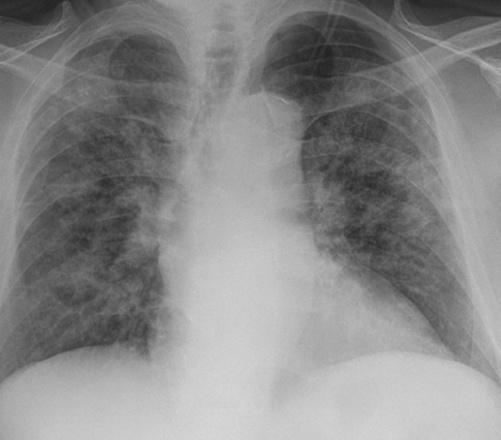
21 A 39-year-old man presents with a cold right leg and complains of nine days of symptoms. Following a thromboembolectomy and fasciotomy, he develops hypoxia with saturation of 87% and respiratory distress. An arterial blood gas shows: pH 7.47, paO2 = 50 mm Hg, HCO3 = 22 mmol/L, pCO2 = 30 mm Hg. Chest x-ray shows patchy consolidations bilaterally and he reports fever prior to admission and that he works in a skilled nursing facility during the pandemic.
Based on the above results, his A-a gradient is (at sea level, water vapor pressure = 47 mm Hg):
A 150 mm Hg
B 10 mm Hg
C 38 mm Hg
D 50 mm Hg
E 62 mm Hg
A-a gradient equals PAO2−PaO2. His PaO2 from the ABG is 50. The PAO2 can be calculated from this equation:
PaO FiOP PPaCORQ
PaO BH 22 02 2 2 021 760 47 30 08 11 / ./ 2 25.mmHg
Therefore, A-a gradient (PaO2−PAO2) = 62.5 mm Hg.
Answer: E
Marino P. The ICU Book, 3rd ed., Lippincott Williams & Wilkins, Philadelphia, PA, chapter 19 2007.
22 The patient above is placed on a nonrebreather mask with minimal improvement. What is the most likely etiology of the above patient’s respiratory failure and appropriate intervention?
A Pulmonary embolism, anticoagulation.
B Hyperventilation from anxiety, benzothiazines.
C COVID-19 pneumonia, dexamethasone, and high-flow nasal canula.
D Neuromuscular weakness, reversal of paralytic.
E Pulmonary edema, acute kidney injury from rhabdomyolysis.
Hypoxemia occurs in four conditions: low inspired oxygen, shunt, V/Q mismatch, and hypoventilation. Hypoventilation would present with high CO2 and normal A-a gradient. This could occur with oversedation, neuromuscular weakness, and residual anesthesia. Hyperventilation would cause tachypnea, low CO2, but not hypoxia, so A-a gradient should be normal. Low inspired oxygen should have a low PO2 and normal gradient. An acute PE or asthma exacerbation presents with V/Q mismatch with elevated A-a gradient and normal PCO2. It should correct with administration of oxygen. Shunting (pulmonary edema or pneumonia) has an elevated A-a gradient that does not improve with oxygen administration. The patient is young for postoperative MI and has risk factors and a chest x-ray consistent with COVID-19 pneumonia, which could also increase his risk of thrombotic events since as an arterial thrombus.
Answer: C
Weinberger SE, Cockrill BA and Mande J. Principles of Pulmonary Medicine, 5th ed., W.B. Saunders, Philadelphia, PA, (2008).
NIH COVID-19 Treatment Guidelines. Therapeutic management of patients with COVID-19. www. covid19treatmentguidelines.nih.gov/therapeuticmanagement/ (accessed 15 December 20).
23 A 63-year-old patient with history of hypertension and type 2 diabetes presents with acute respiratory distress syndrome from pneumococcal pneumonia and is being managed by the ICU team for severe ARDS. After appropriate sedation and analgesia, which of the following is NOT an appropriate strategy for management?
A Low tidal volume ventilation (4–8ml/kg IBW).
B Prone positioning <6 hours/day.
C Use of recruitment maneuvers.
D Higher PEEP levels with plateau pressures <30 cm H2O.
E Very select use of high-frequency oscillatory ventilation.
Acute respiratory distress syndrome management guidelines target management with low tidal volume ventila-
tion, low inspiratory pressures with plateau pressures <30 cm H20, high PEEP levels are better than low PEEP levels, and prone positioning for at least 12-hour periods per day with improved mortality. Less than 6 hours of prone position per day would not be recommended as it is too short a time period.
Answer: B
Fan E., Del Sorbo L, Goligher EC, et al. An Official American Thoracic Society/European Society of Intensive Care Medicine/Society of critical care medicine clinical practice guideline: mechanical ventilation in adult patients with acute respiratory distress syndrome. Am J Respir Crit Care Med. 2017; 195 9: 1253–1263. https://www.thoracic.org/statements/resources/cc/ ards-guidelines.pdf.
Cardiopulmonary Resuscitation, Oxygen Delivery and Shock
Kevin W. Cahill, MD, Harsh Desai, MD, and Luis Cardenas, DO, PhD
Department of Surgery, Christiana Care Health Care System, Newark, DE, USA
1 A 72-year-old woman with a history of Child’s B cirrhosis and supraventricular tachycardia is in the ICU following laparotomy for strangulated ventral hernia. She begins to complain of rapid heartbeat and is noted to be in an irregular, wide-complex ventricular tachycardia on EKG. She maintains pulse and adequate blood pressure. Which of the following is the best initial therapy to administer?
A Synchronized cardioversion.
B Adenosine 6 mg IV.
C Amiodarone 150 mg IV.
D Defibrillation.
E Vagal maneuvers.
The 2020 ACLS guidelines differentiate between regular and irregular wide-complex tachycardia with and without pulse. In this instance, the patient is in an irregular wide-complex tachycardia, symptomatic, but stable as evidence by pulse and pressure. Given this hemodynamic stability, synchronized cardioversion and defibrillation are not the initial therapies (choices A, D). Adenosine and vagal maneuvers may be effective in regular ventricular tachycardia (choices B, E). Therefore, amiodarone is the best initial medication to administration often followed by infusion (choice C). Individuals with hemodynamically unstable ventricular tachycardia should not initially receive amiodarone. These individuals should be cardioverted. Amiodarone can be used regardless of the individual’s underlying heart function and the type of ventricular tachycardia. It can be used in individuals with monomorphic ventricular tachycardia, but is contraindicated in individuals with polymorphic ventricular tachycardia as it is associated with prolonged QT intervals, which will be made worse with anti-arrhythmic drugs. Amiodarone is categorized as a class III anti-arrhythmic
agent, and prolongs phase 3 of the cardiac action potential. Amiodarone slows conduction rate and prolongs the refractory period of the SA and AV nodes. It also prolongs the refractory periods of the ventricles, bundles of His, and the Purkinje fibers without exhibiting any effects on the conduction rate. Serious side effects include interstitial lung disease and liver dysfunction with elevated liver enzymes.
Answer: C
Littmann L, Olson EG,Gibbs MA. Initial evaluation and management of wide-complex tachycardia: a simplified and practical approach. Am J Emerg Med. 2019; 37: 1340–1345.
Panchal AR, Bartos JA, Cabanas JG et al. Part 3: Adult basic and advanced cardiac life support: 2020 American Heart Association guidelines for cardiopulmonary resuscitation and emergency cardiovascular care. Circulation. 2020; 142 (suppl 2): S366–S468.
2 Which of the following techniques has not been shown to be effective in airway management during cardiac arrest?
A Head tilt – chin lift
B Jaw thrust
C Cricoid pressure
D Nasopharyngeal airway
E Oropharyngeal airway
Of the above maneuvers, cricoid pressure has not been shown to be effective during airway management in cardiopulmonary resuscitation. It may impede ventilation or placement of airway adjuncts such as a supraglottic airway as well as contribute to increased airway trauma.
Surgical Critical Care and Emergency Surgery: Clinical Questions and Answers, Third Edition. Edited by Forrest “Dell” Moore, Peter M. Rhee, and Carlos J. Rodriguez. © 2022 John Wiley & Sons Ltd. Published 2022 by John Wiley & Sons Ltd. Companion website: www.wiley.com/go/surgicalcriticalcare3e
Jaw thrust is preferred in patients with suspected spinal injury. Nasopharyngeal and oropharyngeal airways are particularly useful in cases of facial trauma though care must be taken with possible basilar skull fractures.
Answer: C
Carauna E, Chevret S, Pirracchio R. Effect of cricoid pressure on laryngeal view during prehospital tracheal intubation: a propensity-based analysis. Emerg Med J. 2017; 34 (3): 132–137.
Panchal AR, Bartos JA, Cabanas JG et al. Part 3: Adult basic and advanced cardiac life support: 2020 American Heart Association guidelines for cardiopulmonary resuscitation and emergency cardiovascular care. Circulation. 2020; 142 (suppl 2): S366–S468.
3 In a patient experiencing PEA arrest, which of the following would not be a likely etiology?
A Hypoglycemia
B Hypoxia
C Hypovolemia
D Hypokalemia
E Hypocalcemia
Pulseless electrical activity is so named due to evidence of cardiac mechanical activity on echocardiogram or rhythm on EKG. The algorithm is similar to the asystole algorithm utilizing compressions and epinephrine. The traditional etiologies are described as “Hs” and “Ts.” The “Hs” include hypoglycemia, hypoxia, hyper/hypokalemia, hypovolemia, acidosis, and hypothermia. Hypocalcemia can present with muscular and neurologic symptoms such as perioral numbness, cramping, fatigue, seizures, and irritability. Hypocalcemia may also be associated with increased risk of arrhythmias, but is not typically considered high on the initial differential of PEA arrest. The “Ts” taught as etiologies include tension pneumothorax, cardiac tamponade, toxins, pulmonary thrombosis, or coronary thrombosis. Evaluation for pneumothorax or tamponade includes rapid bedside physical exam as well as point of care ultrasound for rule out. Ultrasound may also reveal signs of thrombosis with right ventricular enlargement or free-floating thrombus.
Answer: E
Andersen LW, Holmberg MJ, Berg KM et al. In hospital cardiac arrest: a review. JAMA. 2019; 321 (12): 1200–1210.
Panchal AR, Bartos JA, Cabanas JG et al. Part 3: Adult basic and advanced cardiac life support: 2020 American Heart Association guidelines for cardiopulmonary resuscitation and emergency cardiovascular care. Circulation. 2020; 142 (suppl 2): S366–S468.
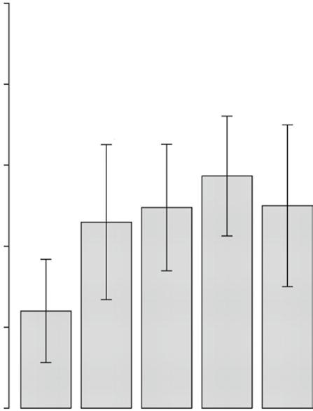
4 Which of the following is the minimum chest compression fraction (defined as amount of time spent delivering chest compressions during CPR) shown to be associated with improved survival?
A 0–20%
B 21–40%
C 41–60%
D 61–80%
E 81–100%
Optimal outcomes have been demonstrated with minimal pauses between compressions for pulse checks and breaths given during high-quality CPR. A compression fraction of at least 60% has been shown to be necessary for best outcomes. Animal studies previously conducted have demonstrated decreased coronary and cerebral perfusion when chest compressions are not being conducted resulting in worsened outcomes. Multiple retrospective analyses and cohort studies have resulted in many emergency agencies targeting a compression fraction of between 60 and 80% as a quality metric. This involves delivery of high-quality compressions of appropriate depth, 2 inches, and rate, at least 100/min.
Pe r centage sur viving
CCF categor y
Answer: D
Christenson J, Andrusiek D, Everson-Stewart S et al. Chest compression fraction determines survival in patients
FinalvitalstatusbyCCFcategory
with out of hospital ventricular fibrillation. Circulation. 2009; 120: 1241–1247.
Panchal AR, Bartos JA, Cabanas JG et al. Part 3: Adult basic and advanced cardiac life support: 2020 American Heart Association guidelines for cardiopulmonary resuscitation and emergency cardiovascular care.
Circulation. 2020; 142 (suppl 2): S366–S468.
5 Which of the following is considered the highest predictor of survival for in- and out-of-hospital CPR?
A Age.
B Shockable rhythm.
C Arrest at home.
D Arrest at night vs during the day.
E Delayed EMS response time.
On the whole, survivability is dependent on patient, system, event, and therapeutic factors. With increasing comorbidity and age, survivability decreases. System factors include time to arrival of EMS, time to initiation of CPR, and time to defibrillation. Event factors include preceding symptoms. Finally, therapeutic factors include availability of medications to treat suspected cause, time to ER, time to cath lab should it be required, etc. The greatest mortality risk with out of hospital cardiac arrest stems from unwitnessed arrests without bystander CPR often occurring at night in the elderly. Highest survivability stems from witnessed arrests with rapid initiation of bystander CPR and initial shockable rhythm, such as ventricular fibrillation.
Answer: B
Myat A, Song K- J, Rea T. Out of hospital cardiac arrest: current concepts. Lancet. 2018; 391: 970–79.
Navab E, Esmaelli M, Poorkhorshidi N et al. Predictors of out of hospital cardiac arrest outcomes in pre-hospital settings; a retrospective cross-sectional study. Arch Am Emerg Med. 2019; 7 (1): e36.
6 A 70-year-old man is 2 weeks status-post laparoscopic sleeve gastrectomy and he undergoes witnessed cardiac arrest at home after complaint of new onset chest pain. Bystander CPR achieves ROSC after 10 minutes. He is now in the ICU, intubated, and on vasopressors for associated hypotension. Which of the following interventions has the strongest associated survival benefit in post-arrest care according to current resuscitation guidelines?
A Maintain 100% FiO2.
B Pursuit of cardiac intervention when STEMI identified.
C Use of corticosteroids.
D Targeted temperature management to prevent fever.
E Seizure prophylaxis.
If a cardiac cause is suspected, pursuit of cardiac intervention such as with percutaneous coronary intervention (PCI) is strongly recommended. Hyperoxygenation therapy, the use of corticosteroids, and seizure prophylaxis have thus far shown no survival benefit (choices A, C, and E). Finally, targeted temperature management is currently recommended for post-arrest care with target of 32–36 ° C. This is based on several studies showing potential neurologic benefit. Preventing fever has not yet been proven to improve outcome though the 2020 AHA guideline (choice D). Ischemic heart disease is a major cause of out of hospital cardiac arrest. Among patients who had been successfully resuscitated after out of hospital cardiac arrest and had no signs of STEMI, immediate angiography was not found to be better than a strategy of delayed angiography with respect to overall survival at 90 days.
Answer: B
Panchal AR, Bartos JA, Cabanas JG et al. Part 3: Adult basic and advanced cardiac life support: 2020 American Heart Association guidelines for cardiopulmonary resuscitation and emergency cardiovascular care. Circulation. 2020; 142 (suppl 2): S366–S468. Yannapoulos D, Bartos JA, Aufderheide TP et al. The evolving role of the cardiac catherization laboratory in the management of patients with out of hospital cardiac arrest: a scientific statement from the American Heart Association. Circulation. 2019; 139 (12): e530–e552. Lemkes JS, Janssens GN, van der Hoeven NW et al. Coronary angiography after cardiac arrest without ST-Segment elevation. April 11, 2019. N Engl J Med. 2019; 380: 1397–1407. DOI: https://doi.org/10.1056/ NEJMoa1816897
7 A 35-year-old, 26 week pregnant woman has cardiac arrest with CPR ongoing in the ED. CPR has been ongoing for 5 minutes. Which of the following has been shown to provide greatest benefit for achieving ROSC?
A Corticosteroids.
B Targeted temperature management.
C Left lateral uterine displacement.
D Fetal monitoring.
E C-section.
In conditions of cardiac arrest after pregnancy, rapid delivery of the fetus, typically by C-section, termed perimortem cesarean delivery (PMCD), has been shown to be associated with improved outcomes when CPR does not achieve ROSC. However, the decision must be made quickly as a review article states that if done within 10 minutes of arrest, it was associated with better maternal outcomes. It was also thought that it was beneficial to
the mother in 31% of cases and was not harmful in any case. The review of the cases resulted in only 94 cases supporting that PMCD is rare. Corticosteroids have shown no benefit and targeted temperature management may be used after achievement of ROSC (choices A and B).The left lateral uterine displacement alleviates aortocaval compression in patients with hypotension, but delivery achieves this much more effectively (choice C). Fetal monitoring during maternal CPR is a distraction and may hinder care (choice D).
Answer: E
Einav S, Kaufman N, Sela HY. Maternal cardiac arrest and perimortem caesarean delivery: evidence or expert based? Resuscitation. 2012; 83 (10): 1191–1200.
Panchal AR, Bartos JA, Cabanas JG et al. Part 3: Adult basic and advanced cardiac life support: 2020 American Heart Association guidelines for cardiopulmonary resuscitation and emergency cardiovascular care. Circulation. 2020; 142 (suppl 2): S366–S468.
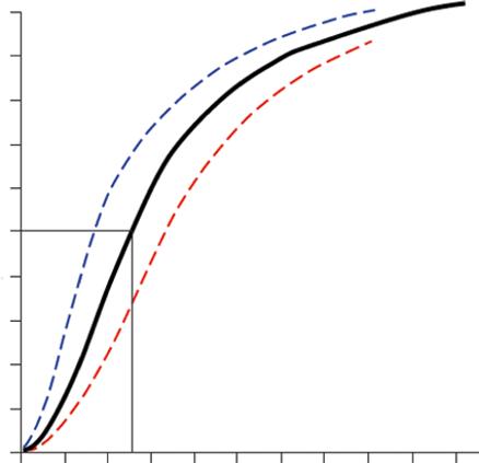
8 Which of the following scenarios causes a shift of the oxygen dissociation curve to the left?
A A patient found unconscious in a basement apartment with malfunctioning heater.
B Patient with pneumonia and fever of 102 ° C.
C Patient with lactic acidosis from mesenteric ischemia.
D Patient with depressed mental status taking slow, shallow breaths.
E Patient returning from climbing Mt Everest where he had to stop and be treated for hypoxia after leaving base camp.
Everest where he had to stop and be treated for hypoxia after leaving base camp. The oxygen–hemoglobin dissociation curve is sigmoidal in shape based on allosteric interactions of each globin monomer binding oxygen. A shift to the right indicates decreased affinity favoring unloading of oxygen while a shift to the left achieves the opposite effect. The strength by which oxygen binds to hemoglobin is affected by several factors and can be represented as a shift to the left or right in the oxygen dissociation curve. A rightward shift of the curve indicates that hemoglobin has a decreased affinity for oxygen, thus, oxygen actively unloads. A shift to the left indicates increased hemoglobin affinity for oxygen and an increased reluctance to release oxygen. Several physiologic factors are responsible for shifting the curve left or right, such as pH, carbon dioxide (CO2), temperature, and 2,3-Disphosphoglycerate. Carbon monoxide exposure, as can be seen in enclosed spaces with a malfunctioning heater, can result in a leftward shift. If the patient
was hypothermic or alkalotic, these conditions would also shift it toward the left.
Answer: A
Woodson, RD. Physiologic significance of oxygen dissociation curve shifts. Crit Care Med. 1979; 7 (9): 368–373.
9 You are caring for a patient in the SICU, currently intubated after undergoing left upper lobectomy for tumor. Patient’s current hemoglobin is 10 g/dL, oxygen saturation 95%, and PaO2 of 92 mmHg. What is the expected oxygen content (CaO2)?
A 0.9 mL/dL
B 9 mL/dL
C 13 mL/dL
D 21 mL/dL
E 140 mL/dL
Blood oxygen content is based on the following formula influenced by oxygen saturation, partial pressure of arterial oxygen, and patient’s hemoglobin:
CaO Hb 22SaOPaO 2 1340 003 134100 95 0 003 92 .. .. .
12 73 028
13 01 13 ./ or mL dL
The single biggest factor for oxygen content is hemoglobin. Doubling of hemoglobin would double the oxygen content. Increasing the partial pressure of oxygen from 60 mmHg to 100 would increase saturation from 90 to 100% and would not be a large change in content. The doubling of partial pressure of oxygen from 60 mmHg to 120 mmHg would still only increase the