Buy ebook Rook’s dermatology handbook 1st edition christopher e. m. griffiths cheap price
Rook’s
1st Edition Christopher E. M. Griffiths
Visit to download the full and correct content document: https://ebookmass.com/product/rooks-dermatology-handbook-1st-edition-christophere-m-griffiths/
More products digital (pdf, epub, mobi) instant download maybe you interests ...
All rights reserved. No part of this publication may be reproduced, stored in a retrieval system, or transmitted, in any form or by any means, electronic, mechanical, photocopying, recording or otherwise, except as permitted by law. Advice on how to obtain permission to reuse material from this title is available at http://www.wiley.com/go/permissions.
The right of Christopher E. M. Griffiths, Tanya O. Bleiker, Daniel Creamer, John R. Ingram, Rosalind C. Simpson to be identified as the authors of the editorial material in this work has been asserted in accordance with law.
Registered Offices
John Wiley & Sons, Inc., 111 River Street, Hoboken, NJ 07030, USA
John Wiley & Sons Ltd, The Atrium, Southern Gate, Chichester, West Sussex, PO19 8SQ, UK
Editorial Office
9600 Garsington Road, Oxford, OX4 2DQ, UK
For details of our global editorial offices, customer services, and more information about Wiley products visit us at www.wiley.com.
Wiley also publishes its books in a variety of electronic formats and by print‐on‐demand. Some content that appears in standard print versions of this book may not be available in other formats.
Limit of Liability/Disclaimer of Warranty
The contents of this work are intended to further general scientific research, understanding, and discussion only and are not intended and should not be relied upon as recommending or promoting scientific method, diagnosis, or treatment by physicians for any particular patient. In view of ongoing research, equipment modifications, changes in governmental regulations, and the constant flow of information relating to the use of medicines, equipment, and devices, the reader is urged to review and evaluate the information provided in the package insert or instructions for each medicine, equipment, or device for, among other things, any changes in the instructions or indication of usage and for added warnings and precautions. While the publisher and authors have used their best efforts in preparing this work, they make no representations or warranties with respect to the accuracy or completeness of the contents of this work and specifically disclaim all warranties, including without limitation any implied warranties of merchantability or fitness for a particular purpose. No warranty may be created or extended by sales representatives, written sales materials or promotional statements for this work. The fact that an organization, website, or product is referred to in this work as a citation and/or potential source of further information does not mean that the publisher and authors endorse the information or services the organization, website, or product may provide or recommendations it may make. This work is sold with the understanding that the publisher is not engaged in rendering professional services. The advice and strategies contained herein may not be suitable for your situation. You should consult with a specialist where appropriate. Further, readers should be aware that websites listed in this work may have changed or disappeared between when this work was written and when it is read. Neither the publisher nor authors shall be liable for any loss of profit or any other commercial damages, including but not limited to special, incidental, consequential, or other damages.
Library of Congress Cataloging‐in‐Publication Data
Names: Griffiths, C. (Christopher), editor. | Bleiker, Tanya, 1969- editor. | Creamer, Daniel, editor. | Ingram, John R., editor. | Simpson, Rosalind C., editor.
Title: Rook’s dermatology handbook / edited by Christopher E. M. Griffiths, Tanya O. Bleiker, Daniel Creamer, John R. Ingram, Rosalind C. Simpson.
Other titles: Dermatology handbook
Description: Hoboken, NJ : Wiley-Blackwell, 2020. | Includes index.
Identifiers: LCCN 2020024268 (print) | LCCN 2020024269 (ebook) | ISBN 9781119428190 (paperback) | ISBN 9781119428350 (adobe pdf) | ISBN 9781119428374 (epub)
Where necessary, the original material has been rewritten to suit a different readership. Thanks also to John Ingram’s colleagues in Cardiff for supplying additional images: Drs Mabs Chowdhury, Manju Kalavala, Colin Long, Richard Motley, Catherine Roberts, Rachel Abbott and Ru Katugampola, and Professors Andrew Finlay and Vincent Piguet.
Preface
Rook’s Textbook of Dermatology is a four volume behometh of information, arguably THE dermatological reference. First published in 1968 Rook has evolved from the visionary text produced by Arthur Rook, Darrell Wilkinson and John Ebling and is now in its 9th edition. Indubitably this is indispensable as a detailed reference on dermatology but is neither a pocket book for ready access in clinic nor a skin disease “101” for trainees, non‐dermatologists and hard pressed consultants. The Rook editors had discussed the relative merits of a “Rook Handbook” for several years and eventually decided, by popular demand, that the time had come for the talking to stop and work to begin on such a book.
Rook’s Dermatology Handbook is a 1000 page, fully illustrated guide to facilitate the rapid diagnosis of skin diseases and provide ready access to key relevant facts about them. The format comprises epidemiology, pathophysiology, clinical features, differential diagnosis, investigations and management. The basic science is kept to a minimum, there are no photomicrographs of histology and management is top line only. The reader would be expected to turn to more detailed references for
in‐depth information. We have pared down the text from Rook’s textbook, retained many of its high‐quality images and introduced new tables and new sections, including dermatological vocabulary and differential diagnosis of common clinical presentations such as blisters, hair loss and erythroderma. Three of the editors of the textbook – Chris Griffiths, Tanya O. Bleiker and Daniel Creamer – have been joined in the venture by two next generation clinicians, John Ingram and Rosalind Simpson. We are delighted with the book but the proof will come from how it performs in the clinic as an aid to the diagnosis, understanding and management of dermatological disease. We are indebted to all of the authors from the Rook 9th edition who freely gave us permission to recycle and synopsise their chapters.
The book would not have seen the light of day without the unstinting support and expertise of Claire Bonnett, Jenny Seward and Nick Morgan of Wiley, and Production team.
Chris Griffiths
Tanya O. Bleiker
Daniel Creamer
John Ingram
Rosalind Simpson
Glossary
Alopecia Decreased density or thickness of hairs
Artefact Induced by exogenous injury, sometimes self‐inflicted
Callus Reactive hyperkeratosis, usually due to friction and/or pressure, leading to enhanced skin markings
Comedone Open: dilated hair (open and infundibulum with oxidised closed) (black) keratinous debris (‘blackhead’)
Closed: expansion of hair infundibulum by keratinous debris, usually with no connection to skin surface (‘whitehead’)
Dysaesthesia Inappropriate sensations, e.g. paraesthesias
Exanthem Acute widespread eruption, usually due to a viral infection or drug reaction
Fissure Linear disruption of stratum corneum; may extend into the dermis
Infarct Ischaemia of tissue due to arterial occlusion
Induration Deep thickening of the skin can result from oedema, inflammation, or infiltration
Keratoderma Thickening of the stratum corneum and/or epidermis of the palms and soles, often inherited
Keratosis Focal thickening of the epidermis, especially the stratum corneum
Kerion Boggy plaque, due to infection, that often contains pustules
Lichenification Accentuation of skin markings, often due to rubbing
Poikiloderma Simultaneous presence of atrophy, telangiectasia and hypo‐ and hyperpigmentation
Prurigo Papules or nodules due to scratching or picking
Purpura Haemorrhage into the skin due to pathological processes, primarily of blood vessels
Stria Linear atrophy along tension lines; initially can be red to purple in colour
Telangiectasia Permanently dilated capillaries and venules which are visible to the naked eye
Abbreviations
ACTH adrenocorticotropic hormone
AE atopic eczema
AGEP acute generalised exanthematous pustulosis
AIDS acquired immune deficiency syndrome
AK actinic keratoses
ALP alkaline phosphatase
ALT alanine aminotransferase
ANA antinuclear antibody
ANCA antineutrophil cytoplasmic antibodies
ASOT antistreptolysin O titre
AST aspartate aminotransferase
ATP adenosine triphosphate
BCC basal cell carcinoma
BCG bacille Calmette–Guérin
BP blood pressure
BSA body surface area
CBT cognitive behavioural therapy
CMV cytomegalovirus
CNS central nervous system
CREST calcinosis, Raynaud phenomenon, oesophageal dysmotility, sclerodactyly and telangiectasia
CRP C‐reactive protein
CT computerised tomography
CTCL cutaneous T‐cell lymphoma
CVI chronic venous insufficiency
CXR chest X‐ray
DEJ dermal–epidermal junction
DLE discoid lupus erythematosus
DLQI Dermatology Life Quality Index
DNA deoxyribonucleic acid
EBV Epstein–Barr virus
EGFR epidermal growth factor receptor
ELISA enzyme‐linked immunosorbent assay
ENA extractable nuclear antigen
ESR er ythrocyte sedimentation rate
FBC full blood count
5‐FU 5‐fluorouracil
G6PD glucose‐6‐phosphate dehydrogenase
GvHD graft‐versus‐host disease
H&E haematoxylin and eosin
Hb haemaglobin
HHV human herpesvirus
HIV human immunodeficiency virus
HLA human leukocyte antigen
HPV human papilloma virus
HSV herpes simplex virus
IF immunofluorescence
IFN interferon
IgE immunoglobulin E
IL interleukin
IMF immunofluorescence
IV intravenous
KC keratinocyte carcinoma
LDH lactate dehydrogenase
LE lupus erythematosus
LFT liver function test
LP lichen planus
MHC major histocompatibility complex
MM Investigations malignant melanoma
MMR mumps measles rubella vaccination
MRI magnetic resonance imaging
MRSA meticillin‐resistant Staphylococcus aureus
NSAID non‐steroidal anti‐inflammatory drug
PAS periodic acid–Schiff
PASI Psoriasis Area Severity Index
PCR polymerase chain reaction
PUVA psoralen and ultraviolet A
PVL Panton–Valentine leukocidin
PXE pseudoxanthoma elasticum
RAS Recurrent aphous stomatitis
RNA ribo nucleic acid
QoL quality of life
SCC squamous cell carcinoma
Sinus Tract leading from a deeper focus to the skin surface
SLE systemic lupus erythematosus
SPF sun protection factor
TCR T‐cell receptor
TFT thyroid function test
TNF tumour necrosis factor
TPMT thiopurine methyltransferase
U&E urea and electrolytes
UV ultraviolet
UVR ultraviolet radiation
VZV varicella‐zoster virus
WCC white cell count
WHO
World Health Organization
Introduction
1
Human skin consists of a stratified, cellular epidermis and an underlying dermis of connective tissue, separated by a dermal–epidermal basement membrane (Figure 1.1). Beneath the dermis is a layer of subcutaneous fat, which is separated from the rest of the body by a vestigial layer of striated muscle. The skin performs a number of functions, including:
• Providing a physiological barrier against the external environment.
• Maintaining fluid balance by restricting water loss through the skin.
• Forming an innate immune defence against bacteria, fungi and viruses through keratinocyte-derived endogenous antibiotics, defensins and cathelicidins. Langerhans cells have a primary role in epidermal immune surveillance.
• Supporting thermoregulation: vasodilatation or vasoconstriction of the blood vessels in the deep and superficial plexuses helps to regulate body temperature. Eccrine sweat glands, found at all skin sites, also play a role in heat control.
• Providing insulation and trauma protection: subcutaneous fat limits excessive heat loss and shields internal structures from physical trauma.
Fat also has an endocrine function, releasing the hormone leptin, which acts on the hypothalamus to regulate hunger and energy metabolism.
• Vitamin D production.
• Performing a psychosocial function: the appearance of human skin and its associated structures, especially scalp hair, has a major impact on self-image and thus on interpersonal relationships.
• Protection from ultraviolet (UV) radiation through production of melanin from melanocytes. Variation in response to sunlight has historically been divided into six categories according to the Fitzpatrick classification (Table 1.1). It is acknowledged that whilst these ‘skin types’ describe response to UV exposure, they do not adequately encompass the wide variation of tones seen in skin of colour.
Table 1.1 Fitzpatrick classification of skin types
Skin
History taking
Clinical assessment of a patient presenting with a skin disorder follows the standard approach of history-taking and physical examination. Investigations may be needed to supplement information obtained at the consultation. Although a dermatological diagnosis might be swiftly apparent, it is essential to take a full history before examining the skin. The history of the presenting complaint will yield the story of the symptoms and the clinical features of the dermatosis (Box 1.1). Further questions will give details about any associated clinical problems and the patient’s background medical history (Table 1.2). The act of questioning (and listening to the answers) contributes to a consultation’s therapeutic function.
Box 1.1 History of the presenting complaint
Essential points in the history: rashes
• Location: Where did the rash start, where did it spread to?
• Temporal: When did it start, does it come and go, if so, how long does it last?
• What are the predominant symptoms: itch, pain, disfigurement?
• Occupational factors: Does it get worse at work? Does it improve away from work?
• Open questions: Do you have any thoughts as to what has caused this? What concerns you most about this problem?
Essential points in the history: lesions
• Where is the lesion? Is it single or multiple?
• Was there any trauma before it arose?
• Temporal: How long has it taken to develop to this size? Is it still growing or is it resolving?
• Symptoms: Is it tender, painful or itchy? Are there any exacerbating factors? Has it bled?
• Past history: Have you had anything similar before?
• Sun exposure: How much sun exposure have you had, including living/ working abroad and use of sun beds? Any history of blistering sun burns?
Table 1.2 Background medical history
Further historyRationale
General medical history
Systemic diseases may have cutaneous features
Medication history Certain dermatoses are induced by drugs
Allergy historyCertain dermatoses are caused by an allergy to a food or drug or contact allergen
Example
Dermatomyositis and other connective tissue disorders
Drug‐induced exanthem
Oral allergy syndrome
Family historyCertain dermatoses are inheritedBasal cell carcinoma syndrome (Gorlin syndrome)
Occupational history
The trigger for a dermatosis may be found only at the patient’s workplace
Leisure historyCertain dermatoses are related to leisure activities
Travel historyCertain dermatoses are more commonly encountered abroad
Social historyCertain dermatoses are associated with lifestyle habits
Ethnicity Certain disorders are more prevalent in particular ethnic groups
Quality of lifeEffects of dermatosis on work, relationships, activities etc. can be quantified
Clinical examination
As in any medical consultation, examination follows history-taking and the correct assessment of skin signs is only achieved in the context of a patient’s symptoms. Effective interpretation of cutaneous clinical features relies on the principle that most skin diseases have characteristic lesions with a predilection for certain body sites. An understanding of these diseasespecific patterns is intrinsic to diagnosis in dermatology, an assertion which is especially true in the appraisal of a rash.
The patient should always be examined in a good light, preferably daylight, and with magnification of lesions if necessary. A mobile light on a flexible stand can be helpful in illuminating areas that are in the shade from overhead lights, such as the mouth and the flexures. Ideally, the
Allergic contact dermatitis in a hairdresser
Allergic contact dermatitis to plants in gardening
Leishmaniasis
Palmo‐plantar pustulosis is associated with smoking
Sarcoidosis and lupus erythematosus occur more frequently in patients with black skin
Dermatology Life Quality Index
entire skin should be examined in every patient, including the scalp and nails. Full skin examination may also reveal suspicious skin lesions that the patient was not aware of, for example lesions on the back.
Examination of a lesion
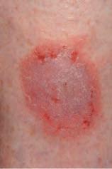
When assessing a solitary skin lesion there are a number of features relating to its morphology which direct the physician to a diagnosis. All skin lesions can be assigned to one of the descriptive entities defined in Table 1.3 (illustrated in Figure 1.2). Recognition of the lesion type is the basis of clinical examination in dermatology. Thereafter a more detailed appreciation of the lesion’s properties will enhance the assessment. Use of the 5Ss is helpful in describing, and thus identifying, a lesion: Site, Size, Symmetry, Shape and Surface.
Table 1.3 Descriptive terms for cutaneous lesions (adapted from Nast et al. 2016)
Term Definition
Bulla (Figure 2a)A circumscribed lesion >1 cm in diameter that contains liquid (clear, serous or haemorrhagic)
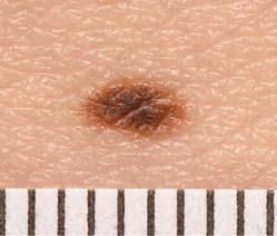
Macule (Figure 2b) A flat, circumscribed, non‐palpable lesion that differs in colour from the surrounding skin
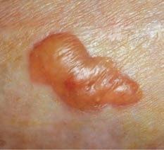
Nodule (Figure 2c)An elevated, solid, palpable lesion >1 cm usually located primarily in the dermis and/or subcutis
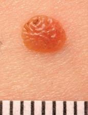
Papule (Figure 2d)An elevated, solid, palpable lesion that is ≤1 cm in diameter
Example
Bullous pemphigoid
Junctional naevus
Squamous cell carcinoma
Intradermal naevus
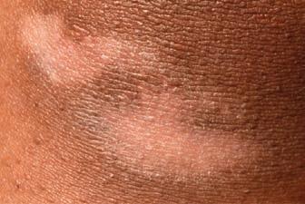
Patch (Figure 2e)A flat circumscribed area of discoloration, >1 cmVitiligo
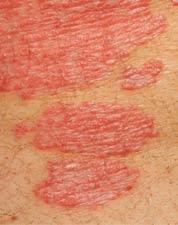
Plaque (Figure 2f)A circumscribed, palpable lesion >1 cm in diameter; most plaques are elevated
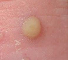
Pustule (Figure 2g)A circumscribed lesion that contains pus
Vesicle A circumscribed lesion ≤1 cm in diameter that contains liquid (not pus)
Weal
A transient elevation of the skin due to dermal oedema
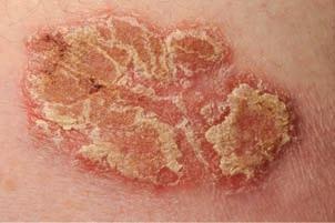
Scale (Figure 2h)A visible accumulation of keratin, forming a flat plate or flake
Crust
Erosion
Excoriation
Ulcer
Psoriatic plaque
Palmoplantar pustulosis
Herpes simplex
Urticaria
Psoriasis
Dried serum, blood or pus on the surface of the skinImpetigo
Loss of either a portion of the epidermis or the entire epidermis
A loss of the epidermis and a portion of the dermis due to scratching or an exogenous injury
Full‐thickness loss of the epidermis plus at least a portion of the dermis
Pemphigus vulgaris
Scratching from any cause
Venous leg ulcer
Source: Adapted from A. Nast et al. The 2016 International League of Dermatological Societies’ revised glossary for the description of cutaneous lesions. British Journal of Dermatology, 2016, 174, pp. 1351–1358. John Wiley & Sons Ltd on behalf of the British Association of Dermatologists.
Examination of a rash
In the initial assessment of a rash the type of primary lesion, from which the dermatosis is constituted, needs to be identified. Thereafter the configuration of lesional skin on the skin’s surface will point to the diagnosis, a deductive process termed pattern recognition. The description of a rash should comment on the morphology of individual lesions, including colour and shape (Table 1.4), as well as information on body sites of involvement and distribution
pattern (Table 1.5) (illustrated in Figure 1.3). Palpation of lesional skin imparts additional information about texture, skin thickness, tenderness and temperature.
Medical photographs
Medical photography is a useful tool to record the current state of a dermatosis and to permit serial comparisons over time to assess change. Patient consent and secure image storage are important considerations.
Figure 1.2 Types of cutaneous lesion: (a) bulla: bullous pemphigoid; (b) macule: junctional naevus; (c) nodule: squamous cell carcinoma; (d) papule: intradermal naevus; (e) patch: vitiligo; (f) plaque: psoriasis; (g) pustule: palmoplantar pustulosis; (h) scale: psoriasis. (Source: Reproduced with permission of Cardiff and Vale University Health Board.)
Bedside tests
Dermoscopy
(Syn. Dermatoscopy)
A dermatoscope (Syn. Dermoscope) provides polarised light and magnification to aid examination of the skin. The dermatoscope lens is applied to the skin surface with a film of oil on the lesion to enhance examination of subcorneal structures. Analysis of the colours and appearances of structural elements, such as the pigment network, is especially useful in the
diagnosis of pigmented lesions. The images may be viewed directly, photographed or recorded digitally for subsequent or sequential analysis. Dermatoscopes can also be useful in distinguishing haemangiomas, angiokeratomas, pigmented basal cell carcinomas and seborrhoeic keratoses. Additional uses include the identification of scabies mites and other parasitic infections. Trichoscopy is use of a dermatoscope for hair and scalp lesions, and aids diagnostic accuracy in scalp disorders.
(a)
(b)
(c)
(d)
(e)
(f)
(g) (h)
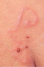
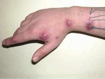
Table 1.4 Shapes of cutaneous lesions (adapted from Nast et al. 2016)
Term Definition Example
Acuminate Elevated lesion with a sharp pointCutaneous horn
Annular (Figure 3a)Shape of a ring (clear centrally)Tinea corporis
Arciform, arcuateA segment of a ring; arch‐likeErythema annulare centrifugum
Digitate
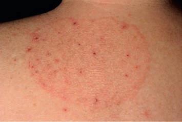
Discoid; nummular (Figure 3b)
Finger‐shaped
Circular or coin‐shaped
Digitate dermatosis, a form of parapsoriasis
Nummular eczema
Linear Lesional skin forming a band or lineLichen striatus
Papillomatous Lesion with multiple surface projections
Pedunculated
Polymorphic
Papule or nodule attached by a thinner stalk
Variable sizes and shapes as well as types of lesions
Epidermal naevus
Skin tag
Acne vulgaris
Polycyclic Coalescence of several ringsSubacute cutaneous lupus erythematosus
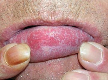
Reticulate (Figure 3e)Net‐like or lacy pattern
Mucosal lichen planus
Serpiginous (Figure 3c) Wavy pattern, reminiscent of a snake Cutaneous larva migrans
Targetoid Lesion composed of concentric rings
Umbilicated Lesion with a small surface depression
Verruciform Lesion with multiple projections resembling a wart
Erythema multiforme
Molluscum contagiosum
Viral wart
Source: Adapted from A. Nast et al. The 2016 International League of Dermatological Societies’ revised glossary for the description of cutaneous lesions. British Journal of Dermatology, 2016, 174, pp. 1351–1358. John Wiley & Sons Ltd on behalf of the British Association of Dermatologists.
A dermatoscope can also be used in the assessment of nail fold capillaries in connective tissue diseases (e.g. dermatomyositis).
Skin swabs (bacterial/viral)
In a suspected bacterial infection skin swabs for bacteriology should be sent to confirm the organism and provide antibiotic sensitivities to guide antibiotic selection.
Detection of viral skin infection, for example herpes simplex, requires a specific viral swab for viral culture. Increasingly polymerase chain reaction (PCR) amplification of viral DNA/RNA can provide a diagnosis within hours.
Mycological sample collection
Skin. S craping samples of surface scale from an active margin are taken with a disposable scalpel blade or banana-shaped scalpel. The scrapings should be transported in folded paper, which keeps the specimen dry, thus preventing overgrowth of bacterial contaminants.
Hairs. If tinea capitis is suspected, the hairs should be plucked with the roots intact; cut hairs are unsuitable. Brush samples from the scalp are excellent for culture, but with this technique microscopy is not possible.
Table 1.5 Distribution patterns of cutaneous lesions (adapted from Nast et al. 2016)
Term
Acral
Asymmetrical
Blaschkoid; along Blaschko lines
Dermatomal (zosteriform)
Disseminated
Exposed skin
Extensor sites
Flexural sites
Definition
Lesions involving distal extremities (e.g. ears, nose, fingers and toes)
Distribution pattern which lacks symmetry along an axis (e.g. the midline)
Lesions occurring on embryonic growth lines (Blaschko lines)
Lesions confined to one or more dermatome (a segment of skin innervated by a single spinal nerve)
Lesions distributed randomly over most of the body surface area
Areas exposed to external agents (e.g. airborne allergens, irritants, sunlight)
Areas overlying muscles and tendons involved in extension (e.g. dorsal forearm, elbow, posterior upper arm)
Areas overlying muscle and tendons involved in flexion of joints (e.g. antecubital fossa)
Example
Acrocyanosis
Lichen striatus
Incontinentia pigmenti
Shingles (herpes zoster)
Viral exanthem
Airborne allergic contact dermatitis
Psoriasis
Atopic dermatitis
Follicular Lesions located within or around hair folliclesKeratosis pilaris
Generalised/ widespread
Intertriginous
Kobnerised (displaying Kobner phenomenon)
Palmo‐plantar
Periorificial (e.g. perioral, periorbital)
Seborrhoeic
Sporotrichoid (Figure 3d)
Symmetrical
Distributed over most of the body surface area (see above)
Present in major body folds (axillae, submammary folds, inguinal creases, natal cleft)
Lesions arranged in a distribution which reflects physical stimuli (e.g. scratching, sunburn)
Involving palmar and plantar skin
Involving the skin around orifices
Involving areas with the highest density of sebaceous glands (e.g. scalp, face, upper trunk)
Lesions occurring along lymphatic vessels, usually of arm or leg
Lesions occurring with symmetry along an axis, commonly the midline
Viral exanthem
Flexural psoriasis
Lichen planus
Palmo‐plantar pustulosis
Peri‐oral dermatitis
Seborrhoeic dermatitis
Mycobacterium marinum infection
Psoriasis
Source: Adapted from A. Nast et al. The 2016 International League of Dermatological Societies’ revised glossary for the description of cutaneous lesions. British Journal of Dermatology, 2016, 174, pp. 1351–1358. John Wiley & Sons Ltd on behalf of the British Association of Dermatologists.
Figure 1.3 Shapes and distribution patterns of skin lesions: (a) annular: tinea cruris; (b) discoid (round): nummular eczema; (c) serpiginous: cutaneous larva migrans; (d) sporotrichoid: Mycobacterium marinum infection; (e) reticulate: lichen planus on the lower lip. (Source: (a), (b) and (c) reproduced with permission of Cardiff and Vale University Health Board.)
Nails. Isolation of the pathogen from nail material is more difficult than in other samples. The full thickness of the nail should be sampled. Debris from under the nail is a fruitful source of material.
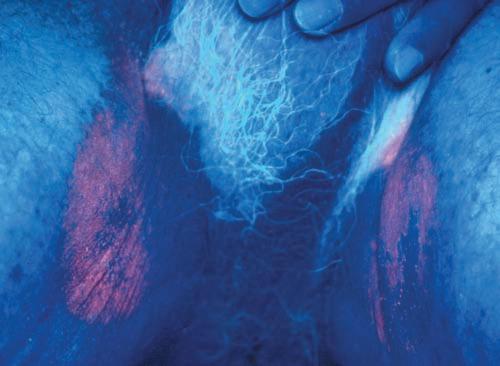
Wood’s light
This is a source of UV light from which visible light has been excluded by a Wood’s (nickel oxide) filter. Variations in epidermal pigmentation are more apparent under Wood’s light than under visible light, whereas variations in dermal pigment are less apparent. For example, Wood’s light accentuates the epidermal depigmentation of vitiligo, whereas the pallor from localised dermal vasoconstriction in naevus anaemicus disappears under Wood’s lamp. Some organisms produce chemicals that fluoresce under Wood’s lamp, including Corynebacterium minutissimum, the bacterium responsible for erythrasma
(Figure 1.4). Wood’s light examination is a useful tool in the diagnosis of superficial mycoses, particularly infections due to Microsporum species which fluoresce green (Table 1.6).
Specific investigations
Biopsy
A biopsy of lesional skin provides essential information on the dermatopathology of almost all skin disorders. Sections from a paraffinembedded biopsy specimen are usually stained with haematoxylin and eosin (H&E) for standard histopathological reporting. Special stains can be used to detect the presence of microorganisms (e.g. dermatophytes), the distribution of particular components of the skin (e.g. elastin) and the deposition of pathological substances (e.g. amyloid). Antibody deposition in the skin in immunobullous diseases is assessed
(a)
(b)
(c)
(d)
(e)
Table 1.6 Colour under Wood’s light linked to clinical examples (adapted from Nast et al. 2016)
Colour under Wood’s light
Clinical example(s)
Blue‐green to yellow‐green Tinea capitis due to Microsporum spp.
Coral pink Erythrasma
Red
White
Urine in some forms of porphyria
Well‐developed lesions of vitiligo
Yellow to yellow‐green Pityriasis (tinea) versicolor
Source: Adapted from A. Nast et al. The 2016 International League of Dermatological Societies’ revised glossary for the description of cutaneous lesions. British Journal of Dermatology, 2016, 174, pp. 1351–1358. John Wiley & Sons Ltd on behalf of the British Association of Dermatologists.
by direct immunofluorescence. Skin biopsies are processed specifically for this purpose; the immunofluorescence laboratory technique is performed on frozen sections.
Several different types of biopsy can be performed under local anaesthetic. Punch biopsies provide a small full thickness sample of skin, shave biopsies provide information on superficial skin layers and incisional biopsies may be used to sample a full-thickness section of a larger skin lesion or rash.
Patch tests
Patch tests are typically used to detect contact allergy of the delayed hypersensitivity type.
Multiple chemicals are applied to the patient’s back to assess for an eczematous reaction. Patch tests are usually read at 2 days and 4 days (see Chapter 69). A patch test technique can be used to detect contact urticaria when the results are read at 15–30 min.
Prick tests
Prick testing investigates immediate type I hypersensitivity reactions. A small quantity of the test solution is placed on the skin and a prick is made through it with a sharp needle. The size of the weal and flare is measured after 15 min and compared with an adjacent control solution.
Figure 1.4 Wood’s light illumination of erythrasma of the groins. The fluorescence in erythrasma is coral pink.
Introduction to dermatological therapeutics 2
Treatment modalities for skin disease comprise:
• Topical treatment
• Dressings (not discussed further in this book)
• Local injection
• Systemic agents (Chapter 82)
• Phototherapy
• Surgical removal of tissue
• Physical destruction of tissue such as with cryotherapy, cautery/diathermy, hyfrecation, and laser.
Topical treatment
There are different ‘vehicles’ (Box 2.1) that can be used to apply topical medication or emollients to the skin. As a rule, acutely inflamed skin is best treated with bland preparations that are least likely to irritate. Moist or exudative
Box 2.1 Different types of vehicle
eruptions are conventionally treated with lotions or creams, whilst dry skin responds well to ointments.
Prescribing topical treatment
The following should be considered when making a prescription for a topical treatment:
Prescription requirements: In general, a prescription of topical agent should comprise drug name, vehicle (cream or ointment), quantity to be dispensed, frequency, site of application and duration of treatment.
Frequency: Emollients should be applied frequently enough to maintain their physical effect, which may mean several applications per day. Active preparations are usually applied once or twice daily. Twice-daily application of topical corticosteroids is only marginally more effective than once-daily application.
Ointments: Semi-solid vehicles composed of lipid, such as white soft paraffin BP (petrolatum). Contain fewer preservatives than other vehicles. Occlusive and emollient properties.
Creams: Semi-solid emulsions containing both lipid and water. Emollient, lubricant and mildly occlusive.
Pastes: Semi-solid preparations containing a high proportion of finely powdered material such as zinc oxide or starch. Occlusive, protective and hydrating.
Lotions: Liquid formulations, usually simple suspensions or solutions of medication in water, alcohol or other liquids. Suitable for treating the scalp and other hairy areas of skin.
Gels: Thickened lotions. Suitable for treating the scalp and other hairy areas of skin.
Powders : Occasionally used to deliver drugs such as antifungal agents applied to the feet.
Paints: Liquid preparations which are usually applied with a brush to the skin or mucous membranes.
Dressings: Impregnated dressings, e.g. bandages containing ichthammol or zinc oxide, or tapes containing topical steroid.
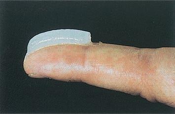
Figure 2.1 The fingertip unit: from the distal crease of the forefinger to the ventral aspect of the fingertip
Quantity: The quantity of active topical agent (such as topical corticosteroids) needed for effective treatment should be explained to the patient. ‘Fingertip units’ are a useful guide for topical corticosteroid application (see Figure 2.1 and Table 2.1). Emollients should be applied more liberally.
Timing of application: Leave a suitable amount of time between emollient and active agent. This avoids dilution of the active medication and prevents spread over areas of skin where it is not required.
Potential hazards: Localised irritant or allergic reactions are the most frequent adverse effects (see Chapter 69). All topically applied drugs are absorbed to some degree, but systemic side effects are relatively rare. Absorption varies considerably depending on the region of skin being treated (absorption greatest from the genital area and least from the soles and palms). Occlusion greatly enhances drug penetration. Inflammation of the skin significantly increases drug absorption, especially in erythrodermic patients. Bath oils tend to make the bath slippery; paraffin-based ointments are flammable, which is a particular risk in smokers.
Table 2.2 lists frequently used topical treatments, but is not intended to provide an exhaustive list.
Topical corticosteroids
Topical corticosteroids (TCS) are the mainstay of treatment in eczematous dermatoses and are used either regularly or occasionally in the management of most inflammatory skin
diseases. TCS vary in potency from mild to very potent (Table 2.3). Classification of potency is important to predict response and possible adverse effects. Penetration of TCS is increased by occlusion using polythene film, dressings, gloves or bandages. This improves beneficial effects but also increases the potential for adverse effects.
Side effects
Significant side effects are rare, especially with short term use. The most common side effects are localised to application sites: skin atrophy, striae, erythema, telangiectasia and purpura; atrophic changes can become irreversible with long-term use of potent preparations. Areas most vulnerable to atrophy are those where the skin is already relatively thin, e.g. flexures and face.
Other local side effects are development of contact allergy, exacerbation of infection and if used on the face acneiform eruptions (see Chapter 40). It is advisable to avoid the use of topical corticosteroids in the presence of active viral infection, including herpes simplex, viral warts or molluscum contagiosum.
It is recommended that patients should use no more than 50 g of a superpotent steroid or 100 g of a potent steroid preparation per week and that prolonged usage at this high rate should be avoided to minimise the risk of systemic absorption leading to Cushing syndrome and hypothalamic-pituitary-adrenal axis suppression.
Rebound worsening of disease may occur when topical corticosteroids are withdrawn, particularly in psoriasis. This is most likely after withdrawal of potent or very potent corticosteroids.
‘Steroid phobia’ describes the fear of using topical corticosteroids which is out of proportion to the likelihood of side effects developing.
Topical calcineurin inhibitors
Topical calcineurin inhibitors (TCIs) have been developed for topical treatment of atopic eczema and have numerous additional applications. TCIs (e.g. tacrolimus and pimecrolimus) exhibit their anti-inflammatory effect by inhibition of calcineurin, which suppresses lymphocyte activation. They do not induce cutaneous atrophy. Theoretically, the local immunosuppression related to these
Table 2.1 Fingertip units required for a single treatment of various regions in children and adults (the unit is measured using an adult finger)
Source: Finlay AY, Edwards PH, Harding KG. ‘Fingertip Unit’ in dermatology. Lancet, 1989, 11, 155 and Long CC, Mills CM, Finlay AY. A practical guide to topical therapy in children. Br J Dermatol 1998, 138, 293–296.
Table 2.2 Different types of topical treatments and their main uses
Type of agentCommon example(s)
Antimicrobial agents
Alcohols: isopropyl alcohol, ethanol and n-propanol
Coal tar solution BP 5% w/w in betamethasone valerate 0.025% w/w ointment 100 g
Salicylic acid 5% w/w/propylene glycol 47.5% w/w in Dermovate® cream 100 g
Reflectant (Dundee) sunscreens (available in coffee, coral pink, beige) 50 g
Indication
Acne
Early plaque stage mycosis fungoides
Treatment of alopecia areata and warts
To help soothe itching and discomfort
For moderate-severe psoriasis of trunk and limbs when other treatments such as vitamin D analogues have been ineffective For use on palmoplantar skin for hyperkeratotic eczema, palmopustular pustulosis and psoriasis not responding to Clobetasol propionate and emollients alone
To treat photosensitivity disorders where the patient is sensitive to visible light, e.g. solar urticaria and porphyrias