Contents
1 Introduction to Dental Anatomy, 1
Formation of the Dentitions (Overview), 1
Nomenclature, 2
Formulae for Mammalian Teeth, 2
Tooth Numbering Systems, 2
Division into Thirds, Line Angles, and Point Angles, 9
Tooth Drawing and Carving, 10
Measurement of Teeth, 11
Summary, 11
2 Development and Eruption of the Teeth, 21
Clinical Considerations, 21
Variability, 22
Malformations, 22
Chronology of Primary Dentition, 23
Development and Eruption/Emergence of the Teeth, 23
The Dentitions, 26
Neuromuscular Development, 27
Transitional (Mixed) Dentition Period, 28
Loss of Primary Teeth, 29
Permanent Dentition, 30
Size of Teeth, 31
Dental Pulp, 31
Cementoenamel Junction, 32
Dental Age, 33
Tooth Formation Standards, 35
Chronologies of Human Dentition, 35
Types of Chronologies, 35
Stages of Tooth Formation, 35
Age of Attainment, 35
Age Prediction, 37
Maturity Assessment, 37
Duration of Root and Crown Formation, 40
Summary of Chronologies, 41
Sequence of Eruption, 41
Estimating Time of Enamel Hypoplasia, 41
3 The Primary (Deciduous) Teeth, 45
Life Cycle, 45
Importance of Primary Teeth, 45
Nomenclature, 45
Major Contrasts between Primary and Permanent Teeth, 47
Pulp Chambers and Pulp Canals, 48
Detailed Description of Each Primary Tooth, 50
4 Forensics, Comparative Anatomy, Geometries, and Form and Function, 67
Forensic Dentistry, 67
Comparative Dental Anatomy, 69
Facial and Lingual Aspects of All Teeth, 73
Summary of Schematic Outlines, 75
Form and Function of the Permanent Dentition, 75 Alignment, Contacts, and Occlusion, 76
5 Orofacial Complex: Form and Function, 81
Form and Function, 81
Form Follows Function, 81
Articulation of Teeth, 81
Physiological Form of the Teeth and Periodontium, 82
Fundamental Curvatures, 82
Proximal Contact Areas, 82
Interproximal Spaces (Formed by Proximal Surfaces in Contact), 84
Embrasures (Spillways), 86
Contact Areas and Incisal and Occlusal Embrasures from the Labial and Buccal Aspect, 88
Contact Areas and Labial, Buccal, and Lingual Embrasures from the Incisal and Occlusal Aspects, 89
Facial and Lingual Contours at the Cervical Thirds (Cervical Ridges) and Lingual Contours at the Middle Thirds of Crowns, 91
The Height of Epithelial Attachment: Curvatures of the Cervical Lines (Cementoenamel Junction [CEJ]) Mesially and Distally, 94
6 The Permanent Maxillary Incisors, 99
Maxillary Central Incisor, 99
Maxillary Lateral Incisor, 106
7 The Permanent Mandibular Incisors, 113
Mandibular Central Incisor, 113
Mandibular Lateral Incisor, 119
8 The Permanent Canines: Maxillary and Mandibular, 125
Maxillary Canine, 125
Mandibular Canine, 132
9 The Permanent Maxillary Premolars, 141
Maxillary First Premolar, 141
Maxillary Second Premolar, 151
10 The Permanent Mandibular Premolars, 157
Mandibular First Premolar, 157
Mandibular Second Premolar, 165
11 The Permanent Maxillary Molars, 171
Maxillary First Molar, 171
Maxillary Second Molar, 181
Maxillary Third Molar, 184
12 The Permanent Mandibular Molars, 189
Mandibular First Molar, 189
Mandibular Second Molar, 199
Mandibular Third Molar, 203
13 Pulp Chambers and Canals, 209
Pulp, Chamber, and Canals, 209
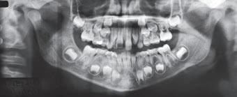
Radiographs, 209
Foramen, 211
Demarcation of Pulp Cavity and Canal, 211
Pulp Horns, 212
Clinical Applications, 212
Pulp Cavities of the Maxillary Teeth, 213
Pulp Cavities of the Mandibular Teeth, 222
Radiographs: Pulp Chamber and Canals, 233
Crown and Root Fractures, 233
14 Dento-osseous Structures, Blood Vessels, and Nerves, 239
The Maxillae, 239
The Mandible, 245
Arterial Supply to the Teeth, 252
Nerve Supply to the Jaws and Teeth, 256
15 The Temporomandibular Joints, Teeth, and Muscles, and Their Functions, 259
Temporomandibular Articulation, 259
Muscles, 265
Mandibular Movements and Muscle Activity, 270
16 Occlusion, 275
Concepts of Occlusion, 275
Development of the Dentitions, 276
Primary Dentition, 276
Mixed (Transitional) Dentition, 279
Permanent Dentition, 282
Cusp, Fossa, and Marginal Ridge Relations, 288
Lateral Occlusal Relations, 294
Biomechanics of Chewing Function, 298
Neurobehavioral Aspects of Occlusion, 298
Oral Motor Behavior, 303
Swallowing, 304
Summary, 304
Appendix A Review of Tooth Morphology, 309
Appendix B Tooth Traits of the Permanent Dentition, 327 Index, 335 Flash Cards
1 1
Introduction to Dental Anatomy
Dental anatomy is defined here as, but is not limited to, the study of the development, morphology, function, and identity of each of the teeth in the human dentitions, as well as the way in which the teeth relate in shape, form, structure, color, and function to the other teeth in the same dental arch and to the teeth in the opposing arch. Thus, the study of dental anatomy, physiology, and occlusion provides one of the basic components of the skills needed to practice all phases of dentistry.
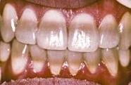
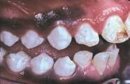
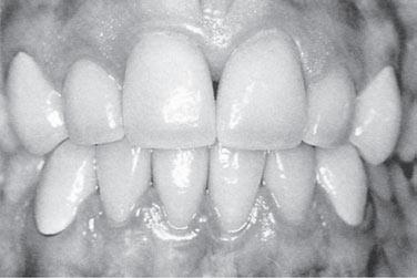
The application of dental anatomy to clinical practice can be envisioned in Figure 1-1, A where a disturbance of enamel formation (considered briefly in Chapter 2) has resulted in esthetic, psychological, and periodontal problems that may be corrected by an appropriate restorative dental treatment such as that illustrated in Figure 1-1, B. The practitioner has to have knowledge of the morphology, occlusion, esthetics, phonetics, and functions of these teeth to undertake such treatment.
Formation of the Dentitions (Overview)
Humans have two sets of teeth in their lifetime. The first set of teeth to be seen in the mouth is the primary or deciduous dentition, which begins to form prenatally at about 14 weeks in utero and is completed postnatally at about 3 years of age. In the absence of congenital disorders, dental disease, or trauma, the first teeth in this dentition begin to appear in the oral cavity at the mean age of 6, and the last emerge at a mean age of 28 ± 4 months. The deciduous dentition
remains intact (barring loss from dental caries or trauma) until the child is about 6 years of age. At about that time the first succedaneous or permanent teeth begin to emerge into the mouth. The emergence of these teeth begins the transition or mixed dentition period in which there is a mixture of deciduous and succedaneous teeth present. The transition period lasts from about 6 to 12 years of age and ends when all the deciduous teeth have been shed. At that time the permanent dentition period begins. Thus, the transition from the primary dentition to the permanent dentition begins with the emergence of the first permanent molars, shedding of the deciduous incisors, and emergence of the permanent incisors. The mixed dentition period is often a difficult time for the young child because of habits, missing teeth, teeth of different colors and hues, crowding of the teeth, and malposed teeth.
The permanent, or succedaneous, teeth replace the exfoliated deciduous teeth in a sequence of eruption that exhibits some variance, an important topic that will be considered in Chapter 16.
After the shedding of the deciduous canines and molars, emergence of the permanent canines and premolars, and emergence of the second permanent molars, the permanent dentition is completed (including the roots) at about 14 to 15 years of age, except for the third molars, which are completed at 18 to 25 years of age. In effect, the duration of the permanent dentition period is 12+ years. The completed permanent dentition consists of 32 teeth if none are congenitally missing, which may be the case. The development of the teeth, dentitions, and the craniofacial complex are considered in Chapter 2. The development of occlusion for both dentitions is discussed in Chapter 16.
Figure � - � A, Chronological developmental disorder involving all the anterior teeth. B, Illustration of restored teeth just after completion, taking in account esthetics, occlusion, and periodontal health. Note that the gingival response is not yet resolved.
(From Ash MM, Ramfjord S: Occlusion, ed 4, Philadelphia, 1995, Saunders.)
Nomenclature
The first step in understanding dental anatomy is to learn the nomenclature, or the system of names, used to describe or classify the material included in the subject. When a significant term is used for the first time here, it is emphasized in bold. Additional terms will be discussed as needed in subsequent chapters.
The term mandibular refers to the lower jaw, or mandible. The term maxillary refers to the upper jaw, or maxilla. When more than one name is used in the literature to describe something, the two most commonly used names will be used initially. After that they may be combined or used separately as consistent with the literature of a particular specialty of dentistry, for example, primary or deciduous dentition, permanent or succedaneous dentition. A good case may be made for the use of both terms. By dictionary definition,1 the term primary can mean “constituting or belonging to the first stage in any process.” The term deciduous can mean “not permanent, transitory.” The same unabridged dictionary refers the reader from the definition of deciduous tooth to milk tooth, which is defined as “one of the temporary teeth of a mammal that are replaced by permanent teeth. Also called baby tooth, deciduous tooth.” The term primary can indicate a first dentition and the term deciduous can indicate that the first dentition is not permanent, but not unimportant. The term succedaneous can be used to describe a successor dentition and does not suggest permanence, whereas the term permanent suggests a permanent dentition, which may not be the case due to dental caries, periodontal diseases, and
trauma. All four of these descriptive terms appear in the professional literature.
Formulae for Mammalian Teeth
The denomination and number of all mammalian teeth are expressed by formulae that are used to differentiate the human dentitions from those of other species. The denomination of each tooth is often represented by the initial letter in its name (e.g., I for incisor, C for canine, P for premolar, M for molar). Each letter is followed by a horizontal line and the number of each type of tooth is placed above the line for the maxilla (upper jaw) and below the line for the mandible (lower jaw). The formulae include one side only, with the number of teeth in each jaw being the same for humans.
The dental formula for the primary/deciduous teeth in humans is as follows:
ICM 2 2 1 1 2 2 10 =
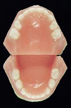
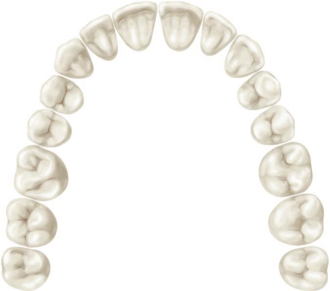
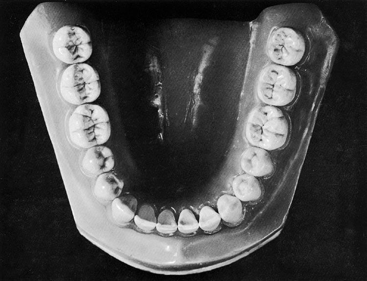
This formula should be read as: incisors, two maxillary and two mandibular; canines, one maxillary and one mandibular; molars, two maxillary and two mandibular—or 10 altogether on one side, right or left (Figure 1-2, A).
A dental formula for the permanent human dentition is as follows: ICPM 2 2 1
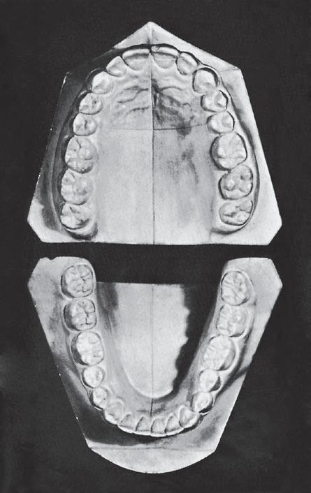
Premolars have now been added to the formula, two maxillary and two mandibular, and a third molar has been added, one maxillary and one mandibular (Figure 1-2, B).
Systems for scoring key morphological traits of the permanent dentition that are used for anthropological studies are not described here. However, a few of the morphological traits that are used in anthropological studies2 are considered in the following chapters, (e.g., shoveling, Carabelli’s trait, enamel extensions, and peg-shaped incisors). Some anthropologists use di1, di2, dc, dm1, and dm2 notations for the deciduous dentition and I1, I2, C, P1, P2, M1, M2, and M3 for the permanent teeth. These notations are generally limited to anthropological tables because of keyboard incompatibility.
Tooth Numbering Systems
In clinical practice some “shorthand” system of tooth notation is necessary for recording data. There are several systems in use in the world, but only a few are considered here. In 1947 a committee of the American Dental Association (ADA) recommended the symbolic (Zsigmondy/Palmer) system as the numbering method of choice.3 However, because of difficulties with keyboard notation of the symbolic notation system, the ADA in 1968 officially recommended the “universal” numbering system. Because of some
Central incisor (first incisor)
Lateral incisor (second incisor)
Canine (cuspid)
First molar
Second molar
Second molar
First molar
Canine
Lateral incisor (second incisor)
Central incisor (first incisor)
Central incisor (first incisor)
Lateral incisor (second incisor)
Canine (cuspid)
First premolar (first bicuspid)
Second premolar (second bicuspid)
First molar
Second molar
Third molar
Third molar
Second molar
First molar
Second premolar (bicuspid)
First premolar (bicuspid)
Canine (cuspid)
Lateral incisor (second incisor)
Central incisor (first incisor)
- �
limitations and lack of widespread use internationally, recommendations for a change sometimes are made.4
The universal system of notation for the primary dentition uses uppercase letters for each of the primary teeth: For the maxillary teeth, beginning with the right second molar, letters A through J, and for the mandibular teeth, letters K through T, beginning with the left mandibular second molar. The universal system notation for the entire primary dentition is as follows: Midsagittal Plane Right
The symbolic system for the permanent dentition was introduced by Adolph Zsigmondy of Vienna in 1861 and then modified for the primary dentition in 1874.
Figure �
A, Casts of deciduous, or primary, dentition. B, Casts of permanent dentition. (A from Berkovitz BK, Holland GR, Moxham BJ: Oral anatomy, histology and embryology, ed 3, St. Louis, 2002, Mosby.)
Independently, Palmer also published the symbolic system in 1870. The symbolic system is most often referred to as the Palmer notation system in the United States and less frequently as the Zsigmondy/Palmer notation system. In this system the arches are divided into quadrants with the entire dentition being notated as follows:
national Association for Dental Research. The FDI system of tooth notation is as follows.
For the primary teeth:
Thus, for a single tooth such as the maxillary right central incisor the designation is A . For the mandibular left central incisor, the notation is given as A . This numbering system presents difficulty when an appropriate font is not available for keyboard recording of Zsigmondy/Palmer symbolic notations. For simplification this symbolic notation is often designated as Palmer’s dental notation rather than Zsigmondy/Palmer notation.
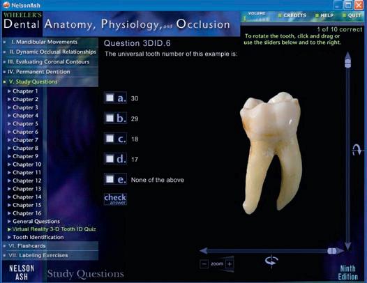
In the universal notation system for the permanent dentition, the maxillary teeth are numbered from 1 through 16, beginning with the right third molar. Beginning with the mandibular left third molar, the teeth are numbered 17 through 32. Thus, the right maxillary first molar is designated as 3, the maxillary left central incisor as 9, and the right mandibular first molar as 30. The following universal notation designates the entire permanent dentition.
12345678910111213141516 323130 29 282726252423222120191817
The Zsigmondy/Palmer notation for the permanent dentition is a four-quadrant symbolic system in which, beginning with the central incisors, the teeth are numbered 1 through 8 (or more) in each arch. For example, the right maxillary first molar is designated as 6 , and the left mandibular central incisor as 1 . The Palmer notation for the entire permanent dentition is as follows:
87654322345678
8765432 11 112345678
Viktor Haderup of Denmark in 1891 devised a variant of the eight-tooth quadrant system in which plus (+) and minus ( ) were used to differentiate between upper and lower quadrants and between right and left quadrants; in other words, +1 indicates the upper left central incisor and 1 indicates the lower right central incisor. Primary teeth were numbered as follows: upper right, 05+ to 01+; lower left, 01 to 05. This system is still taught in Denmark.5
The universal system is acceptable to computer language, whereas the Palmer notation is generally incompatible with computers and word processing systems. Each tooth in the universal system is designated with a unique number, which leads to less confusion than with the Palmer notation.
A two-digit system proposed by Fédération Dentaire Internationale (FDI) for both the primary and permanent dentitions has been adopted by the World Health Organization and accepted by other organizations such as the Inter-
Upper Right 5554535262636465 85848382 5161 8172737475
Lower Right 71
Upper Left Lower Left
Numeral 5 indicates the maxillary right side, and 6 indicates the maxillary left side. The second number of the two-digit number is the tooth number for each side. The number 8 indicates the mandibular right side, and the number 7 indicates the mandibular left side. The second number of the two-digit system is the tooth number. Thus, for example the number 51 refers to the maxillary right central incisor.
For the permanent teeth:
Upper Right
1817161514131222232425262728 48474645444342 1121 413132333435363738
Lower Right
Upper Left Lower Left
Thus, as in the two-digit FDI system for the primary dentition, the first digit indicates the quadrant: 1 to 4 for the permanent dentition and 5 to 8 for the primary dentition. The second digit indicates the tooth within a quadrant: 1 to 8 for the permanent teeth and 1 to 5 for the primary teeth. For example, the permanent upper right central incisor is 11 (pronounced “one one,” not “eleven”).
THE CROWN AND ROOT
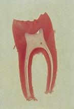
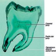
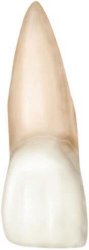
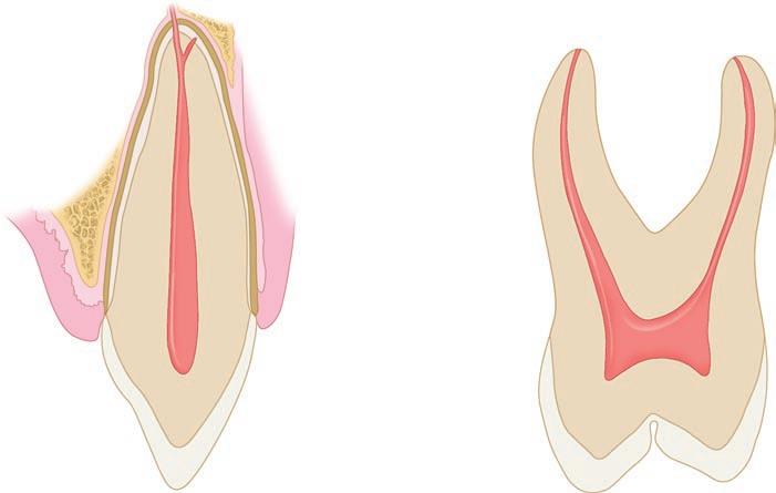
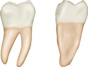
Each tooth has a crown and root portion. The crown is covered with enamel, and the root portion is covered with cementum. The crown and root join at the cementoenamel junction (CEJ). This junction, also called the cervical line (Figure 1-3), is plainly visible on a specimen tooth. The main bulk of the tooth is composed of dentin, which is clear in a cross section of the tooth. This cross section displays a pulp chamber and a pulp canal, which normally contain the pulp tissue. The pulp chamber is in the crown portion mainly, and the pulp canal is in the root (Figure 1-4).The spaces are continuous with each other and are spoken of collectively as the pulp cavity.
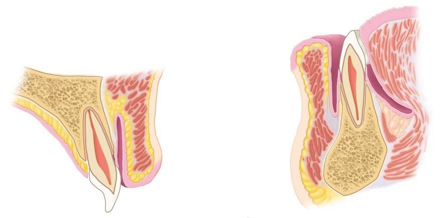
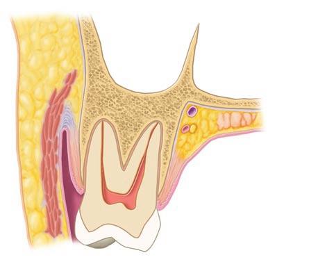
The four tooth tissues are enamel, cementum, dentin, and pulp. The first three are known as hard tissues, the last as soft tissue. The pulp tissue furnishes the blood and nerve supply to the tooth. The tissues of the teeth must be considered in relation to the other tissues of the orofacial structures (Figures 1-5 and 1-6) if the physiology of the teeth is to be understood.
The crown of an incisor tooth may have an incisal ridge or edge, as in the central and lateral incisors; a single cusp, as in the canines; or two or more cusps, as on premolars and molars. Incisal ridges and cusps form the cutting surfaces on tooth crowns.
The root portion of the tooth may be single, with one apex or terminal end, as usually found in anterior teeth and
some of the premolars; or multiple, with a bifurcation or trifurcation dividing the root portion into two or more extensions or roots with their apices or terminal ends, as found on all molars and in some premolars.
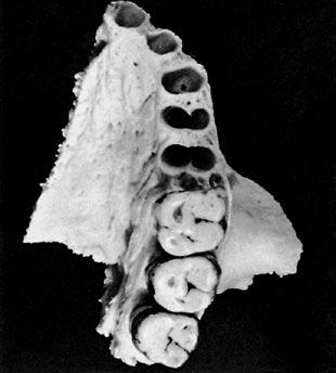
The root portion of the tooth is firmly fixed in the bony process of the jaw, so that each tooth is held in its position relative to the others in the dental arch. That portion of the jaw serving as support for the tooth is called the alveolar process. The bone of the tooth socket is called the alveolus (plural, alveoli) (Figure 1-7).
The crown portion is never covered by bone tissue after it is fully erupted, but it is partly covered at the cervical third in young adults by soft tissue of the mouth known as the gingiva or gingival tissue, or “gums.” In some persons, all of the enamel and frequently some cervical cementum may not be covered by the gingiva.
SURFACES AND RIDGES
The crowns of the incisors and canines have four surfaces and a ridge, and the crowns of the premolars and molars have five surfaces. The surfaces are named according to their positions and uses (Figure 1-8). In the incisors and canines, the surfaces toward the lips are called labial surfaces; in the premolars and molars, those facing the cheek are the buccal surfaces. When labial and buccal surfaces are spoken of collectively, they are called facial surfaces. All surfaces facing toward the tongue are called lingual surfaces. The surfaces of the premolars and molars that come in contact (occlusion) with those in the opposite jaw during the act of closure are called occlusal surfaces. These are
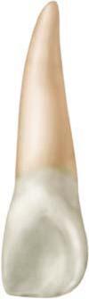
Figure � - � Maxillary central incisor (facial aspect). A, Apex of root; R, root; CL, cervical line; C, crown; IE, incisal edge.
Figure � - � Schematic drawings of longitudinal sections of an anterior and a posterior tooth. A, Anterior tooth. A, Apex; AF, apical foramen; SC, supplementary canal; B, bone; C, cementum; PM, periodontal ligament; PC, pulp canal; G, gingiva; GC, gingival crevice; GM, gingival margin; PCH, pulp chamber; D, dentin; E, enamel; CR, crown. B, Posterior tooth. A, Apices; PC, pulp canal; PCH, pulp chamber; PH, pulp horn; F, fissure; CU, cusp; CEJ, cementoenamel junction; BI, bifurcation of roots.
IV A(8), B
Vestibular mucosa
Attached gingiva
Labial mucosa
Free gingival margin
Attached gingiva
Anterior oral vestibule
� - � Sagittal
Palatine vein
Palatine artery
Palatine glands
Palatine nerve
Figure � - � Section through the second maxillary molar and adjacent tissues.
Figure � -7 Left maxillary bone showing the alveolar process with three molars in place and the alveoli of the central incisor, lateral incisor, canine, and first and second premolars. Note the opening at the bottom of the canine alveolus, an opening that accommodates the nutrient blood and nerve supply to the tooth in life. Although they do not show up in the photograph, the other alveoli present the same arrangement. called incisal surfaces with respect to incisors and canines.
The surfaces of the teeth facing toward adjoining teeth in the same dental arch are called proximal or proximate surfaces. The proximal surfaces may be called either mesial or distal. These terms have special reference to the position of the surface relative to the median line of the face. This line is drawn vertically through the center of the face, passing between the central incisors at their point of contact with each other in both the maxilla and the mandible. Those proximal surfaces that, following the curve of the arch, are faced toward the median line are called mesial surfaces, and those most distant from the median line are called distal surfaces.
Four teeth have mesial surfaces that contact each other: the maxillary and mandibular central incisors. In all other instances, the mesial surface of one tooth contacts the distal surface of its neighbor, except for the distal surfaces of third
molars of permanent teeth and distal surfaces of second molars in deciduous teeth, which have no teeth distal to them. The area of the mesial or distal surface of a tooth that touches its neighbor in the arch is called the contact area.
Central and lateral incisors and canines as a group are called anterior teeth; premolars and molars as a group, posterior teeth.
OTHER LANDMARKS
To study an individual tooth intelligently, one should recognize all landmarks of importance by name. Therefore, at this point it is necessary to become familiar with additional terms, such as the following:
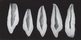
Figure
sections through the maxillary and mandibular central incisors.
1. Central incisor (first incisor)
2. Lateral incisor (second incisor)
3. Canine (cuspid)
4. First premolar (first bicuspid)
5. Second premolar (second bicuspid)
6. First molar
7. Second molar
8. Third molar
There are eight tooth names included in each quadrant of the dental arches they are repeated to include right, left, maxillary and mandibular, making a total of thirty-two teeth in all.
Figure � -8 Application of nomenclature. Tooth numbers 1 to 8 indicating left maxillary teeth. Tooth surfaces related to the tongue (lingual), cheek (buccal), lips (labial), and face (facial), apply to four quadrants and the upper left quadrant. The teeth or their parts or surfaces may be described as being away from the midline (distal) or toward the midline (mesial).
cusp triangular ridgedevelopmental groove tubercletransverse ridgesupplemental groove cingulumoblique ridgepit ridge fossa lobe marginal ridgesulcus
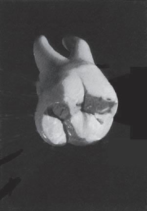
A cusp is an elevation or mound on the crown portion of a tooth making up a divisional part of the occlusal surface (Figures 1-4 and 1-9).
A tubercle is a smaller elevation on some portion of the crown produced by an extra formation of enamel (see Figure 4-14, A). These are deviations from the typical form.
A cingulum (Latin word for “girdle”) is the lingual lobe of an anterior tooth. It makes up the bulk of the cervical third of the lingual surface. Its convexity mesiodistally resembles a girdle encircling the lingual surface at the cervical third (see Figures 1-10 and 4-13, A).
A ridge is any linear elevation on the surface of a tooth and is named according to its location (e.g., buccal ridge, incisal ridge, marginal ridge).
Marginal ridges are those rounded borders of the enamel that form the mesial and distal margins of the occlusal
Figure � -9 Some landmarks on the maxillary first molar. BCR, Buccocervical ridge; BG, buccal groove; MBC, mesiobuccal cusp; SG, supplemental groove; TF, triangular fossa; MLC, mesiolingual cusp; DG, developmental groove; DLC, distolingual cusp; OR, oblique ridge; DMR, distal marginal ridge; DBC, distobuccal cusp; CF, central fossa.
IV A(3), B
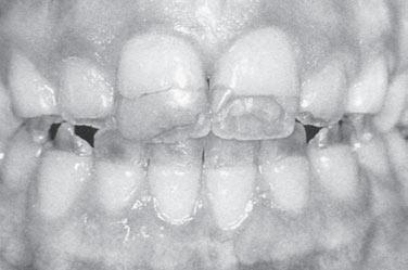
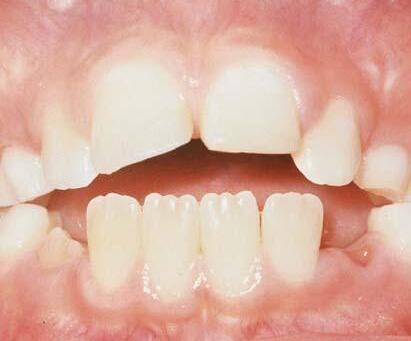
Figure 1-10 A, Maxillary right lateral incisor (lingual aspect). CL, Cervical line; CI, cingulum (also called the linguocervical ridge); MR, marginal ridge; IR, incisal ridge; LF, lingual fossa. B, Mamelons on erupting, noncontacting central incisors.
(B from Bath-Balogh M, Fehrenbach MJ: Illustrated dental embryology, histology, and anatomy, ed 2, St. Louis, 2006, Saunders.)
Figure 1-11
A, Mesial view of a maxillary right first premolar. MR, Marginal ridge; S, sulcus traversing occlusal surface; CR, cusp ridge; BCR, buccocervical ridge. B, Occlusal view of maxillary right first premolar. CR, Cusp ridge; TR, triangular ridges; Trans R, transverse ridge, formed by two triangular ridges that cross the tooth transversely. C, Occlusal view of a maxillary right first molar. Trans R, Transverse ridge; TR, triangular ridge; P, pit formed by junction of developmental grooves; SG, supplemental groove; DG, developmental groove; TR, triangular ridge.
surfaces of premolars and molars and the mesial and distal margins of the lingual surfaces of the incisors and canines (Figures 1-10, A, and 1-11).
Triangular ridges descend from the tips of the cusps of molars and premolars toward the central part of the occlusal surfaces. They are so named because the slopes of each side of the ridge are inclined to resemble two sides of a triangle (Figures 1-11, B and C, and 1-12). They are named after the cusps to which they belong, for example, the triangular ridge of the buccal cusp of the maxillary first premolar.
When a buccal and a lingual triangular ridge join, they form a transverse ridge. A transverse ridge is the union of two triangular ridges crossing transversely the surface of a posterior tooth (Figure 1-11, B and C).
The oblique ridge is a ridge crossing obliquely the occlusal surfaces of maxillary molars and formed by the union of the triangular ridge of the distobuccal cusp and the distal cusp ridge of the mesiolingual cusp (Figure 1-9).
A fossa is an irregular depression or concavity. Lingual fossae are on the lingual surface of incisors (Figure 1-10).
Central fossae are on the occlusal surface of molars. They are formed by the convergence of ridges terminating at a central point in the bottom of the depression where there is a junction of grooves (Figure 1-12). Triangular fossae are found on molars and premolars on the occlusal surfaces mesial or distal to marginal ridges (Figure 1-9). They are sometimes found on the lingual surfaces of maxillary incisors at the edge of the lingual fossae where the marginal ridges and the cingulum meet (see Figure 4-14, A).
A sulcus is a long depression or valley in the surface of a tooth between ridges and cusps, the inclines of which meet at an angle. A sulcus has a developmental groove at the junction of its inclines. (The term sulcus should not be confused with the term groove.)
A developmental groove is a shallow groove or line between the primary parts of the crown or root. A supplemental groove, less distinct, is also a shallow linear depression on the surface of a tooth, but it is supplemental to a developmental groove and does not mark the junction of primary parts. Buccal and lingual grooves are developmental grooves found on the
Figure � - �� Mandibular right first molar. MLC, Mesiolingual cusp; MMR, mesial marginal ridge; MBC, mesiobuccal cusp; MBG, mesiobuccal groove; BCR, buccocervical ridge; CF, central fossa; DBG, distobuccal groove; DBC, distobuccal cusp; DC, distal cusp; TR, triangular ridge; DLC, distolingual cusp; TRR, transverse ridge.
buccal and lingual surfaces of posterior teeth (Figures 1-9 and 1-12).
Pits are small pinpoint depressions located at the junction of developmental grooves or at terminals of those grooves. For instance, central pit is a term used to describe a landmark in the central fossa of molars where developmental grooves join (Figure 1-11, C).
A lobe is one of the primary sections of formation in the development of the crown. Cusps and mamelons are representative of lobes. A mamelon is any one of the three rounded protuberances found on the incisal ridges of newly erupted incisor teeth (Figure 1-10, B). (For further description of lobes, see Figures 4-11 through 4-14).
The roots of the teeth may be single or multiple. Both maxillary and mandibular anterior teeth have only one root each. Mandibular first and second premolars and the maxillary second premolar are single rooted, but the maxillary first premolar has two roots in most cases, one buccal and one lingual. Maxillary molars have three roots, one mesiobuccal, one distobuccal, and one lingual. Mandibular molars have two roots, one mesial and one distal. It must be understood that description in anatomy can never follow a hardand-fast rule. Variations frequently occur. This is especially true regarding tooth roots, for example, facial and lingual roots of the mandibular canine.
Division into Thirds, Line Angles, and Point Angles
For purposes of description, the crowns and roots of teeth have been divided into thirds, and junctions of the crown surfaces are described as line angles and point angles. Actually, there are no angles or points or plane surfaces on the teeth anywhere except those that appear from wear (e.g., attrition, abrasion) or from accidental fracture. Line angle and
Figure � - �� Division into thirds.
point angle are used only as descriptive terms to indicate a location.
When the surfaces of the crown and root portions are divided into thirds, these thirds are named according to their location. Looking at the tooth from the labial or buccal aspect, we see that the crown and root may be divided into thirds from the incisal or occlusal surface of the crown to the apex of the root (Figure 1-13). The crown is divided into an incisal or occlusal third, a middle third, and a cervical third. The root is divided into a cervical third, a middle third, and an apical third.
The crown may be divided into thirds in three directions: inciso- or occlusocervically, mesiodistally, or labio- or buccolingually. Mesiodistally, it is divided into the mesial, middle, and distal thirds. Labio- or buccolingually it is divided into labial or buccal, middle, and lingual thirds. Each of the five surfaces of a crown may be so divided. There will be one middle third and two other thirds, which are named according to their location, for example, cervical, occlusal, mesial, lingual.
A line angle is formed by the junction of two surfaces and derives its name from the combination of the two surfaces that join. For instance, on an anterior tooth, the junction of the mesial and labial surfaces is called the mesiolabial line angle.
The line angles of the anterior teeth (Figure 1-14, A) are as follows: mesiolabial distolingual distolabial labioincisal mesiolingual linguoincisal
IV A(30), B
Labioincisal line angle
Mesiolingual line angle
Mesiolabial line angle
Lingual Labial Distal
Linguoincisal line angle
Distolingual line angle
Distolabial line angle
Mesio-occlusal line angle
Mesiolingual line angle
Mesiobuccal line angle
Bucco-occlusal line angle
Mesial Lingual Buccal Distal
Linguo-occlusal line angle
Distolingual line angle
Distobuccal line angle
Disto-occlusal line angle
Mesiolabioincisal point angle
Mesiolinguoincisal point angle
Mesial Lingual Labial Distal
Distolabioincisal point angle
Distolinguoincisal point angle
A
Mesiolinguo-occlusal point angle
Mesiobucco-occlusal point angle
Distolinguo-occlusal point angle
Distobucco-occlusal point angle
Mesial Lingual Buccal Distal
B
Because the mesial and distal incisal angles of anterior teeth are rounded, mesioincisal line angles and distoincisal line angles are usually considered nonexistent. They are spoken of as mesial and distal incisal angles only.
The line angles of the posterior teeth (Figure 1-14, B) are as follows:
mesiobuccaldistolingualbucco-occlusal distobuccalmesio-occlusallinguo-occlusal mesiolingualdisto-occlusal
A point angle is formed by the junction of three surfaces. The point angle also derives its name from the combination of the names of the surfaces forming it. For example, the junction of the mesial, buccal, and occlusal surfaces of a molar is called the mesiobucco-occlusal point angle.
The point angles of the anterior teeth are (Figure 1-15, A):
mesiolabioincisal
mesiolinguoincisal distolabioincisal distolinguoincisal
The point angles of the posterior teeth are (Figure 1-15, B):
mesiobucco-occlusal
mesiolinguo-occlusal distobucco-occlusal distolinguo-occlusal
Tooth Drawing and Carving
The subject of drawing and carving of teeth is being introduced at this point because it has been found through experience that a laboratory course in tooth morphology (dissection, drawing, and carving) should be carried on simultaneously with lectures and reference work on the subject of dental anatomy. Illustrations and instruction in tooth form drawing and carving, however, are not included here.
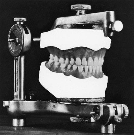
The basis for the specifications to be used for carving individual teeth is a table of average measurements for permanent teeth given by Dr. G. V. Black.6 However, teeth carved or drawn to these average dimensions cannot be set into place for an ideal occlusion. Therefore, for purposes of producing a complete set of articulated teeth (Figures 1-16, 1-17, and 1-18) carved from Ivorine, minor changes have been made in Dr. Black’s table. Also, carving teeth to natural size, calibrated to tenths of a millimeter, is not practical. The adjusted measurements are shown in Table 1-1. The only fractions listed in the model table are 0.5 mm and 0.3 mm in a few instances. Fractions are avoided whenever possible to facilitate familiarity with the table and to avoid confusion.
A table of measurements must be arbitrarily agreed upon so that a reasonable comparison can be made when appraising the dimensions of any one aspect of one tooth in the
Figure � - �� Line angles. A, Anterior teeth. B, Posterior teeth.
Mesial
Figure � - �� A, Point angles on anterior teeth. B, Point angles on posterior teeth.
Figure � - �� Carvings in Ivorine of individual teeth made according to the table of measurements (see Table 1-1). Because skulls and extracted teeth show so many variations and anomalies, an arbitrary norm for individual teeth had to be established for comparative study. Hence, the 32 teeth were carved at natural size and in normal alignment and occlusion, and from the model a table of measurements was drafted.
mouth with that of another. It has been found that the projected table functions well in that way. For instance, if the mesiodistal measurement of the maxillary central incisor is 8.5 mm, the canine will be approximately 1 mm narrower in that measurement; if by chance the central incisor is wider or narrower than 8.5 mm, the canine measurement will correspond proportionately.
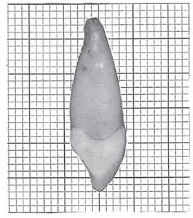
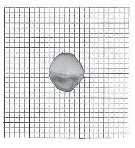
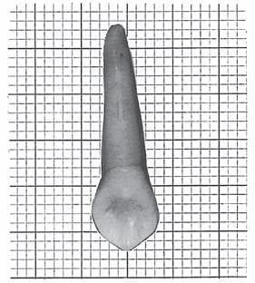
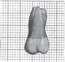
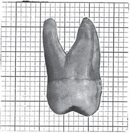
Photographs of the five aspects of each tooth (mesial, distal, labial or buccal, lingual, and incisal or occlusal) superimposed on squared millimeter cross-section paper reduces the tooth outlines of each aspect to an accurate graph, so that it is possible to compare and record the contours (Figures 1-19 and 1-20).
Close observation of the outlines of the squared backgrounds shows the relationship of crown to root, extent of curvatures at various points, inclination of roots, relative widths of occlusal surfaces, height of marginal ridges, contact areas, and so on.
It should be possible to draw reasonably well an outline of any aspect of any tooth in the mouth. It should be in good proportion without reference to another drawing or threedimensional model.
For the development of skills in observation and in the restoration of lost tooth form, some specific criteria are suggested:
1. Become so familiar with the table of measurements that it is possible to make instant comparisons mentally of the proportion of one tooth with regard to another from any aspect.
2. Learn to draw accurate outlines of any aspect of any tooth.
3. Learn to carve with precision any design one can illustrate with line drawings.
Measurement of Teeth
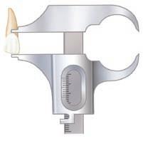
Readers who are not familiar with the Boley gauge should study its use before reading the following instructions on the application of the table of measurements.
To understand the table, let us demonstrate the calibrations as recorded and the landmarks they encompass. There are eight calibrations of each tooth to be remembered. These measurements are shown in the accompanying example for the maxillary central incisor (see the example included in Table 1-1).
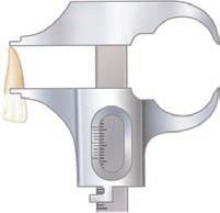
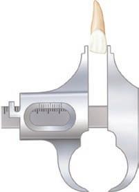
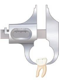
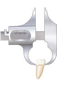
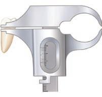
The method for measuring an anterior tooth is shown in Box 1-1 (Figures 1-21 through 1-27), and the posterior method is shown in Box 1-2 (Figures 1-28 through 1-34).
Summary
Terminology is an established basis for communication, and therefore the importance of learning the nomenclature for dental anatomy cannot be minimized. The terms used in describing the morphology of teeth are used in every aspect of dental practice.
Although there is no such thing as an established invariable norm in nature, in the study of anatomy it is necessary that there be a starting point; therefore, we must begin with an arbitrary criterion, accepted after experimentation and due consideration. Since restorative dentistry must approach the scientific as closely as manual dexterity will allow, models, plans, photographs, and natural specimens should be given preference over the written text on this subject.
Text
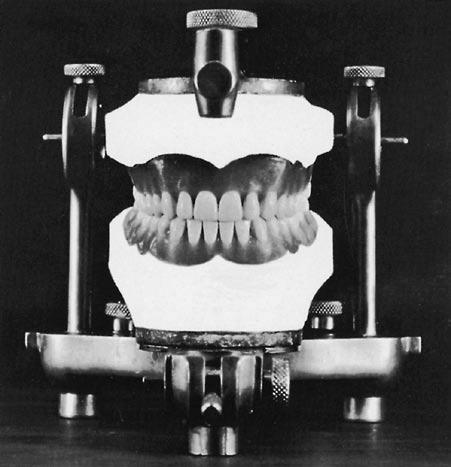
Figure � - � 7 Another view of the models shown in Figure 1-16.
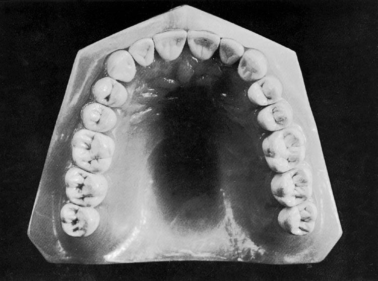
� - � 8 Occlusal
Figure
view of the models shown in Figures 1-16 and 1-17.
Table � - � Measurements of the Teeth: Specifications for Drawing and Carving Teeth of Average Size*
B, Buccal; L, lingual.
*In millimeters. This table has been “proved” by carvings shown in Figures 1-16 and 1-17.
†The sum of the mesiodistal diameters, both right and left, which gives the arch length, is maxillary, 128 mm; mandibular, 126 mm.
‡Lingual measurement is approximately 0.5 mm longer.
Table � - �
Measurements of the Teeth: Specifications for Drawing and Carving Teeth of Average Size—cont’d
Measurements of the Teeth: an Example*
Length of Crown Length of Root
Maxillary Teeth
Mesiodistal Diameter of Crown †
Mesiodistal Diameter of Crown at Cervix
Labio- or Buccolingual Diameter of Crown
Central incisor10.513.08.57.0 7.0
*In millimeters.
Labio- or Buccolingual Diameter of Crown at Cervix
†The sum of the mesiodistal diameters, both right and left, which gives the arch length, is maxillary, 128 mm; mandibular, 126 mm.
Curvature of Cervical Line—Mesial
Curvature of Cervical Line—Distal
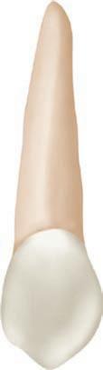
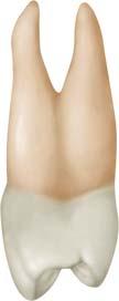
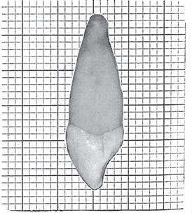
� - � 9 Maxillary left canine. When viewing the mesial and distal aspects, note the curvature or bulge on the crown at the cervical third below the cementoenamel junction. This is called the cervical ridge, or the cervicoenamel ridge.
Figure
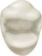
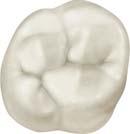
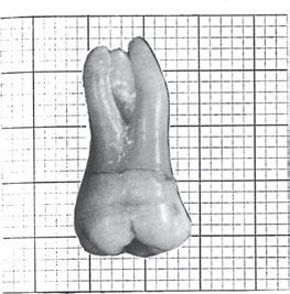
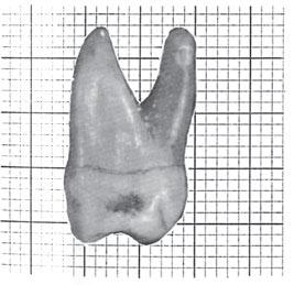
Figure � - � 0 Maxillary right first molar. When viewing the mesial and distal aspects, note the curvature or bulge on the crown at the cervical third below the cementoenamel junction.
BOX
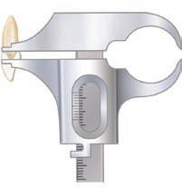
� - � Method of Measuring an Anterior Tooth
(Keep the long axis of the tooth vertical.)
1. LENGTH OF CROWN (LABIAL)*
Measurement { Crest of curvature at cementoenamel junction
Incisal edge
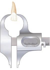
2. LENGTH OF ROOT
Measurement { Apex
Crest of curvature at crown cervix F IGURE � - �� Length of crown.
� - �� Length of root.
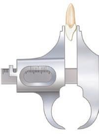
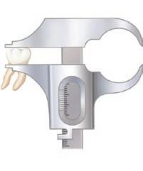
3. MESIODISTAL DIAMETER OF CROWN
Measurement { Crest of curvature on mesial surface (mesial contact area)
Crest of curvature on distal surface (distal contact area)
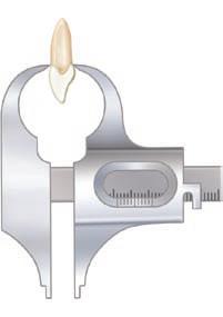
4. MESIODISTAL DIAMETER OF CROWN AT THE CERVIX
Measurement { Junction of crown and root on mesial surface
Junction of crown and root on distal surface (use caliper jaws of Boley gauge in this instance instead of parallel beaks)
� - �� Mesiodistal diameter of crown.
� - �� Mesiodistal diameter of crown at cervix.
*Use the parallel beaks of the Boley gauge for measurements whenever feasible. The contrast of the various curvatures with the straight edges will help to make the close observer more familiar with tooth outlines.
Figure
Figure
Figure
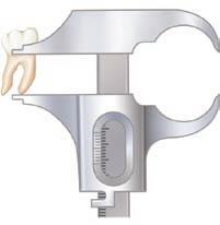
5. LABIOLINGUAL DIAMETER OF CROWN
Measurement { Crest of curvature on labial surface Crest of curvature on lingual surface
6. LABIOLINGUAL DIAMETER OF CROWN AT THE CERVIX
Measurement { Junction of crown and root on labial surface
Junction of crown and root on lingual surface (use caliper jaws in this instance also)
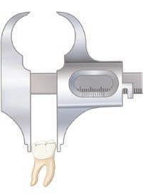
7. CURVATURE OF CEMENTOENAMEL JUNCTION ON MESIAL†
Measurement { Crest of curvature of cementoenamel junction on labial and lingual surfaces
Crest of curvature of cementoenamel junction on mesial surface
8. CURVATURE OF CEMENTOENAMEL JUNCTION ON DISTAL (Turn the tooth around and calibrate as in Figure 1-27.)
� - �� Labiolingual diameter of crown.
� - �� Labiolingual diameter of cervix.
Measurement { Crest of curvature of cementoenamel junction on labial and lingual surfaces
Crest of curvature of cementoenamel junction on distal surface
†This measurement is most important because normally it represents the extent of curvature approximately of the periodontal attachment when the tooth is in situ.
Figure
Figure
Figure � - � 7 Curvature of cementoenamel junction on mesial.
BOX
� - � Method of Measuring a Posterior Tooth
(Keep the long axis of the tooth vertical.)
1. LENGTH OF CROWN (BUCCAL)
Measurement { Crest of buccal cusp or cusps
Crest of curvature at cementoenamel junction
� � 8 Length
2. LENGTH OF ROOT
Measurement { Crest of curvature at crown cervix
Apex of root
� - � 9 Length
3. MESIODISTAL DIAMETER OF CROWN
Measurement { Crest of curvature on mesial surface (mesial contact area)
Crest of curvature on distal surface (distal contact area)
4. MESIODISTAL DIAMETER OF CROWN AT THE CERVIX
Measurement { Junction of crown and root on mesial surface
Junction of crown and root on distal surface (use caliper jaws of Boley gauge instead of parallel beaks)
� - � 0 Mesiodistal
� - �� Mesiodistal
Figure
of crown.
Figure
of root.
Figure
diameter of crown.
Figure
diameter of crown at cervix.
5. BUCCOLINGUAL DIAMETER OF CROWN
Measurement { Crest of curvature on buccal surface
Crest of curvature on lingual surface
6. BUCCOLINGUAL DIAMETER OF CROWN AT THE CERVIX
Measurement { Junction of crown and root on buccal surface
Junction of crown and root on lingual surface (use caliper jaws)
7. CURVATURE OF CEMENTOENAMEL JUNCTION ON MESIAL
Measurement { Crest of curvature of cementoenamel junction on mesial surface
Crest of curvature of cementoenamel junction on buccal and lingual surfaces
8. CURVATURE OF CEMENTOENAMEL JUNCTION ON DISTAL (Turn tooth around and measure as in Figure 1-34.)
Measurement { Crest of curvature of cementoenamel junction on distal surface
Crest of curvature of cementoenamel junction on buccal and lingual surfaces
Every curve and segment of a normal tooth has some functional basis, and it is important to reproduce them accurately. The successful clinician in dentistry or, for that matter, any designer of dental restorations should be able to mentally create pictures of the teeth from any aspect and relate those aspects of dental anatomy to function. Complete pictures can be formed only when one is familiar with the main details of tooth form.
References
1. Webster’s new universal unabridged dictionary, New York, 1996, Barnes & Noble Books.
2. Turner II CG, Nichol CR, Scott GR: Scoring procedures for key morphological traits of the permanent dentition: The Arizona State University Dental Anthropology System. In Kelley MA, Larsen CS, editors: Advances in dental anthropology, New York, 1991, Wiley-Liss.
Figure � - �� Buccolingual diameter of crown.
Figure � - �� Buccolingual diameter of crown at cervix.
Figure � - �� Curvature of cementoenamel junction on mesial.