LAS CUATRO CASAS DE MEDIO
Según lo decretado en 33 Vanir por el Senado Imperial en la Ciudad Eterna
CASA DE TIERRA Y SANGRE
Cambiaformas, humanos, brujas, animales comunes y muchos otros a quienes Cthona comanda, así como algunos de los elegidos de Luna.
CASA DE CIELO Y SOPLADO
Malakim (ángeles), hadas, elementales, elfos * y aquellos bendecidos por Solas, así como algunos favorecidos por Luna
CASA DE MUCHAS AGUAS
Espíritus de río, sirenas, bestias acuáticas, ninfas, kelpies, nøkken y otros protegidos por Ogenas
CASA DE LLAMA Y SOMBRA
Daemonaki, segadores, espectros, vampiros, draki, dragones, nigromantes y muchas criaturas malvadas y sin nombre que ni siquiera Urd puede ver.
Nota
* Los Goblins fueron expulsados de su Casa como castigo por su participación en la Caída, y ahora se les considera Menores, aunque muchos se niegan a aceptar el hecho.
Another random document with no related content on Scribd:
to the ordinary dermal bones which invest the cranial roof there is a transverse row of supra-temporal plates crossing the cranial roof behind the paired parietals (Fig. 132, A). Fringing the outer margins of the frontals and parietals a row of pre- and post-spiracular ossicles extends nearly to the orbits, and between two of them, which form a valve, is the spiracular aperture itself. There is a dentigerous splenial on the inner surface of the lower jaw. The hyoid arch has no separate symplectic bone. An operculum and a suboperculum are present, but no inter-operculum; and unless the hinder part of the large cheek-plate, which is traversed by the mandibulo-hyoid sensory canal, represents a pre-operculum, the latter is wanting. Branchiostegal rays are absent, but there is a single pair of large jugular plates.
Very little is certainly known about the cranial cartilage-bones in the fossil members of the group, but the investing dermal bones, which bear a general resemblance to those of Polypterus, are often somewhat more numerous, and they form a very complete dermal armature for the entire head. There is a very complete ring of circum-orbital bones, and very often a ring of sclerotic plates. Two large cheek-plates are often present. Nothing comparable to preand post-spiracular ossicles is known, but squamosal and supratemporals can often be identified. To the ordinary bones of the lower jaw there may be added a series of infra-dentary plates, and besides the paired principal jugular plates there may also be present a small anterior median plate and a series of small lateral jugular plates on each side, as in the Carboniferous Rhizodopsis (Fig. 274). Most of the superficial dermal bones, both in the living and extinct Crossopterygii, are invested externally by a granulated or rugose layer of enamel-like ganoin.
In the Holostei, and especially in Amia, the skull approximates more closely to the normal Teleostean type as represented by the Salmon's skull. In Amia[201] all the occipital cartilage-bones are present—a basi-occipital, two exoccipitals, and a supra-occipital;
and, except for the absence of a pterotic, the periotic series of bones is also complete. Paired ali- and orbito-sphenoids form the lateral walls of the inter-orbital portion of the cranial cavity. Above, the complete cartilaginous roof of the cranial cavity is invested by a shield of suturally united and ganoin-covered dermal plates. The hyomandibular element has a symplectic bone at its distal extremity. There is a complete series of opercular bones, and the branchiostegal rays are numerous. A single median jugular plate is present. The lower jaw has on each side five dentigerous splenial bones in addition to dentary and angular bones, while cartilagebones are represented by articular and mento-Meckelian elements. In its essential structure the skull of Lepidosteus[202] resembles that of Amia, but it has obviously undergone much specialisation. In some species (e.g. L. osseus) its appearance is greatly modified by the exceptional length and tapering shape of the beak, due to the elongation of that part of the skull which lies between the orbital and nasal regions; but in L. platycephalus the reduced length and greater width of the beak, combined with its somewhat flattened condition, impart an almost Crocodilian aspect to the head. Amongst other points of difference it may be mentioned that in Lepidosteus the continuity of the chondro-cranial roof is interrupted by a large superior fontanelle. There is no supra-occipital, and there are no lateral ethmoids, at all events in the usual position. The inter-orbital portion of the cranial cavity is largely obliterated by the formation of an inter-orbital septum, consisting of a thin vertical plate of bone, which either represents a pair of fused orbito-sphenoids or a pair of similarly modified lateral ethmoids. In addition to the ordinary investing dermal bones, including circum-orbitals, squamosal, and supra-temporals, there are numerous scale-like ossicles which take the place of the cheek-plates of Polypterus. The maxillae are segmented into numerous dentigerous bones fringing the margins of the upper jaw. The lower jaw has no mento-Meckelian bones, but there is a very complete series of dermal elements, including dentary, coronary, splenial, angular, and supra-angular bones in addition to an articular cartilage-bone. One of the most remarkable
features in the skull of Lepidosteus is the existence of a secondary articulation between the metapterygoid bones and a pair of transversely elongated condyles formed on each side by a lateral outgrowth from the parasphenoid and alisphenoid bones. By a horizontal sliding movement of the former on the latter, provision is made for the lateral expansion and contraction of the walls of the oral cavity and the separation and approximation of the lateral halves of the upper jaw.[203]
The generality of Teleosts[204] more or less closely agree with Amia in the main features of their cranial structure. There are, however, certain minor features which are characteristic if not always distinctive of the group. As a rule, to which, nevertheless, there are notable exceptions, there is little of the primary cartilaginous cranium in the adult, nearly the whole of it having become absorbed or converted into cartilage-bones. A supraoccipital is invariably present, and usually a mesethmoid and a basisphenoid. An additional bone is added to the periotic series, viz. a pterotic. Supratemporal bones and jugular plates are always absent, and it may be doubted if mento-Meckelian bones and dentigerous splenials are ever developed in the lower jaw. Within the group itself the skull exhibits many notable modifications, of which only a few can here be mentioned. The shape, size, and character of the mouth and jaws, the extent to which they can be protruded and retracted, and the nature of the dentition, are the source of many characteristic modifications in the structure and appearance of the fore-part of the skull, and these again largely depend upon differences of habit and food. A protrusible mouth, or a mouth which is projected forwards, is usually associated with a suspensorium (hyomandibular) of considerable length, and so greatly inclined forwards as to make a more or less acute angle with the forepart of the cranium.
The presence or absence of an inter-orbital septum is also a feature in which considerable variation occurs. In some Teleosts there is no
septum, and the cranial cavity is prolonged forwards between the orbits, where its lateral walls are formed by well-developed, paired ali- and orbito-sphenoid bones, as, for example, in the Carp and other Cyprinidae. In others the fusion of the cranial walls is accompanied by the median union of the orbito-sphenoids, so that a partly bony and partly cartilaginous inter-orbital septum is found, and the cranial cavity becomes largely obliterated in this region, as in the Salmon; or the orbito-sphenoids may be non-existent, the cartilage may undergo absorption, and the inter-orbital septum may become reduced to a vertical fibrous sheath extending between the frontals above and the parasphenoid below, as is the case in the Cod (Gadus).
An interesting modification of certain of the bones of the primary and secondary upper jaw occurs in the Siluridae. In these Fishes the maxillae are very small and edentulous, and serve no other purpose than forming basal supports for the maxillary barbels, while the rodlike palatine bone, losing its connexion with the pterygoid portion of the primitive upper jaw, but retaining its articulation with the lateral ethmoid, serves to support the maxilla, and at the same time receives the insertion of the muscles by which the barbel is moved in various directions.
In the Plectognathi the premaxillae are co-ossified with the maxillae. Many other interesting cranial modifications occur in Teleosts, and to some of them reference is made in subsequent chapters.
In some respects the skull of Dipnoi[205] is remarkably like that of the Holocephali, especially in its typical autostylism; but in possessing both cartilage- and membrane-bones it in some measure approaches the Teleostome skull. The investing dermal bones are not always easy to identify with those of other Fishes. In Neoceratodus an anterior median membrane-bone or dermal mesethmoid covers the ethmo-nasal region, and, on each side of it,
forming the anterior boundary of the orbit, there is situated a preorbital or dermal lateral ethmoid. Behind the mesethmoid there is a much larger posterior median bone, and on each side a singular backward prolongation of the dermal lateral ethmoid separates it from a squamosal element. The latter bone descends on the outer surface of the quadrate portion of the palato-quadrate cartilage as far as the condyle for the lower jaw. Collectively, these bones form a fairly complete investment to the upper surface of the cranium, but the posterior median bone and the adjacent portions of the dermal lateral ethmoid and the squamosal are widely separated from the underlying chondrocranium by the powerful jaw muscles, and in this respect they differ from the ordinary roofing bones of other Fishes.
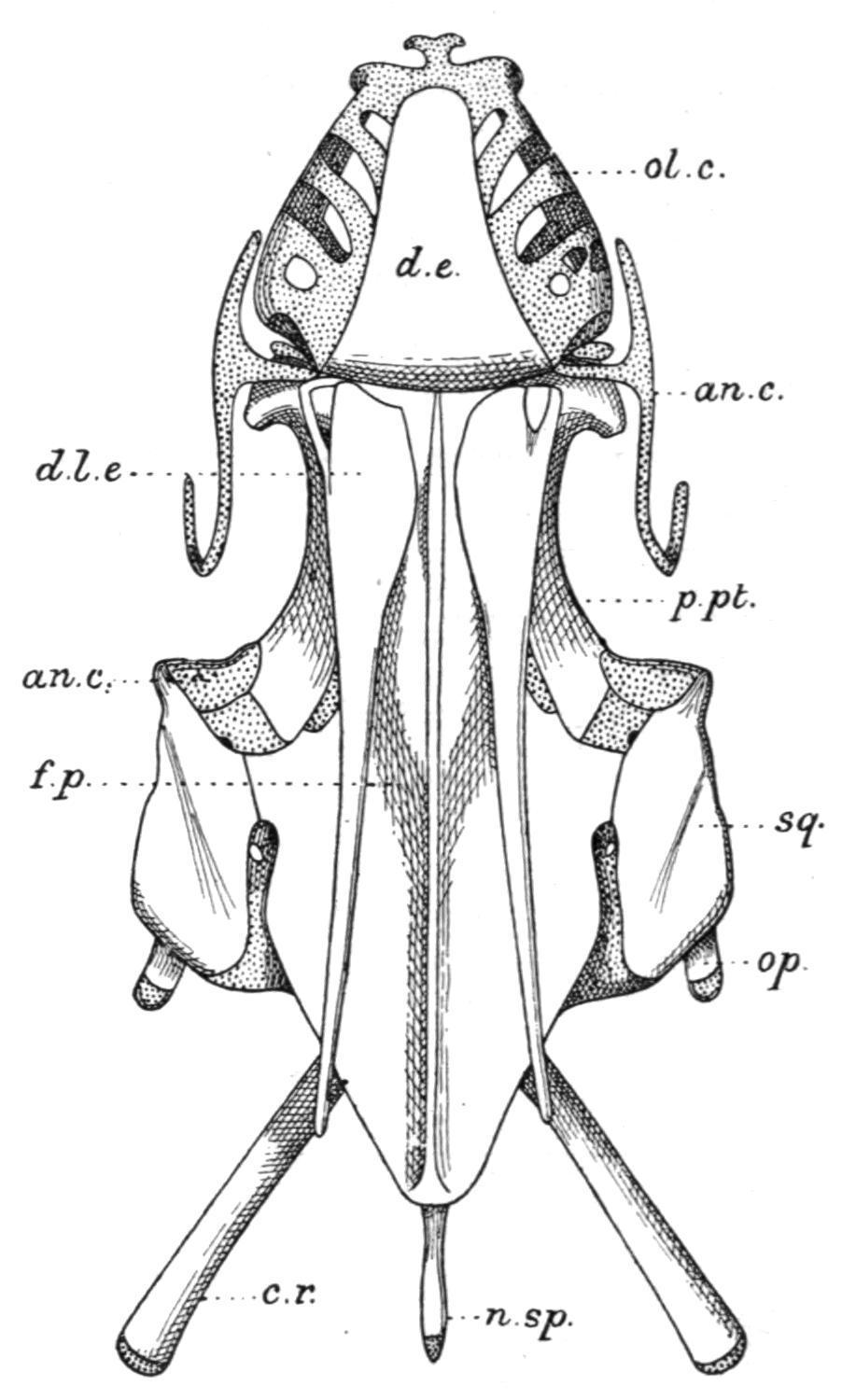
In Protopterus (Fig. 133) and Lepidosiren (Fig. 134) the posterior median bone is non-existent, and its place is taken by a large frontoparietal, which forms the greater part of the cranial roof, internal to the jaw muscles, and is much larger in the latter Dipnoid than in the former. Circum-orbital bones are present only in Neoceratodus. A large parasphenoid supports the cranial floor. Vomers are absent, although there are two small vomerine teeth.
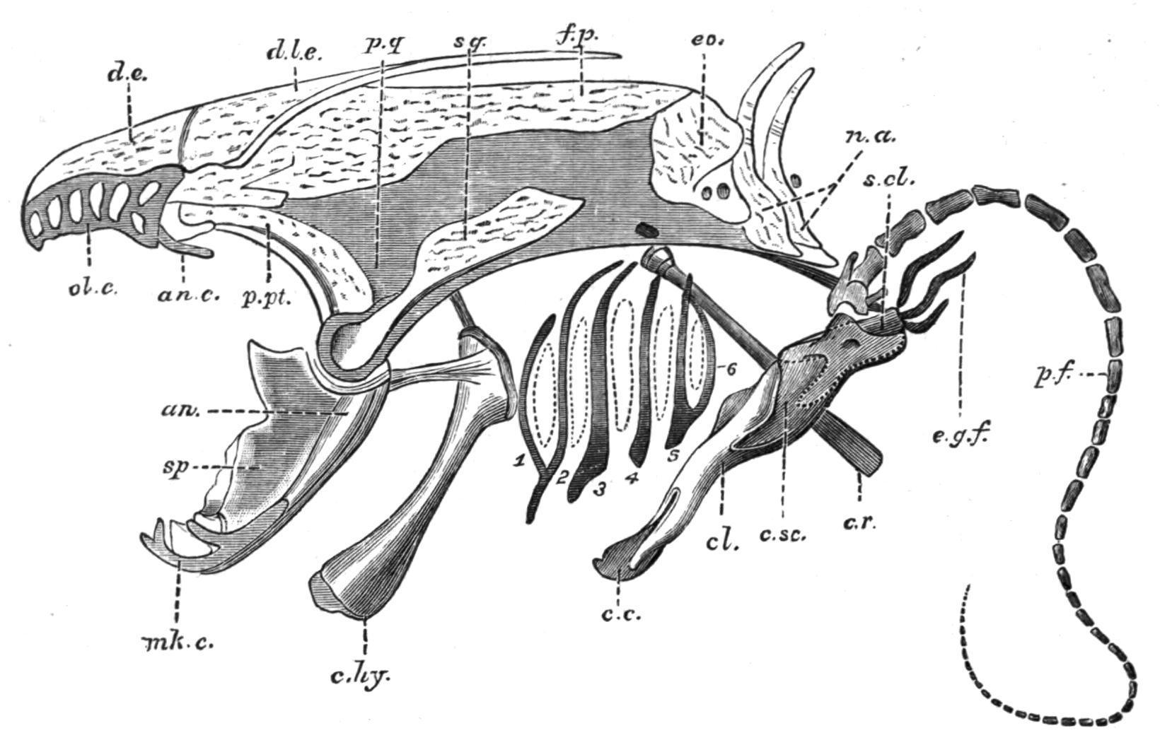
FIG. 133. Side view of the skull of Protopterus, with the pectoral girdle and fin. an, Angular; an.c, antorbital cartilage; c.c, coracoid cartilage (epi-coracoid); c.hy, cerato-hyal; cl, clavicle; c.r, cranial rib; c.sc, coraco-scapular cartilage; d.e, dermal ethmoid; d.l.e, dermal lateral ethmoid; e.g.f, external gills; eo, exoccipital; f.p, fronto-parietal; mk.c, Meckel's cartilage; n.a, neural arches; ol.c, fenestrated roof of the olfactory capsule; p.f, skeleton of the pectoral fin; p.pt, palato-pterygoid bone; p.q, palato-quadrate cartilage; s.cl, supra-clavicle; sp, splenial; sq, squamosal; 1-6, the branchial arches; the segmentation of the second and third arches is not shown. (From Wiedersheim.)
Relatively small opercular and inter-opercular bones are present, and on the inner surface of each may be found vestigial remains of cartilaginous hyoidean rays. The chondrocranium is complete in Neoceratodus, but in the remaining genera it has undergone considerable absorption in the inter-orbital region, so that the roof and floor, and, in part, even the side walls of the cranial cavity, are formed by the fronto-parietal and parasphenoid bones. Two exoccipitals are present in all Dipnoi. There are small labial cartilages in relation with the ventrally-placed nostrils, and large lateral outgrowths from the ethmoid cartilage furnish the olfactory organs with conspicuous lattice-like roofs. A pair of strong palato-pterygoid bones fringe the lower margins of the palato-quadrate cartilage, and meeting in front beneath the ethmoid region their symphysial extremities support the large palatal teeth. The Meckelian cartilages are persistent in all Dipnoi. In Neoceratodus each is flanked by a dentary and an angular externally, and internally by a splenial; but in Protopterusand Lepidosirendistinct dentary bones are wanting. The hyoid arch is best developed in Neoceratodus,[206] and includes a
small hyomandibular cartilage, a partially bony cerato-hyal and cartilaginous hypo-hyal and basi-hyal element. In the other genera (Fig. 133) only a cerato-hyal is retained. The branchial arches are but feebly developed in the Dipnoi. Neoceratodushas five, of which the first four are divided into epi-branchial and cerato-branchial segments, while the fifth is undivided. Protopterus has six, but only the second and third are segmented as in Neoceratodus.[207] In Lepidosirenall the arches are simple undivided rods.
In all three genera the skull conforms to the same general type of structure, but it is much more primitive in Neoceratodusthan in the other two genera.
FIG. 134. Dorsal view of the skull of Lepidosiren. an.c, Condyle on the quadrate cartilage for the lower jaw; n.sp, neural spine; op, operculum. For other reference letters see Fig. 133. (From Bridge.)
With reference to the fossil Dipnoi, it may be stated that, so far as they are known, the cranial roofing bones are more numerous than in the existing genera, and they cannot readily be compared with those of the latter, or with the numerically reduced and more definitely arranged bones of most Teleostomi. There is also evidence that in some fossil Dipnoi (e.g. Dipterus) the chondrocranium and the mandibular suspensorium (palato-quadrate) must have been
replaced by cartilage bones to an extent which has no parallel in any of the surviving types.[208] Jugular bones were present in Dipterus and Phaneropleuron.
Median Fins and Appendicular Skeleton
FIG. 135.—The cartilaginous radialia of the first dorsal fin of Mustelus antarcticus. (From Mivart.)
The Median Fins.—Whether existing in the form of a continuous fin, or as discontinuous isolated fins, the median fins are provided with skeletal supports, and also with muscles, primitively formed from intrusive clusters of cells derived from a variable number of the neighbouring myotomes, for their varied movements. The skeletal structures of the dorsal and anal fins consist of a series of bony or cartilaginous, rod-like, and typically tri-segmented radial elements or pterygiophores,[209] supporting distally a series of dermal structures in the shape of numerous slender horny fibres or ceratotrichia, as in the Elasmobranchii and Holocephali, or a smaller number of bony dermal fin-rays, which are probably modified scales or lepidotrichia, [210] as in the Teleostomi. The typical tri-segmented character of the radialia is often retained in many existing Elasmobranchs (Fig. 135) and in Pleuracanthus, in Neoceratodus amongst the Dipnoi, in the Chondrostei, in existing Holostei (Fig. 136), and to a greater or less extent in several families of Teleosts (e.g. Salmonidae, Esocidae, Cyprinidae, and some Acanthopterygii); but in the latter group the radialia are greatly prone to reduction, and hence they are more
generally bi-segmented, and sometimes consist of a single proximal segment only (e.g. Gymnotus). In all these Fishes the proximal segments are the longest and the most persistent, and when reduction occurs it is at the expense of the middle and distal segments.
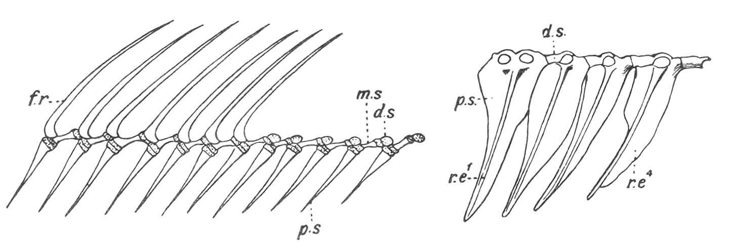
FIG. 136.—The tri-segmented radialia and the fin-rays of part of the dorsal fin of Amia calva. p.s, m.s, and d.s, The proximal, middle, and distal segments of a radial; f.r, fin-rays. (From Bridge.)
FIG. 137. The first four radialia of the dorsal fin of Mesoprion gembra, showing the chain-links for the ring-like bases of the fin-rays. r.e1 , r.e4 , First and fourth proximal radialia.
The cause of this reduction is often, but not always, to be found in the fact that, whenever the dermal fin-rays take the form of stout spines, as in the anterior dorsal fin in many Acanthopterygian Teleostei, the segmentation of their radialia would obviously detract from their value as skeletal supports, and hence they rarely consist of more than their proximal segments, although the radialia which in the same Fish support soft rays may be bi-segmented or trisegmented. The radialia are, however, unsegmented, even slightly branched, cartilaginous rods in the Cyclostomata; short simple rods in the Holocephali; and equally simple bony rods in the dorsal fin of Polypterus, where they support the strong spines of the numerous finlets; but they are bi-segmented in the soft-rayed anal fin. As previously mentioned, the proportional share taken by the radialia and the horny fibres or the dermal fin-rays in the support of the fins differs greatly in different Fishes. In the Cyclostomata radialia are the sole, and in Elasmobranchs the main supports, and they may extend nearly to the free margin of the fin. In the more specialised Fishes, as in most Teleostomi, the reverse is the case. The radialia sink into the muscles of the body-wall and leave the strongly
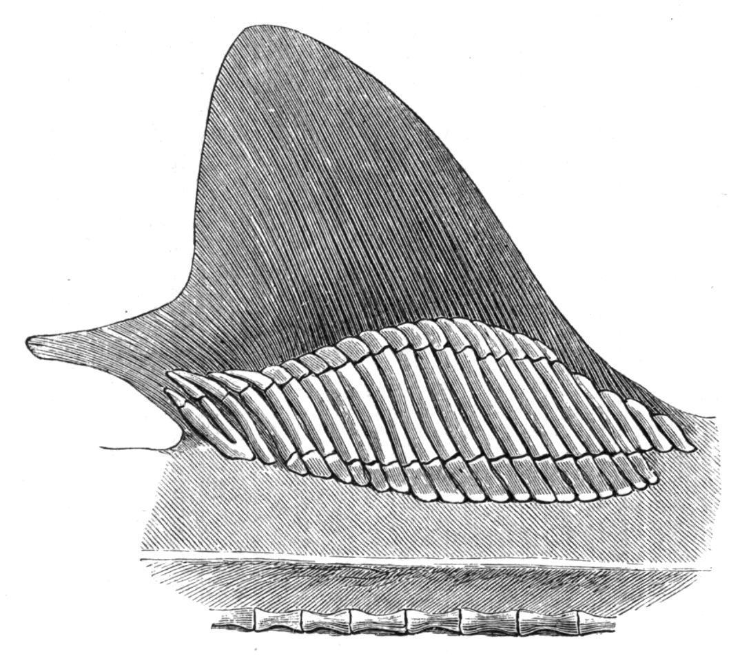
developed fin-rays as the sole support of the visible portions of the fins. In not a few Fishes there is an obvious segmental correspondence between the radialia and the vertebral neural or haemal spines, to the extent that the former equal the latter in number and articulate with their distal extremities, as, for example, in the caudal region of Pleuracanthus and in existing Dipnoi. In others again, as in most Teleostomi, there is no such segmental relation, and the radialia are more numerous than the vertebrae whenever the two are co-extensive. The exoskeletal fin-supports exhibit similar relations to their radialia, but in inverse order. Much more numerous than the radialia in the Elasmobranchs, Holocephali, and the Dipnoi, the former become gradually reduced in the Teleostomi, until in the Holostei and Teleostei they correspond in number with the supporting radialia. Complete numerical correspondence between the neural and haemal spines and the radialia and fin-rays is very rare, and has only been observed in the caudal region of certain Crossopterygii (e.g. the Coelacanthidae). [211]
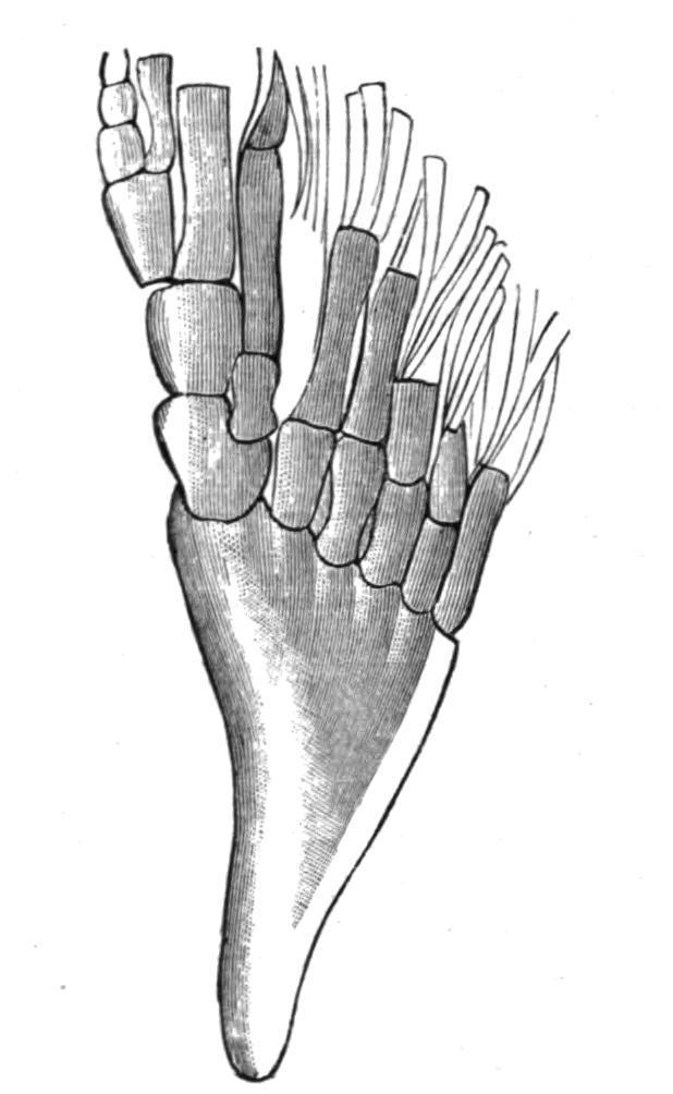
FIG. 138.—The posterior dorsal fin of Holoptychius leptopterus from the old Red Sandstone of Nairnshire. Traces of dermal fin-rays may be seen at the distal margin of the fin. (After Smith Woodward.)
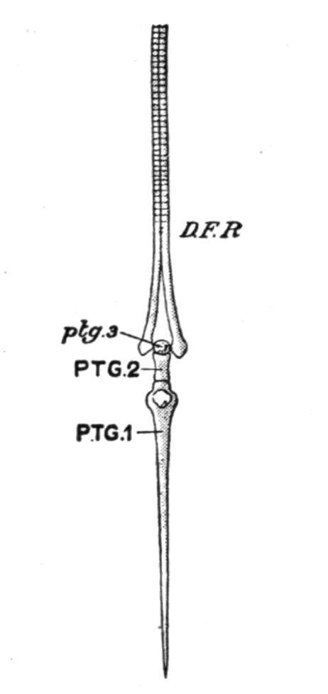
FIG. 139.—A dermal fin-ray and its supporting radial or pterygiophore in the Trout (Salmo fario). D.F.R, Dermal fin-ray; PTG.1, PTG.2, ptg.3, the proximal, middle, and distal segments of which the trisegmented radial consists; ptg.3 is cartilaginous; the other two are bony. (From Parker and Haswell.)
In not a few Fishes the radialia of the median fins undergo modifications which offer an interesting parallel to an early stage in the evolution of the paired fins from primitively continuous lateral fins. The concentration of radialia which occurs in isolated median fins often results, through growth pressure, in the complete fusion of the proximal segments of more or fewer of the radialia into two or three basal supports, or even into a single basal piece. Examples of such basal fusion are frequent in the dorsal fins of Elasmobranchs, and the same modification may also be seen in the anal fin of Pleuracanthus, and especially in the dorsal fin of the Devonian Crossopterygian, Holoptychius[212] (Fig. 138), where several radialia, which are free distally, have their bases united into a single basal piece, or basipterygium. In most Teleostomi elevator and depressor muscles arise from the radialia, and are inserted into different points on the bases of the fin-rays, and by their contraction the latter may either be elevated into an erect position, or folded back like a fan along the middle line of the body, where, as in some Teleosts, there is a groove for their reception. When fin-rays are only capable of simple elevation or depression, the connexion between a radial element and its fin-ray is usually by some form of a hinge-joint, the cleft base of the ray clipping the distal segment of the radial (Fig. 139). In some Teleosts the articulation of the two is by means of a kind of chain-link (Fig. 137). In those Fishes in which the median
fins are capable of lateral undulatory movements the articulation is of a more mobile character.
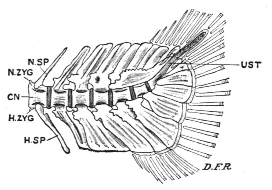
FIG. 140. Caudal end of the vertebral column of a Trout (Salmo fario).
CN, Centrum; D.F.R, dermal fin-rays; H.SP, haemal spine; H.ZYG, haemal zygapophysis; N.SP, neural spine; N.ZYG, neural zygapophysis; UST, the up-tilted, partly ossified, and unsegmented terminal portion of the notochord, or urostyle.
(From Parker and Haswell.)
In the different types of caudal fin, diphycercal, heterocercal, and homocercal, the supporting elements of the ventral lobe are formed by the haemal spines of the terminal caudal vertebrae which are inclined backwards, and are often greatly expanded for the purpose (Fig. 140). The dorsal lobe may be supported either by the adjacent neural spines, or by radialia, or by both.
The Appendicular Skeleton.[213]—It is probable that the skeleton of the paired fins and the pectoral and pelvic girdles have been formed from the supporting radialia of the isolated and enlarged anterior and posterior portions of primitively continuous lateral fins, by a sequence of structural modifications in the same direction as in the median fins. The initial stage was probably marked by the fusion of the proximal portions of the radialia to form a basal support or basipterygium for the free distal portions. Subsequently, it may be, a rudiment of the future limb-girdle became segmented off from the inner extremity of the basipterygium, and by its dorsal and ventral growth in the body-wall the lateral half of a girdle was developed. The subsequent union of the two halves across the mid-ventral line resulted in the evolution of the dorsally incomplete hoop of cartilage which is the primary form of the complete limb-girdle in Craniates.
The primitive fin skeleton or "archipterygium" was formed from the residue of the basipterygium in conjunction with the free distal radialia which it carried. The precise structure of the archipterygium is purely hypothetical. Possibly it was a biserial fin of the Pleuracanthus or Neoceratodus type, consisting of a cartilaginous segmented axis, fringed along its anterior and posterior, or pre-axial and post-axial margins, by a series of slender, simple, or jointed radialia (Fig. 147); or it may have been a uniserial structure, somewhat resembling the pelvic fin of Pleuracanthus, or the pectoral and pelvic fins of existing Elasmobranchs (Figs. 250, 141), in which an axis formed by the residue of the basipterygium or metapterygium had a fringe of radialia on its anterior or preaxial side only. If the archipterygium was biserial then the uniserial fin was probably derived from it by the subsequent suppression of all the post-axial radialia; or, if uniserial, the biserial fin was evolved by a later extension of radialia on to the post-axial margin. The evidence of comparative anatomy is not conclusive as to the nature of the archipterygium, and palaeontology seems to support either view with puzzling impartiality.[214] It may be admitted that the lateral fin theory offers the best solution of the problem of the origin of the paired fins, but it must be borne in mind that no Fish, living or fossil, is known to possess fins of this nature, unless the singular lateral lobes of some Ostracodermi (e.g. the Coelolepidae) are kindred organs[215]; neither do continuous lateral fins ever exist as vestiges, unless, indeed, the bilateral series of spines, which extend between the pectoral and pelvic fins, in some of the Lower Devonian Acanthodei (e.g. Climatius), may be regarded in that light.
ThePectoralandPelvicGirdles. The pectoral girdle is more primitive in Cladoselacheand Pleuracanthusthan in any other Elasmobranch. In the former (Fig. 145, A) it may be doubted if the girdle has passed beyond the basipterygial stage, and although a definite girdle is present in the latter genus (Fig. 250) its lateral halves retain their primitive distinctness. Existing Elasmobranchs, including the Holocephali, have a pectoral girdle in the form of a dorsally
incomplete hoop of cartilage imbedded in the muscles of the bodywall, close behind the last branchial arch (Fig. 141). The upper or dorsal portion of each half is the scapula, and the ventral is the coracoid. Between these two portions of the girdle, and defining their limits, there are articular surfaces for the basal cartilages of the pectoral fin.
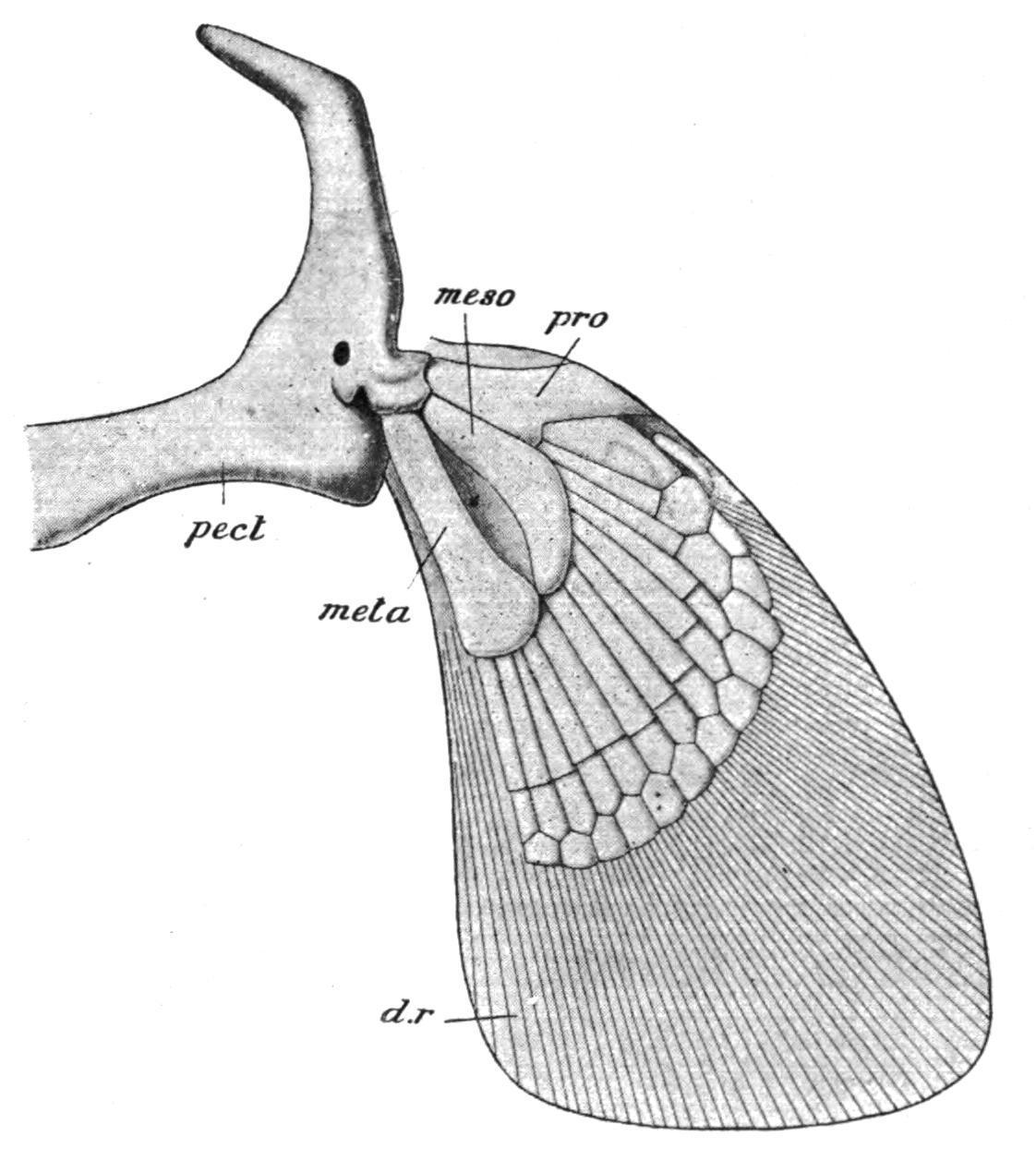
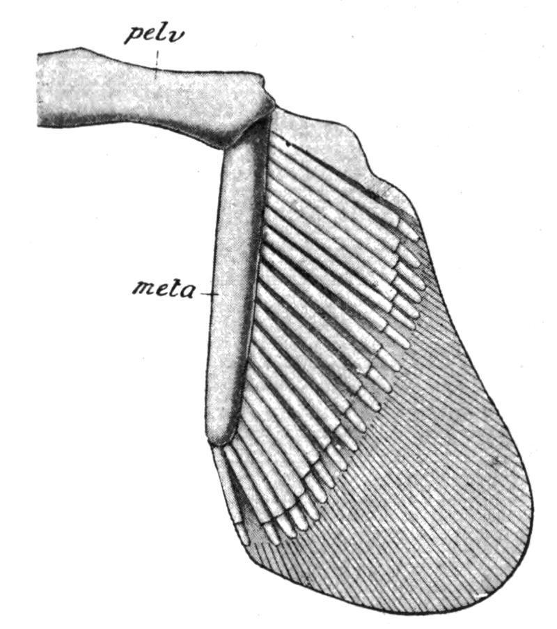
FIG. 141. The right half of the pectoral girdle and the fin of an Elasmobranch (Chiloscyllium). d.r, Dermal horny fibres; meso, mesopterygium; meta, metapterygium; pect, pectoral girdle; pro, propterygium. (From Parker and Haswell.)
Cladoselache (Fig. 145, B) had no pelvic girdle, nor does it appear that this primitive Elasmobranch had acquired even a basipterygium. Pleuracanthus, on the contrary, had a pair of pelvic rudiments distinct from well-developed basipterygia. In other Elasmobranchs there is a distinct girdle, formed by the median union of primitively distinct lateral rudiments, consisting of a simple transverse bar of cartilage, imbedded in the ventral abdominal wall, just in front of the cloacal aperture, and having articulated to each of its outer extremities the basal cartilage (metapterygium) of the pelvic fin.
FIG. 142.—The left half of the pelvic girdle and the right pelvic fin of Chiloscyllum. meta, Metapterygium; pelv, pelvic girdle. (From Parker and Haswell.)
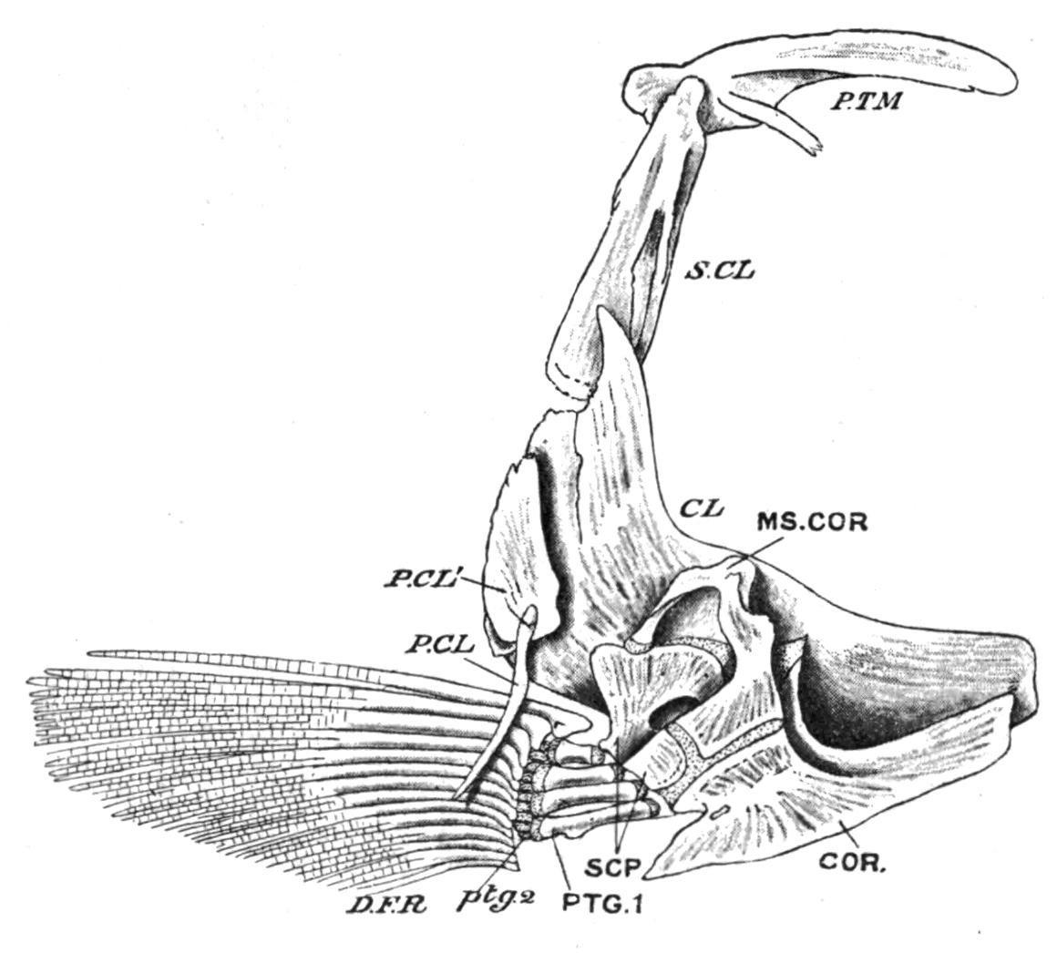
FIG. 143.—Left half of the pectoral girdle of a Trout (Salmo fario), seen from the inner surface. CL, Clavicle (cleithrum); COR, coracoid; D.F.R, dermal fin-rays; MS.COR, meso-coracoid; P.CL, P.CL′, postclavicles; PTG.1, proximal; ptg.2, distal pterygiophores; P.TM, post-temporal; S.CL, supra-clavicle; SCP, scapula. (From Parker and Haswell.)
Sometimes there is a rudiment of a dorsally-directed "iliac" process at each extremity of the girdle, but in no Fish do these processes ever acquire a dorsal connexion with the vertebral column. In the Holocephali the iliac processes are better developed than in any other Fishes, but ventrally the lateral halves of the girdle are united by ligament alone. In the Teleostomi important differences are observable in both girdles. The primary cartilaginous pectoral girdle now consists of distinct lateral halves which have no ventral connexion with each other. In addition, there is developed on the outer surface of each half a series of membrane bones, which form a secondary girdle (Fig. 143). From above downward the series includes a supraclavicle and a cleithrum (clavicle of Teleosts) which
are always present, and to these may be added in the Crossopterygii and Chondrostei an infraclavicle or clavicle proper, while one or two "post-clavicles" may be present in relation with the hinder margin of the cleithrum. The infraclavicles, or in their absence the cleithra (e.g. Holostei and most Teleostei), usually meet in a median ventral symphysis, so that the secondary girdle tends to acquire the characteristic hoop-like arrangement of its parts which has been lost in the primary girdle. With the development of a bony secondary girdle, the primary girdle (scapula and coracoid) becomes much reduced, and, as a rule, does little more than connect the fins with the cleithra. The secondary girdle acquires a dorsal connexion with the skull on each side by means of the post-temporal bone, which is attached below to the supra-clavicle and above to the periotic capsule. In the Chondrostei and the Dipnoi the primary girdle retains its primitive cartilaginous condition, but in the Crossopterygii, Holostei, and in all Teleosts it is ossified as distinct scapulae and coracoids. To these may be added in some Teleosts a mesocoracoid formed by a separate ossification of the coracoid cartilage (Fig. 143). [216]
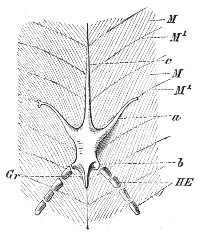
FIG. 144. Ventral view of the pelvic girdle of Protopterus. a, Prepubic process; b, lateral process for the fin; c, epipubic process; Gr, ridge for the origin of the fin muscles; HE, skeleton of the fin; M, myotomes; M′, myocommata. (From Wiedersheim.)
With the possible exception of small paired or median cartilages inserted between the inner extremities of the basipterygia in Polypterusand a few other Teleostomi, the pelvic girdle is absent in all the existing members of this group, having either become
completely suppressed, or remaining unseparated from the basipterygia of the pelvic fins.[217] In the Dipnoi (Fig. 144) there is a true pelvic girdle which has some points of resemblance to that of certain of the caudate Amphibia. It is represented by a median, lozenge-shape, cartilaginous plate, produced in front into a long tapering epipubic process, and on each side of this into a forwardly inclined prepubic process. The hinder part of the plate bears two short processes for the basal cartilages of the pelvic fins. There is no trace, however, of iliac processes.
The Pectoral Fins.—The skeleton of the pectoral fins exhibits remarkable structural variations in different Elasmobranchs. In the existing members of the group two large basal cartilages, the propterygium and the mesopterygium, are formed by the concentration and fusion of the proximal portions of certain of the preaxial radialia, and they, with the metapterygium, articulate with the pectoral girdle; hence the fin is tribasal as well as uniserial (Figs. 141 and 146, A, B). In striking contrast to all other Elasmobranchs the pectoral fin of Cladoselache (Fig. 145, A) is far more primitive than in any other Fish. Each fin is supported by a distal series of slender, more or less parallel, unjointed, cartilaginous radialia, and basally by a similar series of shorter, stouter, and less numerous cartilages, which apparently were imbedded in the body-wall, the entire fin skeleton presenting a striking resemblance to an isolated median fin in which the supporting radialia have concentrated by growth pressure, and their proximal portions have been reduced in number by partial fusion.[218] Pleuracanthus, on the other hand, had a biserial fin, the preaxial and postaxial radialia supporting fan-like clusters of horny fibres at their distal ends (Fig. 250).
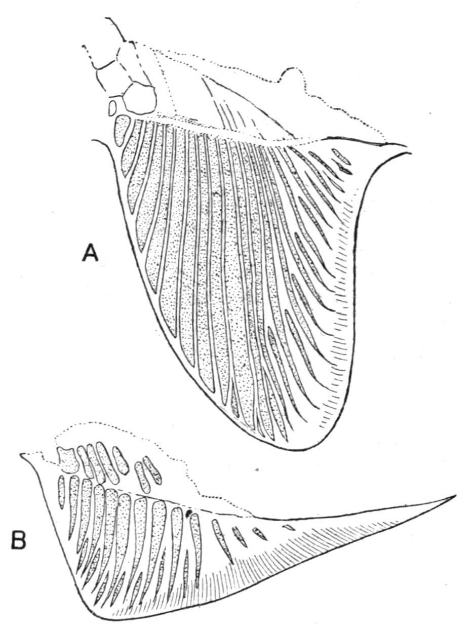
FIG. 145.—A, Pectoral fin, and B, pelvic fin of Cladoselache. (From Bashford Dean.)
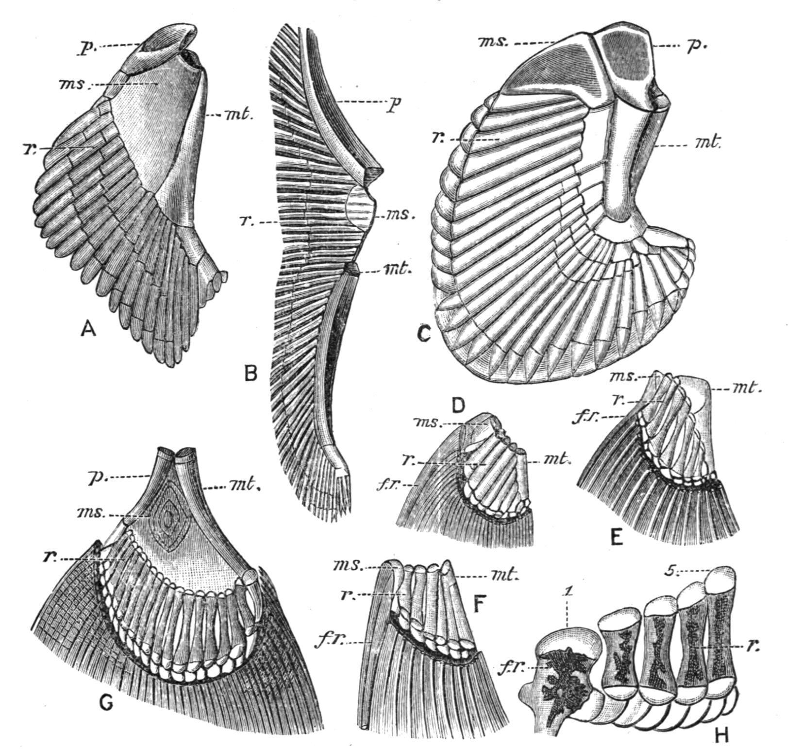
FIG. 146. Pectoral fins of various Fishes. A, Acanthias vulgaris; B, Raia sp.; C, Chimaera monstrosa; D, Acipenser rhynchaeus; E, Amia calva; F, Lepidosteus platyrhynchus; G, Polypterus bichir; H, Salmo salvelinus. The preaxial side of each fin is to the left and the postaxial to the right. f.r, Dermal fin-ray; ms, mesopterygium; mt, metapterygium; p, propterygium; r, free radialia; 1, 5, the preaxial and postaxial basal elements in a Teleost, which may be mesopterygial and metapterygial pieces respectively, the three remaining basal pieces probably being intrusive metapterygial radialia directly articulating with the pectoral girdle. In B, D, E, and F, similar intrusive radialia are shown. (From Gegenbaur.)
The broadly lobate pectoral fin of the existing Crossopterygii (Fig. 146, G) is uniserial, closely resembling that of the more typical Elasmobranchs.[219] There are three basal elements, a
propterygium, a mesopterygium, and a metapterygium, each of which supports a series of partially ossified radialia. Little is known of the endoskeletal elements of the broadly or acutely lobate fins of the fossil Crossopterygii, but it seems probable that their disposition was uniserial and abbreviate in obtusely lobate fins and biserial in acutely lobate fins. In the remaining Teleostomi (Actinopterygii) the endoskeletal elements become gradually reduced in number and importance, their place as fin-supports being usurped by the dermal fin-rays. In addition, more than three, usually several, basal elements articulate directly with the pectoral girdle, and hence the fins become multi-basal. In the Chondrostei and the Holostei a metapterygium is always recognisable, supporting several radialia along its preaxial border, as in Acipenser(Fig. 146, D) and Amia(Fig. 146, E), or only a single one, as in Lepidosteus (Fig. 146, F). The anterior part of the fin is supported by a variable number of cartilaginous or bony radialia, which, with the metapterygium, articulate with the limb-girdle. In Teleosts the process of reduction reaches its maximum. Usually there is but a single row of short, hour-glass-shaped ossicles, of which the postaxial one may represent a vestigial metapterygium, and sometimes there is also a distal row of small cartilages or ossicles, partially hidden in the cleft bases of the dermal fin-rays (Fig. 146, H). In all these Fishes the fin is a much reduced uniserial fin, in which more or fewer of the preaxial radialia have acquired a direct secondary connexion with the pectoral girdle. Of living Dipnoids Neoceratodus has a nearly typical biserial fin, but, as seems to be the case in all fins of this type at present known, there is a marked absence of symmetry in the number and disposition of the radialia on the two sides of the axis. There is also much individual variation. No two fins are precisely alike, and the radialia may sometimes divide. In the very acutely lobate fins of the remaining Dipnoids it is evident that great reduction has taken place. Protopterushas lost all trace of postaxial radialia, and in Lepidosireneven the preaxial have atrophied, leaving only the long jointed axis to represent the originally biserial fin.
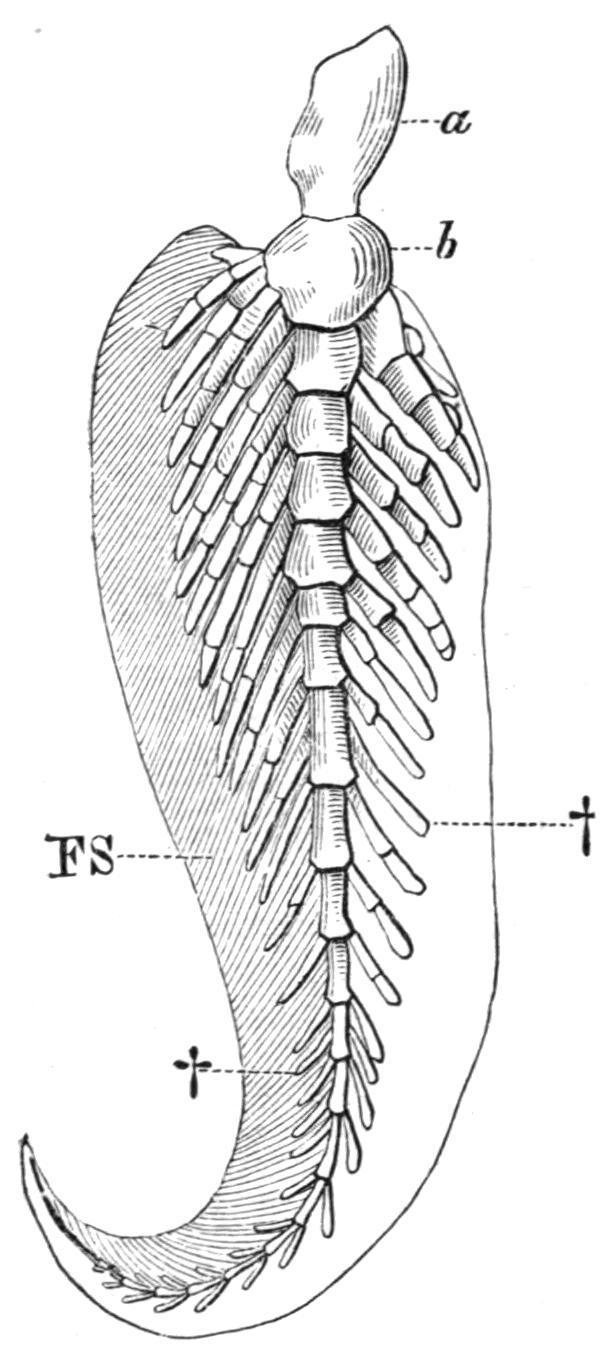
FIG. 147.—The left pectoral fin of Neoceratodus. a, b, First two segments of the axis; FS, preaxial horny fibres; †, †, pre- and post-axial radialia. (After Wiedersheim.)
The Pelvic Fins.—In the simplicity of their endoskeletal supports the pelvic fins of Cladoselacheare the most primitive type of paired fins at present known (Fig. 145, B). In general structure they resemble the pectorals, but the radialia are fewer in number, less modified by concentration, and exhibit little, if any, trace of basal fusion. Add to such features as these the apparent absence of any trace of pelvic rudiments, or of basipterygia, and it will be obvious that the pelvic fins differ but little from the median fins of the same Fish except that they are paired. In Pleuracanthus the pelvic fins differ from the corresponding pectorals in being uniserial instead of biserial (Fig. 250). All other Elasmobranchs, including the Holocephali, have uniserial fins, which consist of a large metapterygium, supporting a preaxial fringe of segmented radialia. A propterygium is sometimes present, notably in some of the Skates and Rays, and, like the metapterygium, it is directly connected with the pelvic girdle.
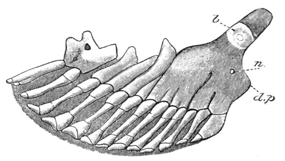
FIG. 148.—Skeleton of a pelvic fin of Polyodon folium, ventral view, with the anterior margin of the fin to the right; to show the partial
fusion of the proximal portions of primitively distinct radialia to form a basipterygium. b, Inner or mesial extremity of the basipterygium; d.p, dorsally directed, rudimentary iliac process; n, foramen for nerves. (After Rautenfeld.)
The skeleton of the pelvic fins of the Teleostomi is often extremely degenerate. It is perhaps best developed in the Chondrostei,[220] where each fin is supported by numerous segmented radialia, more or fewer of which fuse towards the base of the fin, and those form a large and slightly ossified basipterygium (Fig. 148). In the living Crossopterygii, Holostei, and Teleostei, the pelvic fins are similar in essential structure, but are very degenerate. The basipterygium is usually well developed and is always bony (Fig. 149), and in many Teleosts it acquires so extensive a sutural connexion with its fellow that, physiologically, it supplies the place of a true pelvic girdle. At its distal end there may be a single row of small cartilaginous or bony nodules, representing vestigial radialia, as in the Crossopterygii, Holostei, and Teleostei, but even these may be absent, and the dermal fin-rays then articulate directly with the basipterygium. Little is known of the skeleton of the pelvic fins in the fossil Crossopterygii, but there is evidence of the existence of a higher grade of structure than in their surviving allies. In Eusthenopteron,[221] for example, the fin is supported by an axis of at least three bony segments, with at least three ossified preaxial radialia; hence, it has obviously undergone less degeneration than in Polypterus, where the finskeleton is essentially Teleostean. In the Dipnoi the pelvic fins are similar to the corresponding pectoral fins, but individual variation is more marked and even the central axis may divide.[222] In the males of all existing Elasmobranchs, including the Holocephali, certain of the more distally situated metapterygial radialia become modified to form a supporting skeleton for the copulatory organs, the claspers, or mixipterygia. In the latter group the anterior claspers are also provided with cartilaginous supports articulating with the pelvic girdle directly in front of the pelvic fins.
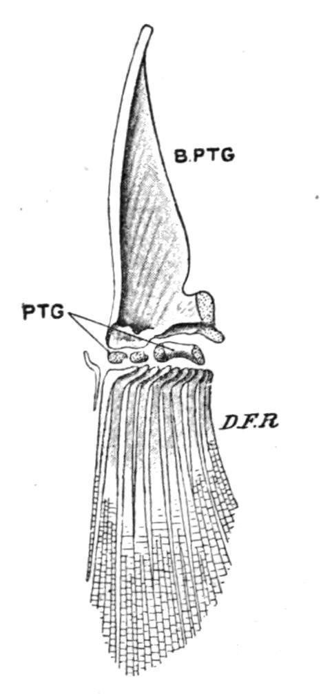
FIG. 149. Skeleton of the left pelvic fin of a Trout (Salmo fario), seen from the dorsal surface. B.PTG, Basipterygium; D.F.R, dermal fin rays; PTG, distal radialia. (From Parker and Haswell.)
CHAPTER IX
THE DENTITION, ALIMENTARY CANAL, AND DIGESTIVE GLANDS
The alimentary canal is a muscular tube with an epithelial lining, formed for the reception and the digestion of the food. It begins with a mouth, and from thence it extends backwards through the coelom, finally communicating with the exterior either by a cloacal or by an anal orifice. The oral or buccal cavity into which the mouth leads is a stomodaeum, and is lined by inpushed epidermis, while the hinder portion of the cloaca and the anus are lined by a somewhat similar inpushing of the epidermis which forms the proctodaeum. The rest of the alimentary canal, consisting in succession of a pharynx, an oesophagus, a stomach, and an intestine, constitutes the mesenteron, and is lined by endoderm. Teeth are developed from the walls of the stomodaeum, and glands for the secretion of digestive fluids from the endoderm of the mesenteron.
Dentition.
In the Lampreys among the Cyclostomata teeth are developed in the form of yellow conical structures on the inner surface of the buccal funnel, and on the extremity of the rasping "tongue" (Fig. 91, A). Each tooth consists of an axial papilla of the dermis, sometimes enclosing a pulp-cavity, and invested by the epidermis, and also by a stratified horny cone which forms the projecting hard part of the tooth. The dermal papilla with its ectodermal investment bears a superficial resemblance to the germ of a true calcified tooth, but no odontoblasts are formed, nor any calcic deposit, the laminated horny teeth being formed by the gradual conversion of the successive strata of the epidermic cells into horny layers.[223] The old teeth are vertically replaced by new teeth developed beneath the functional teeth. With the exception of a median tooth above the oral aperture, Myxine and its allies have only lingual teeth. These are comb-like, and they are formed by the basal fusion of primitively distinct toothgerms. The structure and development of the teeth of the Cyclostomes lend no support to the view that the teeth are degenerate calcified structures. With greater probability they represent a stage in the evolution of teeth and dermal spines, which has been succeeded by a later stage in which calcification superseded cornification as a method of hardening.

FIG. 150. Vertical section of developing tooth in Petromyzon marinus, showing a successional tooth, which is just beginning to cornify at its apex beneath the functional tooth. d, Dermis; d.p, dermal papillae; ep, epidermis lining buccal funnel; ep1 , epidermis which has formed the horny functional tooth ht; ep2 , epidermis forming the horny cone of the successional tooth ht1 . (From Warren.)
True calcified teeth first make their appearance in Fishes, where they assume the form of modifications of exoskeletal structures.[224] The teeth of Elasmobranchs are identical in essential structure, as well as in the manner of their development, with the ordinary dermal spines of the skin, and in the embryo the dermal spines form a continuous series with those which invest the jaws and eventually become teeth (Fig. 151). It is only later, when lips become apparent, that the continuity of the teeth and dermal spines is interrupted, and the two structures assume their distinctive characters.
The tissues of which the teeth of Fishes are composed are (1) dentine, which is a non-vascular, calcified tissue, traversed by numerous radiating, branched, dentinal tubuli, into which extend protoplasmic prolongations from the cells (scleroblasts) by which the dentine is secreted. Dentine forms the greater part of the body of a tooth. (2) vasodentine and (3) osteodentine are modifications of ordinary dentine, the former containing blood-vessels ramifying in its substance but no dentinal tubules, and the latter more closely resembling bone. (4) enamel, an exceptionally dense, non-vascular, non-tubular tissue, which may or may not exhibit traces of the prismatic structure so characteristic of this tissue in the higher Vertebrates, forms the outer investment of the teeth.
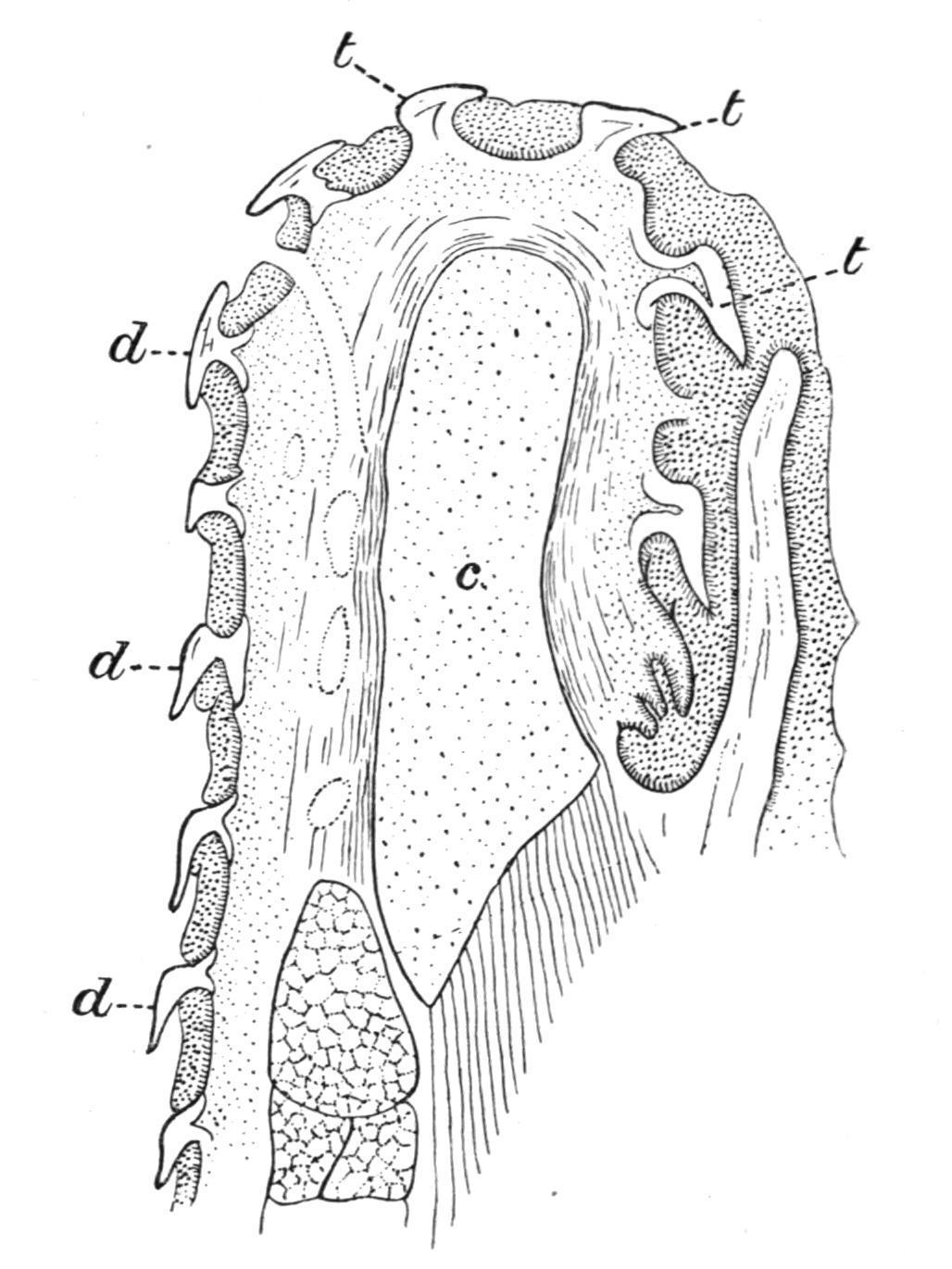
FIG. 151. Transverse section through the lower jaw of an embryo Scyllium, to show the gradual transition from dermal spines (d, d,
d) on the outer surface of the jaw to teeth (t, t, t) on the oral surface. c, Cartilage of the lower jaw. (From Gegenbaur.)
As regards their fixation, the more primitive forms of teeth, such as those of Elasmobranchs, are simply embedded in the gums, and are only connected with the jaws by fibrous tissue; but in some of the older fossil Sharks the fixation of the teeth is effected by the mutual articulation of the basal plates of the teeth with one another. The Chondrostean Polyodon, so shark-like in many other respects, also has teeth implanted basally in the gums, and quite free from any special connexion with the jaw-bones. In some Teleosts with movable teeth, the latter are merely attached to the jaws by fibrous, and often elastic, ligaments, as in the Pike (Esox) and the AnglerFish (Lophius). As a rule, however, the teeth are directly ankylosed to the bones developed in relation with the jaws. Very rarely, as, for example, in some Characinidae, are the teeth implanted in sockets.
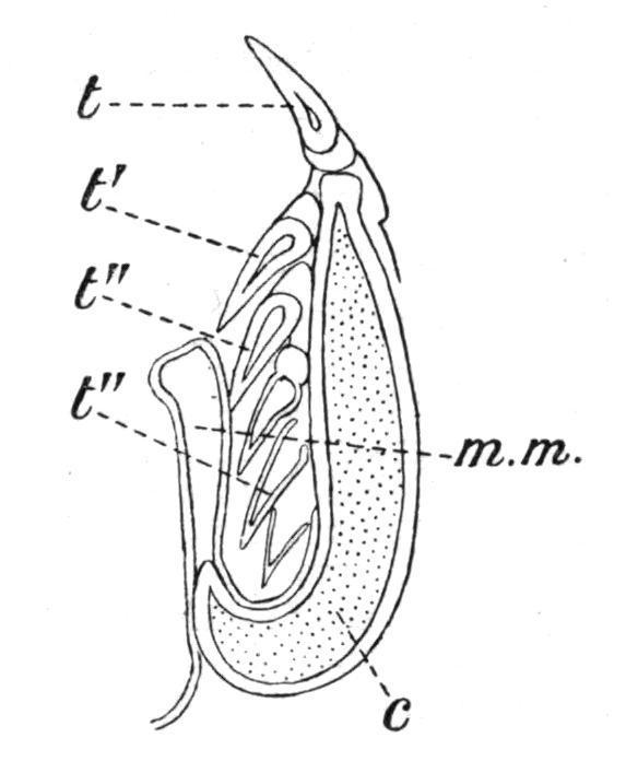
Nearly all Fishes are polyphyodont, that is, the old teeth are constantly replaced by new teeth as fast as they become worn down or fall out. In the Sharks and Dog-Fishes, for example, where the teeth are arranged in rows parallel to the axis of each jaw, the functional teeth along the upper edge of the jaw are usually erect, while those in the rows more internally situated point inwards towards the oral cavity; and behind these again there are rows of developing teeth in different stages of growth, and partially hidden beneath a projecting fold of the oral mucous membrane (Fig. 152). As the teeth in use become lost they are successively replaced by the inner rows, which, with the mucous membrane in which they are embedded, move forwards to the edge of the jaw, where they become erect and functional. The teeth of the Holocephali and of the Dipnoi are not shed, but the loss which they sustain through wear and tear is made good by persistent growth at their bases. In the Teleostomi the succession is less regular, new teeth being formed between or at the bases of the old teeth. In the case of socketed teeth the succession is usually vertical, the new teeth being
formed at the sides of the old ones; and by the absorption of the bases of the latter, the former come to lie directly below them, and eventually they occupy the same sockets.
FIG. 152. Transverse section through the jaw of a Shark (Carcharias), showing how the teeth are replaced. c, Cartilage of the jaw; t, functional tooth; t′, its immediate successor; t", t", still younger teeth, covered by the fold of mucous membrane, m. m. (From Ridewood.)
As might be expected from the remarkable diversity in the habits and in the food of different Fishes, the teeth exhibit an equally striking diversity in form, size, and structure. The most primitive type of tooth resembles an ordinary dermal spine, and is little more than a simple pointed cone. A few Elasmobranchs and many Teleostomi possess teeth of this kind. By the flattening of the cone parallel to the axis of the jaw, the tooth becomes triangular, and then the margins may either remain smooth and trenchant, or they may become complicated by the formation of marginal serrations or of accessory basal cusps, and by such modifications the characteristic teeth of most Elasmobranchs are formed. The simple cone may also be modified to form crushing teeth—short, blunt, more or less hemispherical teeth—or even transformed into a mosaic of hexagonal plates, as in the Myliobatidae amongst Elasmobranchs. Massive, flattened, scroll-like crushing teeth are also formed by the fusion of adjacent teeth, or of several successional teeth, and of such composite teeth we have examples in the Heterodontidae and in the Palaeozoic Cochliodontidae. By a somewhat similar process of concrescence the anomalous composite teeth of such Teleosts as the Diodons and Tetrodons, and of the Parrot-Fish (Scarus), have been evolved. The singular dental structures of the Holocephali are
probably composite teeth, and it is certain that the highly characteristic teeth of the Dipnoi have resulted from the basal fusion of primitively distinct simple conical denticles. The dentition is often heterodont. In Heterodontus (Cestracion), for example, the anterior teeth in each jaw are pointed and prehensile, while the hinder ones are scroll-like and crushing. Prehensile and crushing molar-like teeth are also present in such Teleosts as many of the Sparidae, and in the Wolf-Fish (Anarrhichas). The existence of sexual differences in the dentition is illustrated in the Skates and Rays (Raia), where teeth which are simple and pointed in the male become flattened and plate-like in the female. A few Teleosts, like the Syngnathidae, Cyprinidae, and some Siluridae, are entirely devoid of jaw-teeth.
In addition to jaw-teeth, many Teleosts possess pharyngeal or gillteeth, developed in connexion with the inner margins of the branchial arches, to which they are usually firmly ankylosed (Figs. 352, 412 and 413). As a rule "the pharyngeal dentition is inversely proportional to the extent of tooth development on the jaws."[225] Pharyngeal teeth differ greatly in size and structure in different Teleosts, and, like the jaw-teeth, they are capable of replacement by vertical succession. The teeth are sometimes restricted to the inferior pharyngeal bones (cerato-branchials of the last branchial arch), and then, as in the Carp (Cyprinus), they may bite against a callous pad on the under surface of the basioccipital bone; or, as in some of the Wrasses (Labrus), the inferior teeth are opposed to superior teeth on the upper pharyngeal bones (pharyngo-branchials of more or fewer of the branchial arches). When pharyngeal teeth are present it is probable that they are the principal masticatory organs, the jaw-teeth being used for seizing or holding the prey.
Alimentary Canal.
A protrusible tongue is never developed in Fishes. A rudiment of that organ is present in the Elasmobranchs (Fig. 153) and Dipnoi, and
also in the Crossopterygii, and usually consists of an elevated area of mucous membrane provided with free lateral edges and a forwardly projecting apex; it is supported by the basi-hyal element of the hyoid arch. In the Crossopterygii (e.g. Polypterus) the tongue contains muscle fibres, and in the Dipnoi, where the organ is better developed than in any other Fishes, special lingual muscles are present.
The pharynx succeeds the oral cavity, and is perforated on each side by the branchial clefts (Figs. 153, 154). The rest of the alimentary canal differs considerably in various Fishes in the degree of distinctness of its several regions, and in the extent to which it is convoluted. As a rule the pharynx is followed in succession by an oesophagus, a stomach, and an intestine (Fig. 153), the latter terminating in a portion usually termed the "rectum." The boundaries of these regions are not always very obvious, but are indicated by variations in calibre, by changes in the character of the lining epithelium, by special valves or sphincter muscles, or by the entrance of the ducts of certain glands like the pancreas and liver.
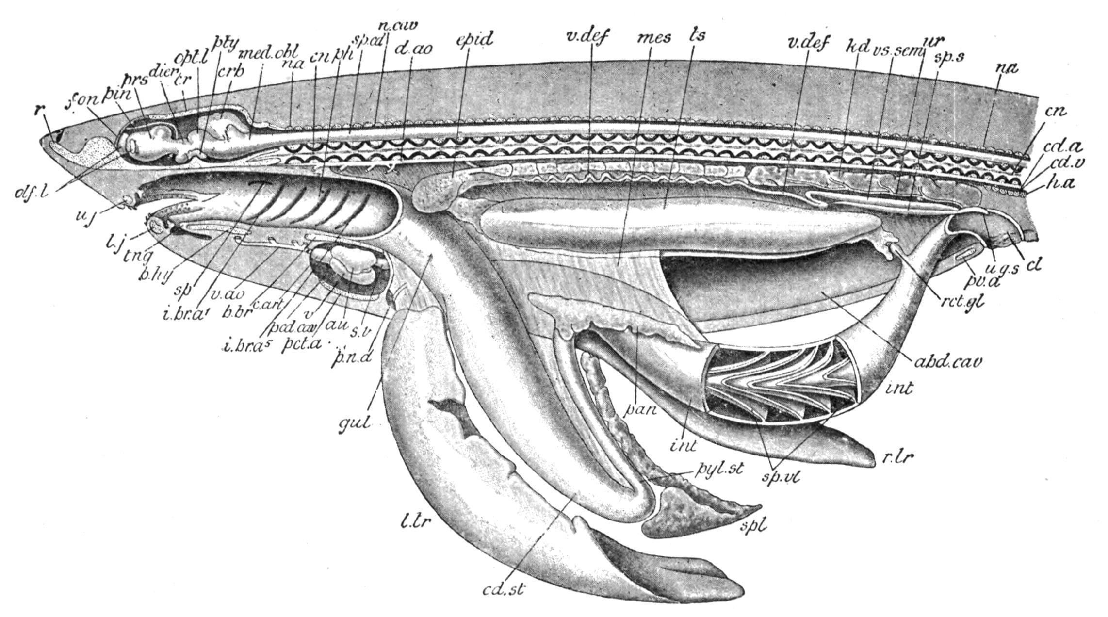
FIG. 153. Dissection of a male Dog-Fish (Scyllium). The left side of the body is cut away to the median plane so as to expose the abdominal and pericardial cavities and the neural canal in their whole length. The alimentary canal and the liver have been drawn downwards, and the oral cavity, the pharynx, part of the intestine, and the cloaca have been opened. The cartilaginous parts of the skeleton are dotted, and the calcified portions of the vertebral centra are black. abd.cav, Abdominal cavity; au, auricle; b.br,
basi-branchial; b.hy, basi-hyal; c.art, conus arteriosus; cd.a, caudal artery; cd.st, cardiac part of the stomach; cd.v, caudal vein; cl, cloaca; cn, centrum; cr, cranium; crb, cerebellum; d.ao, dorsal aorta; dien, thalamencephalon; epid, epididymis; fon, fontanelle; gul, oesophagus; h.a, haemal arch; i.br.a1-i.br.a5 , internal gill-clefts; int, intestine; kd, kidney; l.j, lower jaw; l.lr, left lobe of liver; med.obl, medulla oblongata; mes, mesentery; n.a, neural arch; n.cav, neural canal; olf.l, olfactory lobes; opt.l, optic lobes; pan, pancreas; pcd.cav, pericardial cavity; pct.a, pectoral arch; ph, pharynx; pin, pineal body; p.n.d, vestigial Müllerian duct; prs, prosencephalon; pty, pituitary body; pv.a, pelvic arch; pyl.st, pyloric portion of the stomach; r, rostrum; r.lr, right lobe of liver; rct.gl, rectal gland; sp, spiracle; sp.cd, spinal cord; spl, spleen; sp.s, sperm sac; sp.vl, spiral valve; s.v, sinus venosus; tng, tongue; ts, testis; u.g.s, urino-genital sinus; u.j, upper jaw; ur, metanephric duct; v, ventricle; v.ao, ventral aorta; v.def, vas deferens or mesonephric duct; vs.sem, vesicula seminalis. (From Wiedersheim, after T. J. Parker.)
The oesophagus is occasionally separated from the stomach by a slight constriction, but more frequently the replacement of the squamous epithelium of the oesophagus by the columnar epithelium of the stomach and the appearance of gastric glands in the wall of the latter cavity afford the only distinction between the two regions. The commencement of the intestine is usually indicated by a pyloric "valve" (Fig. 155, A, B), in the form of a ring-like, inwardly projecting thickening of the circularly-disposed muscle fibres of the terminal extremity of the stomach, and usually also by the entrance of the distinct or united ducts of the liver and pancreas; sometimes, as in certain Elasmobranchs and in the Dipnoi, by a special dilatation or "Bursa Entiana" (Fig. 155, A). The rectum, or terminal portion of the intestine, is distinguished from the rest of the gut by its straight course to the cloacal aperture or the anus, and sometimes by an increase in calibre. In Box vulgaris and a few other Teleosts[226] a caecal diverticulum indicates the commencement of the rectum, while in a few cases the pre-rectal portion of the intestine communicates with the enlarged rectal segment by a much constricted valvular orifice which is suggestive of the ileo-colic valve
of the higher Vertebrates,[227] as in the Teleosts Amiuruscatus,[228] Triglagurnardus, and Cyclopteruslumpus.
The relation of the regional divisions of the intestine in Fishes to those of other Vertebrates are somewhat difficult to determine. If we may regard the "rectal" gland of Elasmobranchs and the intestinal caecum of certain Teleosts as homologous with each other, and with the caecum coli of the higher Vertebrates, then it would seem that by far the greater part of the intestine of Fishes, including that portion in which a spiral valve may be developed, is homologous with the pre-caecal segment of the gut or small intestine in other Vertebrates, and that the post-caecal section, or large intestine, of the latter is represented in Fishes only by that relatively short portion of the gut which lies posterior to the rectal gland or its homologue in Teleosts, the equivalent of the colon of Mammalia being, as in Amphibia, Reptiles, and Birds, practically undifferentiated.[229]
In the Cyclostomata the alimentary canal retains much of its primitive simplicity. It pursues a straight course from mouth to anus, and the usual regions are very obscurely indicated. The same remarks apply also to the Holocephali and a few Teleosts, although in these Fishes the limits of the different regions are somewhat more clearly defined. In the Dipnoi (Fig. 155, A), a contracted sigmoid curve between the somewhat dilated stomach and the spacious intestine is the only departure from the straight course of the preceding groups.