Assistant Professor, Coordinator Medical Laboratory Technician Program School of Health Professions, Wellness and Physical Education Anne Arundel Community College Arnold, Maryland
R ICHARD H. M ILLER , P H D, MBA, MLS(ASCP)CM
Former Program Director & Instructor Clinical Laboratory Technology Program Southwest Georgia Technical College Thomasville, Georgia
C AMELLIA S T . J OHN , ME D , MT(ASCP)SBB
Associate Professor Department of Clinical Laboratory Sciences School of Health Professions University of Texas Medical Branch at Galveston Galveston, Texas
Contributors
T HOMAS S. A LEXANDER , P H D, D(ABMLI)
Immunologist, Summa Health System
Professor of Clinical Immunology
Northeast Ohio Medical University
Rootstown, Ohio
Chapter 3 Immunology
L YNNE S. G ARCIA , MS, CLS, FAAM
Director, LSG & Associates
Santa Monica, California
Chapter 7, Section 7.11 Parasitology
D ENISE H ARMENING , P H D, MT(ASCP)
Director of the Online Masters in Clinical Laboratory Management
Adjunct Professor, Department of Medical Laboratory Sciences
College of Health Sciences
Rush University
Chicago, Illinois
Chapter 1 Hematology
R OBERT H ARR , MS, MLS(ASCP)
Associate Professor
Department of Public and Allied Health
MLS Program Director
Bowling Green State University
Bowling Green, Ohio
Chapter 5 Clinical Chemistry
Chapter 6 Urinalysis and Body Fluids
Chapter 8 Molecular Diagnostics
Chapter 10 Photomicrographs and Color Plate Examination
V IRGINIA C. H UGHES , MS, MLS(ASCP)SBB
Assistant Professor and Director
Medical Laboratory Science Program
Dixie State College of Utah
St. George, Utah
Chapter 4 Immunohematology
P AMELLA P HILLIPS , ME D , MT(ASCP)SM
Education Coordinator, Program in Medical Laboratory Science
Bowling Green State University
Bowling Green, Ohio
Chapter 7 Microbiology
Chapter 9 Education and Management
M ITRA T AGHIZADEH , MS, MT(ASCP)
Former Assistant Professor (Retired) University of Maryland School of Medicine
Baltimore, Maryland
Chapter 2 Hemostasis
Preface v Introduction xiii
Design of Questions xiii
Prepare for Your Certification Examination xiv
Test-Taking Skills xiv
CHAPTER 1 Hematology 1
1.1 Basic Hematology Concepts and Laboratory Procedures 3
1.2 Normocytic and Normochromic Anemias 9
1.3 Hypochromic and Microcytic Anemias 14
1.4 Macrocytic and Normochromic Anemias 16
1.5 Qualitative and Quantitative White Blood Cell Disorders 18
1.6 Acute Leukemias 21
1.7 Lymphoproliferative and Myeloproliferative Disorders 26
1.8 Hematology Problem Solving 31
CHAPTER 2 Hemostasis 41
2.1 Coagulation and Fibrinolytic Systems/Reagents and Methods 43
2.2 Platelet and Vascular Disorders 48
2.3 Coagulation System Disorders 53
2.4 Inhibitors, Thrombotic Disorders, and Anticoagulant Drugs 57
2.5 Hemostasis Problem Solving 65
CHAPTER 3 Immunology 75
3.1 Basic Principles of Immunology 77
3.2 Immunologic Procedures 83
3.3 Infectious Diseases 88
3.4 Autoimmune Diseases 99
3.5 Hypersensitivity 103
3.6 Immunoglobulins, Complement, and Cellular Testing 106
3.7 Tumor Testing and Transplantation 109
3.8 Immunology Problem Solving 113
CHAPTER 4 Immunohematology 121
4.1 Genetics and Immunology of Blood Groups 123
4.2 ABO Blood Group System 126
4.3 Rh Blood Group System 130
4.4 Testing for Antibodies 134
4.5 Compatibility Testing 140
4.6 Transfusion Reactions 145
4.7 Components 149
4.8 Donors 154
4.9 Hemolytic Disease of the Newborn (HDN) 158
4.10 Serological Testing of Blood Products 162
4.11 Immunohematology Problem Solving 164
CHAPTER 5 Clinical Chemistry 171
5.1 Instrumentation 173
5.2 Blood Gases, pH, and Electrolytes 190
5.3 Glucose, Hemoglobin, Iron, and Bilirubin 205
5.4 Calculations, Quality Control, and Statistics 222
5.5 Creatinine, Uric Acid, BUN, and Ammonia 238
5.6 Proteins, Electrophoresis, and Lipids 246
5.7 Enzymes and Cardiac Markers 263
5.8 Clinical Endocrinology 282
5.9 Toxicology and Therapeutic Drug Monitoring 294
5.10 Tumor Markers 304
5.11 Clinical Chemistry Problem Solving 310
CHAPTER 6 Urinalysis and Body Fluids 327
6.1 Routine Physical and Biochemical Urine Tests 329
6.2 Urine Microscopy and Clinical Correlations 342
6.3 Cerebrospinal, Serous, and Synovial Fluids 352
6.4 Amniotic, Gastrointestinal, and Seminal Fluids 362
6.5 Urinalysis and Body Fluids Problem Solving 371
CHAPTER 7 Microbiology 381
7.1 Specimen Collection, Media, and Methods 383
7.2 Enterobacteriaceae 389
7.3 Nonfermentative Bacilli 400
7.4 Miscellaneous and Fastidious Gram-Negative Rods 406
7.5 Gram-Positive and Gram-Negative Cocci 416
7.6 Aerobic Gram-Positive Rods, Spirochetes, Mycoplasmas and Ureaplasmas, and Chlamydia 427
7.7 Anaerobic Bacteria 433
7.8 Mycobacteria 438
7.9 Mycology 445
7.10 Virology 456
7.11 Parasitology 463
7.12 Microbiology and Parasitology Problem Solving 477
CHAPTER 8 Molecular Diagnostics 495
8.1 Molecular Methods 497
8.2 Molecular Diagnostics 510
CHAPTER 9 Education and Management 523
CHAPTER 10 Photomicrographs and Color Plate Examination 537
The Medical Laboratory Science (MLS) Review has been designed to provide a challenging personal assessment of practical and theoretical knowledge needed by medical laboratory scientists and technicians. The MLS Review will help you identify strengths, weaknesses, and gaps in your knowledge base. Because taxonomy level is a part of the assessment, you will also be able to concentrate on the type of question that causes the most difficulty. The suggested approach to maximizing use of the MLS Review is to read the explanation that follows each question thoroughly, regardless of whether you answered it correctly or not. Highlight the content you did not know, and study it until committed to memory.
This MLS Review was developed as a tool to facilitate both self-assessment and new learning. The units are arranged in a logical sequence corresponding to the organization of a textbook, and follow the pattern of presentation used in laboratory science lectures. The questions within a unit are related, and can be used by students as they progress through their courses in order to improve understanding. The sections are comprehensive, and suitable for all certification levels although some questions may be more appropriate for one certification level than another. The MLS Review is intended to supplement courses in the curriculum and assist technologists and technicians who are re-entering the laboratory. In addition, it is designed to improve performance on generalist, categorical, and specialist certification exams.
Design of Questions
Test questions used in certification examinations are multiple choice. Each consists of a question, incomplete statement, or problem to be solved called the stem and four alternative responses. One of the alternatives is the correct response and the remaining three are incorrect (these may be wrong, incomplete, partially correct, or less correct than the most appropriate response). Incorrect alternatives
that appear plausible are called distractors. The difficulty of a question is determined by how close the distractors are to the correct response. Some questions were written for assessment of your knowledge, and others for learning. For pedagogic reasons, the latter may contain an “all of these options” alternative. This makes such questions into three true or false statements that are related by the subject (stem) of the question. If you are reasonably sure that two of the responses are true, then the correct response must be “all of these options.” For this reason, such questions are not used on certification exams. Questions involving combinations of statements (multiple, multiple choice) are not used on certification examinations or in this book. All of the questions in this book are multiple choice. Each question is followed by the test item classification. Alongside each question is the correct answer and an explanation. The test item classification consists of the subject category, task, and taxonomy level of the question. A question in Blood Banking, for example, that asks for an interpretation of an ABO problem, may have a test item classification, “Blood Bank/Evaluate laboratory data to recognize problems/ABO discrepancy/3.” The test item classification places the question in the major category of blood banking; the question asks for an evaluation of data; the subcategory is ABO discrepancy; and the taxon omy level classifies the question as problem solving. Taxonomy level 1 questions address recall of information. Taxonomy 2 questions require calculation, correlation, comprehension, or relation. Taxonomy 3 questions require problem solving, interpretation, or decision making. This question design allows you to compute a score, which helps you to identify strengths and weaknesses in various content areas and tasks. You may then focus study time on a particular content area or on practicing with questions of a specific taxonomy level. For example, if you answer several mycology questions incorrectly, then you should devote extra time to studying this content area. If,
however, you miss several recall questions (taxonomy 1 level) over several different content areas such as hematology, chemistry, and immunology, then repetitive review is indicated for all of these sections. Poor performance with questions that require mathematical solutions (taxonomy 2 level) requires you to review formulas used for lab calculations, and to practice using them. If interpretation or problem solving (taxonomy 3 level) is identified as a weakness, then the best approach is to study the explanation that follows each question in order to understand the logic or reasoning behind the answer.
Because the answers and explanations appear on the same page as the questions, it is recommended that you tear off the perforated flap and use it as a blocker to cover the answers while answering the questions. When you have answered a question, slide the blocker down the page to reveal the answer and explanation. The blocker is printed with a compilation of reference ranges for common analytes that will assist you with answering some questions.
Prepare for Your Certification Examination
Ideally, an examination score should reflect your knowledge of the content without the influence of other variables. However, variables such as stress, wellness, selfconfidence, emotional state, and alertness all influence performance. In addition, examination skills often factor into exam scores and can be decisive. A single question answered correctly can make the difference between passing or failing, the only two meaningful scores for a certification exam.
Certification exams are usually delivered by computer. There are two types of computer-based examinations, traditional and adaptive. Traditional exams are of fixed length and content. Therefore, everyone taking the exam does so at the same time and receives the same set of questions. Computer adaptive exams may be fixed or variable in length, but every exam is different because the difficulty of the next question is determined by whether you answer the current question correctly. Since the difficulty of the questions answered correctly—not the number answered correctly—determines passing or failing, you should always give your best answer the first time. Although every examinee’s question set is different, all questions come from a common database, and therefore there is some overlap between the questions used. The examination is constructed so that the number of questions in each category (e.g., hematology) is within the specifications published for the exam; however, the distribution of questions and the order of questions will vary significantly. Certification exams are criterion referenced. This means that examination performance is scored passing or failing independently of the performance of other candidates taking the examination. The minimum passing score for certification examinations is normalized in order to minimize the variance between examinations. However, the
minimum passing score usually falls within the range of 65%–70% correct responses. A score below 65% on any content area in the MLS Review is a strong indicator that you have not mastered the material in this area, and that further study is required.
Preparation for a certification exam requires a study plan. Begin with a review of the exam content outline that is made available by the certification agency. For example, if 20% of the exam is Microbiology but only 2% of the exam is Laboratory Management, you should spend significantly more time studying the former. Within each content area will be subcategories (e.g., Bacteriology and Parasitology under Microbiology). If 60% of the Microbiology content is Bacteriology and only 10% is Parasitology, then devote significantly more time to studying the former.
Allow yourself sufficient time prior to the exam to review each content area no less than three times. Begin studying your strongest subject, then progress to your weakest. Study your class notes first, then use this review book to test your knowledge of the respective content area. Devote time to reading the explanation for each question, regardless of whether you answered it correctly or not. Highlight information you did not know and review it before answering the questions in this book a second time. Rarely, will you encounter any of the same questions on your certification exam; however, you are likely to encounter variants of the questions, and the explanations will help prepare you to answer these correctly. When finished with the second round, take the comprehensive exam included with this book. Evaluate your performance by both subject and taxonomy. If you score lower in Clinical Chemistry, devote more time to it in your third round of study. If you are weakest in recall-type questions, make note cards with charts and tables, and study them regularly until the information on them is committed to memory. Note your progress from the first to the second round. If your progress is significant, use the same approach on the third round. If not, devote more time to studying your weakest content areas. Plan your third round of study so that you end with your weakest subject. Then, repeat each chapter in the MLS Review a final time. Finish by taking the examinations on the CD included with this book. These questions are all different than those in the book, and will give you exposure to many more based on interpreting photomicrographs.
Test-Taking Skills
Before the Exam
First, make a study plan such as the one suggested earlier. You cannot expect to review all of this material in only a few days. Allow yourself at least 1 month to study all areas completely and carefully. Set aside an allotted time period of at least 1 hour each day when you are alert and can stay focused.
Assemble all of your study materials before you begin your review. Searching for old notes or textbooks may become time consuming and frustrating. You may have a tendency to “give up” looking for needed materials, if you do not have them readily available. Therefore, you may neglect or not study a major content area.
Provide a study environment. Choose a quiet, comfortable area for your study. Find a place where you will not be distracted or disturbed. Simulate test conditions. Regardless of your study plan, you should take some portion of the review process, for example, the mock examination, under simulated test conditions. These examinations should be timed, uninterrupted, and designed to observe realistic testing practices. For example, you should take the mock examination with only a sheet of scratch paper, pencil, and a basic calculator at your disposal.
A few days before the exam, be sure to again read through the instructions sent to you by the certification agency. Some types of calculators (e.g., graphing or programmable calculators) may be prohibited and you should know what you can and cannot bring with you. Make your travel arrangements and familiarize yourself with directions to the site. Finally, go to sleep early the night before the exam, and leave yourself extra time if you have to travel a long distance to the examination site.
On Exam Day
Eat properly and, if possible, engage in some light physical activity such as walking prior to leaving for the exam. Dress comfortably with layered clothing that you may remove, if the examination room is too warm. Make sure you bring two forms of signed identification including 1 photo identification card (driver’s license or state issued ID card). These must not be expired, and the name on them must match the name on your letter of admission to the exam that you should also have with you.
Wear a watch so that you can keep track of time. Do not take notes or books with you. If you have not prepared prior to the examination day, you will not succeed by trying to cram last-minute facts. If you become anxious before or during the exam, close your eyes and breathe deeply for a few seconds. Perhaps focus on a
special activity that you may have planned as a reward for yourself after the examination.
Have confidence in your abilities. At this point, you have successfully completed a rigorous course of classroom and clinical training and the examination represents merely the last step in this long process. Tell yourself that you have adequately practiced and prepared for the examination and that you are ready.
During the Exam
Read all directions. Make sure you understand how to take the examination. Read the questions carefully and note key words. Accept the question as you first read it; do not read your own thoughts into the question and do not look for hidden meanings.
Quickly look at all of the answers. Next, carefully read all choices. You may wish to mentally place a T for true or an F for false beside each alternative, or to reject outright obviously wrong choices. Select your first choice and do not change your answer. Answer all of the questions. There is no penalty for guessing on certification examinations. Always answer to the best of your ability the first time. A computer-adapted exam selects the next question based upon your previous answer.
Apply a few simple rules to those questions you cannot answer. Consistently choose the same letter on those questions. “B” is the most common correct answer. Choose one of the longest answers. Pick items that are more specific or detailed than the others.
Do not overlook words such as not, never, always, most, least, best, worst, except. Statements that contain unqualified absolutes (always, never) are usually incorrect. In contrast, alternatives that are worded to contain exceptions (usually, generally) are often true. Do not panic if you do not know an answer. Continue the test and do not allow anxiety to make you forget items that you know.
Work steadily and do not spend too much time on questions you do not know; keep an eye on the time. Try to pace yourself so that sufficient time remains after completing the test to review all of your answers. Do not change your original answer unless you are certain that you made a mistake when you answered the question initially.
1.1 Basic Hematology Concepts and Laboratory Procedures
1.2 Normocytic and Normochromic Anemias
1.3 Hypochromic and Microcytic Anemias
1.4 Macrocytic and Normochromic Anemias
1.5 Qualitative and Quantitative White Blood Cell Disorders
1.6 Acute Leukemias
1.7 Lymphoproliferative and Myeloproliferative Disorders
1.8 Hematology Problem Solving
1. Insufficient centrifugation will result in:
A. A false increase in hematocrit (Hct) value
B. A false decrease in Hct value
C. No effect on Hct value
D. All of these options, depending on the patient
Hematology/Apply principles of basic laboratory procedures/Microscopic morphology/Differential/2
2. Variation in red cell size observed on the peripheral smear is described as:
A. Anisocytosis
B. Hypochromia
C. Poikilocytosis
D. Pleocytosis
Hematology/Apply knowledge of fundamental biological characteristics/Microscopic morphology/RBCs/1
3. Which of the following is the preferable site for bone marrow aspiration and biopsy in an adult?
A. Iliac crest
B. Sternum
C. Tibia
D. Spinous processes of a vertebra
Hematology/Apply knowledge of fundamental biological characteristics/Bone marrow/1
4. Mean cell volume (MCV) is calculated using the following formula:
A. (Hgb ÷ RBC) × 10
B. (Hct ÷ RBC) × 10
C. (Hct ÷ Hgb) × 100
D. (Hgb ÷ RBC) × 100
Hematology/Calculate/RBC indices/2
5. What term describes the change in shape of erythrocytes seen on a Wright’s-stained peripheral blood smear?
A. Poikilocytosis
B. Anisocytosis
C. Hypochromia
D. Polychromasia
Hematology/Apply knowledge of fundamental biological characteristics/Microscopic morphology/ RBCs/1
Basic Hematology Concepts and Laboratory Procedures
Answers to Questions 1–5
1. A Insufficient centrifugation does not pack down the red blood cells; therefore, the Hct, which is the volume of packed cells, will increase.
2. A A mature erythrocyte is approximately 7–8 μm in diameter. Variation in normal size is denoted by the term anisocytosis. Hypochromia is a term that indicates increased central pallor in erythrocytes, and poikilocytosis denotes variation in red cell shape.
3. A The iliac crest is the most frequently used site for bone marrow aspiration and biopsy. This site is the safest and most easily accessible, with the bone just beneath the skin, and neither blood vessels nor nerves are in the vicinity.
4. B MCV is the average “volume” of the red cells. This is obtained by dividing the Hct or packed cell volume (PCV) by the red blood cell (RBC) count in millions per microliter of blood and multiplying by 10. The MCV is expressed in cubic microns (μm3) or femtoliters (fL).
5. A Variation in shape of the erythrocytes on a peripheral blood smear is poikilocytosis. Anisocytosis refers to a change in size. Hypochromia is an increase in central pallor in erythrocytes. Polychromasia describes the bluish tinge of the immature erythrocytes (reticulocytes) circulating in the peripheral blood.
6. Calculate the mean cell hemoglobin concentration (MCHC) using the following values:
Hgb: 15 g/dL (150 g/L) Hct: 47 mL/dL (0.47)
RBC: 4.50 × 10⁶/μL (4.50 × 10¹²/L)
A. 9.5% (.095)
B. 10.4% (.104)
C. 31.9% (.319)
D. 33.3% (.333)
Hematology/Calculate/RBC indices/2
7. A manual white blood cell (WBC) count was performed. A total of 36 cells were counted in all 9-mm2 squares of a Neubauer-ruled hemacytometer. A 1:10 dilution was used. What is the WBC count?
A. 0.4 × 10⁹/L
B. 2.5 × 10⁹/L
C. 4.0 × 10⁹/L
D. 8.0 × 10⁹/L
Hematology/Calculate/Manual WBCs/2
8. When an erythrocyte containing iron granules is stained with Prussian blue, the cell is called a:
A. Spherocyte
B. Leptocyte
C. Schistocyte
D. Siderocyte
Hematology/Apply knowledge of fundamental biological characteristics/RBCs microscopic morphology/Stain/1
9. A 7.0-mLethylenediaminetetraacetic acid (EDTA) tube is received in the laboratory containing only 2.0 mL of blood. If the laboratory is using manual techniques, which of the following tests will most likely be erroneous?
A. RBC count
B. Hemoglobin (Hgb)
C. Hct
D. WBC count
Hematology/Apply knowledge to identify sources of error/Specimen collection and handling/CBCs/2
10. A 1:200 dilution of a patient’s sample was made and 336 red cells were counted in an area of 0.2 mm2. What is the RBC count?
A. 1.68 × 10¹²/L
B. 3.36 × 10¹²/L
C. 4.47 × 10¹²/L
D. 6.66 × 10¹²/L
Hematology/Calculate/Manual RBCs/2
11. What phagocytic cells produce lysozymes that are bacteriocidal?
A. Eosinophils
B. Lymphocytes
C. Platelets
D. Neutrophils
Hematology/Apply knowledge of fundamental biological characteristics/Leukocytes/1
Answers to Questions 6–11
6. C MCHC is the average concentration of Hgb in red cells expressed as a percentage. It expresses the ratio of the weight of Hgb to the volume of erythrocytes and is calculated by dividing Hgb by the Hct, and then multiplying by 100. A decreased MCHC indicates that cells are hypochromic. In this example, (15 ÷ 47) × 100 = 31.9%. The reference range for MCHC is 32%–36%.
7. A The formula used for calculating manual cell counts using a hemacytometer is: Number of cells counted × dilution factor × depth factor (10) divided by the area. In this example, 36 × 10 × 10 = 3600 ÷ 9 = 400/mm3 or 0.4 × 10⁹/L.
8. D Siderocytes are red cells containing iron granules and are visible when stained with Prussian blue.
9. C Excessive anticoagulant causes shrinkage of cells; thus, the Hct will be affected. RBC and WBC counts remain the same, as does the Hgb content.
10. B RBC count = number of cells counted × dilution factor × depth factor (10), divided by the area. In this example, 336 × 200 × 10 = 672,000 ÷ 0.2 = 3.36 × 10⁶/mm3 = 3.36 × 10¹²/L.
11. D Neutrophils are highly phagocytic and release lysozymes, peroxidase, and pyrogenic proteins. Eosinophils migrate to sites where there is an allergic reaction or parasitic infestation, releasing peroxidase, pyrogens, and other enzymes, including an oxidase that neutralizes histamine. They are poorly phagocytic and do not release lysozyme.
Another random document with no related content on Scribd:
sometimes fecal matter. Cystic dilatation of the portion of the duct originally connected with the ileum is also sometimes seen.
2. Allantoic Cysts.
—These are connected with the urachus, which should ordinarily be found as a fibrous cord, but which occasionally persists in a pervious condition, in whole or in part. At birth it is often traversed by a narrow canal lined with epithelium continuous with that of the bladder. The urachus lies outside the peritoneum, and may be dilated at any point between its two extremities. When the entire urachus is pervious urine is discharged from the navel.
3. Cysts Connected with Remains of the Wolffian Body.
—The Wolffian body, or the mesonephros, is intimately related with the development of the kidney, the ovary, and the testis. In the two latter locations glandular elements may be met, persisting in adult life.
In the male the tubulespersist as excretoryductsfrom thetestis, but in the female they persist, in a vestigial condition, as the parovarium and Gärtner’s ducts. The ovary proper consists of the oöphoron and the paroöphoron, the former being the egg-bearing portion, the latter receiving the tubules from the adjoining structure known as the parovarium. The paroöphorongives rise to cysts which burrow deeply between the layers of the broad ligament, make their way alongside the uterus, and raise the peritoneum. It is a peculiarity of these cysts that their inner walls often become papillomatous, and may even develop such a crop of warty outgrowths that these make their way through the cyst wall and protrude into the abdominal cavity, where they sometimes become detached and are dropped as loose bodies into the peritoneal sac. The condition is also often accompanied by warty growths upon the peritoneal surfaces. These need give rise to no alarm, because they usually disappear spontaneously with removal of the tumor. Paroöphoritic cysts are to be distinguished from parovarian cysts, which develop from the parovarium, this latter consisting of a number of tubules situated between the layers of the mesosalpinx, composed of an outer series known as Kobelt’s, an inner set, about a dozen in number, known as the verticaltubules, with a straight tube, running at right angles to these through the broad ligament to the
vagina, known as Gärtner’s duct, which is homologous with the vas deferens in the male. Cystic dilatation of Kobelt’s tubes is often seen, these cysts being very small and having no clinical importance. Cysts arising from the vertical tubules are usually transparent until they attain considerable size, when their walls thicken. Their contained fluid is not harmful, and after rupture of such cysts internally the fluid is absorbed. Such cysts may rupture and refill several times. As betweentheparoöphorous and parovariancyststhe latter are easily enucleated, carry the ovary upon one side, and have the Fallopian tube stretched over them without communication.
The internal sections of Gärtner’s ductare more often involved in animals than in women, but excellent illustrations of cystic dilatation of its various portions have been observed, usually in the walls of the vagina.
Corresponding to the above-mentioned conditions in the female there are in the male, as the result of changes in the Wolffian body, two conditions—encysted hydrocele of the testicle, and general cystic degeneration of the same. Like the ovary, the testicle is a complex organ with remnants of the mesonephros persisting among its ducts, while only a few of the Wolffian tubules remain. True encysted hydroceles arise sometimes in the efferent tubes of the testis and sometimes in Kobelt’s tubes (the same structures which in the female give rise to parovarian cysts), the two conditions, therefore, being analogous and homologous. These cysts, though closely associated with the testis, lie outside its tunica vaginalis. Their contained fluid is usually clear or of a milky whiteness, due to fat globules. Sometimes it contains spermatozoa. Another variety is cystic dilatation of one or more of Kobelt’s tubules, which is often described as involving the hydatidofMorgagni.
Generalcystic disease of the testis, known also as adenomatous degeneration, was formerly referred to as hydatid disease of the same organ. The multiple cysts appear to originate in the remnant of the mesonephros still persisting, known as the paradidymis. The cavities are lined with epithelium, and papillomatous intracystic formation is not uncommon. These tumors have been called by a number of improper names, such as “cystic sarcoma,” etc.
Hydroceles.
—The term hydrocele has covered numerous conditions. At present, when no other locality is designated, hydrocele of the tunica vaginalis is understood. (The term implies a collection of watery fluid in a previously existing serouscavity.) This is the most common form.
Possibility of its formation depends upon the prolongation of the peritoneal cavity which takes place in advance of or along with the descending testicle, and which in many of the lower animals remains connected with the general cavity throughout life. In men only is it expected to close, even before birth. When the portion which extends along the spermatic cord is not completely obliterated there is encysted hydrocele of the cord, or funicular hydrocele, which is not common. The common form of hydrocele is constituted by serous effusion into the tunica vaginalis, and occurs usually without recognizable exciting cause. It will be treated more fully in its appropriate place.
The corresponding process of peritoneum in the female is known as the canal of Nuck; and, when persistent, this also becomes distended with fluid and forms a cyst known as hydrocele of the canalofNuck, occupying the inguinal canal.
In many of the lower animals the ovaries are contained within a serous sac derived from the peritoneum, which is so connected with the opening of the Fallopian tubes that when the ova escape from the ovary they enter these tubes and pass to the uterus without entering the general peritoneal cavity. This ovarian sac is subject to serous distention, and constitutes a condition called by Sutton an ovarianhydrocele. An homologous condition occurs sometimes in the human female, by pathological adhesion, and such cysts may attain large size. They project from, and are intimately connected with, the posterior layer of the broad ligament.
Hydroceles of the Neck.
—Hydroceles of the neck, so called, are cystic collections of congenital origin found in the cervical region, due to dilatation of ducts or clefts which should have disappeared at or before birth. The forms of cyst to which the name “hydrocele of the neck” are usually limited are recognizable at or soon after birth, and constitute fluctuating tumors, often extending beneath the
clavicle into the axilla or down upon the thorax. They may occupy the entire lateral region of the neck, and may be unilateral or bilateral—may be single or multilocular, and may even intercommunicate.
They originate always beneath the deep fascia. Some of these cysts are undoubtedly due to dilatation of lymph spaces. This is particularly true of the multilocular forms. There is noted in many of them a tendency toward spontaneous recovery, but many again require operative measures for their eradication. Occasionally their walls are extremely vascular, even to the degree meriting the term nevoid.
Some of these cysts are considered by Sutton to be essentially examples of the laryngeal saccules which are met with as diverticula from the laryngeal mucous membrane, which undermine the deep cervical fasciæ of certain monkeys. These air chambers, which are normal in the monkey, communicate with the larynx through the thyrohyoid membrane, and occasionally run down beneath the upper border of the thorax. Many of the cysts having this resemblance are closely related to the hyoid bone and to the larynx, and there is much to substantiate the view thus quoted.
Glandular Cysts.
—Ranula is an altogether too comprehensive term which has long been used in surgery, alluding to cysts in the floor of the mouth, and not indicating minutely their character nor their exact location. At present this term should either be restricted in signification or be eliminated. If used, it should be confined to retention cysts due to obstruction of the submaxillaryor sublingualducts. Such obstruction is often caused by salivary calculi impacted in the duct orifices. In other instances it is due to cohesion of the margins of the outlet. A similar condition in the parotid duct is known, but is less common. Aside from this, certain other cysts originate from minute beginnings in and about the floor of the mouth, being due to dilatation of the mucous glands, particularly one near the tip of the tongue, sometimes known as Nuhn’s gland. Dermoid cysts in this locality are not uncommon. Formerly cysts of the floor of the mouth were described as ranula.
Pancreaticcysts correspond in large degree to salivary cysts, the pancreatic duct becoming dilated by retention when its orifice is obscured; and, indeed, the condition has been referred to as pancreaticranula. Sometimes the canal is dilated in distinct portions, so that the condition resembles a string of cysts; at other times it is the terminal portion which is most enlarged. Such cysts attain large size and contain mainly mucoid material. Examples have been reported showing that they have attained a capacity of two gallons.
In the mesentery there sometimes develop cysts which are known as chyle cysts, whose sacs appear to be formed of separate mesenteric layers, their cavity being occupied by fluid identical with chyle. Such tumors also sometimes attain great size.
In the eyelids one occasionally meets with cystic dilatations of the lacrymal ducts. These are known as dacryopic cysts or dacryops. Fistulas result when they are opened through the skin, and if meddled with at all they should be radically extirpated.[13]
[13] In the treatment of cysts, as of many abscesses (e. g., those of the gland of Bartholin), it will be of advantage to empty the cavity through a small trocar or needle and then to fill it with melted paraffin, as suggested by Pozzi. When it has thus been distended it can be dissected out with much more deliberation and more easily than would be otherwise possible.
Pseudocysts.
In his elaborate work on tumors Sutton has made a distinct classification of pseudocysts, which lack some of the characteristics of genuine cysts, yet, nevertheless, are entitled to consideration in this place. Among these are included intestinal diverticula and vesical diverticula, in either of which instances hernial protrusions of the mucous membrane through the outer coating of the bowel or of the bladder occur, thus forming pouches. These are common in the bowel, rare in the bladder; especially in the former locality they are often multiple. This condition is often referred to as sacculation, and sacculation of the bladder may even be confounded with true urachus cyst. They are of little consequence so long as foreign materials, such as feces, urinary calculi, etc., do not lodge in them. But they occasionally cause serious trouble. Diverticula have been mistaken for
appendices, while diverticula from the bladder have been encountered in hernia operations.
Pharyngeal diverticula give rise to rare but most interesting tumors. It is well known that the branchial clefts, which in early fetal life connect with the pharynx, are sometimes not completely closed, and that a portion of one may persist abnormally, giving rise to a condition known as the pouch of Rathke. There may also occur sacculation of the pharyngeal wall where it joins the esophagus, or hernial protrusions, especially in Rosenmüller’s fossa.
Cystic dilatation of Rathke’s pouch occurs near the upper part of the pharynx, and may attain the size of a marble. Hernial pouches are seldom mistaken for cysts, and are of importance mainly because of the fact that food or other foreign material gathers and lodges in them. Most of the other cystic abnormalities of the pharynx pertain to dermoids, and will be considered shortly. In a general way, these pharyngeal tumors have been grouped as pharyngoceles.
Similarly in the esophagus and trachea hernialprotrusions occur, and lesions closely resembling retention cysts may be seen.
Synovialcysts(i.e., those containing synovial fluid) may arise (1) by protrusion of synovial sheaths, (2) by distention of bursæ in the vicinity of joints, or (3) by hernial protrusions of joint membranes. They are often met with in connection with the larger joints, more particularly about the knee. In this way tumors as large as gooseeggs may be formed, while their location may be so shifted that they present themselves in perplexing ways. To that form produced by hernial protrusion of the lining of a tendon sheath has been given the name ganglion.
The simple ganglion is frequently seen on the back of the wrist, and, while it is always connected with the tendon sheath, it undoubtedly often connects with the synovial membrane of the carpal joints. The compound ganglion, so called, is a much more serious and extensive affair, being one which has prolongations in two or more directions, and containing peculiar bodies, known as melon-seed bodies, which appear to be fibrinous concretions worn round and smooth by attrition. These are present sometimes in
enormous numbers. (See Tuberculosis of Synovial Structures, Chapter IX.)
Bursæ are normal in many well-known situations in the body, but may undergo cystic dilatation and become annoying tumors. In many other places, under the influence of friction or mechanical irritation, there develop bursæ which are known as adventitious. These are sometimes subtendinous, and may communicate alike with joint sheaths and tendon sheaths. These are true cysts of new formation not developed from a pre-existing cavity.
They are largely the effect of peculiar occupation, as in housemaids and carpet-layers there are formed frequently prepatellar bursæ, while miners get them upon the elbow, porters upon the shoulder, plasterers upon the forearm, etc. In the same way, by the pressure of ill-fitting boots, an adventitious bursa is developed over the expanded head of the first metacarpal bone, thus forming a condition known as bunion.
Neural Cysts.
—This term has been applied by Sutton to pseudocystic dilatation of certain cavities found in the brain and central nervous system. Hydrocephalusis in one sense a pseudocyst of this variety. Corresponding to it in fetal life is hydramnios. Hydrocele or cystic dilatation of the fourth ventricle is well known. Cranialmeningoceles, which are hernial protrusions of brain membranes, are also pseudocysts, to be included in this category. They will be considered in Chapter XXXVI. Cephalhematoma may be also included in the same way. Spina bifida, a condition which will be described in Chapter XXXVIII, is, nevertheless, practically a cyst of congenital origin involving the spinal meninges. One form of spina bifida is constituted by cystic dilatation of the central canal of the spinal cord, and produces syringomyelocele. These conditions will be treated more fully in their appropriate places.
Sutton has rendered a great service by showing that the brain and spinal cord are evolved from a segment of the primary intestines, and that the intestinal canal and the neural canal communicate in fetal life at their lower terminations; while it has been shown by several that in the earlier forms of mammalian life they were also
connected by their anteriorterminations. It is in this way that certain complex tumors of the sacral and coccygeal region are to be explained. So also is the collection of lymphoid tissue in the vault of the pharynx, known as Luschka’stonsil, and in the coccygeal region, known as Luschka’sgland, it being a curious and instructive fact that lymphoid tissue of this character is always met with in the neighborhood of obsolete canals.
Hydatid Cysts.
These cysts are the indirect product of the eggs of the Tænia echinococcus, a form of tape-worm which infests the alimentary canal of dogs. The eggs reach in some direct or indirect way the food or water taken into the human stomach and are there hatched; the young animals migrate through vessel walls and are deposited in some tissue or organ where the cyst later develops. These cysts have a thick, elastic wall, with a lining containing cells, involuntary muscle fibers, and a watervascular system. After such a cyst has attained the size of an inch or more, small vesicles, or “brood capsules,” begin to develop, which present at one point a retractable head, with scolices so arranged in crown form as to produce sucking disks. According to the date at which the cyst is opened appearances will differ. Sometimes a large cavity will be filled with multiple “daughter cysts,” and sometimes these will have disappeared, so that the cyst fluid contains nothing distinctive. After having ceased to develop, hydatids frequently undergo atrophy and even become calcified; the characteristic hooklets are the last of the distinctive features to disappear. These growths may be rapid, even to the point of producing necrosis and rupture, or may be very slow and persist almost unchanged for years. The disease is uncommon among the nativeborn population of the United States, and most of its examples are seen in emigrants. It is exceedingly prevalent in Iceland and in New Zealand. It occurs most often in the liver, but is frequently met with in these countries in the lungs, the brain and spinal canal, and the bones, but may be encountered in any part of the body. When located near the intestinal tract or the air tract the cysts are more liable to penetration by ordinary germs of sepsis, and then may suppurate. It is not infrequent to have conversion of an hydatid cyst
into an abscess. Before or after such change it may undergo rupture, spontaneous or traumatic, and this, according to the nature and amount of its contents, and the location of the opening, will promptly produce more or less grave symptoms. While spontaneous recovery has, in rare instances, followed rupture, it has perhaps more often led to fatal result. At all events, it will produce serious and perhaps distressing symptoms.
The only radical treatment for hydatid cysts is extirpation. When this is not possible the cyst may be opened and the margins of the opening attached to those of the skin wound. After being evacuated it should be packed and drained, and then may be expected to slowly contract, perhaps even to the point of obliteration. The contents of such a cyst should not be allowed to escape into any of the body cavities, since their sterility can not be always relied upon.
Cystic Degeneration.
Hematocele is an expression meaning a tumor composed originally of effused blood which has undergone chemical and other changes, which consist of lamination and thickening of its exterior portion and fluidification of the interior, until in course of time such an internal blood clot may be converted into a distinct and plainly walled cyst. This condition may be seen in two locations—namely, in the pelvis and between the cranium and the brain, or in the brain. The hemoglobin gradually disappears, and the contents of these cysts are translucent or even watery in appearance. Hematoceles may form where there has been internal hemorrhage in certain locations which has failed to absorb, and where no pyogenic infection has occurred.
Pseudocystic changes occur in other tumors and in other parts of the body as the result of mucoid and colloid liquefactions. In the midst even of apparently dense and entirely defined tumor masses changes of this kind occur, and lead to formation of cavities containing fluid of variable consistence, causing the tumor when divided to present the appearance of the geodes or quartz rocks, containing cavities lined with quartz crystals. The occurrence of such cystic changes is indicated, in naming such a tumor, by prefixing the term cysto-, as cystosarcoma, cystofibroma, etc.
2. Dermoids.
Dermoids are cysts or tumorscontaining tissues andappendages which are developed from theepiblast, and which occur when skin and mucous membrane are not normally found. The simplest form of dermoid is a cyst whose interior is lined with modified skin, containing sebaceous glands and hair follicles, from which often numerous long hairs are produced. Even sweat glands may be present. Its cavity is occupied by mixed material, pultaceous in character, made up of sebum, cholesterine, and growing hairs which are often rolled into balls. The sebum is the product of the glands contained in the cyst wall.
A complex form of so-called dermoid cyst is met with in which there are unstriped muscle fiber, teeth, mammary glands, etc. These belong rather to the class of teratomas, as they contain more or less tissue not of epiblastic origin.
A dermoidtumor is one lacking cystic characteristics, made up of tissue largely developed from the epiblast, with more or less tissue of mesoblastic origin. Such a tumor may contain much connective tissue, fat, fetal hyaline cartilage, and nerve tissue, while from its exterior long hair may grow, and teeth project from its surface or be embedded within its substance. Such tumors are generally found in the pharynx and about the rectum.
The explanation of dermoids and teratomas may be gleaned from embryology, and rests upon the arrangement of the different blastodermic layers of the developing ovum, and upon the facts already alluded to in explaining Cohnheim’s hypothesis of the origin of tumors. Strictly speaking, a dermoid should contain only that which may be developed from the epiblastic layer. It is well known that teeth and hair, as well as sebaceous material, are epiblastic products. Consequently such material may be found within a dermoid and needs no further explanation than an epiblastic inclusion, according to Cohnheim’s views. But so soon as such a tumor contains bone, muscle, etc. (i. e., tissues of mesoblastic origin), we should drop the term dermoidand consider it a teratoma. Such is the distinction between these two terms. According to Wilm’s
researches, any tumor of this sort which contains epithelial products as teeth or hair is sure to contain also mesoblastic elements, and thus to belong to the latter. The term epidermoidshas been applied to the former.
The most prominent characteristics of dermoid cysts are: (1) Skin, which may be thick or thin, lined with papillæ, containing more or less pigment, its deeper layers possessing a quantity of fat. (2) Hair, which next to skin is the most constant structure found in dermoids; this may be present in trifling amount or in long coils or balls. It is of interest that in dermoids found in animals covered with wool we find the same character of hairy structure, while in birds dermoids contain feathers rather than hairs. (3) Sebaceous glands and their peculiar secretion are invariably found. These may be of large size, and sebaceous retention cysts may be seen in the walls of dermoids. Sometimes horny matter or tissue is found in these, indicating the same relation between horn and sebaceous structures, as we see upon the external skin in other instances. So, too, material resembling the texture of finger-nails is occasionally found projecting into the cavity.
The fluid or semifluid contents of these cysts consist usually of sebaceous material, cholesterin, epithelial debris, etc. Sometimes it is thick, sometimes thin—and occasionally consists almost entirely of mucus.
It is not uncommon to find structures in ovarian dermoids closely analogous to, or actually resembling, mammary glands. These may be mere nipple-like processes of skin, or completely developed mammæ, well formed, but without ducts or gland tissue, may occupy such a cyst. These really are pseudomammæ, because they have no ducts. Nevertheless, glandular tissue is not always absent. This resemblance proceeds even farther, in that in some of these ovarian mammæ changes occur analogous to those which take place in normal breasts.
The epiblast seems to have the power of developing mammary glands or supernumerary mammæ in many locations—in fact, upon any part of the body surface. About the thorax they are common;
upon the abdomen they are rarely observed; and they have been found even upon the labia.
Sweatglandsare infrequent in dermoids. Teethare quite common. These may vary in number from two or three up to several hundred —may be embedded in definite sockets or simply sprout from the cyst wall. Occasionally bone material, lodging such teeth and crudely resembling a jaw, will be found.
Dermoids containing mucous membrane are found, especially in connection with the ovary and with the postanal gut (i. e., the original communication between the spinal and alimentary canals).
It is curious that under these circumstances mucous membrane is sometimes furnished with hair, as it normally is in the stomach or other cavities of some of the lower animals. Mucous glands and retention cysts of these glands are also found in ovarian dermoids. This will be more readily understood if the mutability of skin and mucous membrane be not forgotten. The transition from one to the other is not difficult, and we find all intermediate stages between the two extremes—if not in man, at least in animals. This will account for the fact that skin-covered dermoid tumors are found in certain parts of the alimentary canal, and particularly in the pharynx. These tumors grow also from the mucous membrane of the bowel, of the rectum, or even of the small intestine.
Sutton has made a division of dermoids into three classes:
1. Sequestration;
2. Tubulodermoids;
3. Ovarian.
1. Sequestration Dermoids.
—Sequestration dermoids occur chiefly in situations where during embryonic life coalescence takes place between two surfaces possessing an epiblastic covering, although sometimes this coalescence practically occurs late in life and by implantation.
Dermoids of the trunk occur particularly where opposite halves of the body wall coalesce—that is, in the midline of the trunk and head. Dermoid cysts are rarely found in connection with spina bifida, and certain tumors spoken of as spina bifida undoubtedly are dermoids. Anteriorly dermoids occur frequently in the scrotum, and
occasionally in the testicle. At the umbilicus they are rarely found— usually as pedunculated tumors projecting externally. In the midline of the thorax and neck they are most common opposite the manubrium, dropping down behind it to invade the anterior mediastinum. Near the hyoid bone they occur relatively frequently; about the head they are met with most commonly at the angles of the orbits—more so at the outer than at the inner angle. Dermoid cysts are known to oculists as growing upon the iris or springing from the conjunctiva. About the ear they are not infrequent; in the roof of the mouth, especially if this be incomplete, we frequently find cysts of epiblastic origin.
Sequestration dermoid cysts are also undoubtedly found in connection with the dura mater, in the scalp, most commonly at the anterior fontanelle, at the root of the nose, and at the external occipital protuberance, where they may be confounded with sebaceous cysts or with meningoceles. In order that a dermoid of the dura may communicate with the skin there must of course be osseous defect.
Sequestration dermoids upon the limbs have been mostly reported as sebaceous cysts. They are rare, and usually associated with antecedent injury, by which epiblastic structures are driven in and implanted in such a way that as they develop they give rise to these peculiar tumors. These are what Sutton calls implantationdermoids. They are found upon the fingers and elsewhere.
2. Tubulodermoids.
—These are largely connected with obsolete canals and ducts. It is a great service which Sutton has rendered in proving, apparently beyond the possibility of doubt, that the central canal of the nervous system is really of intestinal origin, and may be regarded as a disused segment of the primary alimentary canal. He has also shown how it behaves occasionally as do other functionless ducts, and that cysts and dermoids in connection with it are to be thus explained. He and others have also shown the anterior as well as the posterior communication of these canals, and the pituitary body are to be regarded in this light as the same formation of lymphoid tissue
around an obsolete canal which we see in Luschka’s tonsil close by, and in Luschka’s gland at the other extreme of the canal.
The primary alimentary canal was a continuous tube lined with a continuous layer of columnar epithelium. That portion connected with the yolk sac develops into the intestine, the balance into the central nervous canal. Portions of this canal are in postnatal life absolutely obsolete; others persist in a rudimentary condition. Dermoid cysts and dermoid tumors develop in connection with each
FIG. 72
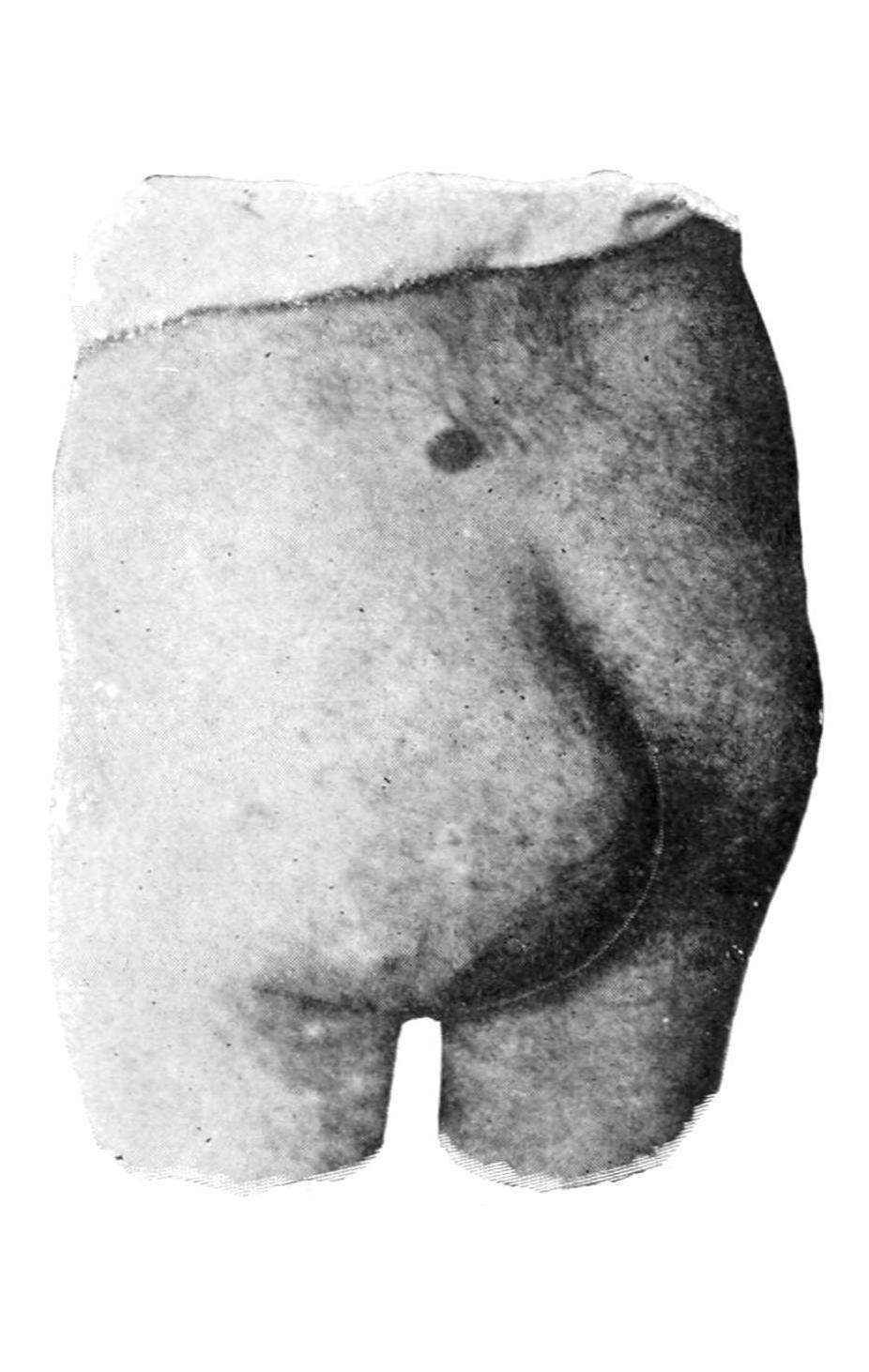
Solid dermoid escaping from pelvis. (Original.)
FIG. 73
Congenital dermoid cyst of pelvis. (Ahlfeld.)
of these. In some there is a large central cavity; others are almost absolutely solid. Thus we find dermoids in the coccygeal region, which have been variously regarded as sarcomas, adenomas, etc., which are really of origin as stated above and should be considered simply as dermoid tumors. Most of these project outwardly; some of them arise and develop within the pelvis. Dermoid cysts and tumors are also met with in connection with the rectum—sometimes between the rectum and the bladder, and between the rectum and the spine. Dermoid tumors are also found in connection with the pituitary body. These sometimes develop within the cranium, or, again, protrude perhaps into the orbit, perhaps into the pharynx.
Thyroid dermoids are tumors of great interest. They develop sometimes about the craniopharyngeal canal, which may be detected as a small canal in the macerated sphenoid bone of a fetus, and which before birth is filled with fibrous tissue. It connects with a recess in the middle line and at the base of the skull, presenting in the pharynx, which is often referred to as the bursapharyngea. It is around this recess that the lymphoid tissue known as the “pharyngeal tonsil” develops. It may be thus expected that the roof of the pharynx should be the occasional site of dermoids. It is from the pharynx or the floor of the mouth that in vertebrates the thyroid body arises. In higher forms it becomes dissociated from the pharynx and shifts its position. The thyroid body is developed around the thyroid duct, which first appears as the thyrohyoid duct, which later becomes divided, that portion in relation with the tongue becoming the thyrolingual duct, the remaining portion persisting as the thyroid duct. These are present about once in every ten subjects, according to Sutton, the canal when persistent being lined with epithelium. When the extremities of these ducts become occluded retention cysts may form. In the same way dermoids of the tongue are formed, similar to those occurring on the scalp. These are frequently mistaken for sebaceous cysts. They may be unilateral, central, or even bilateral. The lingualduct is also of interest, because it would appear that certain cases of epithelioma of the tongue arise along this duct, and perforating malignant ulcer of the tongue is thus produced. Dermoid tumors of the lingual or thyroid ducts resemble
in structure the thyroid body. The thyroid duct may also be detected in many adults running from the isthmus of the thyroid body to the posterior aspect of the hyoid bone, and surrounded by muscle tissue. Sometimes the space usually occupied by this duct is represented by a series of detached bodies known as accessory thyroids. These are not infrequently the seat of cysts, sometimes of considerable size. (The accessory thyroids often enlarge when the main thyroid has been extirpated for disease.) Thus cysts in close relation to the hyoid bone are common. Some of them grow slowly, while others grow rapidly and contain much fluid. Many of them are unilateral, and are often mistaken for enlargements of one lobe of the thyroid. Cysts growing from accessory thyroids are often filled with papillomatous masses, and are occasionally the seat of malignant degeneration.
In the omphalomesenteric duct or its remains, especially in relation with the umbilicus, we often meet with small cysts or tumors in infants and young children. When the duct is persistent it presents normal intestinal structure, and, like the appendix, possesses much adenoid or lymphoid tissue.
Another and very important form of tubulodermoids develops in connection with the branchial clefts of the neck. Congenital fistulas of the neck have been long known, but only comparatively recently understood. Of the branchial clefts it is well known that the first alone should persist, as the Eustachian tube. Occasionally, however, they fail to become obliterated, and then we have congenital tumors or cysts, which may, perhaps, not develop to appreciable size until somewhat late in life; or there may be fistulous passages opening either into the pharynx or externally, forming canals varying in length from half an inch to two inches, secreting a little fluid because lined with epithelium. When these become inflamed an abscess results. When they open externally the opening is often marked by a little tag of skin containing a fragment of yellow cartilage. These are often referred to as cervicalauricles. They open along the line of the sternomastoid muscle. The internal openings of these fistulas frequently form diverticula from the pharynx or esophagus. Thus it will be seen that dermoid cysts about the neck are principally relics
of openings or ducts, which are normal in embryonic life, but which should have been obliterated at or long before birth. Congenital fistulas, however, may be met with in the middle line of the neck, which are not to be confounded with branchial fistulas, but rather with the ducts previously described.
3. Ovarian Dermoids and Teratomas.
—These may be unilocular or multilocular cysts, usually the latter. They are lined with epithelium, and contain mostly mucoid fluid, the inner coat being practically identical with mucous membrane. Occasionally, however, the skin is furnished with hair, sebaceous glands, teeth, and even nipples. The multilocular cysts are practically an aggregation of those just described. They are surrounded by dense capsules, often attain great dimensions, and are made up of primary cysts resembling large cavities in a honeycomb-like mass, which itself is occupied by secondary cysts, and belong rather to the class of mucous retention cysts, these being occupied by still smaller ones, which are histologically indistinguishable from distended ovarian follicles. In these large tumors we find in some cases hair, in others teeth, and in others sebaceous glands, etc., the dermoid constituents being scattered throughout. As Wilms has shown, in almost every tumor of this character a projection may be found whose summit is covered with epiblastic elements, which when cut in serial transverse sections will show in its deeper portion other epithelial collections representing a feeble attempt to develop a nervous system, or lung tissue, while mesoblastic elements, like connective tissue, cartilage, and bone, appear scattered throughout, as though a very crude effort had been made to reproduce an atypical embryo.
3. Teratomas.
So far the endeavor has been to limit the term dermoidto tumors which are essentially of epiblastic formation, their location being explained on the inclusion theory of Cohnheim. There is also a still more complicated type of tumor, composed of tissues of both epiblastic and mesoblastic origin, perhaps even hypoblastic. Their
consideration belongs to that department of pathology known as teratology, which is supposed to deal especially with monsters. Strictly speaking a teratoma refers to an irregular tumor or mass containing tissues and fragments of viscera of a suppressed fetus which is attached to an otherwise normal individual. Nevertheless the term is often applied to growths which are the result of luxuriant mesoblastic development in which neither form nor member of a suppressed fetus is present.
The presence of supernumerary members is largely connected with what is called dichotomy, alluding thereby to cleavage either at the anterior or posterior end of the developing embryo. When the whole embryonic axis divides twins may be produced, but should cleavage be partial we may have a monster with two heads if it be anterior, or one with three or more limbs if it be posterior. Children born with these deformities are usually called monsters, and the study of such cases belongs entirely to teratology. But in certain tumors small portions of a suppressed fetus may develop, as, for instance, from the posterior portion of the sacrum, or within the abdomen or thorax, or upon the neck or face, which on dissection may contain a few vertebrae or processes resembling fingers associated perhaps with a structure resembling intestine or liver. This should be called a teratoma. Such tumors possess for the pathologist the greatest value. In surgery, however, they are rare, and there are scarcely two cases alike. The question of operation will often arise, as it does with supernumerary limbs, and each case should be studied upon its own merits. Sometimes they are amenable to extirpation.
Embryonal Adenosarcoma.
—Embryonal adenosarcoma is a term given to certain teratomatous tumors peculiar to renal and adrenal structure, which present peculiar characteristics in the mixture of elements which enter into their composition. At various times these tumors have been called adenoma, sarcoma, rhabdomyoma, congenital cystic kidney, etc. They have been also likened to the thyroid. They comprise a group of neoplasms, always congenital in origin, which usually appear early in life, but occasionally occur in advanced adult life. One of the most
marked specimens of this kind the writer removed from a man over fifty years of age. Most of the specimens, however, described in literature pertain to the young. On minute examination they often present a strange, mixed picture of voluntary muscle elements intermingled with epithelium arranged to imitate acinous glands, with cystic dilatations of the true kidney tissue. They often attain enormous size, and undergo such proliferation of mesoblastic elements as to resemble sarcoma. Their occurrence is to be explained only on the principles of Cohnheim’s hypothesis. When the original Wolffian body is being differentiated from the elements about it a confusion of the same with the excretory tubular beginnings, which are to empty into the Wolffian duct, occurs. Thus we have the commencement of a mixed mass which presents itself as a more or less rapidly growing tumor, in which even cartilage or other mesoblastic structures may be met with. It is scarcely possible that any two specimens should yield exactly the same microscopic picture, much depending on whether one element or the other prevail. In a few of them there may occur also a mixture of adrenal elements. Sometimes the renal structure itself is more or less distinct, and rides, as it were, upon the surface of the tumor; at other times it is entirely mixed up with it. While the condition is usually limited to one side it may be a double affection, so that the second kidney becomes useless and the patient succumbs. The only treatment is extirpation.
Teratomatous tumors are sometimes found hanging in the pharynx, attached by a small pedicle, where they may be confounded with dermoids unless carefully examined after removal. Many instances of this type of tumor are found in animals. Here no false sentiment will prevent complete examination and preservation of the specimen. They are also encountered in the sacral and coccygeal regions.
4. Tumors of Connective-tissue Type.
Lipoma.
—Lipomas, or tumors composed of fat, are the most common of the neoplasms. Their normal type is the
ordinary adipose tissue of the body, and may be divided into the encapsulatedand the diffuse, the former of which are surrounded by fibrous tissue. The diffuse lipomas are those which have no capsule, and where the pathological collection of fat merges into that normally present—in other words, they are not circumscribed.
Subcutaneous Lipomas.
—Subcutaneous lipomas are perhaps the most common of all, and are usually irregularly lobulated and encapsulated, adherent rather to the skin than to the deeper tissues. Usually but one is found in an individual, though instances of multiple lipomas are not rare. They develop sometimes to enormous size, cases being on record where the tumor has even weighed one hundred pounds. They may be met with at any point on the surface of the body. The lobules often burrow between the muscles, and those found in the palm of the hand penetrate even beneath the palmar fasciæ. They are sometimes markedly pedunculated, and often hang by a small stem. The diffuse subcutaneous lipoma is most common about the neck, in the groin, and in the axilla.
Subserous Lipomas.
Subserous lipomas are mostly retroperitoneal, and large tumors of this character, mistaken for ovarian, have been successfully removed by operation. They also occur in the hernial canals and spaces. They develop beneath the peritoneum covering the intestines, and in this location give rise occasionally to intussusception. Here in their pathological development they have the general form and significance of appendicesepiploicæ.
Subsynovial Lipomas.
—Subsynovial lipomas occur about various joints and tendon sheaths; within the knee they assume a distinctive type which has been called lipomaarborescens, where they take on a dendritic appearance and arrangement. Submucous lipomas are rare. Intermuscular fatty tumors are occasionally met with, an interesting variety being that which develops between the masseter and buccinator muscles. Intramuscularforms rarely occur, as well as a variety known as parosteal, which arises in connection with the periosteum. Fatty tumors also occur within the spinal dura, as well as outside of it within the spinal canal, and more or less lipomatous alterations are common in connection with spina bifida.
Lipomas are ordinarily easy of recognition, save when deeply located. The subcutaneous forms are intimately related with the overlying skin, and have a dough-like consistence which is usually pathognomonic. Tumors, suspected to be fatty, in the middle line of the back or cranium are always to be viewed with suspicion, as they are often connected with congenital meningeal protrusions.
An encapsulated lipoma when thoroughly removed will not return.
Mixed forms of fibrous and fatty neoplasm are occasionally seen, and are referred to as lipomafibromatosumor fibromalipomatosum, according as one or the other tissue predominates. These growths are innocent in their character, but call for thorough extirpation. They frequently give rise to considerable discomfort or pain, and are called lipomadolorosa.
Fibroma.
—Fibromas are tumors composed of fibrous tissue, which, when of pure type, are found to be not so common as was formerly supposed, the majority of tumors hitherto roughly grouped as fibromas containing either muscle tissue or sarcomatous elements, which takes them out of the category of pure fibroma. A typical fibroma is ordinarily dense, and is composed of wavy bundles of fibrous tissue whose cells are long and slender and closely packed together, the mass being permeated by distinct bloodvessels.
Fibroma occurs most commonly in the ovary, the uterus, the intestine, the gum (epulis), in nerve sheaths, and in the skin in the form of so-called painful subcutaneous tubercles and molluscum fibrosum. There is also a fibrous tumor of the skin, known as keloid, sustaining to fibroma the same relation that exists between exostosis and osteoma.
Painful subcutaneous tubercle is a sample of pure fibroma in the shape of a small, flattened, pea-like tumor which never attains great size. It is situated loosely in the subcutaneous structure and may form a visible prominence. Insignificant as it would thus appear, it becomes the seat of exasperating pain, particularly when touched or handled, which may radiate to considerable distances. The etiology of these growths is unknown.
In the ovary, the uterus, the intestine, and the larynx true fibrous tumors are pathological curiosities rather than common lesions.
Keloid of external ear: a, dense tissue of skin; b, fibrous connective tissue; c, epidermis. (Klebs.)
Epulis.
—Epulis means any tumor growing upon the gum. The term was formerly applied in an indistinct and too comprehensive way, although it is still retained in literature. But pure fibromas do spring from the fibroösseous structure of the gum and alveolar process. They are covered with the gingival mucous membrane and seem to spring from the periodontal membrane. They seldom attain large size, and then only by neglect. By the pressure of such tumors teeth
may be separated and distortion of the mouth produced. They should be promptly extirpated.
—Keloid is a fibrous neoplasm arising mainly in cicatricial tissue, which is essentially fibroid in structure. It is a neoplasm which often follows the general outline of the scar in which it grows, consists in elevation of the surface, ordinarily quite smooth, sometimes of a delicate pink from the dilated vessels which it contains. Keloid is the bête noir of surgeons, as it frequently complicates and disfigures scars which have at first been satisfactory, and since it indicates a condition which it is discouraging to deal with, because when it is removed there is usually recurrence of growth within a few months after cicatrization. It often occurs in stitch-hole scars and upon the site of extensive burns, and may be observed after puncture of the ears for ear-rings, and has also been observed in scars left by smallpox, acne, etc. It is more prevalent in the colored race than in the white. In negroes multiple keloid tumors are often seen, occasionally in large numbers. Their explanation is unknown, and it may be that some trifling injury has preceded each individual tumor (Fig. 74).
The treatment of keloid will be considered in the chapter on the Surgical Diseases of the Skin.
Keloid. Desmoids.
This term has been applied to tumors of a certain clinical type which arise from the fibrous structures, usually of the abdominal wall, and produce neoplasms like the fibromas of other parts of the body. The use of the term should be restricted to those tumors which proceed primarily only from muscles, tendons, and aponeuroses, or perhaps from ligamentous and periosteal tissues. These tumors are usually single, attain sometimes considerable size, grow slowly, rarely involve other structures, and not infrequently develop to such an extent as to encroach upon either pelvis or the abdomen, or both. They have been known to attain to the weight even of ten pounds or more. They are usually more or less encapsulated, and are firm and dense in structure. Under the microscope they have the general appearance of cellular fibroma. Sarcomatous elements may be met, while they occasionally undergo
cystic degeneration. Their occurrence may be explained, at least in some instances, on the embryological theory of Cohnheim.
Multiple enchondromas.
The treatment of desmoids consists in their complete extirpation. They should not be allowed to attain large size because their removal may entail a serious weakening of the abdominal wall. There should be such plastic rearrangement of abdominal protecting membranes as to reduce the resulting weakening to a minimum.
Psammoma.
—Psammoma is a term applied to a form of hard fibroma met with in the dura mater, in which there has occurred a petrefaction of some of the cells—i. e., a deposition of calcareous salts, which gives it a gritty or sandy appearance.
Chondroma.
The true chondroma is a tumor composed of hyalinecartilage. It occurs in the long bones, usually in relation with epiphyseal cartilages, and is often noted during the earlier years of life. While it is usually a solitary tumor, multiple chondromas are often seen, especially upon the hands. These tumors are often encapsulated and form deep hollows, in which they rest. Unless pressing upon nerve trunks they are painless and slow of growth. They are exceedingly dense and hard, and ordinarily immovable. Mucoidsoftening(i.e., cysticdegeneration) is common, and the softened areas may give rise to fluctuation. There may be coincident calcification or ossification in any of these growths. It is noted as a curious circumstance by Sutton that their tissue resembles histologically the bluish, translucent, epiphyseal cartilage seen in progressive rickets.
To the small local hypertrophies of cartilage which are seen especially about joints, about the laryngeal cartilages, and the triangular cartilage of the nose, are given the term ecchondroses. They are most common in the knee in connection with rheumatoid arthritis, and occur as prominences along the margins of the joint cartilage. They may project to such an extent as to be detached by accident, after which they become movable and floating bodies in the joints. Many of the floating cartilages or bodies found in joints are detached ecchondroses, which may be smoothed off by attrition, and may be found singly or multiple, even several hundred existing in one joint.
Chondromatouschangesas occurring in sarcomatoustumorshave been alluded to. It seems to be easy for connective tissue to form hyaline cartilage, and mixed tumors may thus be seen in connection either with sarcoma, fibroma, or other forms.
Treatment.
—The treatment of chondroma is solely operative. Unless the integrity of a member or a limb be compromised, such a tumor can usually be shelled out from its location, but requires that the matrix be completely extirpated; all of which may call for the use of powerful bone instruments. At other times amputationis the only measure which may relieve from deformity, pain, and disability. The ecchondroses occurring within joints necessitate incision and evacuation, with the most rigid aseptic precautions, with or without drainage. When practised according to modern technique this is almost invariably successful. In former times many lives were lost because of septic infection.
Osteoma.
—Under the head of nomenclature I have already endeavored to distinguish as between exostosis, or irregular bone outgrowth, and osteoma, as a distinct tumor which is composed of bone tissue, with the subvariety odontoma, or tumors of dental origin and structure. Osteoma is regarded by some as ossifying chondroma, for it is nearly always found near epiphyseal lines, and is always covered by hyaline cartilage when thus found. Nevertheless it is not invariably such. We speak of compactor ivory osteoma and of a cancellous form. The former is identical with the compact tissue of the shafts of long bones, and may occur anywhere, but is most common about the cranium, at the frontal sinus, the external meatus, and the mastoid process. Osteomas growing into the frontal sinus of oxen form large, lobulated, bony masses, sometimes weighing several pounds, and as dense as ivory. Some of these tumors growing into the cranial cavity have been regarded as ossified brains. Osteomas in connection with the external auditory meatus may partially obscure this channel and cause deafness. They constitute ivory-likegrowths, which sometimes defy the finest steel instruments with which the surgeon can supply himself.
Cancellous osteomas grow in the cranium as well as in the long bones, and, like the compact forms, only occasion pain by pressure upon nerve trunks.
Exostoses.
—Exostoses are classed by Sutton as—
1. Those formed by ossificationoftendonsand their attachments. There should be excluded from this group such natural or evolutionary processes as the supracondyloid process, the third trochanter of the femur, etc. Over or around such exostoses bursæ will form to mitigate as much as possible the effect of friction. Such an outgrowth is known as an exostosis bursa; it is most frequently seen on the inside of the femur immediately above the knee.
2. Subungualexostoses, occurring usually beneath the nail of the big toe.
3. Exostoses due to calcification of inflammatory exudations, including the rare condition known as myositisossificans.