Agriscience programs vary nationwide and most have undergone extensive curricular changes within the past decade. Many include advanced placement-type coursework, such as veterinary science. While teaching agricultural education at Greenwood High School in Millerstown, Pennsylvania, Dr. Baker searched for materials to be used in a new veterinary science course. After a futile hunt, and hearing similar concerns from other instructors, Dr. Baker teamed with Dr. Lawhead, a practicing veterinarian who served the local area where she taught, in an effort to author a veterinary science text that was both student and teacher friendly.
The authors believe that two of the most useful features in this book are the “A Day in the Life” of a veterinarian, coupled with the “Clinical Practice” chapter features. These two elements tie the real-life work of a veterinarian, which can have less than desired outcomes, with the technical and, sometimes, dry and dif cult text material. Therefore, the next time a student says, “I want to be a veterinarian,” a venture into Introduction to Veterinary Science will provide the learner with a realistic preview of both veterinary work and the academic rigor needed to achieve success in the profession.
Simply put, the goals of this text are to afford learners a base knowledge of veterinary science by moving through topics ranging from the cell to surgery, and to provide a view of the practice of veterinary medicine through the eyes of an experienced practitioner. Chapters 1 and 2 begin the text with a comprehensive investigation of cells and tissues. Following chapters examine the musculoskeletal, circulatory, respiratory, renal, digestive, reproductive, nervous, endocrine, and immune systems. The basic physiology learned in the beginning of the text is then applied in concluding chapters covering nutrition, species differentiation in nutrition, principles and prevention of infectious disease, disease classi cation, zoonotic diseases, disease diagnosis, and surgery.
NEW TO THIS EDITION
• Additional hands-on activities that use easyto- nd materials have been added to the chapters.
These new activities will help instructors reinforce student learning using a variety of applications.
• The new safety chapter provides guidelines to help teachers ensure student safety in the classroom and eld laboratories, while another new chapter further explores veterinary careers.
• Technical material has been further explained by the author, Dr. James Lawhead. These expanded and updated explanations will help students grasp more advanced material.
• Additional photos and gures bring the veterinary practice into the classroom, helping to keep students engaged.
• The new edition discusses the most current technology used in veterinary practice, providing a look into recent advances in the eld of veterinary medicine.
EXTENSION TEACHING/LEARNING MATERIALS
Instructor’s Companion Website
The Instructor Resources are now available on the companion website. Updated for the third edition, this robust suite of teaching resources includes the following components to help minimize instructor prep time and engage students:
• Instructor’s Guide to Text—The Instructor’s Guide provides answers to the end-of-chapter questions and additional material to assist the instructor in the preparation of lesson plans.
• PowerPoint—Chapter outlines with images for each textbook chapter.
• Computerized Test Bank in Cognero—Hundreds of modi able questions for exams, quizzes, inclass work, or homework assignments, in an online platform.
• Image Gallery—Hundreds of images from the textbook that can be used to easily customize the PowerPoint outlines.
Each chapter in the textbook begins with clear educational objectives to be learned by the student in the reading, a list of important key terms, and an introduction overview of the chapter content.
CHAPTER 1
Basic Cell Biology
Objectives
Upon completion of this chapter, you should be able to:
■ Explain the molecular makeup of cells.
■ Identify the basic structures of the cell and their corresponding functions.
■ Review the basic function of the cell.
■ Describe the process of protein synthesis.
Key Terms
■ Discuss mitosis and its clinical signi cance in diseases such as cancer.
■ Detail meiosis in mammalian reproduction.
■ Connect cellular parts and function to clinical veterinary practice.
Introduction
The cell is the basic structure of animal life. However, the cell contains other structures and molecules. Cells conduct many functions and are also able to reproduce. Animals not only have millions of cells that comprise
A Day in the Life ADR—Ain’t Doin’ Right…
I remember the day in veterinary school when our stethoscopes arrived. The air lled with excitement as we listened to our own heartbeats. This instrument became a necessary tool in everyday life as I began to examine animals. I must admit I felt cool walking around the hospital in a white lab coat with a stethoscope draped around my neck! It seems like yesterday, even though more than a few years have passed.
Several months ago I examined a cow that was ADR—ain’t doin’ right. As I walked into the pen, I could see she obviously wasn’t feeling well at all. She appeared quite droopy, had lost a lot of weight, and had developed a swelling under her jaw. During the physical, I listened to her heart. It sounded like the noise from a washing machine in midcycle. The heart made a sloshing sound with every beat. Using the stethoscope, I diagnosed hardware disease . The cow had eaten a piece of metal that migrated from the stomach and lodged close to the heart. The location and structure of the heart provided me with the information necessary to interpret the symptoms of this disease. Hardware disease is often found during my appointed rounds. The next diagnosis is not.
This week, Dr. Deppen and I were both doing evening small animal appointments at the office. It was snowing heavily and we were hoping to nish at a reasonable hour. Dr. Deppen was seeing Lucky, a 12-yearold Schnauzer mix that had a history of having what the owners thought was a seizure. She detected that the dog’s heart rate was too slow and the rhythm was very irregular. I had a chance to listen to the dog’s heart as well and agreed that we should do more tests to detect the underlying problem.
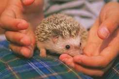
The author James Herriot portrayed veterinary work in his best-selling collection of stories, All Creatures Great and Small. Times have changed considerably since Herriot practiced. Much more information and sophisticated medicines and techniques are now readily available. Still, I cannot possibly be an expert on all animals. Last year our of ce received a call from a local school. The sixth grade class mascot, Sonic the hedgehog, had a sore foot. In this case, my experience with hedgehogs was limited to reading just one obscure
journal article. I had never even met one in real life. Therefore, I advised the teacher of my lack of experience but agreed to examine Sonic. Sonic arrived at the of ce in a cage (Figure 4–1). He looked just like a miniature porcupine. Because hedgehogs are nocturnal animals, Sonic was apparently taking his afternoon nap when he arrived at the of ce.
I disturbed him as I tried to examine his leg. Sonic jumped and snorted in an attempt to scare me. To be honest, it worked! His prickly quills were quite sharp.
My assistant and I then put on thick leather gloves and proceeded with the examination. Sonic countered with another protective measure. He rolled himself into a tight ball, so tight his legs were completely hidden. I referred to the journal article for help.
Following the recommendations, I anesthetized Sonic with an inhalant anesthetic. We placed him in the large clear mask. The anesthetic was slowly delivered with every breath. Finally Sonic relaxed enough so I was able to have a more thorough look. Once Sonic’s leg was exposed, the problem was quite obvious. The rags that Sonic used as a nest had tattered edges with loose strings. One of these strings had wrapped tightly around his foot and stopped the circulation. The foot had turned dark and was oozing. All mammals rely on circulation to maintain their bodies. What happened to Sonic’s foot when the blood supply was stopped?
the body but also many different cell types. The combination of these cell types makes an animal function. This chapter will discuss the structure of cells, and how they work.
Each chapter features “A Day in the Life” of a veterinarian vignette that relays James Herriot–type stories with relevance to clinical practice and the real-life work of a veterinarian.
55 Chapter 4 The Circulatory System
FIGURE 4–1 A hedgehog.
attach to these vertebrae, forming a sling that supports internal organs.
The sacrum, a group of three sacral vertebrae, fuses to support the pelvis (Figure 3–14). In addition, the sacrum articulates with the last lumbar vertebra and the rst caudal vertebra. The sacrum then joins with the pelvis, allowing the hind limbs to support the weight of the body. This connection can be damaged. The pelvis may split away from the sacrum when dogs and cats are hit by cars (HBC). During this type of accident, fracture of the pelvis itself is also common. Very painful lameness often results from a split pelvis or pelvic fracture. Many of these fractures heal if the animal’s activities are restricted. In severe cases, surgeries may be required.
The nal group of vertebrae is called caudal. These small vertebrae comprise the tail. As mentioned, the numbers of vertebrae vary among species and within
41 Chapter 3 The Musculoskeletal
System
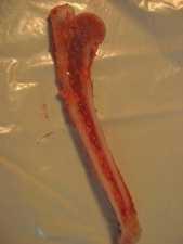
a species. The typical dog has 20 caudal vertebrae, but this can range from 6 to 23. The appendicular skeleton includes the bones of the forelimbs and hind limbs. A study of this part of the skeleton provides a clear examination of comparative anatomy. Although the same anatomic terms are used for all mammals, great differences exist in the numbers and sizes of bones in the mammalian appendicular skeleton. For instance, a dog has four or ve toes, whereas a horse has only one. The forelimb, or thoracic limb, does not have a bony connection to the axial skeleton. The scapula, or shoulder blade, lies at against the rib cage (Figure 3–15). The scapula connects to the axial skeleton with a group of muscles. This attachment allows the scapula to move over the rib cage. This rotation ranges as high as 25 degrees in animals such as cats while running. This exibility is also useful in cats as they land after a jump. As the cat falls, it extends its front legs fully at both the scapula and the elbow. As the front feet hit the ground, the elbow exes and the scapula rotates. The cat makes this very coordinated act look quite graceful. Clinically, this is of signi cance when cats fall from extreme heights. In large cities, this happens often as cats tumble from balconies or windows of tall buildings. In high-rise syndrome, the falling cat rarely breaks a leg; however, it will often break its lower jaw. The high speed of the falling cat forces the jaw to contact the ground.
Each chapter contains combinations of charts, illustrations, photographs, radiographs, and the like that help to illustrate and enhance the concepts presented.
5 Chapter 1 Basic Cell Biology
allowing veterinarians to diagnose what speci c organism is causing the sickness.
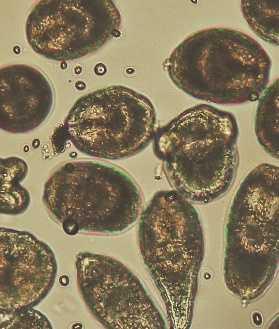
Nucleic acids provide plans for the differing construction of proteins. Nucleic acids are fabricated with a series of nucleotides. The nucleotides are made up of a ve-carbon sugar, a phosphate group, and a nitrogen-containing base (Figure 1–5). Ribonucleic acid (RNA) claims ribose as its sugar, whereas deoxyribonucleic acid (DNA) has deoxyribose as its sugar. There are four different bases for RNA and DNA (Table 1–1).
Notice that the bases are the same except for thymine and uracil. The order of base combination determines what amino acids are used to make proteins. This information is stored in the cell’s genetic material. Both DNA and RNA have a backbone of sugar alternating with phosphate. The nitrogenous bases are attached to this backbone. In DNA, a double-stranded molecule is formed as the bases are loosely bonded together. The molecule has a twisted structure, which is described as a double helix (Figure 1–6). The bases join, speci cally, thymine to adenine and cytosine to guanine. Later in the chapter, a process of transcription will be described, in which the sequence of DNA nitrogenous bases is converted to a molecule of RNA. In this situation, adenine in the DNA molecule bonds to a uracil base of RNA. The sequence of nitrogenous bases is used to de ne the amino acids used in protein synthesis. A group of three nitrogenous bases is the code for a speci c amino acid. The order of the nitrogenous bases makes up the genetic code of the animal. Each gene provides the code for one peptide chain.
FIGURE 1–5 Chemical structure of a nucleotide.
Table 1–1 RNA and DNA Bases
Thoracic Vertebrae
Rib Sternum Lumbar Vertebrae
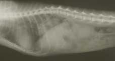
FIGURE 3–13 Radiograph of a cat, showing the thoracic and lumbar spine. Ribs and sternum are also visible.
Pelvis
Lumbar Vertebrae
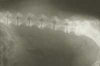
FIGURE 3–14 Radiograph of the lumbar spine of a dog. A portion of the pelvis is also visible. This dog is showing an age-related change called spondylosis. In spondylosis, bone spurs are formed that can eventually bridge between vertebrae.
FIGURE 3–15 The scapula. Spine
cat from that of a horse. Having muscles closely associated with the skeleton provides movement of the bones at a joint. The movement of bones allows locomotion and function of the animal.
The strength of bones also protects more fragile tissues. The rib cage gives protection to the heart and lungs, whereas the skull protects the delicate brain.
Bone acts as a reservoir for calcium and phosphorus. In times of need, the minerals are moved from the bone and sent into the bloodstream. Excess minerals can be stored in the bone. Calcium plays an essential role in muscle contraction and enzyme activity. Phosphorus is necessary for energy metabolism within the cell. Bone, in response to several hormones, maintains a tight regulation on the blood level of these minerals. These hormones, calcitonin and parathyroid hormone, will be discussed in much greater detail in Chapter 10. The long bones are present in the legs (and arms in humans). The femur and humerus are classi ed as long bones. They have a dense outer shell and a hollow shaft. Bone marrow is made in this hollow center, the medullary cavity. Bone marrow in turn produces blood cells.
BONE STRUCTURE
Objective
■ Detail the Structure of Bone
Splitting a long bone along its length shows the typical structure of bone (Figure 3–3). The outer shell is composed of dense or compact bone. The term cortical bone is also used for this region. The greater the forces placed on a bone, the thicker this layer will be. In the femur, this compact bone is thickest in the middle of the shaft, where greatest strain occurs. Within compact bone lies a more loosely arranged bone, called spongy or cancellous bone. Spongy bone is found within the long bones but not inside the at bones of the skull or pelvis. It only lls the ends of these long bones. Spongy bone is made up of tiny spicules and plates of bone. The spicules look random but are actually arranged to maximize strength. The spongy arrangement keeps the weight of the bones much lighter than that of a solid bone of the same dimension. The medullary cavity is located in the hollow center of the shaft. The bone marrow lies within the medullary cavity and the spaces of the spongy bone. As mentioned earlier, bone marrow produces blood cells. Bones are covered with a thin connective tissue called the periosteum. The periosteum blends into tendons and ligaments, binding them to the bone. The periosteum has an extensive blood and nerve supply. Hence trauma to the periosteum is quite painful. The portion of bone within the joint is covered with cartilage and not by periosteum. This articular cartilage
Articular Cartilage
Bone Marrow Cancellous or Spongy Bone
Medullary Cavity
Compact Bone Tissue Endosteum
Chapter 3 The Musculoskeletal System
Proximal Epiphysis
Physis Metaphysis
1–12
Periosteum
FIGURE 3–3 A. Illustration of bone structure. B. Photograph of the internal structure of bone.
provides protection as the bones move against one another within a joint. The open spaces within bone are covered with a similar connective tissue, the endosteum. Both the periosteum and endosteum provide cells necessary for the repair of damage.
Extracellular Fluid Cytoplasm
enzyme begins at a speci c series of bases (thymine, adenine, cytosine) called a promoter. The RNA polymerase moves along the length of the DNA molecule, creating a complementary strand of RNA. The RNA bases are added in the speci c order that bonds to the bases of the DNA. The corresponding bases were discussed earlier in the chapter. This process continues until the polymerase reaches a terminator series of bases (adenine, thymine, thymine). The mRNA is released and the DNA helix reconnects.
Extracellular Fluid Cytoplasm
(Figure 1–12). During endocytosis, the cell membrane wraps around the particle, pinches off, and moves into the cytoplasm as a vacuole. Lysosomes then join with the vacuole, providing the enzymes necessary to break down the particle. The smaller fragments produced are then released into the cell.
In cells producing protein, the opposite process occurs. In exocytosis, a membrane-bound sac containing the protein joins with the cell membrane and releases it into the ECF (Figure 1–13). These sacs are produced within the Golgi apparatus. In intestinal cells, fat droplets can be taken into the cell through endocytosis. The vacuole is transported across the cell and released into the bloodstream by exocytosis.
PROTEIN SYNTHESIS
Objective
■ Describe the Process of Protein Synthesis
As mentioned previously, every cell contains all the genetic material of the animal. The expression of certain genes produces speci c proteins that allow cell specialization. Protein synthesis begins within the nucleus on the basis of the DNA structure. During transcription, information within the DNA is transferred to a strand of messenger RNA (mRNA) that moves into the cytoplasm.
An enzyme called RNA polymerase binds to DNA, causing a separation of the double-helix strands (Figure 1–14). This pulling apart exposes a gene. The
Each chapter is further enhanced by the addition of repeat objectives to aid in student comprehension.
FIGURE
Endocytosis: A large particle is engulfed by the cell membrane and brought into the cytoplasm within a vacuole.
FIGURE 1–13
Exocytosis: A membrane-bound sac joins with the cell membrane to release the particle.
FIGURE 1–14 Transcription of mRNA: RNA polymerase separates the strands of DNA and creates a strand of mRNA coded by the nucleotides of the DNA molecule.
88 Unit 1 Comparative Anatomy and Physiology
SUMMARY
Being able to identify respiratory structures and their associated functions, from the nose to the lungs, allows veterinarians to diagnose and treat such disease conditions as pneumonia and roaring. Moreover, respiratory
REVIEW QUESTIONS
1. De ne any 10 of the following terms: respiration palpated endotracheal tube inspiration expiration cyanosis pneumonia pleural friction rub contagious roaring heaves bronchodilators
2. True or False: Mucus lines the epithelial tissue in the nostrils.
3. True or False: The cartilage rings of the trachea are shaped like an O
4. The is the common area shared by the nose and throat.
ACTIVITIES
Materials needed for completion of the activities: stethoscope balloons Y-shaped polypropylene connecting tubes
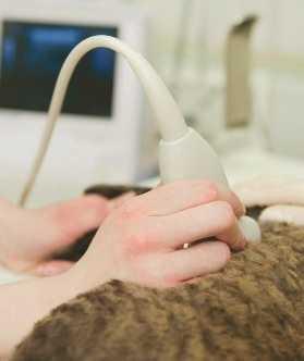
1. Use the stethoscope to listen to normal lung sounds. Have the “patient” take deep, slow breaths. The patient should breathe quietly, not making noise through the nose and mouth. The stethoscope can detect these noises. Listen to different areas on the chest, from both the front and the back.
2. Take two identical balloons and in ate them to different sizes. Slip a balloon onto an end of Y-shaped polypropylene connecting tubes. Do not
rate provides a key piece of information to practitioners when assessing the overall health of animals. The status of the respiratory system affects the breathing and therefore the total health of animals.
5. The human larynx is sometimes called the
6. The trachea branches into two
7. Gas exchanges occur in the smallest openings of the respiratory system. These openings are called the
8. The muscles between the ribs are called the
9. Name the re ex action that occurs when there is an irritation in the nose.
10. What substance lines the lungs, making them easier to in ate?
11. What controls the rate of respiration?
12. What is the normal respiration rate for a dog?
13. What plays a more signi cant role in the control of respiration, oxygen, or carbon dioxide?
14. What medical tool is used to evaluate breathing?
15. What species can develop a condition referred to as roaring?
release the balloons yet. Plug the third opening of the Y piece. Hypothesize what will happen when the balloons are released. Will the large balloon de ate and ll the smaller balloon to equalize the size? Or will the smaller balloon de ate into the other balloon? Surfactant prevents this problem from occurring between alveoli. Even though the alveoli may be of different sizes, the pressure in each is similar. Without it, the small alveoli would de ate.
3. Observe the respiratory rates of classmates and pets or livestock. Compare to the normal rates listed in Table 5–1.
A chapter summary highlights the topics that have been presented, and the end of each chapter is also followed by a series of review questions and student activities.
The new chapter on careers investigates occupations in veterinary science, including veterinary technicians, veterinary assistants, private practitioners, and veterinary specialists.
methods for prevention of rabies, and responds to questions about rabies from other public health professionals and the public.
Veterinary Surgeon
Dr. David Sweet graduated in 1989 from the University of Pennsylvania School of Veterinary Medicine. Following his graduation, Dr. Sweet pursued further training as an intern at the University of Pennsylvania and a surgical residency at the North Carolina State University. Following that training, Dr. Sweet accepted an instructorship at Washington State University and returned to the University of Pennsylvania as an assistant professor. During his training, Dr. Sweet met the rigorous quali cations necessary to become a diplomate in the American College of Veterinary Surgeons. This honor earned by Dr. Sweet distinguishes him as a surgical specialist.
Dr. Sweet works at a referral practice. The center employs veterinary specialists in many elds, including surgery. The veterinary practice provides a service that allows private practitioners to refer dif cult cases for more specialized treatment. Dr. Sweet performs both soft tissue and orthopedic surgery (Figure 21–4). He performs many complicated and dif cult surgeries. As with all veterinarians, he attends continuing education conferences to learn new procedures and information.
EDUCATIONAL REQUIREMENTS FOR VETERINARY CAREERS
Objective
■ Explain the Educational Requirements for a Variety of Veterinary Careers
Veterinary technicians must complete either a twoyear associate degree or four-year bachelor of science degree program. Further, they must pass a state licensing exam. The number of institutions offering such coursework has grown signi cantly over the past several years. Veterinary assistants are not required to complete any formal classes. However, increasing numbers of technical schools and community colleges offer veterinary assistant programs.
Both technicians and assistants help the veterinary practice by performing a wide range of tasks (Figure 21–5). These individuals may greet patients, keep records, bill clients, and restrain animals, as well as feed, exercise, and provide basic health care for patients. The responsibilities vary from employer to employer with technicians performing more technical duties. Numbers of available jobs for veterinary assistants and technicians will continue to grow with the demand for veterinarians.
Level of degree separates veterinary assistants from veterinary specialists. Almost 30 programs grant degrees in veterinary specialties. Most of these programs deliver master’s and doctorate degrees, although a few award associate and bachelor’s degrees.
Specialists may provide such supportive services as nutrition counseling, ration balancing, or radiology expertise to veterinary clinics. Conversely, other specialists may be employed in academia, where they perform research or extension duties in veterinary-related
355 Chapter 21 Careers and Decision Making in
Courtesy of Dr Cathy Hanlon.
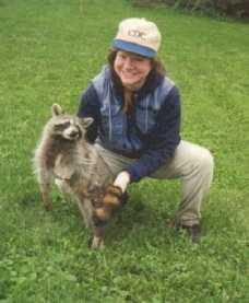
FIGURE 21–3 Dr. Hanlon working with a sedated raccoon that had been captured in a live trap.
Courtesy of Dr David Sweet.
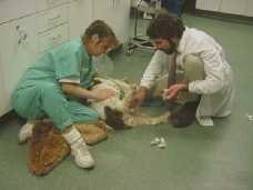
FIGURE 21–4 Dr. David Sweet, with assistance of registered veterinary technician Michele Antoch, examines a surgical incision on a dog.
A BO U T THE AUTHOR S
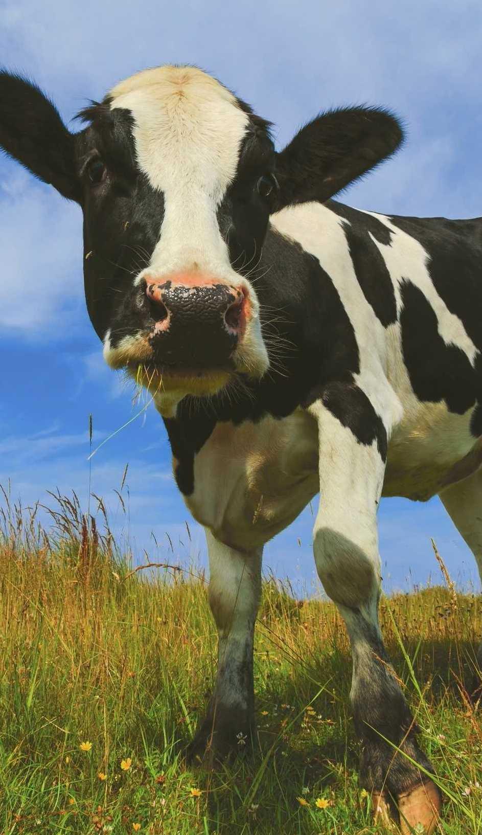
Dr. James Lawhead is a veterinarian in a private mixed animal practice located in Millerstown, Pennsylvania. As lead partner, he works primarily with dairy cattle, dogs, and cats. Dr. Lawhead joined this practice in 1987 following graduation from the University of Pennsylvania School of Veterinary Medicine. He gained acceptance to veterinary school following completion of his bachelor’s degree at Juniata College.
Dr. Lawhead has a special interest in dairy cattle nutrition, providing nutritional services to a number of his clients. Dr. Lawhead enjoys teaching as well and actively supports local school districts with lectures and demonstrations.
Dr. MeeCee Baker owns Versant Strategies, an agricultural and rural affairs rm that serves clients in
Harrisburg, Pennsylvania, and Washington, D.C. In addition, Dr. Baker serves as an adjunct professor at the North Carolina State University. She earned both her bachelor’s and doctorate degrees from Pennsylvania State University in agricultural education and a master’s of science degree from the University of Delaware in agricultural economics. Dr. Baker was the rst woman to be elected president of the National Vocational Agriculture Teachers’ Association (now known as the National Association of Agricultural Educators). Formerly, she taught high school agriculture and worked in the executive of ce of the Pennsylvania Department of Agriculture as coordinator of agricultural education. Dr. Baker lives on her family beef farm with her husband, Jim, and daughter, Libby.
A CKNOWLE D GMENT S
Although only two authors are listed for this text, the number of people responsible for the nal product is quite large. The authors would like to thank all of those people who supported and contributed to the text, especially the Cengage Learning Team. Cengage Learning deserves special recognition for faith in the authors.
We would like to thank all the veterinarians and staff at Millerstown Veterinary Associates for their assistance and contributions. Their help in obtaining case material and photographs for the text was invaluable. Likewise, we appreciate the support of the clients who encouraged the use of their case material for the text. Special thanks are in order for Leesa Landis, Dr. Robert Mikesell, and Krista Pontius for their long hours of technical help in putting together the text. Caleb Wright, a Versant intern and newly minted agricultural education teacher, helped to
freshen objectives and questions for the third edition. We appreciate the use of reference material supplied by Mechelle Regester. The veterinary science students at Greenwood High School completed activities, lessons, and accompanying assignments to help netune the text and ancillary material. We appreciate their thoughtful consideration.
In addition, we would like to thank Dr. David Sweet, Dr. Cathy Hanlon, Dr. Abby Maxson Sage, and Dr. Lawrence Hutchinson for their contributions of photographs and support to the project.
Having input from experts in various elds helped to strengthen the core material of the text. Our utmost thanks to Dr. William Bacha Jr., Dr. Linda Bacha, and Dr. Arthur Hattel for the photographic material provided. The histology and pathology photographs are a tremendous bene t to the text.
Comparative Anatomy and Physiology Unit I
CHAP TER 1
Basic Cell Biology
Objectives
Upon completion of this chapter, you should be able to:
■ Explain the molecular makeup of cells.
■ Identify the basic structures of the cell and their corresponding functions.
■ Review the basic function of the cell.
■ Describe the process of protein synthesis.
Key Terms
■ Discuss mitosis and its clinical signi cance in diseases such as cancer.
■ Detail meiosis in mammalian reproduction.
■ Connect cellular parts and function to clinical veterinary practice.
anesthetize antibiotics cancer lipid hydrophilic hydrophobic glucose diabetes glycogen enzymes antibodies exocytosis metabolism anabolism catabolism homeostasis diffusion osmosis active transport endocytosis benign malignant pathologists
Introduction
The cell is the basic structure of animal life. However, the cell contains other structures and molecules. Cells conduct many functions and are also able to reproduce. Animals not only have millions of cells that comprise the body but also many different cell types. The combination of these cell types makes an animal function. This chapter will discuss the structure of cells, and how they work.
A Day in the Life There Just Never
Seems to be a Typical Day
I headed to the of ce with the thought of doing only cow work on this particular day. However, those plans were short lived. Shortly after I arrived at work, two nervous owners walked through the door with their Labrador retriever. Poor Jake had just been run over by the owner’s car! Amazingly, Jake was doing very well, although he was a bit excited. Apart from a couple of cuts on his jaw, he was ready to go home and play.
Then, at my rst farm call of the day, the farmer wanted me to look at his dog, Millie. Millie had a grapefruit-size lump under her jaw. The lump felt like it was full of uid. I asked him to bring Millie to the of ce so I could work on her there. I nished my farm calls and headed back to the small animal clinic.
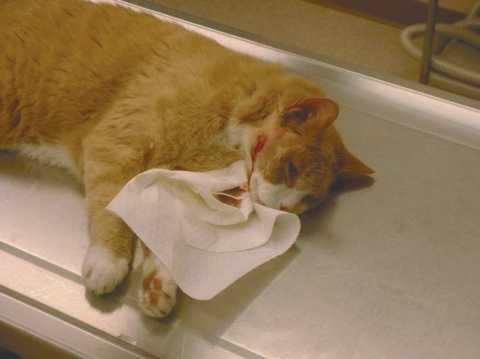
Once there, I anesthetized Millie and made an incision into the skin. Pus owed from the lump (Figure 1–1). I ushed the large pocket left behind and started Millie on a course of antibiotics , drugs that ght bacterial infections. Although I do not know why it started, I do know Millie was ghting an infection with her body’s cells.
Next I had the opportunity to remove a tumor from Penny, a 12-year-old cocker spaniel. Last week I gave Penny a physical examination and administered blood tests. Penny appeared healthy, and we elected to do
CELL MAKEUP
Objective
■ Explain the Molecular Makeup of Cells
Cells and their structures are composed of molecules. Biochemistry is the study of these molecules in living creatures. One goal of this chapter is to identify the differing types of molecules and their properties.
Lipids or fats combine hydrogen, carbon, and oxygen in a form that is poorly dissolvable in water (this is why fat oats to the top of water). Fat consists of a molecule of glycerol and three fatty acid molecules (Figure 1–2). Fats are stored in the cells of the body as a source of high energy.
Phospholipids are similar but have only two fatty acid groups and a phosphate group (PO4). This is signi cant because one end of the molecule is attracted to or soluble in water (hydrophilic) and the other end is repelled by water (hydrophobic). These characteristics of phospholipids are important in the structure of the cell membrane.
Carbohydrates supply energy and provide structure within the cell. Monosaccharides are the simplest
. . .
surgery. The surgery went well, and I was able to remove the entire lump.
In private practice, cells affect me every day. Today I saw Millie’s cells attacking the bacteria in her neck. Penny, on the other hand, had cancer-causing cells dividing uncontrollably. To understand how mammals work and how to treat them, I rst had to learn how cells function.
of these molecules. They possess the basic structure of (CH2O)n (Figure 1–3). In this formula, n describes the number of carbon atoms in the molecule. The genetic material in the cell has the ve-carbon sugars ribose and deoxyribose. Glucose (blood sugar), a six-carbon sugar, is used for energy in the cells. The amount of glucose in blood is routinely monitored. If there is too much or too little glucose in the blood, the animal will not function normally. In diabetes, the blood sugar increases to very high levels, but the animal does not utilize it properly. Diabetes requires treatment to lower the blood sugar.
Polysaccharides are composed of many monosaccharides. One example of a polysaccharide is starch, such as glycogen, which is used to store energy within the cell. Glycogen is made when monosaccharides are taken into the cell and then assembled into a long chain. Polysaccharides can be joined with protein molecules to form glycoproteins, which assist in building the cell structure.
Proteins play a key role in the structure and function of cells. Proteins make up 50% of the dry weight of animals. Proteins are large molecules of many amino acids. (Twenty-two different amino acids are used to
FIGURE 1–1 Draining an abscess on the side of the face of an anesthetized cat.
make proteins; Figure 1–4.) A single protein can include 200 to 300 of these amino acids. It was mentioned earlier that proteins could be joined to sugars. They may also be joined with lipids and phosphate groups. Protein molecules are not only very large but also quite complex molecules. Chemical bonding between amino acids will fold the amino acid chains into a three-dimensional structure. This complex structure is essential for the function of certain protein molecules.
Proteins have many functions in cells. Muscle is largely composed of protein that is specially arranged to allow cells to contract and move. Further, enzymes are protein molecules that speed the chemical reactions in the body (i.e., enzymes act as catalysts). Proteins also add strength to many of the structures in the body. Proteins are found within the cell membrane and are commonly found in the intercellular matrix of tissues. Protein can bind with other molecules to aid in their transport in the bloodstream. In addition, proteins found in blood help to carry oxygen, stop bleeding, and ght off infection. These infection- ghting proteins are called antibodies. In practice, antibodies speci c to different diseases are measured in the blood, thus
R Long Hydrocarbon Chain Fatty Acid
FIGURE 1–2 Chemical structure of glycerol, a fatty acid, and a typical lipid.
FIGURE 1–3 Chemical structure of selected sugars.
FIGURE 1–4 Chemical structure of selected amino acids.
allowing veterinarians to diagnose what speci c organism is causing the sickness.
Nucleic acids provide plans for the differing construction of proteins. Nucleic acids are fabricated with a series of nucleotides. The nucleotides are made up of a ve-carbon sugar, a phosphate group, and a nitrogen-containing base (Figure 1–5). Ribonucleic acid (RNA) claims ribose as its sugar, whereas deoxyribonucleic acid (DNA) has deoxyribose as its sugar. There are four different bases for RNA and DNA (Table 1–1).
Notice that the bases are the same except for thymine and uracil. The order of base combination determines what amino acids are used to make proteins. This information is stored in the cell’s genetic material.
Both DNA and RNA have a backbone of sugar alternating with phosphate. The nitrogenous bases are attached to this backbone. In DNA, a double-stranded molecule is formed as the bases are loosely bonded together. The molecule has a twisted structure, which is described as a double helix (Figure 1–6). The bases join, speci cally, thymine to adenine and cytosine to guanine. Later in the chapter, a process of transcription will be described, in which the sequence of DNA nitrogenous bases is converted to a molecule of RNA. In this situation, adenine in the DNA molecule bonds to a uracil base of RNA. The sequence of nitrogenous bases is used to de ne the amino acids used in protein synthesis. A group of three nitrogenous bases is the code for a speci c amino acid. The order of the nitrogenous bases makes up the genetic code of the animal. Each gene provides the code for one peptide chain.
or Pyrimidine)
FIGURE 1–5 Chemical structure of a nucleotide.
Table 1–1 RNA and DNA Bases
the inside of the cell contained. The cell membrane is so ne that it cannot be seen with a normal light microscope. The cell membrane is about half protein and half lipid (phospholipid type). One end of phospholipids is attracted to water, whereas the other end is repelled by water. The cell membrane, which is surrounded by water on both sides, has two layers of lipid in its wall (Figure 1–7). The ends of the lipid that are attracted to water face outward. Protein is also included in the membrane, both between the lipid molecules and on the surface. The position of the protein molecules is not rmly established; rather, the molecules are mobile within the membrane. Cholesterol, another molecule in the cell membrane, provides stabilization of the membrane.
Cell membranes are semipermeable, meaning they allow certain substances but not others to pass. Some molecules, such as water, are able to pass through easily. The specialized proteins in the cell membrane in uence which molecules are able to pass readily. In addition, the intrinsic membrane proteins can act as receptors. These receptors can process a signal from the extracellular uid to in uence the cell’s interior (e.g., a hormone can trigger a reaction within the cell). Other molecules, such as proteins, starches, and some ions, are unable to pass.
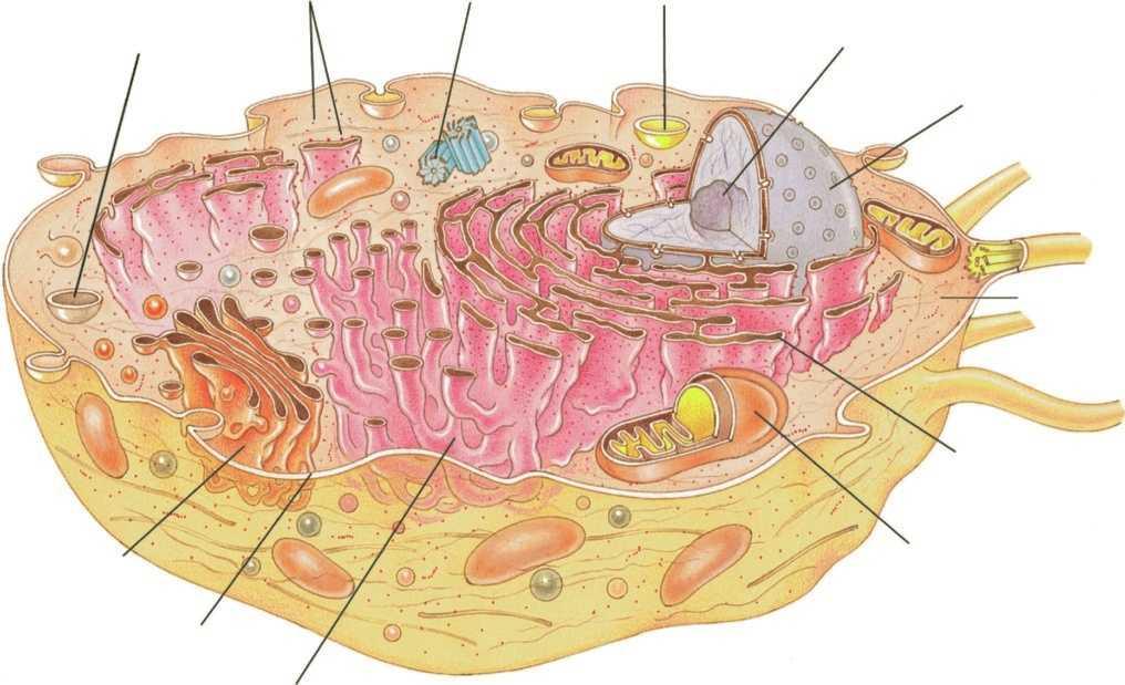
CELL STRUCTURE
Objective
■ Identify the Basic Structures of the Cell and Their Corresponding Functions
Many cell types exist. These cells not only look different but function differently as well. Nevertheless, many features are common among cells. Specialized structures within the cells are called organelles. These organelles are present in most but not all cells. Red blood cells, for example, lack a nucleus.
The cell membrane (or plasma membrane) is common to all cells. It serves as the boundary that keeps
Many of the organelles within the cell are also surrounded by a membrane. The basic structure remains the same for all the membranes. The speci cs of the makeup differ, depending on function.
Cell contents are divided into the nucleus and the cytoplasm. Cytoplasm generally describes the organelles and uid in the cell. A nucleus comes as a standard part of most cells (with a few exceptions such as the red blood cell; Figure 1–8). The nucleus contains the genetic material (i.e., DNA) of the cell, which controls cellular activities by coding for protein synthesis. The DNA in the nucleus is called chromatin. As the cell divides, the chromatin clumps into chromosomes. Identical DNA is passed to all daughter cells. All the cells in the body have the same chromatin. However, cells take on different roles by using certain areas of the chromatin more than others.
FIGURE 1–6 DNA structure: The structure is described as a double helix. Phosphate and sugar groups make up the two strands. The strands are joined by hydrogen bonds between two nitrogenous bases.
FIGURE 1–7 Illustration of cell membrane. The cell membrane has a double layer of phospholipid. In addition, protein molecules are present on and within the phospholipid layers.
A membrane made of two lipid bilayers surrounds the nucleus. This membrane is often joined to other organelles, such as the endoplasmic reticulum and ribosomes. Such a close association helps the nucleus control cell function.
In cells not dividing, a nucleolus is often seen in the nucleus. The nucleolus produces RNA that forms the ribosomes, which in turn produce protein. Cells with large nucleoli actively produce protein.
Ribosomes are small granular-like structures that can be found in the cytoplasm. They contain roughly 60% RNA and 40% other protein. Ribosomes manufacture the protein used in the cell. Growing cells require large amounts of protein and, therefore, have a greater number of ribosomes. The speci c proteins produced by a cell are governed by the nucleus.
The endoplasmic reticulum (ER) is a collection of folded membrane. This membrane attaches to the membrane of the nucleus. The ribosomes often line this membrane, giving it a bumpy appearance and therefore its name, rough endoplasmic reticulum (RER). Protein produced by the ribosomes is then deposited into the RER. These proteins can be further changed in the RER. This protein may be used by the cell or moved to the surface of the cell for secretion. The protein is moved through the membrane in a process called exocytosis, which will be discussed later in the chapter.
Smooth endoplasmic reticulum (SER) has no ribosomes attached. This form is not as common. Some liver cells contain a large amount of SER. The SER in these cells produces glycogen and lipids, and removes toxins.
The Golgi apparatus is formed with large amounts of folded membrane that looks similar to SER. The Golgi apparatus produces polysaccharides and special protein sacs called lysosomes. Protein produced in the RER is moved to the Golgi apparatus. The Golgi apparatus then changes the protein and collects it in the lysosomes. These sacs are pinched from the Golgi apparatus and then moved to the surface of the cell and released.
The proteins contained in the lysosomes are enzymes (remember, enzymes are molecules that help speed chemical reactions in the body). Lysosomes contain enzymes that help to break down other molecules. Varying enzymes match differing molecules. The membrane surrounding lysosome prevents the enzymes from attacking other parts of the cell.
Lysosomes are used to digest food taken in by the cell and to destroy cell structures no longer needed. In Millie, the dog with the abscess, her white blood cells were using lysosomes to destroy bacteria. Cells that die in the body are eliminated when enzymes within lysosomes are released into the cytoplasm. This process of autolysis makes room for replacement cells.
Mitochondria are small rod-shaped organelles found in varying numbers in cells. The more active the cell, the more mitochondria are present. Mitochondria have a double membrane, similar to the cell membrane. The outer membrane is smooth and forms the shape of the mitochondria. The inner membrane is highly folded. These shel ike infolded ridges are called cristae.
The role of mitochondria is well de ned. The mitochondria convert food substances into energy that can be used by the cell. Mitochondria contain the enzymes necessary for this process. Because of this role, mitochondria are called the powerhouses of the cell. The mitochondria are found within cells at their areas of highest activity.
CELL FUNCTION
Objective
■ Review the Basic Function of the Cell
The cell constantly reacts to its environment. Metabolism describes all the reactions going on in cells. Metabolism can be categorized into two main types. Anabolism describes reactions in which smaller molecules are combined into larger ones. The joining of amino acids to form proteins serves as an example. Catabolism, the opposite, occurs when large molecules are broken down into smaller ones. The breaking down of glycogen to release energy is an example of catabolism.
A liquid called extracellular uid (ECF) surrounds living cells. The ECF supplies cells with all the products necessary for their functions. ECF is derived from blood. The outermost skin cells are not covered in liquid; however, they are no longer living.
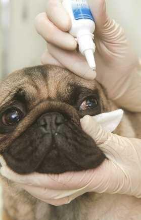
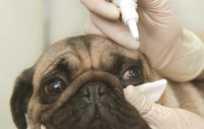
Other cells exposed to the surface, such as those of the eye, need moisture. In the eye, tears produced by glands act as the source of moisture and nutrients. The eyelids help to sweep the tears across the surface of the eye. Certain breeds of dogs, such as the pug, have eyes that bulge from the eye socket. The bulging can be so severe that the eyelids cannot keep the surface of the eye moist with tears. This results in a disease condition on the surface of the eye. Arti cial tears are often used to keep the surface moist.
Table 1–2 summarizes the makeup of ECF. Water is the major component of ECF. Oxygen passes to the cells through the ECF. Conversely, carbon dioxide passes from the cells through it. There are many inorganic ions in the ECF. Some ions, macrominerals, are present in large amounts. Trace minerals are present in much smaller amounts. Both macrominerals and trace minerals are essential for cellular function. Many of the trace minerals are needed for enzymes to function. Organic compounds, including the lipids, proteins, and carbohydrates, are also delivered by the ECF. Metabolism produces waste products, which must be removed from the cells. These waste products are
5. Hormones: compounds produced by glands to in uence metabolism of cells
6. Waste products
eliminated by the ECF. Without elimination, the waste products actually become toxic to the cell.
Many of the products in ECF must be maintained at constant normal concentrations. Cells will be unable to function properly if there is too much or too little of certain products. Glucose provides an excellent example. Small puppies can become low in blood sugar if they have too many parasites robbing them of nutrients. When the sugar in ECF becomes too low, the cells do not have adequate energy. The puppy can become weak or, in severe cases, develop a seizure. Homeostasis is the maintenance of ECF. Homeostasis allows maintenance of normal concentrations of molecules in spite of a wide variety of external conditions.
Cells must be able to obtain products from the ECF. It is not enough that the chemicals just exist in the ECF; there must be means for their exchange with the cell. Table 1–3 summarizes the mechanisms by which materials are exchanged across the cell membrane. The rst mechanism is a process called diffusion (Figure 1–9), in which molecules move from higher to lower concentrations. Because molecules are always moving, there is a greater chance that they will move toward areas of lower concentration. This movement continues until the concentrations are equalized.
The cell membrane does not allow totally free diffusion. Diffusion is in uenced by the size of the molecule, its charge, and its ability to dissolve in lipid. In general, the smaller the molecule, the more easily
Table 1–3 Mechanisms of Cellular Exchange
1. Diffusion
2. Osmosis
3. Active transport
4. Endocytosis
5. Exocytosis
Molecule
Equilibrium
1–9 Diffusion: Random movement of molecules allows equalization of concentrations across a membrane.
the diffusion occurs. Some large molecules such as proteins are unable to diffuse through the membrane and must be transported in other ways.
As previously learned, the property of allowing only certain molecules to diffuse through the membrane is called semipermeability. This characteristic sets the stage for a special type of diffusion, called osmosis
A solvent (in the following case, water) moves across the membrane to equalize the concentration; however, the molecules dissolved in the water (called solutes) cannot pass through the membrane (Figure 1–10). This process can be observed in red blood cells when they are placed in a concentrated solution. The water from the cell moves outward into the solution. Microscopically, the red blood cells can be seen to shrink.
1–10 Osmosis: The semipermeable membrane prevents the passage of large molecules. In this situation, water moves across the membrane to equalize the concentration.
In certain situations, a cell may require a higher concentration of a molecule than is found in the ECF. For example, red blood cells have higher levels of potassium than the surrounding uid. Diffusion constantly attempts to equalize the concentrations (e.g., potassium continually diffuses from the cell). In this case, the potassium is pumped back into the cell, and the higher concentration is maintained. This process is referred to as active transport (Figure 1–11). Active transport requires the cell to burn energy and use enzymes to aid the process. Many different cell types perform the function. Another example occurs in intestinal cells, which transport glucose into the bloodstream, where it is present at higher levels.
Large molecules, such as proteins, must be moved through the membrane in a process called endocytosis
1–11
FIGURE
FIGURE
(Figure 1–12). During endocytosis, the cell membrane wraps around the particle, pinches off, and moves into the cytoplasm as a vacuole. Lysosomes then join with the vacuole, providing the enzymes necessary to break down the particle. The smaller fragments produced are then released into the cell.
In cells producing protein, the opposite process occurs. In exocytosis, a membrane-bound sac containing the protein joins with the cell membrane and releases it into the ECF (Figure 1–13). These sacs are produced within the Golgi apparatus. In intestinal cells, fat droplets can be taken into the cell through endocytosis. The vacuole is transported across the cell and released into the bloodstream by exocytosis.
PROTEIN SYNTHESIS
Objective
■ Describe the Process of Protein Synthesis
As mentioned previously, every cell contains all the genetic material of the animal. The expression of certain genes produces speci c proteins that allow cell specialization. Protein synthesis begins within the nucleus on the basis of the DNA structure. During transcription, information within the DNA is transferred to a strand of messenger RNA (mRNA) that moves into the cytoplasm. An enzyme called RNA polymerase binds to DNA, causing a separation of the double-helix strands (Figure 1–14). This pulling apart exposes a gene. The
enzyme begins at a speci c series of bases (thymine, adenine, cytosine) called a promoter. The RNA polymerase moves along the length of the DNA molecule, creating a complementary strand of RNA. The RNA bases are added in the speci c order that bonds to the bases of the DNA. The corresponding bases were discussed earlier in the chapter. This process continues until the polymerase reaches a terminator series of bases (adenine, thymine, thymine). The mRNA is released and the DNA helix reconnects.
1–14 Transcription of mRNA: RNA polymerase separates the strands of DNA and creates a strand of mRNA coded by the nucleotides of the DNA molecule.
FIGURE 1–12 Endocytosis: A large particle is engulfed by the cell membrane and brought into the cytoplasm within a vacuole.
FIGURE 1–13 Exocytosis: A membrane-bound sac joins with the cell membrane to release the particle.
FIGURE
Nontranscribed
Copy
Translation, which occurs in the ribosomes, is the process in which the code of bases in the mRNA is converted to a series of amino acids. Each series of three bases in the mRNA is a codon (Figure 1–15). The codon provides the signal for a speci c amino acid. The molecule of mRNA is bound by ribosomes. A molecule of transfer RNA (tRNA) that contains the three complementary bases (anticodon) attaches to the mRNA. Each molecule of tRNA carries the amino acid speci c to the codon. Enzymes on the ribosome allow release of the amino acid from the tRNA. A peptide bond is created between adjacent amino acids. This process is repeated along the length of the mRNA molecule, creating a polypeptide. The proteins created may be used within the cytoplasm or processed further within the endoplasmic reticulum.
MITOSIS AND CANCER
Objective
1–15 Translation: The mRNA created in transcription is used to code the amino acid sequence in protein formation. Table 1–4 Stages of Mitosis
■ Discuss Mitosis and its Clinical Signi cance in Diseases Such as Cancer
Cells must reproduce. In mitosis, the cells divide, producing two identical cells. Mitosis is necessary for the growth and maintenance of the animal. Cell division is controlled by a number of factors present within the cell and the extracellular uid. The rate of cell division is adapted to the needs of the animal. Some cells, such
as the epithelia lining the intestinal tract, divide frequently to maintain the integrity of the layer. Other cells, such as skeletal muscle, do not divide in an adult. When these normal controls break down, the cells can begin to undergo frequent mitosis. Uncontrolled mitosis results in cancer. New cells are produced more quickly than needed, resulting in an accumulation or mass of cells in a region. This mass of rapidly dividing cells is called a tumor.
In a nondividing cell, the genetic material is called chromatin. In this form, the chromatin is loosely arranged in the nucleus. The individual chromosomes cannot be seen with a light microscope. These cells are described as being in the interphase. In this stage, the cell is in the process of doubling its DNA. The steps of division are broken down into four phases (Table 1–4). The phases are identi ed to help understand the
FIGURE
FIGURE 1–16 Mitosis. Interphase: Cell in its normal state, as the chromosomes begin to replicate. Prophase: The chromatin thickens and becomes visible, taking on an X shape. The nucleoli and nuclear membrane begin to disappear. Metaphase: The spindle forms between two centrioles. The chromosomes align on the spindle. Anaphase: Chromosomes split at the centromeres, with each half moving to opposite ends. Telophase: The nucleus reforms and a groove divides the two new cells.
process. However, actual cell division is a continuous process, as seen in Figure 1–16.
Prophase begins as the chromatin thickens into visible chromosomes. This is the rst time that the individual chromosomes can be seen with a light microscope. Along with this process, the nucleoli and nuclear membrane begin to disappear. At this point, the chromosomes show the doubling that occurred during interphase. The chromosomes have an X shape. The two identical halves, or chromatids, are joined at a small point called the centromere. Two small organelles, the centrioles, separate and move to opposite ends of the cell.
In metaphase, a spindle is formed between the two centrioles. This is a collection of microtubules that stretch between the two centrioles. The chromosomes move to the center of the cell and align themselves on the spindle.
As anaphase begins, the chromosomes split at the centromere. At this point, the chromosomes are still on the spindle. Each chromatid begins to move outward. The centromere portion moves rst, giving the chromosome a V shape. The chromosomes move to opposite ends of the cell.
Telophase is basically the reverse of prophase. The chromosomes become loosely organized into chromatin. The nuclear membrane and nucleoli return. A groove then forms down the center of the cell. This groove deepens until two identical cells are produced in a process called cytokinesis.
Mitosis is essential in maintaining the population size of cells in the body. The number of cells is established on the basis of the frequency of mitosis, the differentiation of cells, and cell death. An increase in cell number can occur if the rate of cell division increases or the rate of death decreases. In certain instances, a combination of these two changes has a cumulative effect.
The rate of cell division is controlled by soluble factors found in the extracellular uid surrounding
the cells. These factors can either stimulate cell division or inhibit it. Other factors found in the ECF help to control cell death in a process called apoptosis. A classic example of this balance occurs in the cells that line the gastrointestinal tract. Cells at the base of the lining divide frequently at a rate that balances with the cells undergoing apoptosis and death at the surface of the lining. The programmed cell death is designed to occur whenever there is signi cant cell damage such as mutations.
Different cells divide at varying rates. Cells found in certain areas such as the bone marrow and linings of the gastrointestinal tract have stem cells that actively divide on a regular basis. Other cells found in organs such as the liver, kidney, and pancreas do not routinely divide. However, following injury or disease, these cells can become activated into frequent mitosis to allow repair of the organ. A few specialized cells such as nerve and muscle cells have very limited or no ability to divide.
MAMMALIAN REPRODUCTION
Objective
■ Detail Meiosis in Mammalian Reproduction
Mammals rely on sexual reproduction for species survival. In sexual reproduction, a sperm cell and egg cell join to form the new embryo. In this process, half of the genetic material is provided by each of the cells. Meiosis is the division in which the resulting cells contain only half of the genetic material.
There are two cell divisions during meiosis, with only one doubling of the chromatin. The nal result is the formation of four cells, each with half the number of chromosomes. Just as in mitosis, meiosis divides into phases (Figure 1–17).