Supplements for Students
Human Anatomy Laboratory Manual with Cat Dissections, Eighth Edition
Human
Anatomy
By Elaine N. Marieb and Lori A. Smith
0-134-25558-5 / 978-0-134-25558-3
This lab manual contains 30 gross anatomy and histology exercises for all major body systems, featuring 24 cat dissection photos. Illustrated in full color, with convenient spiral binding and an Instructor’s Guide, the lab manual is an excellent accompaniment to Human Anatomy for lab.
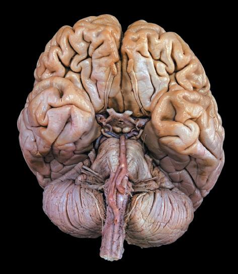
A Photographic Atlas for Anatomy & Physiology
By Nora Hebert, Ruth Heisler, Karen Krabbenhoft, Olga Malakhova, and Jett Chinn 0-321-86925-7 / 978-0-321-86925-8
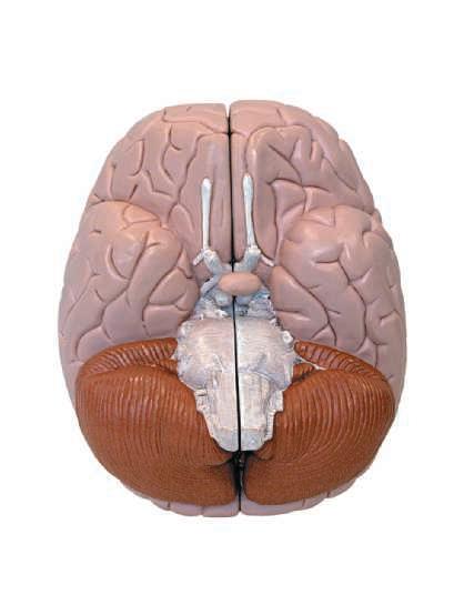
NEW! Visual lab study tool that helps students learn and identify key anatomical structures. Featuring photos from Practice Anatomy Lab™ 3.0 and other sources, the Atlas includes over 250 cadaver dissection photos, histology photomicrographs, and cat dissection photos plus over 50 photos of anatomical models.
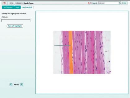
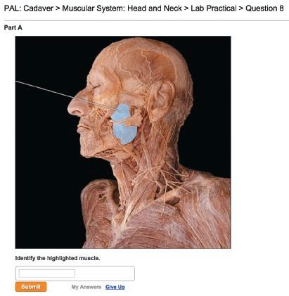
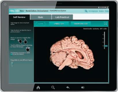
Practice Anatomy Lab™ 3.0 DVD
By Ruth Heisler, Nora Hebert, Jett Chinn, Karen Krabbenhoft, and Olga Malakhova 0-321-68211-4 / 978-0-321-68211-6
PAL 3.0 is an indispensable virtual anatomy study and practice tool that gives students 24/7 access to the most widely used lab specimens, including the human cadaver, anatomical models, histology, cat, and fetal pig. Practice Anatomy™ (PAL™) 3.0 Lab Guide
By
Ruth Heisler, Nora Hebert, Karen Krabbenhoft, Olga Malakhova, and Jett Chinn
Without PAL 3.0 DVD (0-321-84025-9); with PAL 3.0 DVD (0-321-85767-4)
The PAL 3.0 Lab Guide enhances students’ virtual anatomy lab experience by helping them explore anatomical structures through a series of labeling activities and quizzes using the images from PAL. Get Ready for A&P, Third Edition
By Lori K. Garrett
0-321-81336-7 /
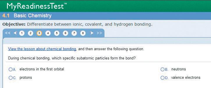
This book and online component were created to help students be better prepared for their course. Features include pre-tests, guided explanations followed by interactive quizzes and exercises, and end-of-chapter cumulative tests. Also available in the Study Area of MasteringA&P®.
The Anatomy Coloring Book, Fourth Edition
By Wynn Kapit and Lawrence M. Elson
0-321-83201-9 / 978-0-321-83201-6
For over 35 years, The Anatomy Coloring Book has been the #1 best-selling human anatomy coloring book. A useful tool for anyone with an interest in learning anatomical structures, this concisely written text features precise, extraordinary hand-drawn figures that were crafted especially for easy coloring and interactive study. Organized according to body systems, each of the 162 spreads featured in this book includes an ingenious color-key system where anatomical terminology is linked to detailed illustrations of the structures of the body.
Hebert Heisler Krabbenhoft Malakhova Chinn
The general philosophy behind this Eighth Edition of Human Anatomy remains the same as in the previous editions. As an instructor, you know that teaching anatomy is not just the presentation of facts. You must provide information in a framework that encourages genuine understanding, devise new presentations to help students remember large amounts of material, and help students apply what they have learned to new situations. All the while you hope that you inspire in the students a love of the subject.
After many years of teaching human anatomy, we became convinced that new approaches to the subject could excite and challenge the students’ natural curiosity. That is why we decided to write this book. We are fortunate to have collaborated with Pearson Education, a publisher that shares our goal: to set a new standard for pedagogical and visual effectiveness in an anatomy text.
This book is designed for one-semester or one-quarter introductory anatomy courses that serve students in prenursing, pre-medical, pre-physical therapy, radiological technology, physician assistant training, pre-dentistry, pharmacy, and other allied-health fields, as well as physical education, athletic training, and nutrition.
Unique Approach to Anatomy
Since its inception, we have worked diligently to distinguish Human Anatomy from the many other anatomy books currently available. This book explains anatomy thoroughly, and its discussions are not merely brief summaries of the art. We have striven to present the basic concepts of anatomy—gross, microscopic, developmental, and clinical—in a manner that is clearly written, effectively organized, up to date, and well illustrated. We realize that learning anatomy involves assimilating gargantuan amounts of material, and we have tried to make our presentation as logical and accessible as possible. To this end, we present anatomy as a “story” that can be explained and understood—convincing the students that the structure of the body makes sense.
Although descriptive gross anatomy is a relatively static science, knowledge is growing quickly in the subfields of functional anatomy, neuroanatomy, developmental anatomy, and the functional aspects of tissue and cellular anatomy. This text strives to keep up with the knowledge explosion in these subfields and to present anatomy in a way that allows modern biology students, whose training is becoming ever more molecular and cellular, to anchor their biochemical and medical training in the physical context of the human body.
Functional Approach
We strongly emphasize the functional anatomy theme, giving careful consideration to the adaptive characteristics of the anatomical structures of the body. Wherever possible, we explain how the shape and composition of the anatomical
structures allow them to perform their functions. Such functional anatomy is not physiology (which focuses on biological mechanisms), but is more akin to “design analysis.” This approach is unique for a text at this level.
Microscopic Anatomy
We have worked to provide an especially effective treatment of microscopic anatomy. Many undergraduate texts treat histology as a specialized and minor subfield that takes a back seat to gross anatomy. This is unfortunate, because most physiological and disease processes take place at the cellular and tissue level, and most allied-health students require a solid background in histology and subcellular structure to prepare them for their physiology courses.
Embryology
Our text is designed to present embryology in the most effective and logical way. We are convinced that the fundamentals should be presented early in the text, before the more advanced discussions of the developing organ systems in the relevant chapters. Therefore, we wrote Chapter 3 as a basic introduction to embryology. Because a comprehensive presentation of embryology early in the book could be intimidating to some students, we have used a “velvet glove approach,” providing only the most important concepts in a concise, understandable way, visually reinforced with exceptionally clear art.
Life Span Approach
Most chapters in this book close with a “Throughout Life” section that first summarizes the embryonic development of organs of the system and then examines how these organs change across one’s life span. Diseases particularly common during certain periods of life are pointed out, and effects of aging are considered. The implications of aging are particularly important to students in the health-related curricula because many of their patients will be older adults.
Helpful Presentation of Terminology
The complex terminology of anatomy is one of the most difficult aspects of the subject to make interesting and accessible. To this end, we highlight important terms in boldfaced type, and we provide the pronunciations of more terms than do many competing texts. Also, we include the Latin or Greek translations of almost every term at the point where the term is introduced in the text. This promotes learning by showing students that difficult terms have simple, logical derivations. The anatomical terms used in this text are consistent with the terms accepted by the International Federation of Associations of Anatomists (IFAA). Clinical terminology is also presented in the Related Clinical Terms section found at
the conclusion of most chapters. A helpful glossary, pronunciation guide, and list of word roots and suffixes are located at the end of the text.
NEW TO THE EIGHTH EDITION
The Eighth Edition builds on the book’s hallmark strengths— art that teaches better, a student-friendly narrative, and easyto-use media and assessment tools—and improves on them.
• Twelve updated body movement photos and seven updated facial movement photos clearly demonstrate movements allowed by synovial joints, as well as actions of muscles of the face, scalp, and neck.
• Two updated Focus figures, Focus Figure 4.11 (Identifying Epithelial and Connective Tissues) and Focus Figure 15.2 (Comparing Somatic Motor and Autonomic Innervation), have been revised to better highlight and teach important, tough-to-understand concepts.
• New and improved in-text media references to PAL 3.0, A&P Flix animations, bone videos, animal organ dissection and cat dissection videos, and art-labeling activities in the Study Area of MasteringA&P® help students easily find helpful study tools as they are reading the book.
More Robust MasteringA&P
MasteringA&P now includes:
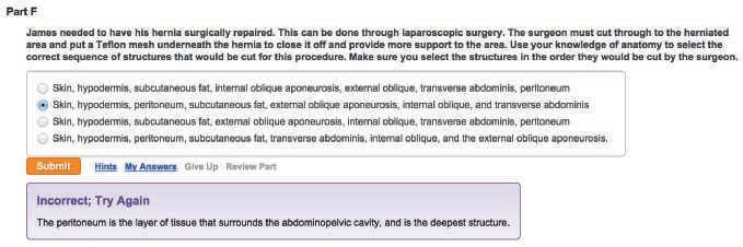
• NEW! Clinical Scenario Coaching Activities that complement lecture and lab, and can be assigned as part of in-class activities or as post-class assignments. Multiple coaching activities for each chapter include an assortment of multiple choice, sorting, labeling, and matching questions.
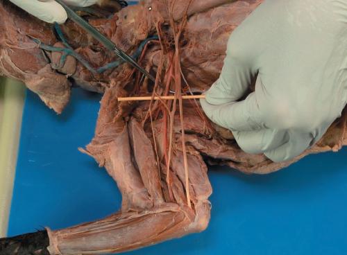
• NEW! Cat Dissection Videos, created by coauthor Patricia Wilhelm, that are assignable in MasteringA&P with hints and wrong-answer feedback. The videos without questions are also available in the Study Area of MasteringA&P.
Video topics cover:
• Superficial Muscles of the Trunk, Dorsal View
• Deep Muscles of the Trunk, Dorsal View
• Posterior Muscles of the Hip and Thigh
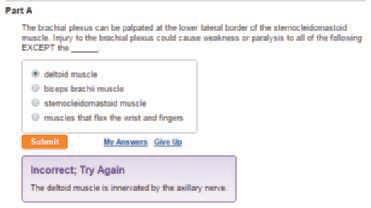
• Brachial Plexus and Innervation of the Muscles of the Arm and Forearm
• Digestive Structures of the Head
• Peritoneum and Mesenteries of the Abdomen
• Structures That Pass Through Mesenteries
• Blood Vessels of the Thorax
• Male Reproductive Structures
• Female Reproductive Structures
• NEW! Dynamic Study Modules that help students study effectively on their own by continuously assessing their activity and performance in real time. Here’s how it works: Students complete a set of questions with a unique answer format that also asks them to indicate their confidence level. Questions repeat until the student can answer them all correctly and confidently. Once completed, Dynamic Study Modules explain the concept using materials from the text. These are available as graded
assignments prior to class, and accessible on smartphones, tablets, and computers.
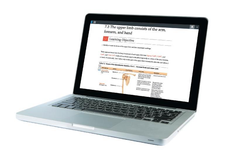
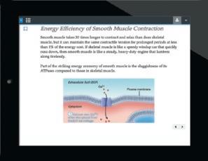
• NEW! eText 2.0, which seamlessly integrates videos and other rich media right into the reading experience. eText 2.0 is available in MasteringA&P and on smartphones and tablets. It is screen-reader ready, includes note-taking, highlighting, bookmarking, and search capabilities, and features customizable settings such as night reading mode.
• Bone and Dissection Video Coaching Activities review all major bones and organ dissections. Each video is supported by activities with hints and specific wrong-answer feedback.
• UPDATED! Focus Figure Coaching Activities expand upon the popular Focus figures in the text by guiding students through complex processes step by step with hints and specific wrong-answer feedback. The Coaching Activities for Focus Figures 4.11 and 15.2 have been updated.
• Get Ready for A&P Diagnostic, Learning Styles, and Cumulative Tests along with Get Ready for A&P Video Tutors feature award-winning teacher Lori Garrett walking students through key basic concepts needed for students to be successful in A&P. Students can take the assignable Diagnostic Test and/or Learning Styles Test in MasteringA&P to assess their base knowledge at the start of the course. Chapter assessments include Reading Questions and Video Tutor Coaching Activities. The key concepts covered include: Learning Styles, Study Skills, Basic Math Review, Terminology, Body Basics, Chemistry, and Cell Biology.
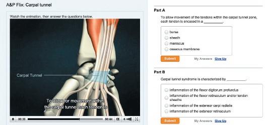
• A&PFlix™ Coaching Activities provide dramatic 3-D animations of key anatomy topics, including individual muscle origins, insertions, actions, and innervations, and key muscle actions and joint movement. Each animation provides practice quizzes and wrong-answer feedback.
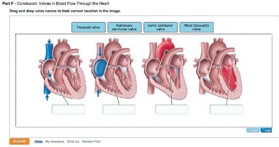
• Drag-and-Drop Art Labeling Activities and Art-Based Questions
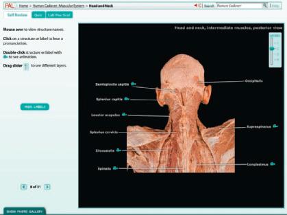
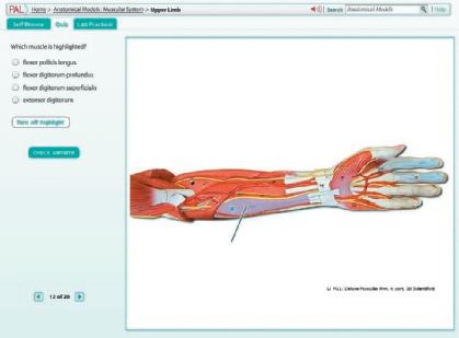
• Practice Anatomy Lab™ 3.0 is an indispensable virtual anatomy study and practice tool that gives students 24/7 access to the most widely used lab specimens including human cadaver, anatomical models, histology, cat, and fetal pig. PAL™ 3.0 includes built-in pronunciation guides, rotatable bones, multiple choice quizzes, and fillin-the-blank lab practical exams.
• Practice Anatomy Lab™ 3.0 Test Bank includes over 4,000 customizable multiple choice and fill-in-the-blank questions. With this test bank, you can assign only the structures you want your students to know.
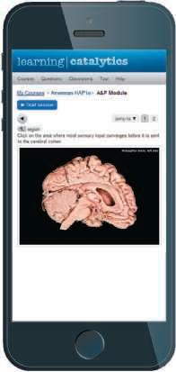
• Learning Catalytics™ is an interactive, classroom tool that uses students’ smartphones, tablets, or laptops to engage them in more sophisticated tasks and thinking. Now included with Mastering with eText, Learning Catalytics enables you to generate classroom discussion, guide your lecture, and promote peer-to-peer learning with real-time analytics. Instructors can:
• Pose a variety of open-ended questions that help your students develop critical thinking skills
• Monitor responses to find out where students are struggling
• Use real-time data to adjust your instructional strategy and try other ways of engaging your students during class
• Manage student interactions by automatically grouping students for discussion, teamwork, and peer-to-peer learning
ACKNOWLEDGMENTS
As we work on each new edition, we are reminded of the great pleasure of working collaboratively with dedicated, competent, and skilled professionals. This experience reinforces the importance of developing collaborative skills in our students. This edition is no different. So many individuals have been involved in the various stages of manuscript preparation, review, and production. Each person mentioned here has directly influenced and improved the final product. More important, each has been a pleasure to work with, and we thank them all.
Serina Beauparlant, Editor-in-Chief, guided the planning and implementation of this new edition and associated electronic media. Serina’s understanding of the needs of faculty and students, focused approach, and dedication to producing the best product available have proved invaluable. We thank her for her leadership and her friendship.
Michele Mangelli, Mangelli Productions, was the project manager for this new edition, coordinating the editorial and production aspects of the project. As always, Michele’s diligence and attention to detail and flexibility with due dates has facilitated the work on this edition. Michele also organized and managed the production of video content that supplements the textbook, along with Amanda Kaufman, Coproducer; Richard Boghosian, Videographer; and Anthony Saccocio, Sound Engineer. The Content Producers, Nicole Tache and Patrice Fabel, oversaw the development of the book’s media program. Stacey Weinberger once again contributed to this text with her manufacturing expertise. Allison Rona and Derek Perrigo, our Marketing Managers, have efficiently kept a finger on the pulse of the marketplace—keeping in touch with professors and students, and providing feedback on what they do or do not like about the text and media products. Thank you all.
Laura Southworth worked on the revision of Focus Figures. Her skill with art and layout, combined with her extensive content knowledge helped to improve select focus figures tremendously.
Kristin Piljay has done an outstanding job as photo researcher. Special thanks to the team at Imagineering for work on the art for this edition. Their skillful work has added significantly to this new edition.
We thank other members of the production team—Karen Gulliver, production coordinator; Jean Lake, art and photo coordinator; Anita Hueftle, copyeditor; and Betsy Dietrich,
proofreader—for their excellent work and attention to detail. Many thanks go out to tani hasegawa for a beautiful interior design and to Tandem Creative for the stunning new cover. We also appreciate the fine work of Cenveo in assembling the pages.
Special thanks to Ruth Heisler for writing Clinical Scenario Coaching Activities for MasteringA&P, Leslie Hendon for her work on the Dynamic Study Modules, Justin Shaffer for writing alt-text and completing correlations for questions in MasteringA&P, and Janet Brodsky for her work on art-labeling activities in MasteringA&P. Leif Saul, Anthony Friscia, and Anthony Weinhaus reviewed revised Focus Figures. Their insight and expertise contributed significantly to the effectiveness of these figures. Thank you. The administration at Johnson & Wales University provided teaching release time for work on this revision. We are also grateful to Eric Leaver, Development Editor, for revising the Test Bank.
We also want to thank the following reviewers for their feedback and advice on MasteringA&P and our new media for the eighth edition:
Joslyn Ahlgren, University of Florida
Gary Allen, Dalhousie University
Kathleen Azevedo, University of California, Berkeley Extension, Las Positas College
David Babb, West Hills College–Lemoore Campus
Elizabeth Co, Boston University
David Conley, Washington State University
Lisa Flick, Monroe Community College
Jill Harper-Judd, St. Petersburg College–Clearwater Campus
Ruth Heisler, University of Colorado, Boulder
Leslie Hendon, University of Alabama at Birmingham
Kerrie Hoar, University of Wisconsin–La Crosse
Steve Hobbs, University of Colorado, Boulder
Dawn Hunter, West Virginia University
Jeremy Ingraham, University of North Carolina, Greensboro
Patricia Phelps, University of California, Los Angeles
Carmen Rodriguez, Virginia Commonwealth University
Justin Shaffer, University of California, Irvine
Judith Tamburlin, University at Buffalo
Shelley Thai, Glendale Community College
Finally, a note of gratitude for the support and encouragement provided by our families. They have been patient and understanding about the time taken from family while we focused on revisions, and we thank them. Our last acknowledgment is a shout-out to our students, who continue to inspire us.
Elaine N. Marieb
Patricia Brady Wilhelm
Jon Mallatt
1
The Human Body: An Orientation 1
An Overview of Anatomy 2
Subdisciplines of Anatomy 2
Gross Anatomy 2
Microscopic Anatomy 2
Other Branches of Anatomy 2
The Hierarchy of Structural Organization 2
Scale: Length, Volume, and Weight 6
Anatomical Terminology 6
Gross Anatomy: An Introduction 6
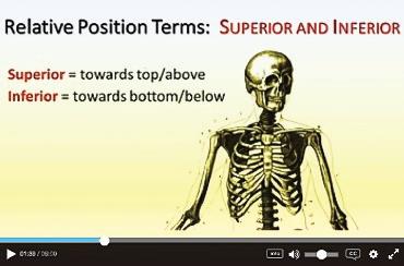
Regional and Directional Terms 6
Body Planes and Sections 7
The Human Body Plan 10
Body Cavities and Membranes 11
Dorsal Body Cavity 11
Ventral Body Cavity 11
Serous Cavities 12
Abdominal Quadrants 13
Anatomical Variability 13
Microscopic Anatomy: An Introduction 13
Light and Electron Microscopy 13
Scanning Electron Microscopy 15
Artifacts 15
Clinical Anatomy: An Introduction to Medical Imaging Techniques 15
X-Ray Imaging 15
Advanced X-Ray Techniques 16
Computed Tomography 16
Angiography 17
Positron Emission Tomography 17
Sonography 18
Magnetic Resonance Imaging 18
FOCUS FIGURE
Focus on Levels of Structural Organization 3
Chapter Summary 19
Review Questions 20
2 Cells: The Living Units
Overview of Cells 23
The Plasma Membrane 24
Structure 26
Functions 26
Membrane Transport 27
The Cytoplasm 28
Cytosol 28
Cytoplasmic Organelles 29
Ribosomes 29
Endoplasmic Reticulum 29
Golgi Apparatus 30
Lysosomes 31
Mitochondria 32
Peroxisomes 32
Cytoskeleton 32
Centrosome and Centrioles 33
Cytoplasmic Inclusions 33
The Nucleus 34
Nuclear Envelope 34
Nucleolus 34
Chromatin and Chromosomes 35
The Cell Life Cycle 37
Interphase 37
Cell Division 37
Mitosis 37
Cytokinesis 40
Developmental Aspects of Cells 40
Cell Differentiation 40
Aging 41
FOCUS FIGURE
Focus on Mitosis 38
22
CLINICAL APPLICATIONS
Hypercholesterolemia 28 Tay-Sachs Disease 31
Related Clinical Terms 42
Chapter Summary 42
Review Questions 44
3 Basic Embryology
46
Stages of Prenatal Development 46
The Basic Body Plan 47
The Embryonic Period 48
Week 1: From Zygote to Blastocyst 48
Week 2: The Two-Layered Embryo 50
Week 3: The Three-Layered Embryo 50
The Primitive Streak and the Three Germ Layers 50
The Notochord 51
Neurulation 52
The Mesoderm Begins to Differentiate 54
Week 4: The Body Takes Shape 54
Folding 54
Derivatives of the Germ Layers 54
Weeks 5–8: The Second Month of Embryonic Development 56
The Fetal Period 59
A CLOSER LOOK
Focus on Birth Defects 60
CLINICAL APPLICATIONS
Conjoined (Siamese)Twins 49 Neural Tube Defects 53
Related Clinical Terms 61
Chapter Summary 61
Review Questions 62
4
Tissues
64
I. Epithelial Tissue 65
Special Characteristics of Epithelia 66
Classification of Epithelia 66
Simple Epithelia 67
Stratified Epithelia 72
Glands 72
Endocrine Glands 72
Exocrine Glands 72
Epithelial Surface Features 74
Lateral Surface Features: Cell Junctions 74
Basal Feature: The Basal Lamina 76
Apical Surface Features: Microvilli and Cilia 76
II. Connective Tissue 77
Special Characteristics of Connective Tissues 78
Structural Elements of Connective Tissues 78
Cells 78
Fibers 80
Ground Substance 80
Classification of Connective Tissues 81
Connective Tissue Proper—Loose Connective Tissues 81
Connective Tissue Proper—Dense Connective Tissue 84
Cartilage 86
Bone 89
Blood 89
Covering and Lining Membranes 89
III. Muscle Tissue 93
IV. Nervous Tissue 93
Tissue Response to Injury 95
Inflammation 95
Repair 97
The Tissues Throughout Life 97
FOCUS FIGURE
Focus on Identifying Epithelial and Connective Tissues 90
A CLOSER LOOK
Cancer—The Intimate Enemy 99
CLINICAL APPLICATIONS
Basement Membranes and Diabetes 76 Kartagener’s Syndrome 77 Scurvy 80
Related Clinical Terms 98
Chapter Summary 100
Review Questions 101
5 The Integumentary System
103
The Skin and the Hypodermis 104
Epidermis 105
Layers of the Epidermis 105
Dermis 108
Papillary Dermis 108
Reticular Dermis 108
Hypodermis 110
Skin Color 110
Appendages of the Skin 111
Nails 111
Hair and Hair Follicles 112
Hair 112
Hair Follicles 112
Types and Growth of Hair 114
Hair Thinning and Baldness 114
Sebaceous Glands 115
Sweat Glands 115
Eccrine Sweat Glands 116
Apocrine Sweat Glands 116
Disorders of the Integumentary System 116
Burns 116
Skin Cancer 118
Basal Cell Carcinoma 118
Squamous Cell Carcinoma 118
Melanoma 118
The Skin Throughout Life 118
A CLOSER LOOK
Tattoos 110
CLINICAL APPLICATIONS
Skin’s Response to Friction 108 Decubitus Ulcer 108
The Patch Drug Delivery System 109 Freckles and Moles 111 Cyanosis 111 Chemotherapy and Hair
Loss 114 Acne 115
Related Clinical Terms 119
Chapter Summary 120
Review Questions 121
6 Bones and Skeletal Tissues 123
Cartilages 124
Location and Basic Structure 124
Types of Cartilage 125
Hyaline Cartilage 125
Elastic Cartilage 126
Fibrocartilage 126
Growth of Cartilage 127
Bones 127
Functions of Bones 127
Bone Tissue 127
Extracellular Matrix 127
Cells 128
Gross Anatomy of Bones 128
Classification of Bones 128
Compact and Spongy Bone 129
Structure of a Typical Long Bone 129
Structure of Short, Irregular, and Flat Bones 131
Bone Design and Stress 131
Microscopic Structure of Bone 132
Compact Bone 132
Spongy Bone 135
Bone Development and Growth 135
Intramembranous Ossification 136
Endochondral Ossification 136
Anatomy of the Epiphyseal Plate 138
Postnatal Growth of Endochondral Bones 139
Bone Remodeling 139
Repair of Bone Fractures 141
Disorders of Bones 143
Osteoporosis 143
Osteomalacia and Rickets 144
Osteosarcoma 144
The Skeleton Throughout Life 145
A CLOSER LOOK
The Marvelous Properties of Cartilage 126
CLINICAL APPLICATIONS
Achondroplasia 139 Paget’s Disease 141
Traction 143
Related Clinical Terms 146
Chapter Summary 146
Review Questions 147
7 Bones, Part 1: The Axial Skeleton 150
The Skull 152
Overview of Skull Geography 152
Cranial Bones 153
Parietal Bones and the Major Sutures 153
Sutural Bones 154
Frontal Bone 155
Occipital Bone 155
Temporal Bones 156
Sphenoid Bone 158
Ethmoid Bone 162
Facial Bones 162
Mandible 162
Maxillary Bones 163
Zygomatic Bones 167
Nasal Bones 167
Lacrimal Bones 167
Palatine Bones 167
Vomer 167
Inferior Nasal Conchae 167
Special Parts of the Skull 167
Nasal Cavity 167
Paranasal Sinuses 168
Orbits 169
The Hyoid Bone 169
The Vertebral Column 170
Regions and Normal Curvatures 170
Ligaments of the Spine 171
Intervertebral Discs 171
General Structure of Vertebrae 172
Regional Vertebral Characteristics 173
Cervical Vertebrae 174
Thoracic Vertebrae 175
Lumbar Vertebrae 176
Sacrum 177
Coccyx 177
The Thoracic Cage 178
Sternum 179
Ribs 179
Disorders of the Axial Skeleton 180
Cleft Palate 180
Stenosis of the Lumbar Spine 180
Abnormal Spinal Curvatures 180
The Axial Skeleton Throughout Life 181
CLINICAL APPLICATIONS
Deviated Septum 167 Herniated Disc 172
The Dens and Fatal Trauma 174
Related Clinical Terms 182
Chapter Summary 182
Review Questions 183
8 Bones, Part 2: The Appendicular Skeleton 185
The Pectoral Girdle 186
Clavicles 186
Scapulae 187
The Upper Limb 187
Arm 187
Forearm 191
Ulna 191
Radius 191
Hand 192
Carpus 192
Metacarpus 192
Phalanges of the Fingers 192
The Pelvic Girdle 194
Ilium 194
Ischium 196
Pubis 196
Pelvic Structure and Childbearing 197
The Lower Limb 197
Thigh 197
Leg 199
Tibia 201
Fibula 202
Foot 202
Tarsus 203
Metatarsus 203
Phalanges of the Toes 203
Arches of the Foot 203
Disorders of the Appendicular
Skeleton 204
The Appendicular Skeleton Throughout Life 205
CLINICAL APPLICATIONS
Fractures of the Clavicle 187 Palpation of Colles’
Fracture 191 Carpal Tunnel Syndrome 194 Hip
Fracture as a Result of Osteoporosis 197 Ankle
Fractures 202 Metatarsal Stress Fracture 203
Related Clinical Terms 205
Chapter Summary 206
Review Questions 206
9 Joints 208
Classification of Joints 209
Fibrous Joints 209
Sutures 209
Syndesmoses 210
Gomphoses 210
Cartilaginous Joints 210
Synchondroses 210
Symphyses 210
Synovial Joints 210
General Structure of Synovial Joints 211
Movements Allowed by Synovial Joints 213
Gliding 213
Angular Movements 214
Special Movements 217
Types of Synovial Joints 217
Factors Influencing the Stability of Synovial Joints 217
Articular Surfaces 217
Ligaments 220
Muscle Tone 220
Selected Synovial Joints 221
Temporomandibular Joint 222
Sternoclavicular Joint 224
Shoulder (Glenohumeral) Joint 224
Elbow Joint 226
Wrist Joint 227
Hip Joint 228
Knee Joint 229
Ankle Joint 232
Disorders of Joints 234
Joint Injuries 234
Torn Cartilage 234
Sprains 234
Dislocations 235
Inflammatory and Degenerative Conditions 235
Bursitis, Tendonitis, and Tenosynovitis 235
Arthritis 235
The Joints Throughout Life 237
FOCUS FIGURE
Focus on Synovial Joints 218
CLINICAL APPLICATIONS
Temporomandibular Disorders 223 Shoulder
Dislocations 226 Elbow Trauma 227 Knee
Injuries 231 Ankle Sprains 234 Autologous Cartilage
Implantation 234 Nursemaid’s Elbow 235
Related Clinical Terms 237
Chapter Summary 238
Review Questions 239
10 Skeletal Muscle Tissue
241
Overview of Muscle Tissue 242
Properties of Muscle Tissue 242
Terminology Specific to Muscle Tissue 242
Functions of Muscle Tissue 242
Types of Muscle Tissue 242
Skeletal Muscle Tissue 242
Cardiac Muscle Tissue 242
Smooth Muscle Tissue 242
Skeletal Muscle 244
Gross Anatomy of a Skeletal Muscle 244
Connective Tissue and Fascicles 244
Nerves and Blood Vessels 245
Muscle Attachments 245
Microscopic and Functional Anatomy of Skeletal Muscle Tissue 246
The Skeletal Muscle Fiber 246
Myofibrils and Sarcomeres 246
Titin and Other Myofibril Proteins 248
Sarcoplasmic Reticulum and T Tubules 248
Mechanism of Contraction 249
Muscle Extension 250
Muscle Fiber Length and the Force of Contraction 250
Innervation of Skeletal Muscle 250
Types of Skeletal Muscle Fibers 252
Disorders of Skeletal Muscle Tissue 255
Muscular Dystrophy 255
Myofascial Pain Syndrome 256
Fibromyalgia 256
Skeletal Muscle Tissue Throughout
Life 257
A CLOSER LOOK
Anabolic Steroid Abuse 257
CLINICAL APPLICATIONS
Delayed-Onset Muscle Soreness 249 Muscle
Twitch 252 Rhabdomyolysis 253
Related Clinical Terms 258
Chapter Summary 258
Review Questions 260
11 Muscles of the Body 262
Arrangement of Fascicles in Muscles 263
Lever Systems: Bone-Muscle Relationships 264
First-Class Lever 265
Second-Class Lever 266
Third-Class Lever 266
Organizational Scheme Based on Embryonic Development 266
Muscle Actions and Interactions 268
Naming the Skeletal Muscles 270
Major Skeletal Muscles of the Body 270
Muscle Compartments of the Limbs 271
Upper Limb 271
Lower Limb 271
Table 11.1 Summary of Actions of Muscles Acting on the Arm, Forearm, and Hand 274
Table 11.2 Summary of Actions of Muscles Acting on the Thigh, Leg, and Foot 276
The Muscle Tables 278
Table 11.3 Muscles of the Head, Part I: Facial Expression 279
Table 11.4 Muscles of the Head, Part II: Mastication and Tongue Movement 282
Table 11.5 Muscles of the Anterior Neck and Throat: Swallowing 284
Table 11.6 Muscles of the Neck and Vertebral Column: Head Movements and Trunk Extension 287
Table 11.7 Deep Muscles of the Thorax: Breathing 291
Table 11.8 Muscles of the Abdominal Wall: Trunk Movements and Compression of Abdominal Viscera 293
Table 11.9 Muscles of the Pelvic Floor and Perineum: Support of Abdominopelvic Organs 296
Table 11.10 Superficial Muscles of the Anterior and Posterior Thorax: Movements of the Scapula 298
Table 11.11 Muscles Crossing the Shoulder Joint: Movements of the Arm (Humerus) 302
Table 11.12 Muscles Crossing the Elbow Joint: Flexion and Extension of the Forearm 305
Table 11.13 Muscles of the Forearm: Movements of the Wrist, Hand, and Fingers 306
Table 11.14 Intrinsic Muscles of the Hand: Fine Movements of the Fingers 312
Table 11.15 Muscles Crossing the Hip and Knee Joints: Movements of the Thigh and Leg 315
Table 11.16 Muscles of the Leg: Movements of the Ankle and Toes 323
Table 11.17 Intrinsic Muscles of the Foot: Toe Movement and Foot Support 329
Regional Surface Anatomy 332
The Head 333
The Cranium 333
The Face 333
The Neck 334
Skeletal Landmarks 334
Muscles of the Neck 335
Triangles of the Neck 335
The Thorax 335
Skeletal Landmarks 335
Muscles of the Thorax 335
The Abdomen 335
Skeletal Landmarks 335
Muscles and Other Abdominal Surface
Features 335
The Back 336
Bones of the Back 336
Muscles of the Back 337
The Upper Limb and Shoulder 337
The Axilla 337
The Shoulder 338
The Arm 338
The Elbow Region 339
The Forearm and Hand 340
The Lower Limb and Gluteal Region 340
The Gluteal Region 340
The Thigh 343
The Leg and Foot 343
FOCUS FIGURE
Focus on Muscle Action 269
CLINICAL APPLICATIONS
Compartment Syndrome 271 Torticollis 287
Hernia 293 Tennis Elbow 306 Lacerations to the Neck 335 The Triangle of Auscultation 337
Gluteal Intramuscular Injections 341
Related Clinical Terms 344
Chapter Summary 344
Review Questions 347
12
Fundamentals of the Ner vous System and Nervous Tissue 349
The Functional Organization of the Nervous System 350
Functions of the Nervous System 350
Basic Divisions of the Nervous System 350
Somatic Sensory (SS) 352
Visceral Sensory (VS) 352
Somatic Motor (SM) 352
Visceral Motor (VM) 352
Nervous Tissue 353
The Neuron 353
The Cell Body 353
Neuron Processes 353
Synapses 354
Classification of Neurons 355
Neuroglia 358
Neuroglia in the CNS 358
Neuroglia in the PNS 359
Myelin Sheaths 359
Gross Anatomy of the Nervous System:
An Overview 361
Gray and White Matter of the CNS 361
Nerves 361
Neuronal Integration 363
Reflex Arcs 363
Monosynaptic Reflex 363
Polysynaptic Reflex 363
Neuronal Circuits 364
Diverging Circuit 365
Converging Circuit 365
Reverberating Circuit 365
Serial Processing 365
Parallel Processing 365
Integration Between the PNS and CNS 366
Disorders of Nervous Tissue 367
Multiple Sclerosis 367
Neuronal Regeneration 368
Nervous Tissue Throughout Life 369
Embryonic Development of Nervous Tissue 369
FOCUS FIGURE
Focus on Neuronal Pathways 366
CLINICAL APPLICATIONS
Gliomas 359 Tic Douloureux 359 Regeneration and Spinal Cord Injuries 368
Related Clinical Terms 371
Chapter Summary 371
Review Questions 372
13 The Central Nervous System 374
The Brain 375
Embryonic Development of the Brain 375
Basic Parts and Organization of the Brain 377
Ventricles of the Brain 377
The Brain Stem 378
The Medulla Oblongata 378
The Pons 380
The Midbrain 381
The Cerebellum 383
The Cerebellar Peduncles 383
The Diencephalon 385
The Thalamus 385
The Hypothalamus 385
The Epithalamus 387
The Cerebrum 388
Lobes of the Cerebral Cortex 388
Functional Areas of the Cerebral Cortex 388
White Matter of the Cerebrum 396
Deep Gray Matter of the Cerebrum 398
Functional Brain Systems 399
The Limbic System 399
The Reticular Formation 400
Protection of the Brain 401
Meninges 403
Cerebrospinal Fluid 404
Blood Brain Barrier 407
The Spinal Cord 407
White Matter of the Spinal Cord 410
Gray Matter of the Spinal Cord and Spinal Roots 411
Protection of the Spinal Cord 412
Sensory and Motor Pathways in the Central Nervous System 413
Ascending Pathways 414
Spinocerebellar Pathway 414
Dorsal Column–Medial Lemniscal Pathway 414
Spinothalamic Pathways 414
Descending Pathways 416
Direct Pathways (Pyramidal Tracts) 418
Indirect Pathways (Extrapyramidal Tracts) 418
Disorders of the Central Nervous System 419
Spinal Cord Damage 419
Brain Dysfunction 419
Cerebrovascular Accidents 419
Alzheimer’s Disease 421
The Central Nervous System Throughout Life 421
Embryonic Development and Congenital Conditions 421
Postnatal Changes in the Brain 422
A CLOSER LOOK
Traumatic Brain Injury 420
CLINICAL APPLICATIONS
Localization of a Brain Stem Injury 383 Injury to the Cerebellum 384 Why Won’t Teenagers Sleep at Night? 387 Phantom Limb Pain 391 Agnosia 394
Neglect Syndrome 395 Dyskinesia 399
Hydrocephalus 407 Flaccid and Spastic Paralysis 412
Meningitis 413 Amyotrophic Lateral Sclerosis (ALS) 419
Related Clinical Terms 423
Chapter Summary 423
Review Questions 425
14 The Peripheral Nervous System
427
Organization of the Peripheral Nervous System 428
Peripheral Sensory Receptors 428
Functional Classification 429
Location of Receptors 429
Stimulus Type 429
Structural Classification 429
Free Nerve Endings 429
Encapsulated Nerve Endings 431
Cranial Nerves 432
I The Olfactory Nerves 435
II The Optic Nerves 435
III The Oculomotor Nerves 436
IV The Trochlear Nerves 436
V The Trigeminal Nerves 437
VI The Abducens Nerves 438
VII The Facial Nerves 439
VIII The Vestibulocochlear Nerves 440
IX The Glossopharyngeal Nerves 441
X The Vagus Nerves 442
XI The Accessory Nerves 443
XII The Hypoglossal Nerves 443
Spinal Nerves 444
Innervation of the Back 446