Modern C++ for Absolute Beginners: A Friendly Introduction to the C++ Programming Language and C++11 to C++23 Standards, 2nd Edition Slobodan Dmitrovi■
No part of this publication may be reproduced or transmitted in any form or by any means, electronic or mechanical, including photocopying, recording, or any information storage and retrieval system, without permission in writing from the publisher Details on how to seek permission, further information about the Publisher’s permissions policies and our arrangements with organizations such as the Copyright Clearance Center and the Copyright Licensing Agency, can be found at our website: www.elsevier.com/permissions.
This book and the individual contributions contained in it are protected under copyright by the Publisher (other than as may be noted herein).
Notices
Knowledge and best practice in this field are constantly changing As new research and experience broaden our understanding, changes in research methods, professional practices, or medical treatment may become necessary.
Practitioners and researchers must always rely on their own experience and knowledge in evaluating and using any information, methods, compounds, or experiments described herein In using such information or methods they should be mindful of their own safety and the safety of others, including parties for whom they have a professional responsibility
With respect to any drug or pharmaceutical products identified, readers are advised to check the most current information provided (i) on procedures featured or (ii) by the manufacturer of each product to be administered, to verify the recommended dose or formula, the method and duration of administration, and contraindications It is the responsibility of practitioners, relying on their own experience and knowledge of their patients, to make diagnoses, to determine dosages and the best treatment for each individual patient, and to take all appropriate safety precautions.
To the fullest extent of the law, neither the Publisher nor the authors, contributors, or editors, assume any liability for any injury and/or damage to persons or property as a matter of products liability, negligence or otherwise, or from any use or operation of any methods, products, instructions, or ideas contained in the material herein
International Standard Book Number: 978-0-323442640
Content Strategy Director: Penny Rudolph
Content Development Manager: Ellen Wurm-Cutter
Content Development Specialist: Alexandra York
Publishing Services Manager: Deepthi Unni
Senior Project Manager: Kamatchi Madhavan
Designer: Patrick Ferguson
Printed in China
Last digit is the print number:
Dedication
For my dear wife, Sarbjit Kaur, son, Pahul Singh, and our dog, Boomrang
Dedicated to our parents, Sardar Modan Singh and Sadarni Gurmail Kaur Sardar Pritam Singh and Sardarni Mohinder Kaur
Contributors
CHAPTER CONTRIBUTORS
Judy Klimek, DVM, MS, Associate Professor, Department of Anatomy and Physiology, Kansas State University College of Veterinary Medicine, Manhattan, KS
Gillian Muir, DVM, PhD, Professor and Head, Department of Veterinary Biomedical Sciences, Western College of Veterinary Medicine, University of Saskatchewan, Canada
Emily J. Reppert, DVM, MS, DACVIM, Assistant Professor, Livestock Services, Department of Clinical Sciences, Kansas State University, Manhattan, KS
FIGURE CONTRIBUTORS
Kalman Czeibert, DVM, Research Fellow, Justanatomy Ltd , Hungary
Ors Petnehazy, DVM, PhD
Research Fellow, Institute of Diagnostic Imaging and Radiation Oncology, Kaposvár University CEO, Justanatomy Ltd., Hungary
Ram S. Sethi, BVSc&AH, MVSc, PhD, Professor, School of Animal Biotechnology, Guru Angad Dev Veterinary and Animal Sciences University, Ludhiana, Punjab
Jaswant Singh, BVSc&AH, MVSc, PhD, Professor, Department of Veterinary Biomedical Sciences, University of Saskatchewan, Saskatoon, Canada
Preface
It is an honour and a true privilege to be entrusted with the task of preparing the 5th edition of Textbook of Veterinary Anatomy by Dyce, Sack and Wensing. When starting as a graduate teaching assistant in veterinary anatomy courses, I never did or could imagine such an opportunity. Of the three highly distinguished anatomists and original authors of this text, I met only Prof Wolfgang Sack at the American Association of Veterinary Anatomists meeting in Knoxville in 1996, when I was a postdoctoral fellow. Naturally, I was in awe of him! But he was highly encouraging of my efforts to become a teacher of veterinary anatomy.
I have taken exceptional care to maintain the foundational integrity of the textbook as developed and nurtured by Profs. Dyce, Sack and Wensing. Students accept this text to be the “go-to” anatomy text for their foundational reading as well as a quick check-in I have also found this book in large numbers of veterinary clinics, and veterinarians attest to its authoritative usefulness in their clinical practice of veterinary medicine. These observations underscore the established value of the text and its fundamental endurance.
There are many current trends that impact the teaching of veterinary anatomy. These trends, including an increase in the volume of veterinary clinical and biomedical information, have resulted in reduced allocation of time to educate students in veterinary anatomical sciences in general Veterinary embryology has nearly been eliminated from the veterinary medical curricula. The time in veterinary curricula to teach histology has also been reduced significantly. There however is resurging realization that these trends are not fostering development of sufficient foundational knowledge of veterinary anatomy and an integrated set of concepts for the students For example, the growth in the use of imaging modalities in veterinary medicine has created a need for better education in veterinary anatomy. Also, reduction in time devoted to the instruction of veterinary anatomy is stimulating interest in more integrated teaching of anatomy, histology, and embryology; the Textbook of Veterinary Anatomy has been at the forefront of such integrated instruction of veterinary anatomy.
The 5th edition of the Textbook of Veterinary Anatomy introduces many changes and makes a gentle pivot to indicate the future direction of the book. In preparing this edition, I have had many discussions with students, and some with fellow teachers. The major changes are as follows:
• significant editing of the text to remove many redundancies;
• removal of text that is not germane to the veterinary medical student;
• addition of nearly 100 new figures;
• addition of many figures at the sub-gross level to create a link between the gross anatomy and histology;
• addition of a new chapter on camelids;
• creation of more than 120 highlighted text boxes to make to easier to grasp important concepts and some clinical features;
• addition of new tables to summarize information;
• creation of a new box called Comprehension Check at the end of each chapter to facilitate group discussion and practice;
• introduction of new contributors: Dr. Gillian Muir has worked on chapters related to the nervous system, and Dr. Judy Klimek and Dr. Emily Reppert have contributed a new chapter on camelids. Dr Jaswant Singh, Dr Ors Petnehazy, Dr Kalman Czeibert, and Dr R S Sethi have contributed illustrations
Taken together, these changes enable the textbook, while maintaining its rigorous content, to start to look toward a new phase of its life.
This work would not be possible without the exceptional support received from the Elsevier team. In particular, I express sincere thanks to Penny Rudolph, who engaged me in the creation of
educational materials for learning veterinary anatomy Having worked with her in creating Elsevier’s Veterinary Anatomy Flash Cards and Veterinary Anatomy Coloring Book, I have enjoyed doing so again in preparing the 5th edition of the Textbook of Veterinary Anatomy. Throughout the preparation of this edition, I received high levels of support and encouragement from Alexandra York, Kamatchi Madhavan, Shelly Stringer, and Brian Loehr
Finally, I thank many teachers and students who been instrumental in my development as a teacher Special thanks to Dr Alastair Summerlee, an exceptional teacher and scholar, who gave me my first opportunity to be a graduate teaching assistant in anatomy laboratories.
I look forward to receiving comments from students and my fellow anatomy teachers to make further improvements to the book and will welcome their opinions on the changes introduced in this edition.
About the Author
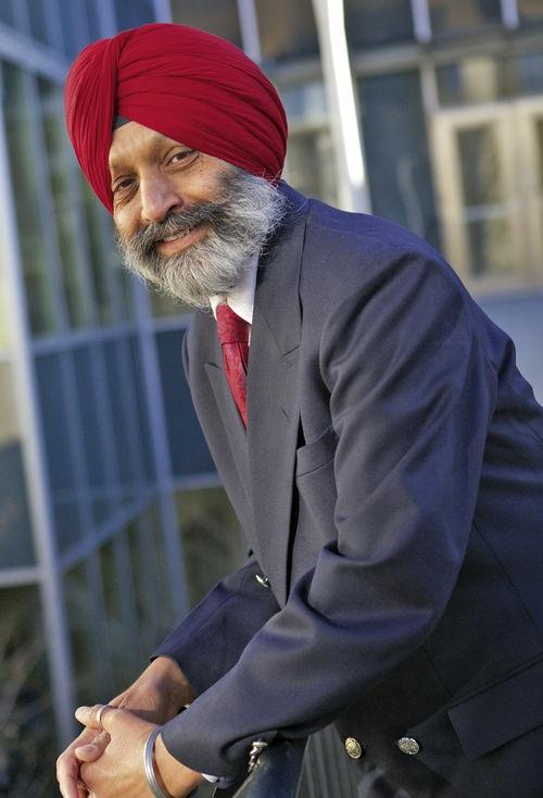
Baljit Singh is Dean and Professor of Veterinary Anatomy at Faculty of Veterinary Medicine, University of Calgary Before this, he was Professor and Associate Dean (Research) at Western College of Veterinary Medicine, University of Saskatchewan. He obtained a BVSc&AH and an MVSc from Punjab Agricultural University and a PhD from the University of Guelph, and he completed postdoctoral training at Texas A&M University and Columbia University in New York He is licensed as a Veterinarian in Canada
Baljit is a recipient of many teaching awards, including Outstanding Veterinary Anatomist Award (2016) from the American Association of Veterinary Anatomists; 3M Canadian National Teaching Fellowship (2009), Canada’s highest teaching honour; and Master Teacher Award of the University of Saskatchewan. He is a Fellow of the American Association of Anatomists. He has
served as the President of the American Association of Veterinary Anatomists (2005-2008), as a member of the Canadian National Examining Board for Veterinary Medicine (2006-2013), and as a member of the Council on Education of the American-Canadian Veterinary Medical Associations.
Baljit’s research program is in the areas of lung inflammation, infectious disease, and nanomedicines. He has trained more than 90 postgraduate, undergraduate, and postdoctoral students and published approximately 95 research papers He is currently Section Editor (Immunology/Inflammation) of Cell and Tissue Research and an editor of Advances in Anatomy, Embryology and Cell Biology. He has received the Pfizer Research Excellence Award, Visiting Professorship at the Centre for Excellence in Immunology, Hannover, Germany, and has given many invited lectures
Sources of Non-Original Illustrations
Figure 1 3: After Feeney DA, Fletcher TF, Hardy RM: Atlas of correlative imaging anatomy of the normal dog, Philadelphia, 1991, Saunders.
Figures 1.14, A; 1.20, A; 1.22, A; 2.1; 2.23; 2.24; 2.27; 2.53; 11.44, A; 12.9; 12.12; 15.13; 16.2; 16.5; 16.13; 17.3; 30.5: Drawn by DS Geary Courtesy Dr A Horowitz, Oregon State University; and from Horowitz A: Guide for the laboratory examination of the anatomy of the horse, Columbus, 1965, The University of Ohio, Dept. of Veterinary Anatomy [Published by the author]; and Horowitz A: The fundamental principles of anatomy: dissection of the dog, Saskatoon, 1970, University of Saskatchewan [Published by the author]
Figure 1 5, A: Courtesy Dr JS Boyd, Glasgow University
Figures 1.5, B; 22.16: Courtesy Dr. BA Ball, Cornell University.
Figure 1.12: After Dawkins MJR, Hull D: The production of heat by fat, Scient Am 213:62–67, 1965
Figure 1.15: After Brookes M, Elkin AC, Harrison RG, Heald CB: A new concept of capillary circulation in bone cortex, Lancet 1:1078–1081, 1961
Figures 2.15; 2.63; 17.5: After Taylor IA: Regional and Applied Anatomy of the Domestic Animals, Edinburgh, 1970, Oliver & Boyd
Figures 2.25; 15.12: Courtesy Dr A Rijnberk, Utrecht University
Figure 2.26: After Bradley OC: Topographic anatomy of the dog, ed 6, Edinburgh, 1959, Oliver & Boyd
Figures 2.37; 5.38; 18.3, B: Based on Nickel R, Schummer A, Seiferle E: Lehrbuch der anatomie der haustiere, Berlin, 1987, Paul Parey
Figure 3.26: From Nickel R, Schummer A, Seiferle E: Lehrbuch der anatomie der haustiere, Berlin, 1987, Paul Parey
Figures 3.37; 10.18; 10.19: Redrawn from Ellenberger W, Baum H: Handbuch der vergleichenden anatomie der haustiere, ed 18, Berlin, 1974, Springer
Figures 4.10; 4.18: After Nickel R, Schummer A, Seiferle E, Sack WO: The viscera of the domestic animals, ed 2, New York, 1978, Springer
Figures 5.40; 5.62; 5.73, B; 15.9; 15.10; 34.3: Courtesy Dr. B Colenbrander, Utrecht University.
Figure 5.68: Courtesy Dr. DF Antczak, Cornell University.
Figures 5.72, A; 37.20: Courtesy Dr JM Fentener van Vlissingen, Rotterdam
Figures 5.73; 11.2; 11.3; 11.4; 15.26: Courtesy M Gaus, Lelystad
Figure 7.2: Redrawn after Noden, DM, and de Lahunta A: The embryology of domestic animals, Baltimore, 1985, Williams & Wilkins.
Figure 7.26: Redrawn after Moore KL: The developing human: clinically oriented embryology, ed 5, Philadelphia, 1993, Saunders
Figures 7.39: After Simoens P, de Vos NE: Angiology. In Schaller O, editor: Illustrated veterinary anatomical nomenclature, Kinderhook, NY, 1992, IBD Ltd.
Figure 7.41: Based on Evans HE, de Lahunta A: Guide to the dissection of the dog, ed 7, Philadelphia, 2010, Saunders
Figures 7.42, 7.44: After Budras KD, Fricke W: Atlas der anatomie des hundes, kompendium für tierärzte und studierende, Hannover, 1993, Schlütersche Verlagsanstalt.
Figures 7.53; 7.54: Based on Frewein J, Vollmerhaus B, editors: Anatomie von hund und katze, Berlin, 1994, Blackwell
Figures 7.55; 7.59: After Baum H: Das lymphgefasssystem des hundes, Berlin, 1918, Hirschwald.
Figure 7.60: Based on Vollmerhaus B: In Nickel R, Schummer A, Seiferle E, editors: The anatomy of the domestic animals, Vol 3, Berlin, 1981, Paul Parey
Figure 7 62: After Steger G: Zur biologie der milz der haussäugetiere, Deutsch Tierärztl Wochenschr 39:609–614, 1939.
Figures 8.12; 8.25: Based on Romer AS: The vertebrate body, ed 3, Philadelphia, Saunders, 1962
Figures 8.20; 8.23; 8.58; 11.19; 11.20: Courtesy Dr J Ruberte, Barcelona
Figure 8.61: From de Lahunta A: Veterinary neuroanatomy and clinical neurology, ed 3, Philadelphia, 2009, Saunders
Figure 8.77: Redrawn from Mizeres, NJ: The anatomy of the autonomic nervous system in the dog, Am J Anat 96:285–318, 1955
Figures 9.4; 9.6; 9.14; 11.37, A-B: Courtesy Dr. F Stades and Dr. M Boeve, Utrecht University.
Figure 9.22: Courtesy Dr P Simoens, Gent University
Figures 11.7, B; 11.10, C; 16.10, E-F; 17.8, B: Courtesy Dr C Poulsen Nautrup, Hannover
Figures 11.18; 11.31, A-B; 11.43, A-B: Courtesy Dr AJ Venker van Haagen, Utrecht University
Figure 11.22: Redrawn from de Lahunta A, Habel RE: Applied veterinary anatomy, Philadelphia, 1998, Saunders.
Figures 11.23; 13.20; 15.23, B; 17.4, D; 17.7, C-D; 37.16, A: Courtesy Dr. BJ Smith, Virginia Technical and State University
Figures 13.13, B; 14.2; 14.3: After Marthen G: Über die arterien der körperwand des hundes, Morph Jahrb 84:187–219, 1939.
Figure 15.20: Redrawn from Christensen GC: Angioarchitecture of the canine penis and the process of erection, Am J Anat 95:227–262, 1954
Figures 16.12; 17.10: Courtesy Dr RL Kitchell, University of California, Davis
Figures 18.21; 18.22: Courtesy Dr. I Kassianoff, Hannover.
Figures 18.24; 18.25: Courtesy Dr. L de Schaepdrijver, Gent University.
Figure 18.33: Courtesy Dr KE Baptiste, Copenhagen
Figures 21.9; 21.15; 23.33; 23.38, A; 24.7, A: From (and based on) Schmaltz R: Atlas der anatomie des pferdes, Vol. 4, Die Eingeweide, Berlin, 1927, Paul Parey; and Schmaltz R: Atlas der anatomie des pferdes, ed 3, Vol. 1. Berlin und Hamburg, 1911, Paul Parey.
Figures 22.4: Modified from Hopkins GS: Guide to the dissection and study of the blood vessels and nerves of the horse, ed 3, lthaca, NY, 1937, [Published by the author]
Figure 22.12: Dr. TAE Stout, Utrecht University.
Figure 23.1: After Blythe LL, Kitchell RL: Electrophysiologic studies of the thoracic limb of the horse, Am J Vet Res 43:1511–1524, 1982
Figure 23 4: After Ellenberger W, Dittrich H, Baum H: Atlas of animal anatomy for artists, New York, 1956, Dover Publications.
Figure 23.14, B: Courtesy Dr. AJ Nixon, Cornell University.
Figures 23.16; 24.4; 24.11, A: After B Volmerhaus, München
Figure 23 35, B: Courtesy Dr N Crevier-Denoix, École National Vétérinaire Alfort
Figure 23.37: Courtesy Dr. H Brugalla, Berlin.
Figure 24.19: After Pohlmeyer K, Redecker, R: Die für die klinik bedeutsamen nerven an den gliedmassen des pferdes einschliesslich möglicher varianten, Deutsche Tierärztl Wschr 81:501–505, 1974
Figures 25.25; 30.14, A; 30.16; 31.9, A; 31.11: Courtesy Dr. JE Smallwood, North Carolina State University.
Figure 26.1, B: Courtesy Dr A Meekma, The Netherlands
Figure 27 1: Courtesy Dr C Pavaux, Toulouse
Figures 28.16, A; 28.17: Courtesy Dr. RR Hofmann, Berlin.
Figure 28.20: After Lagerlöf N: Investigations of the topography of the abdominal organs in cattle, and some clinical observations and remarks in connection with the subject, Skand Vet 19:1–96, 1929
Figure 29.4: Redrawn from Habel RE: Guide to the dissection of domestic ruminants, ed 3, Ithaca, NY, 1983, [Published by the author]
Figure 29.38: Courtesy Dr GH Wentink, Arnhem
Figure 29.44: Courtesy J Peter, Zürich
Figure 30.1: Courtesy Dr. AD McCauley and Dr. FH Fox, Cornell University.
Figure 31.3: Courtesy Dr C Maala, University of the Philippines
Figures 31.7: Courtesy Dr GC van der Weyden, Utrecht
Figures 32.3; 32.14: Drawn by Kramer B, Geary DS: From Sack WO, editor: Horowitz/Kramer atlas of the musculoskeletal anatomy of the pig, Ithaca, NY, 1982, Veterinary Textbooks.
Figure 32.13: After Saar LI, Getty R: The interrelationship of the lymph vessel connections of the lymph nodes of the head, neck, and shoulder regions of swine, Am J Vet Res 25:618–636, 1964
Figure 35.9: After Mollerus FW: Zur funktionellen anatomie des eberpenis, Berlin (FU), 1967, Vet. Diss.
Figure 35.10: After Meyen J: Neue untersuchungen zur funktion des präputialbeute1s des schweines, Zentralbl Vet Med 5:475–492, 1958
Figures 37.2; 37.4: After Lucas AM, Stettenheim PR: Avian anatomy: integument, parts I and II Agriculture handbook 362, Washington DC, 1972, US Government Printing Office.
Figure 37.3: Courtesy Dr M Frankenhuis, Amsterdam Zoo
Figure 37.22: After Komarek V: Die männliche kloake der entenvögel, Anat Anz 124:434–442, 1969
Figure 38.1: Image by Richard Masoner; unmodified from original Available at: https://commons.wikimedia.org/w/index.php?curid=3131886. This work is licensed under the Creative Commons Attribution-Share Alike 2 0 Generic license
Figure 38.2: Image by Andy Farrington; modified from original (cropped) Available at: http://www.geograph.org.uk/reuse.php?id=2525996 This work is licensed under the Creative Commons Attribution-Share Alike 2.0 Generic license.
Figure 38.3: Image by Jaxxon; unmodified from original Available at: https://commons wikimedia org/wiki/File:Andean woman with alpaca jpg This work is licensed under the Creative Commons Attribution-Share Alike 3.0 Unported, 2.5 Generic, 2.0 Generic and 1.0 Generic license.
Figure 38.4A: A, Image by Johann Dréo, Wikimedia Commons Unmodified from original Available at: https://en wikipedia org/wiki/File:Unshorn alpaca grazing jpg This work is licensed under the Creative Commons Attribution-Share Alike 2.0 Generic license
Figure 38.4B: B, Alpaca at Little Durnford Manor, by Trish Steel; modified from original (cropped). Available at: https://commons wikimedia org/wiki/File:Alpaca - geograph org uk - 511843 jpg This work is licensed under the Creative Commons Attribution-Share Alike 2 0 Generic license
Figure 38.10: Image by Arbutus Photography. Available at: https://www.flickr.com/photos/arbutusridge/8672528601/in/pool-1087584@n20. This work is licensed under the Creative Commons Attribution-Share Alike 2 0 Generic license
Figure 38 12: Unmodified from original Available at: https://www.flickr.com/photos/justinlindsay/91878991 This work is licensed under the Creative Commons Attribution-Share Alike 2.0 Generic license.
Figures 38.16, 38.21, 38.22, 38.25, 38.27, 38.31, 38.34, 38.37: From Cebra C, Anderson DE, Tibary A, et al: Llama and alpaca care: medicine, surgery reproduction, nutrition, and herd health, St Louis, 2014, Elsevier, Fig. 38-11.
Figure 38.26: Drawn from Tibary A, and Vaughan J: Reproductive physiology and infertility in male South American camelids: a review and clinical observations, Small Ruminant Res 61:283–298, 2006
Figure 38.30: From Cebra C, Anderson DE, Tibary A, et al: Llama and alpaca care: medicine, surgery reproduction, nutrition, and herd health, St. Louis, 2014, Elsevier.
Figure 38.41: Courtesy of Dr Gheorghe M Constantinescu, University of Missouri IN Constantinescu GM, Reed SK, Constantinescu IA: The Suspensory Apparatus and Digital Flexor Muscles of the Llama (Llama glama) 1. The Pelvic Limb, Int J Morphol 26(3):551-556, 2008.
PART I
Some Basic Facts and Concepts
TheScopeofAnatomy
Anatomy is the study of the form, arrangement, and structure of the tissues and organs that compose the body It is fundamental to the art and practice of veterinary medicine The word, of Greek origin, means “cutting apart,” and the dissection of the dead is the traditional method used in anatomy Anatomists do employ a host of other techniques to supplement the knowledge of gross anatomy obtained by use of the scalpel The use of light microscopy and electron microscopy to study the structures invisible to the eye is a subdiscipline of anatomy known as microscopic anatomy. The discipline is also extended by the study of the stages through which the organism evolves from conception through birth, youth, and maturity to old age; this study, known as developmental anatomy, is rather broader in scope than classic embryology, which confines its attention to the unborn. The central focus of the anatomy now is to understand the relationships between structure and function, which can be described as functional anatomy.
This book is concerned mainly with gross anatomy because of the general practice of presenting microscopic anatomy and developmental anatomy in separate courses Nonetheless, the book draws on microscopic and developmental aspects to promote an understanding of gross anatomy or as a means of enlivening what would otherwise be a rather dry account.
The information obtained by dissection can be arranged and organized in two principal and complementary ways Systematic anatomy is the study of groups of organs that are closely related in their functions to constitute body systems the digestive system, the cardiovascular system, and so forth. Systematic anatomy lends itself to a comparative approach; readily combines gross, microscopic, developmental, and functional aspects; and provides the basis for the study of the other medical sciences Moreover, for the beginner, it is easier to understand than regional anatomy It is the approach employed in Chapters 2 through 10.
The alternative approach, regional anatomy, is used in the second and larger part of this book. Regional (or topographic) anatomy is directly concerned with the form and relationships of all the organs present in particular parts or regions of the body It pays less attention to function than systematic anatomy but has immediate application to clinical work. Because matters of detail that may lack theoretical interest are often relevant to the clinician, it is necessary to give separate consideration to the regional anatomy of the different species Regional anatomy is one of the foundations of clinical practice, and different aspects pursued with particular aims are sometimes known as surface, applied, surgical, and radiographic anatomy terms whose connotations overlap but hardly require definition.
TheLanguageofAnatomy
Anatomic language must be precise and unambiguous In an ideal world each term would have a single meaning, each structure a single name Unhappily, there has long been an alarming superfluity of terms and much inconsistency in their use. In the hope of reducing this confusion, an internationally agreed-on vocabulary Nomina Anatomica Veterinaria (NAV)∗ was introduced in 1968 and has since obtained wide acceptance It was revised most recently in 2012, and we have tried to use it consistently throughout this work. Occasionally, we have included a second, older, and unofficial alternative when such a term appears to be so deeply rooted in clinical use that it is unlikely to be eliminated by edict The terms of the NAV are in Latin, but it is permissible to translate them into vernacular equivalents We have used translations that closely resemble the original Latin and give the Latin name only when the translation could be in doubt or there is no handy English equivalent. Because the names, whether in Latin or in English, are intended to be informative and an aid to comprehension, the reader should look terms up in a medical dictionary when their meaning is not self-evident
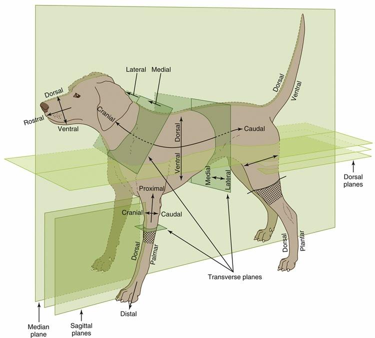
The terms that indicate position and direction must be mastered at once. These official terms are more precise than the common alternatives because they retain their relevance regardless of the actual posture of the subject They are defined in the following list, and their use is illustrated in Fig 1 1 We have not used them pedantically when there is no reasonable prospect of misunderstanding. When we use common terms (above, behind, and so forth), we always have in mind a standard anatomic position, which, for a quadruped, is that in which the animal stands square and alert It differs from the human anatomic position Medical anatomists make much use of the terms anterior and posterior and superior and inferior, all of which have very different connotations when applied to quadrupeds. These terms are therefore best avoided, except for a few specific applications to the anatomy of the head.
FIG 11 Directionaltermsandplanesoftheanimalbody The stippled areas representthecarpusand tarsusonforelimbsandhindlimbs,respectively
The principal recommended terms of position and direction are arranged in pairs, and it should be emphasized that they refer to relative, not absolute, positions. Most of these adjectives, which are listed here, form corresponding adverbs with the addition of the suffix ly Dorsal structures (or positions) lie toward the back (dorsum) of the trunk or, by extension, toward the corresponding surface of the head or tail
Ventral structures lie toward the belly (venter) or the corresponding surface of the head or tail. Cranial structures lie toward the head (cranium, literally skull), caudal ones toward the tail (cauda) Within the head, structures toward the muzzle (rostrum) are said to be rostral; caudal remains appropriate
Medial structures lie toward the median plane (medianus, in the middle) that divides the body into symmetrical right and left “halves ”
Lateral structures lie toward the side (latus, flank) of the animal
Different conventions apply within the limbs Structures that lie toward the junction with the body are proximal (proximus, near), whereas those at a greater distance are distal (distantia, distance) Within the proximal part of the limb (which is defined for this purpose as extending to the proximal limit of the carpus [wrist] or tarsus [hock, ankle]), structures that lie toward the “front” are said to be cranial, those that lie toward the “rear” caudal Within the remaining distal part of the limb, structures toward the “front” are dorsal (dorsum, back of the hand), and those toward the “rear” are palmar (palma, palm of the hand) in the forelimb or plantar (planta, sole of the foot) in the hindlimb Additional terms may be applied to the anatomy of the digits Axial structures lie close to the axis of a central digit, close to the axis of the limb if this passes between two digits; abaxial (ab, away from) positions are at a distance from the reference axis.
The terms external and internal, superficial, and deep (profundus) hardly require explanation or
definition
Sometimes it is necessary to refer to a section through the body or a part of it (see Fig 1 1) The median plane divides the body into symmetrical right and left halves. Any plane parallel to this is a sagittal plane, and those close to the median are sometimes termed paramedian planes A dorsal plane sections the trunk or other part parallel to the dorsal surface A transverse plane transects the trunk, head, limb, or other appendage perpendicular to its own long axis
AnIntroductiontoRegionalAnatomy
Although the first nine chapters that follow deal with systematic anatomy, those readers who are about to begin a laboratory course will find that they require an elementary knowledge of several systems at once. It is the principal purpose of the remainder of this chapter to supply that background. However, devoting some attention to the live animal also has benefits.
StudyoftheLiveAnimal
Regional anatomy is conveniently studied by means of dissection, but this approach has obvious limitations if the goal is knowledge of the anatomy of the living. The embalmed organs are inert and greatly changed in color and consistency from their living state The impressions gained in the dissection room or from prosection must therefore be modified and corrected by frequent reference to fresh material or radiographic images; by observation of surgical operations, whenever possible; and by application of the simpler methods of clinical examination to normal animals. We suggest that the student use many experiential learning opportunities to develop a strong foundation of anatomic knowledge
The simplest method is observation of the contours, the proportions, and the posture of the body Bony projections provide the clearest landmarks, but superficial muscles and blood vessels are also useful, if less striking; reference to these landmarks allows the positions of other structures to be deduced from their known relationships Little experience is required to reveal the importance of breed, age, sex, and individual variation or to show that although some landmarks are fixed and reliable, others are prone to move. Some (e.g., the costal arch) move with each respiration, whereas other features change more gradually, for example, becoming more or less prominent or shifting in position with the deposition or depletion of fat or with the advance of pregnancy
Structures that are not directly visible may be identified by palpation that is, by gentle or firmer touch as circumstances require. Bones may be identified by their rigidity, muscles by their contraction, arteries by pulsation, veins by swelling when the blood flow is interrupted by pressure, and lymph nodes and internal organs by their size, configuration, and consistency Nonetheless, variation is great and is affected by many factors. Palpation through the skin can be supplemented by digital or manual exploration per rectum and per vaginam.
Certain organs may be identified by percussion to elicit resonance when the overlying skin is struck a sharp blow (in a prescribed clinical fashion) Different materials produce different notes; a gas-filled organ is more resonant whereas a solid or fluid-filled emits duller notes. The normal activities of certain organs produce sounds continuously or intermittently. Although the lungs and heart (not forgetting the fetal heart) are the prime examples of organs whose positions can be determined by auscultation, the movement of blood within vessels or of gas or of ingesta within the stomach or intestines can also be a useful source of anatomic information. While applying these techniques one must remember that the vagaries of sound conduction through materials of different densities may provide a distorted indication of the position and dimensions of the source
The study of the anatomy of the live animal can be enlarged by other methods whose exercise requires considerable training and more elaborate apparatus than the simple stethoscope. These additional procedures have provided a variety of new illustrations scattered through later chapters; some elementary knowledge of how these illustrations were obtained may assist their appreciation, but detailed consideration of the various technologies involved is clearly beyond the scope of this book.
Many parts and cavities that are normally out of sight can be brought into view with the use of various instruments Perhaps the most familiar of these are the ophthalmoscope, used to study the fundus of the eye, and the otoscope, used to explore the external ear canal Other instruments for which the generic title “endoscope” is available may be introduced into natural orifices and advanced to allow inspection of deeper parts, such as the nasal cavity, bronchial tree, or gastric
lumen These examples of endoscopy are noninvasive, but other examinations require preparatory surgery Among these are arthroscopy, the inspection of the interior of synovial joints, and laparoscopy, the technique in which an endoscope is passed into the peritoneal cavity through a small opening in the abdominal wall. This latter technique may be employed for diagnostic purposes or for the visual control of (“keyhole”) surgery with the use of instruments introduced through separate portals
The rigidity of the early endoscopes limited their utility. The modern endoscopes use flexible fiberoptic systems and are remotely controlled The essential components of the fiberoptic instrument are two bundles of glass fibers One bundle is used to convey light distally, from an external source to the region to be viewed; the component fibers can be relatively coarse and randomly arranged. The second bundle conveys the image and is composed of finer fibers that maintain fixed positions in relation to one another The image is composed of many tiny units, each corresponding to an individual fiber, and is presented to the eye (or to a camera or video system) at the proximal end of the instrument.
Radiographic anatomy has for some time been an indispensable component of every course of anatomy X rays are produced by bombarding with electrons a tungsten target (focus) housed within a shielded tube Only a narrow x-ray beam is permitted to escape, and this beam is directed toward the relevant region of the subject. The passage of the rays through the body is affected by the tissues they encounter; tissues substantially composed of elements of high atomic weight tend to scatter or absorb the rays, and tissues substantially composed of elements of low atomic weight have proportionately less effect Because of its calcium content, bone clearly belongs to the first (radiopaque) category, whereas soft tissues generally belong to the second (radiolucent) category. Those rays that succeed in passing through the subject are allowed to impinge on a sensitive film (or other detector), which responds to the radiation received When the film is developed, mostly as digital images these days, those areas that were overlain by soft tissues (or gas-filled spaces) appear dark, even black, and those areas that were overlain by bone (or other radiopaque material) appear lighter, even white. The distinction between tissues of similar radiodensity may be enhanced by introducing an appropriate contrast agent to coat a surface or fill a space Specific methods utilizing various materials are available to depict such different features as the gastric lumen, urinary tract, and subarachnoid space.
Radiographic views are appropriately identified by reference to the direction taken by the x-ray beam in its passage through the subject Thus a radiograph of a supine animal, presenting its belly to the x-ray source, is described as a ventrodorsal film; that obtained with the animal turned over, with its belly now facing the film, is described as a dorsoventral film. The convention provides little scope for confusion but occasionally produces an awkward term, such as dorsolateral-plantaromedial, which specifies a particular oblique view of the hock
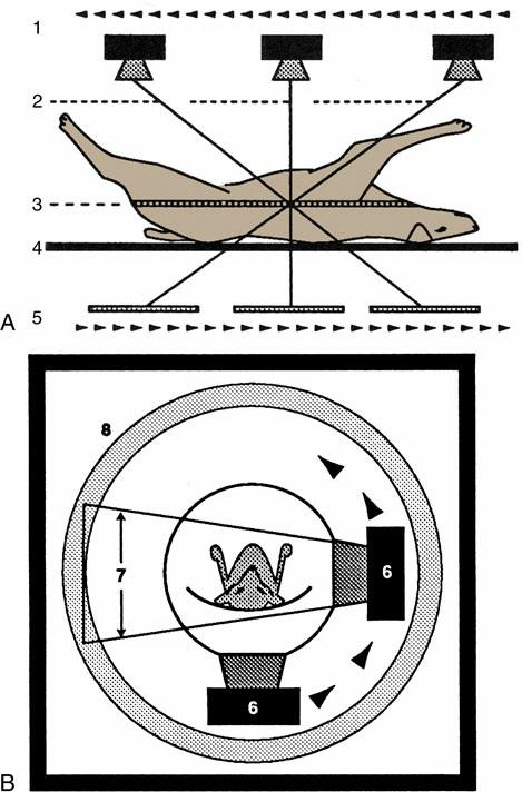
Awareness of certain general principles helps one avoid some common misinterpretations: the image of any structure is always magnified to the degree determined by the ratio between the distance from focus to film and distance from focus to object; the divergence of the x rays produces an apparent shift in position of any object not directly below the focus Two simple diagrams (Fig 1 2) will make these points clear A less easily resolved difficulty results from the superimposition of the images of structures that lie over each other. An ingenious, only partly successful, solution to this problem was sought in the coordinated movement in opposite directions of tube and film during the period of the exposure (Fig 1 3A) In this technique, known as tomography, the axis about which tube and film travel coincides with the plane of the horizontal slice of the subject that is of current interest. Structures contained within this slice remain more or less in focus throughout the exposure, whereas the images produced by structures at other levels are blurred or subsumed within the general background Such tomograms never found much employment in veterinary radiology The later developed and more sophisticated technique known as computed tomography (CT) has a different basis but retains the aim of clearly depicting the parts within one particular body slice while excluding extraneous images Despite the considerable cost of the apparatus and its limited suitability for use with large animals, the technique is now widely offered by veterinary referral centers
In the modern CT scanner, the x-ray source is moved in a circle that is centered on the longitudinal axis of the subject during the procedure, which takes from one to several seconds for its completion (see Fig 1 3B) During this time the movement of the tube is repeatedly arrested for very short periods; during each of these, a burst of radiation is directed through the subject along a different radius. The beams that penetrate the selected, very narrow slice of the subject impinge on
a series of discrete detectors or, in some designs, on portions of a continuous circumferential detector and are photomultiplied After the procedure is completed, these records are analyzed, compared, and combined according to complex formulae (algorithms); from these computations, a single cross-sectional image is constructed in which the forms, locations, and comparative radiodensities of all the tissues within the selected body slice are represented (Fig 1 4) In more complex settings, multiple overlapping or adjacent slices can be imaged in an extended, continuous process. With the amount of information the extended process supplies, it is possible by even more elaborate computation to construct images in other than transverse planes The data may also be manipulated to enhance subtle differences in contrast presented by tissues of very similar radiodensity
CT is, of course, not free of all drawbacks: subjects must be strictly immobilized during the exposure procedure; the total radiation dose may be quite considerable, even though individual exposures are very short, and the resulting images amplified; artifacts may produce deceptive images; and current apparatus designed for medical use is suitable for small animals but must be adapted for application with large animals and is then limited to the investigation of the head and limbs. One by-product of CT is the revival of interest in cross-sectional anatomy, an approach to the discipline that had been regarded as irretrievably passé but is now clearly indispensable to CT interpretation
Our knowledge of anatomy, especially the correlates of structure and function, will continue to grow through the implementation of new tools such as positron emission tomography–magnetic resonance imaging (PET-MRI), which exploits the high-resolution capability of MRI with the use of sophisticated imaging tracers in PET
Familiarity with cross-sectional anatomy is also required for the practice of ultrasonography This technique depends on the capacity of a piezoelectric crystal to convert electrical energy into sound