https://ebookmass.com/product/biochemical-pathways-an-atlasof-biochemistry-and-molecular-biology-2nd-edition/
Instant digital products (PDF, ePub, MOBI) ready for you
Download now and discover formats that fit your needs...
Biochemistry and Molecular Biology 6th edition Snape
https://ebookmass.com/product/biochemistry-and-molecular-biology-6thedition-snape/
ebookmass.com
Disease Pathways: An Atlas of Human Disease Signaling Pathways Anastasia Nesterova Et Al.
https://ebookmass.com/product/disease-pathways-an-atlas-of-humandisease-signaling-pathways-anastasia-nesterova-et-al/
ebookmass.com
BRS Biochemistry, Molecular Biology, and Genetics (Board Review Series) 7th Edition, (Ebook PDF)
https://ebookmass.com/product/brs-biochemistry-molecular-biology-andgenetics-board-review-series-7th-edition-ebook-pdf/
ebookmass.com
Geographical modeling: cities and territories Pumain
https://ebookmass.com/product/geographical-modeling-cities-andterritories-pumain/
ebookmass.com
TypeScript Basics: Learn TypeScript from Scratch and Solidify Your Skills with Projects 1st Edition Nabendu Biswas
https://ebookmass.com/product/typescript-basics-learn-typescript-fromscratch-and-solidify-your-skills-with-projects-1st-edition-nabendubiswas/
ebookmass.com
Dark Wine at the Grave (A Hill Vampire Novel Book 7) Jenna
Barwin
https://ebookmass.com/product/dark-wine-at-the-grave-a-hill-vampirenovel-book-7-jenna-barwin/
ebookmass.com
A Hundred Other Girls Iman Hariri-Kia
https://ebookmass.com/product/a-hundred-other-girls-iman-hariri-kia/
ebookmass.com
Adapting Television Drama: Theory and Industry 1st Edition
Christopher Hogg
https://ebookmass.com/product/adapting-television-drama-theory-andindustry-1st-edition-christopher-hogg/
ebookmass.com
Change of Heart Linda Hill
https://ebookmass.com/product/change-of-heart-linda-hill/
ebookmass.com
Campbell's Operative Orthopaedics 4-Volume Set 14th
Edition Frederick M. Azar
https://ebookmass.com/product/campbells-operativeorthopaedics-4-volume-set-14th-edition-frederick-m-azar/
ebookmass.com
3.6 Nucleotides and Nucleosides 124
Röbbe Wünschiers
3.6.1 Purine Nucleotides and Nucleosides 124
3.6.2 Pyrimidine Nucleotides and Nucleosides 130
3.7 Cofactors and Vitamins 133
Ida Schomburg
3.7.1 Retinol (Vitamin A) 133
3.7.2 Thiamin (Vitamin B1) 134
3.7.3 Riboflavin (Vitamin B2), FMN and FAD 135
3.7.4 Pyridoxine (Vitamin B6) 136
3.7.5 Cobalamin (Coenzyme B12, Vitamin B12) 137
3.7.6 Folate and Pterines 138
3.7.7 Pantothenate, Coenzyme A and Acyl Carrier Protein (ACP) 141
3.7.8 Biotin 141
3.7.9 Nicotinate, NAD+ and NADP+ 143
3.7.10 Ascorbate (Vitamin C) 145
3.7.11 Calciferol (Vitamin D) 146
3.7.12 Tocopherol (Vitamin E) 148
3.7.13 Phylloquinone and Menaquinone (Vitamin K) 148
3.7.14 Other Compounds 149
3.8 Nucleic Acid Metabolism in Bacteria 149
Susanne Peifer and Elmar Heinzle
3.8.1 Bacterial DNA Replication 149
3.8.2 Bacterial DNA Repair 151
3.8.3 Degradation of Nucleic Acids 156
3.9 Nucleic Acid Metabolism in Eukarya 157
Helmut Burtscher
3.9.1 Eukaryotic DNA Replication 157
3.9.2 Eukaryotic DNA Repair 162
3.10 Special Bacterial Metabolism and Biosynthesis of Antimicrobials 164
Julia Garbe, Annika Steen and Max Schobert
3.10.1 Bacterial Envelope 164
3.10.2 Bacterial Protein Export across the Cytoplasmic Membrane 166
3.10.3 Protein Transport across the Outer Membrane of Gram-Negative Bacteria 167
3.10.4 Bacterial Transport Systems 168
3.10.5 Bacterial Fermentations 169
3.10.6 Anaerobic Respiration 173
3.10.7 Chemolithotrophy 175
3.10.8 Quinoenzymes, Alkane and Methane Oxidation 178
3.10.9 Antibiotics 179
3.11 Electron Transfer Reactions and Oxidative Phosphorylation 183
Martina Jahn and Dieter Jahn
3.11.1 General Principles 183
3.11.2 Different types of electron transport chains 183
3.11.3 The Energetic Basis of the Oxidative Phosphorylation 183
3.11.4 Electron Transport System in Mitochondria and Bacteria 184
3.12 Photosynthesis 188
Dieter Oesterhelt and Josef Wachtveitl
3.12.1 Light Reaction 188
3.12.2 Dark Reactions 192
3.13 Plant Secondary Metabolism 193
Antje Chang
3.13.1 Phenolics 194
3.13.2 Terpenoids 198
3.13.3 Nitrogen-containing Secondary Metabolites 201
4 Protein Biosynthesis, Modifications and Degradation 210
4.1 Protein Synthesis in Bacteria 210
Martina Jahn and Dieter Jahn
4.1.1 Bacterial Transcription 210
4.1.2 Regulation of Bacterial Gene Expression 212
4.1.3 Bacterial Protein Synthesis 214
4.1.4 Degradation of Nucleic Acids 217
4.2
Protein Biosynthesis in Eukarya 219
Röbbe Wünschiers
4.2.1 Eukaryotic Transcription 219
4.2.2 Regulation of Eukaryotic Transcription 226
4.2.3 Eukaryotic Translation 228
4.2.4 Translational Regulation 231
4.2.5 mRNA Degradation 231
4.3 Cell Cycle in Eukarya 232
Stefan Ries
4.3.1 Core Components of the Cell Cycle Machinery 232
4.3.2 Cell Cycle Regulation in Yeast 234
4.3.3 G1 to S Transition in Mammalian Cells 234
4.3.4 G2 to M Transition in Mammalian Cells 235
4.3.5 Mitosis in Mammalian Cells 235
4.3.6 Cell Cycle Checkpoints 236
4.4 Posttranslational Modification of Proteins 238
Röbbe Wünschiers
4.4.1 Protein Processing in the Endoplasmic Reticulum 238
4.4.2 Glycosylation Reactions in the Golgi Apparatus 241
4.4.3 Terminal Carbohydrate Structures of Glycoconjugates 243
4.5 Protein Folding, Transport / Targeting and Degradation 244
Petra Dersch
4.5.1 Folding of Proteins 244
4.5.2 Vesicular Transport and Secretion of Proteins 248
4.5.3 Protein Transport into the Nucleus 249
4.5.4 Protein Transport into Mitochondria 252
4.5.5 Protein Transport into Chloroplasts 254
4.5.6 Protein Degradation 256
4.5.7 Protein Degradation by the Ubiquitin-Proteasome System 258
5 Viruses 261
Klaus Klumpp
5.1 General Characteristics of Viruses 261
5.1.1 Genomic Characteristics of Viruses 261
5.1.2 Structure 263
5.2 DNA Viruses 264
5.2.1 Papillomavirus 264
5.3 RNA Viruses 267
5.3.1 Hepatitis C Virus 267
5.4 Retroviruses 268
5.4.1 Human Immunodeficiency Virus (HIV) 268
6 Transport 272
6.1 Transport Through Membranes 272 Wilhelm Just
6.1.1 Systems of Eukaryotic Membrane Passage 272
6.1.2 Channels / Pores 272
6.1.3 Solute Carriers 276
6.1.4 Primary Active Transport Systems 277
6.1.5 Import by Endocytosis and Pinocytosis 278
6.1.6 The Cytoskeleton as Means for Intracellular Transport and Cellular Movements in Eukarya 278
6.2 Transport of Lipids in Plasma 279
Horst Klima
6.2.1 Apolipoproteins (Apo) 279
6.2.2 Plasma Lipoprotein Metabolism 279
6.2.3 Lipid Transport Proteins 281
6.2.4 Lipoprotein Receptors 281
6.2.5 Lipid Metabolic Disorders 282
6.3 Oxygen Transport by Hemoglobin 282
Gerhard Michal
6.3.1 Biosynthesis and Properties of Hemoglobin and Myoglobin 282
6.3.2 Oxygen Binding to Hemo- and Myoglobin 283
6.3.3 Hemoglobin Diseases in Humans 285
7 Signal Transduction and Cellular Communication 286
Gerhard Niederfellner
7.1 Intercellular Signal Transmission by Hormones 286
7.1.1 General Characteristics of Hormones 286
7.1.2 General Characteristics of Receptors 286
7.1.3 Insulin and Glucagon 287
7.1.4 Epinephrine and Norepinephrine (Catecholamines) 287
7.1.5 Hypothalamus-Anterior Pituitary Hormone System 287
7.1.6 Placental Hormones 291
7.1.7 Hormones Regulating the Extracellular Ca++, Mg++ and Phosphate Concentrations 292
7.1.8 Hormones Regulating the Na+ Concentration and the Water Balance 292
7.1.9 Hormones of the Gastrointestinal Tract 293
7.2 Nerve Conduction and Synaptic Transmission 294
7.2.1 Membrane Potential 294
7.2.2 Conduction of the Action Potential along the Axon 294
7.2.3 Transmitter Gated Signalling at the Synapse 294
7.2.4 Voltage Gated Signalling at the Synapse 296
7.2.5 Postsynaptic Receptors 296
7.2.6 Axonal Transport 296
7.3 Principles of Intracellular Communication 296
7.4 Receptors Coupled to Heterotrimeric G-Proteins 299
7.4.1 Mechanism of Heterotrimeric G-Protein Action 300
7.4.2 cAMP Metabolism, Activation of Adenylate Cyclase and Protein Kinase A 302
7.4.3 Activation of Phospholipase C and Protein Kinase C 302
7.4.4 Metabolic Role of Inositol Phosphates and Ca++ 303
7.4.5 Muscle Contraction 305
7.4.6 Visual Process 307
7.4.7 Olfactory and Gustatory Processes 308
7.4.8 Arachidonate Metabolism and Eicosanoids 309
7.5 Receptors Acting Through Tyrosine Kinases 311
7.5.1 Regulatory Factors for Cell Growth and Function 311
7.5.2 Components of the Signal Cascades 311
7.5.3 Receptor Tyrosine Kinases 312
7.5.4. Tyrosine Kinase-Associated Receptors (TKaR) 315
7.6 Programmed Cell Death (Apoptosis) 319
7.7 Receptors for Steroid and Thyroid Hormones, for Retinoids and Vitamin D 321
7.8 Cyclic GMP Dependent Pathways and Effects of Nitric Oxide (NO) 322
7.8.1 Membrane Bound Guanylate Cyclases 323
7.8.2 Soluble Guanylate Cyclases and Their Activation by Nitric Oxide (NO) 323
7.8.3 Protein Kinase G 323
8 Immune System 325
Ernst Peter Rieber
8.1 Components of the Immune System 325
8.1.1 Innate, Non Adaptive Immune System 325
8.1.2 Specific, Adaptive Immune System 328
8.1.3 Development and Maturation of the Cellular Components 328
8.1.4 Antigen Receptor of B Lymphocytes, Antibodies 330
8.1.5 Complement System 334
8.1.6 Antigen Receptor of T Lymphocytes 336
8.1.7 Antigen Presentation by MHC Molecules 337
8.1.8 Cytokines, Chemokines and Receptors 338
8.2
Generation of a Specific Immune Response 343
8.2.1 Activation of T Cells 343
8.2.2 CD4+ T Effector Cells, Regulation of the Immune Response 344
8.2.3 Activation of B Cells 345
8.2.4 Lymphocyte Circulation and Generation of Cellular and Humoral Immune Responses in Lymphoid Tissue 345
8.2.5 Cellular Cytotoxicity and Apoptosis 347
8.2.6 Interactions between the Immune System and the Neuroendocrine System 350
8.2.7 Immunological Tolerance 350
8.2.8 Induction of Specific Immune Responses against Pathogens 351
8.3 Pathologic Immune responses 352
8.3.1 IgE-Mediated Hypersensitivity of the Immediate Type 352
8.3.2 Autoimmunity 353
8.4 Adhesion of Leukocytes 354
Anton Haselbeck
9
Blood Coagulation and Fibrinolysis 357
Peter Müller
9.1 Hemostasis 357
9.2 Initial Reactions 358
9.2.1 Reactions Initiated by the Tissue Factor 358
9.2.2 Contact Activation 358
9.2.3 Generation of Binding Surfaces 358
9.3 Coagulation Propagation and Control 359
9.3.1 Requirements for Protease Activity 359
9.3.2 Pathways Leading to Thrombin 359
9.3.3 Key Events 359
9.3.4 Controlled Propagation 360
9.3.5 Generation of Fibrin 361
9.4 Platelets (Thrombocytes) 362
9.5 Fibrinolysis 364
9.5.1 Pathways of Plasminogen Activation 364
9.5.2 Control of Fibrinolysis 365
10 Biochemical Networks, Bioinformatics and Systems Biology 366
Dietmar Schomburg
10.1 Systems Biology and Networks 366
10.2 Modeling of Metabolic Fluxes 366
10.3 Biochemical Pathways Information Resources 366
10.3.1 Overview 366
10.3.2 Detailed Description of Some Databases 367
Index 374
1 Introduction and General Aspects Gerhard
Michal and Dietmar Schomburg
This book deals with the chemistry of living organisms. However, this topic cannot be considered in an isolated way, but has to be placed into a more general context. In two introductory chapters, a short outline of interconnections with neighboring sciences is given.
Chapter 1 deals with the organic chemistry of important components present in living organisms and with the physical chemistry of reactions.
Chapter 2 describes the overall organization of cells and their organelles as well as the structure of proteins and nucleic acids. This is followed by a discussion of enzyme function, which depends on the protein structure and regulates almost all biological processes.
The topics of Chapter 3 are various aspects of metabolism, showing the complex network with multiple interconnections.
Sections 3.1 … 3.6 are devoted to general metabolism, focusing on small molecules (carbohydrates, amino acids, tetrapyrroles, lipids including glycolipids, steroids, nucleosides and nucleotides).
Figures 1.1-1 … 1.1-3 give a simplified survey of the main metabolic pathways in order to allow quick location of the detailed descriptions in this book. The decimal classification numbers in the various boxes refer to chapters and sections. Figure 1.1-1, which abstracts Chapter 3, shows only biosynthetic pathways and sequences passed through in both directions (amphibolic pathways). This avoids a complicated presentation. (In the text, however, the degradation pathways of these compounds are usually discussed immediately following the biosynthesis reactions.) Most of the compounds mentioned here are ‘key compounds’, which appear in the detailed figures later in this book either at the beginning or at the end of the reaction sequences. The classification of these compounds into chemical groups is indicated by the color background of
Key to the background colors:
green = carbohydrates; blue = amino acids; red = lipids including steroids; orange = nucleotides; brown = tetrapyrroles; none = compounds involved in general interconversions. The colors of the frames are for easy differentiation only.
the names. Section 3.7 deals with cofactors and vitamins, which are involved in many reactions of general metabolism. Sections 3.8 and 3.9 describe the metabolism of DNA in bacteria and eukarya and the repair systems of these essential information carriers. The special metabolism of bacteria (including energy aspects), the biosynthesis and the effects of antibiotics are topics of Section 3.10. Aerobic respiration and its central role in energy turnover, as well as the photosynthetic reactions that are the source of almost all compounds in living beings, are discussed in Sections 3.11 and 3.12. Many special metabolic reactions take place in plants. These are summarized inSection 3.13.
The biosynthesis of proteins in bacteria and eukarya, and their consecutive modification, as well as the cell cycle, are discussed in Chapter 4.Figure 1.1-2 gives a short outline of these reactions, subdivided into bacterial reactions (left) and eukaryotic reactions (right).
Biochemical Pathways: An Atlas of Biochemistry and Molecular Biology, Second Edition. Edited by Gerhard Michal and Dietmar Schomburg. © 2012 John Wiley & Sons, Inc. Published 2012 John Wiley & Sons, Inc.
1.1-1. Biosynthetic Reactions in General Metabolism
1.1-2. Protein Biosynthesis
Figure
Figure 1.1-3. Cellular Communication
Figure
Viruses, which utilize these mechanisms in hosts, are discussed in Chapter 5
Chapter 6 gives a survey of transport mechanisms through membranes and within vessels.
The topic of Chapter 7 is cellular communication and the regulation mechanisms employed by multicellular organisms. Figure 1.1-3 briefly summarizes these multiple interconnections.
Chapter 8 deals with the defense mechanisms of higher animals and Chapter 9 with blood coagulation.
Every presentation can only contain a selection of the present knowledge. For this reason, the final Chapter 10, is intended to assist in obtaining further information from electronic sources, which offer the most comprehensive collection of scientific results available today.
1.1.1 Conventions Used in This Book
1. A decimal classification system is used throughout with the following subdivisions: chapters, sections, subsections. Figures, tables, and formulas are assigned to the relevant sections, e.g., Figure 3.7.6-1.
Reactions:
2. Whenever available, the "Accepted Names" as defined by the IUMB Biochemical Nomenclature Committee are used for enzymes and substrates. The enzyme classification scheme (EC numbers) and the transporter classification scheme (TC numbers) are listed in the index.
3. Substrates of enzymatic reactions are printed in black, enzymes in blue, coenzymes in red. Regulatory effects are shown in orange. This color is also used for pathway names and for information on the location of a reaction. For numbering systems, green is used.
4. The color of the reaction arrows shows where the reaction was observed (or at least where reasonable indications for its occurrence exist): black = general pathway, blue = in animals, green = in plants and yeasts, red = in prokarya (bacteria and archaea).
5. Bold arrows indicate main pathways of metabolism.
6. Points on both ends of an arrow ´ indicate noticeable reversibility of this reaction under biological conditions. Unless expressly noted, this type of arrow does not indicate mesomeric (resonance stabilized) states of a compound, contrary to usage in organic chemistry.
7. Double arrows are used when the interconversion of two compounds proceeds via different reactions in each direction (e.g., for some steps of glycolysis).
8. Dashed reaction arrows show conversions with primarily catabolic (degradative) importance. Full line arrows show either mainly anabolic (biosynthetic) reactions or reactions in biological systems which are frequently passed through in both directions (amphibolic reactions).
Regulation:
9. Necessary cofactors, activating ions etc. are printed in orange next to a reaction arrow.
10. Full line orange arrows with an accompanying or indicate that the respective factor exerts ‘fast’ activation or inhibition of the reaction (by allosteric mechanisms, product inhibition etc.). Dashed arrows are used if the amount of enzyme protein is regulated, e.g., by varied expression or by changes in the degradation rate. If only one of multiple enzymes is regulated in this way, it is indicated by Roman numbers.
Enzymes and Proteins:
11. When enzyme complexes are involved, the respective components are schematically drawn in blue-lined boxes with rounded edges. This does not express the spatial structure. If possible, interacting components are drawn next to each other.
12. When a sequence of domains occurs in a protein, special symbols are used for the individual domains. They are explained next to the drawing.
13. When the peptide chain has to be shown, helices are drawn as (e.g., in transmembrane domains), otherwise they are symbolized as
Abbreviations and Notations:
14. Organic phosphate is generally abbreviated as −P, inorganic phosphate and pyrophosphate as Pi and PPi respectively. In drawings where the reaction mechanism is emphasized, phosphate residues are shown as –O–PO32−.
15. Braces {} are used for atoms or residues which formally enter or leave during a reaction, if the molecular context is unknown.
16. While notations for genes are usually printed with small case letters (e.g., raf), the respective gene products (proteins) are written with a capitalized first letter (e.g., Raf). A number of proteins are defined by their molecular mass in kDa, e.g., p53.
17. When protein names are abbreviated, the notation frequently uses capitalized letters, e.g., cyclin dependent kinases = CDK in accordance with the literature.
18. A list of common abbreviations used throughout the book is given in 1.1.2. Less frequently used abbreviations are defined in the text.
Literature:
19. Only some recent references, primarily review articles and monographs, are listed at the end of the various sections. For more details refer to the literature quoted in these references, to electronic data banks, to review books and journals and to biochemistry textbooks.
20. Chapter 10 contains a survey on electronic data banks and a list of printed sources, which have been used frequently during the writing of this book.
1.1.2 Common Abbreviations
(Other abbreviations are defined in the text)
aaAmino acid
Acc, AccH2
Acceptor, reduced acceptor (unspecified)
ACPAcyl carrier protein
ATP, ADP, AMP, AAdenosine tri-, di-, monophosphate, adenosine bp, kbpbase pair (in DNA), kilobase pairs
cAMPCyclic AMP = adenosine 3,5-monophosphate
cGMPCyclic GMP = guanosine 3,5-monophosphate
CoA-SH, CoA-S-Coenzyme A
CTP, CDP, CMP, CCytidine tri-, di-, monophosphate, cytidine
CytCytochrome
Da, kDaDalton, kilodalton (unit of molecular mass)
dATP, dADP, dAMP, dADeoxyadenosine tri-, di-, monophosphate, deoxyadenosine dCTP, dCDP, dCMP, dCDeoxycytidine tri-, di-, monophosphate, deoxycytidine dGTP, dGDP, dGMP, dGDeoxyguanosine tri-, di-, monophosphate, deoxyguanosine dTTP, dTDP, dTMP, dTDeoxythymidine tri-, di-, monophosphate, deoxythymidine
DNADeoxyribonucleic acid
EEnzyme
EC numberEnzyme classification according to the IUBMB EC classification
EREndoplasmatic reticulum
ETFElectron transferring flavoprotein
F430 A corrinoid coenzyme (Ni)
FAD, FADH2 Flavin-adenine dinucleotide, reduced flavin-adenine dinucleotide
FdFerredoxin
FMN, FMNH2 Flavin mononucleotide, reduced flavin mononucleotide
FpFlavoprotein
DGChange of free energy (see 1.5.1)
G6PGlucose 6-phosphate
GSH, GSSGGlutathione, oxidized glutathione
GTP, GDP, GMP, GGuanosine tri-, di-, monophosphate, guanosine IgImmunoglobulin
ITP, IDP, IMP, IInosine tri-, di-, monophosphate, inosine
kVelocity constant of a reaction (1.5.4)
KEquilibrium constant of a reaction (see 1.5.1)
KS, KI, KD
Dissociation constants (see 1.5.4, 7.1.2)
KM Michaelis constant (see 1.5.4)
kbKilobases (103 bases)
l Wavelength of light
a-Lipoic acid, oxidized a-lipoic acid
NAD+, NADH + H+
NADP+, NADPH + H+
Nicotinamide-adenine dinucleotide, reduced nicotinamideadenine dinucleotide
Nicotinamide-adenine dinucleotide phosphate, reduced nicotinamide-adenine dinucleotide phosphate
ntNucleotide
NTP, NDP, NMP, NAny nucleotide tri-, di-, monophosphate or nucleoside
PAPAdenosine 3¢,5¢-diphosphate
PAPS3¢-Phosphoadenylylsulfate
PEPPhosphoenolpyruvate
P , Ppi
Inorganic phosphate, inorganic pyrophosphate
pHNegative decadic logarithm of the H+ concentration
pKNegative decadic logarithm of a dissociation constant
PQQPyrroloquinoline quinone
PRPP a-D-5-Phosphoribosylpyrophosphate
PyrPPyridoxal phosphate
RNARibonucleic acid
mRNA, rRNA, tRNAMessenger-, ribosomal-, transfer ribonucleic acid
R-S-S-RDisulfide group of amino acids or peptides
SSvedberg units (sedimentation coefficient)
SAHS-Adenosylhomocysteine
SAMS-Adenosylmethionine
THF5,6,7,8-Tetrahydrofolate
THMPT5,6,7,8-Tetrahydromethanopterin
ThPPThiamin pyrophosphate
UDPGUridine diphosphate glucose
UQ, UQH2 Ubiquinone, reduced ubiquinone
UTP, UDP, UMP, UUridine tri-, di-, monophosphate, uridine
Abbreviations for amino acids are listed in Figure 1.3.2, abbreviations for sugars in Figure 4.4.1-1.
1.2 Carbohydrate Chemistry and Structure
Carbohydrate monomers are of the general formula (CH2O)n. They have the chemical structure of aldehydes or ketones with multiple hydroxyl groups (aldoses and ketoses, respectively). A common name of monomers and dimers is ‘sugar’.
The large number of reactive groups, together with the stereoisomers causes a multiplicity of structures and reaction possibilities. Besides ‘pure’ carbohydrate monomers, oligomers (3.1.4) and polymers (3.1.2), carboxylic (3.1.5.1…2) and amino (3.1.7) derivatives, polyalcohols (3.1.5.5), deoxy sugars (3.1.5.6) etc., exist in nature.
Carbohydrates are the primary products of photosynthesis (3.12.2) and function as energy storage forms (e.g., starch, glycogen, 3.1.2), as part of nucleic acid and nucleotide molecules (3.6.1, 3.6.2), in glycoproteins (4.4) and glycolipids (4.4) and as structural elements in cell walls of bacteria (3.10.1), plants (3.4) and in the exoskeleton of arthropods (3.1). They are the most abundant chemical group in the biosphere.
1.2.1 Structure and Classification
The simplest carbohydrates are the trioses (C3 compounds) glyceraldehyde (an aldose) and dihydroxyacetone (glycerone, a ketose). Larger molecules are tetroses (C4), pentoses (C5), hexoses (C6), heptoses (C7) etc.; the C5 and C6 molecules are most common.
Glyceraldehyde is the smallest aldose with an asymmetric C-atom (chirality center). Therefore there are two stereoisomers (enantiomers), which cause right and left rotation of polarized light. By the Fischer convention, they are named D- and L-form, respectively. For details, see organic chemistry textbooks. Tetroses and larger carbohydrate monomers are classified (by comparison of the asymmetric center most distant to the aldehyde or keto group with D- or L-glyceraldehyde) as the D- and L-series of enantiomers (Fig. 1.2-1). With n-carbon aldoses, a total of 2n−2 stereoisomers exist, and with n-carbon ketoses there are 2n−3 stereoisomers. Epimers are stereoisomers, which differ in configuration at only one asymmetric C-atom. Most physiological sugars are of the D-configuration.
Aldopentoses, aldohexoses and ketohexoses (and higher sugars) can form cyclic structures (hemiacetals and hemiketals) by intramolecular reaction of their aldehyde or keto groups respectively with an alcohol group. This results in pyranoses (6-membered rings) and furanoses (5-membered rings, Fig. 1.2-2). In equilibrium, the cyclic structure is more prevalent as compared to the open structure. The ring closure produces another asymmetric C-atom; the respective stereoisomers are named anomers (a- and b-forms).
The nonplanar pyranose rings can assume either boat (in 2 variants) or chair conformation. The substituents extend either parallel to the perpendicular axis (axial, in Fig. 1.2-3 printed in red) or at almost right angles to it (equatorial, printed in green). The preferred
1.2-2.
Figure 1.2-1. Nomenclature of Carbohydrates
The compounds printed in green are formally obtained by epimerization at the indicated positions. The L-enantiomers are the mirror images at the perpendicular mirror plane.
Figure 1.2-3. Chair and Boat Conformations of Hexoses (Top) and Half-Chair (Envelope) Conformation of Pentoses (Bottom)
Figure
Ring Closure of Carbohydrates
conformation depends on spatial interference or other interactions of the substituents.
Although the bond angles of a furanose ring would permit an almost planar structure, the interference of substituents with each other causes a slight bending (puckering), e.g., to a half-chair (= envelope) structure in nucleotides and nucleic acids (Fig. 1.2-3).
The linear form of carbohydrates is usually shown as Fischer projection (ligands drawn horizontally are in front of the plane, ligands drawn vertically are behind the plane, e.g., in Fig. 1.2-1). The ring form is either drawn as Haworth formula (Fig. 1.2-2, disregarding the bent ring structure) or as boat/chair formula.
1.2.2 Glycosidic Bonds (Fig. 1.2-4)
If the hemiacetal or hemiketal hydroxyl of a sugar is condensed with an alcoholic hydroxyl of another sugar molecule, a glycosidic bond is formed and water is eliminated. Since this reaction between free sugars is endergonic (DG¢ 0 = 16 kJ/mol), the sugars usually have to be activated as nucleotide derivatives (3.1.2.2) in order to be noticeably converted. Depending on the configuration at the hemiacetal/ hemiketal hydroxyl (1.2.1), either a- or b-glycosides are formed. Sugar derivatives, which contain a hemiacetal or a hemiketal group (e.g., uronic acids) are also able to form glycosidic bonds.
Since sugar molecules contain several alcoholic groups, various types of bonds are possible. Frequently, 1 Æ 4 or 1 Æ 6 bonds occur. With oligo- or polysaccharides, both linear and branched structures are found. Bond formation may also take place with alcoholic, phenolic or other groups of non-sugar molecules (aglycons).
Literature:
Organic chemistry textbooks.
1.3 Amino Acid Chemistry and Structure
All amino acids present in proteins carry a carboxyl- and an amino group, hydrogen and variable side chains (R) at a single (a-)carbon atom. Thus, this Ca-atom is asymmetric (compare 1.2.1), with the exception of glycine, where R = H. Almost all of the proteinogenic amino acids occurring in nature are of the L-configuration. (The ‘L’ is assigned by comparison with L- and D-glyceraldehyde, which are taken as standards, Fig. 1.3-1). A number of D-amino acids are found in bacterial envelopes (3.10) and in some antibiotics (3.10).
Figure 1.2-4. Examples of Glycosidic Bonds
Unless otherwise stated, all amino acids discussed in the following sections are of the L-configuration.
Chains of amino acids form proteins and peptides. As enzymes, regulatory, mobility and structural compounds, they are the central components in all living beings. Therefore they are the topic of most of this book. Protein synthesis is described in Chapter 4. Their structure is discussed in Section 2.3, which also gives a short listing of their functions.
a) Non-polar, aliphatic amino acids. The non-polar side chains undergo hydrophobic interactions in protein structures. While the small glycine molecule allows high flexibility, the bulky proline confers enhanced rigidity to the structures.
GLYCINE (Gly, G)L-ALANINE (Ala, A) L-VALINE (Val, V)L-LEUCINE (Leu, L)L-ISOLEUCINE (Ile, I)L-PROLINE (Pro, P)
b) Polar, uncharged residues R. These functional groups are hydrophilic and can form hydrogen bonds with water or other polar compounds. Cysteine can easily be oxidized, resulting in intra- or intermolecular interconnections by disulfide bonds.
L-CYSTEINE (Cys, C)
L-SERINE (Ser, S)L-THREONINE (Thr, T)L-METHIONINE (Met, M)L-ASPARAGINE (Asn, A)L-GLUTAMINE (Gln, Q)
c) Aromatic residues R. The aromatic side chains are hydrophobic, while the hydroxyl group of tyrosine and the ring nitrogen of tryptophan form hydrogen bonds, which often play a role in enzyme catalysis.
L-PHENYLALANINE (Phe, F) L-TYROSINE (Tyr, Y)L-TRYPTOPHAN (Trp, W)
e) Negatively charged side chains R. The charged groups contribute in many cases to catalytic mechanisms and are also of influence to the protein structure.
L-ASPARATE (Asp, D) L-GLUTAMATE (Glu, E)
d) Positively charged side chains R. The charged groups contribute in many cases to catalytic mechanisms and also influence the protein structure.
L-LYSINE (Lys, K) L-ARGININE (Arg, R)L-HISTIDINE (His, H)
L-PYRROLYSINE (Pyl, O) f) Special amino acids using unusual genetic codes and found only in a small number of proteins (3.10.6.2, 4.1.3.4).
Figure 1.3-2. Amino Acids With Their 3- and 1-Letter Codes
L-SELENOCYSTEINE (Sec, U)
Figure 1.3-1. Asymmetric Center of Amino Acids
L-ORNITHINE (3.2.9)L-CITRULLINE (3.2.9)L-4-HYDROXYPROLINE (3.2.3)
(3.2.5.2)
(3.2.5.2)
-CARBOXYGLUTAMATE (9.3.1)
Figure 1.3-3. Some Nonstandard Amino acids
1.3.1 Structure and Classification
The individual properties of the amino acids are determined by the side chain R. This is also the criterion for amino acid classification.
There are 20 standard (classical) amino acids, which are incorporated as such into proteins, employing their own codons (4.1, 4.2). These amino acids are shown in Figure 1.3-2. Two additional amino acids, selenocysteine and pyrrolysine, are also incorporated directly by an unusual decoding procedure of mRNA (4.1). Nonstandard amino acids are produced by metabolic conversions of free amino acids (e.g., ornithine and citrulline) or by posttranslational modification of amino acids in proteins (e.g., by hydroxylation, methylation or carboxylation). Examples are given in Figure 1.3-3.
At about neutral pH, the free amino acids are ‘Zwitterion’ dipols with charged carboxylate (dissociation constant pK1 = 1.82 … 2.35) and amino groups (pK2 = 8.70 … 10.70). In seven cases, the side chains R also contain charged groups. Only the pKa of histidine (3.2.8) is in the physiological range. In Figure 1.3-2 and 1.3-3, the charged molecules are shown, while in the rest of the book, un-ionized forms are presented for reasons of simplicity.
1.3.2 Peptide Bonds (Fig.
1.3-1)
Proteins and peptides are linear chains of amino acids connected by peptide bonds between their a-amino and carboxylate groups. Since the formation of these bonds is endergonic, the reactants have to be activated as tRNA derivatives. Details are described in 4.1.3.
The peptide bonds are rigid and planar: The carboxylate-O and the amino-H are in trans conformation, the C–N bond shows partially double bond characteristics. Only peptide bonds followed by proline or hydroxyproline can alternatively be cis (6 … 10 %). To some extent, both bonds in the backbone of the peptide chain extending from Ca can perform rotational movements (although there are still constraints on most conformations, which are shown in Ramachandran diagrams). Flexibility and constraints play a major role in the proper folding of the proteins (1.3.1).
Proteins and peptides carry charged amino- (N-) and carboxy(C-) termini. Additional charges are contributed by the side chains. This allows analytical separation by electrophoresis. It has to be considered, however, that the pK a of amino acids in peptides differ from those in free amino acids due to the effects of neighboring groups.
Literature:
Meister, A. Biochemistry of the Amino Acids. 2 Vols. Waltham, (MA):Academic Press; 1965.
Ramachandran, G.N., Sasisekharan, V. Adv. Prot. Chem. 1968;23:326–367.
Rose, G.D. et al. Adv. Prot. Chem. 1985;37:1–109.
Organic chemistry textbooks.
1.4 Lipid Chemistry and Structure
The common properties of lipids are their hydrophobic character and their solubility in organic solvents. Otherwise, they belong to different chemical classes. The biochemistry of most of them is described in Chapter 3.4, some other lipids are discussed in their metabolic context elsewhere (see cross-references below).
1.4.1
Fatty acids
(Table 1.4-1, Fig. 1.4-1)
Fatty acids are characterized by a carboxylic group with a hydrocarbon ‘tail’. The higher fatty acids are practically insoluble in water and show typical lipid properties. They serve in esterified form astriacylglycerols for energy storage or are, as glycerophospholipids, part of cellular membranes. In contrast, the short-chain fatty acids are water soluble. They act as intermediates of metabolism and are discussed in the respective chapters.
Higher fatty acids can also enter an amide bond (e.g., in ceramides). Some are precusors of other compounds (e.g., of prostaglandins, 7.4.8). Almost none of them occur in free form.
The predominant fatty acids in higher plants and animals have an even number of C atoms in the range of C14 … C20 and are unbranched. Usually, more than half of all fatty acids are unsaturated. Monounsaturated fatty acids mostly contain a cis-double bond between C-9 and C-10. Often additional double bonds exist towards the methyl terminus, usually with two saturated bonds in between (polyunsaturated fatty acids). Some of them cannot be synthesized in animals and have to be supplied by food intake (essential fatty acids). The notation of fatty acids is (number of C atoms) : (number of double bonds), e.g., for linoleic acid 18:2. The location of the double bonds is given as, e.g., D9,12
Polyunsaturated fatty acids are not usually present in bacteria, but cis- and trans-monounsaturated, hydroxylated and branched fatty acids exist in many species.
Table 1.4-1. Higher Fatty Acids Frequently Occurring in Nature
Number of C atoms SaturatedUnsaturated, Number of Double Bonds E = Essential Fatty Acid for Humans
14myristic acid
16palmitic acid
18stearic acid1: oleic acid (D9)
2: linoleic acid (D9,12 ) E 3: a-linolenic acid (D9,12,15) E
20arachidic acid3: dihomo-g-linolenic acid (D5,8,11)
4: arachidonic acid (AA, D5,8,11,14 ) ( E)1
5: eicosapentaeonoic acid (EPA, D5,8,11,14,17)
24lignoceric acid1: nervonic acid (D15)
1 can be synthesized from the essential fatty acid, linoleic acid.
Figure 1.4-1. Structure of Saturated and Unsaturated Fatty Acids (18:0 and 18:1, showing the bend).
While saturated fatty acids tend to assume an extended shape, unsaturated fatty acids show 30° bends at their double bonds (Fig. 1.4-1). This reduces van der Waals interactions between neighboring molecules and lowers the melting point (see organic chemistry textbooks):
18:0 (70°) 18:1 (13°) 18:2 (−9°) 18:3 (−17°)
1.4.2 Acylglycerols and Derivatives (Fig.
1.4-2)
A major proportion of lipids occurring in plants and animals are triesters of glycerol (3.4.2) with higher fatty acids (triacylglycerols = triglycerides = neutral fat). In most of them, the fatty acids are different. Their type and the degree of their unsaturation determine the melting point.
Fats are solid and oils are liquid at room temperature. They are without influence on the osmotic situation in the aqueous phase due to their insolubility and do not bind water as, e.g., glycogen does. Thus, these compounds constitute an effective, convenient storage form of energy (ca.10 kg in adult humans).
Their degree of oxidation is lower than that of carbohydrates or proteins, therefore they provide higher energy during combustion: triolein yields 39.7 kJ/g. This is more than twice the value for anhydrous carbohydrates (17.5 kJ/g) or proteins (18.6 kJ/g) and about six times the energy gained from degradation of these alternative compounds in their physiological state due to their water content.
Triacylglycerols do not contain any hydrophilic groups. If, however, only one or two of the hydroxyl groups of glycerol are esterified (mono- or diacylglycerols), the remaining polar hydroxyl groups allow the formation of ordered structures at water-lipid interfaces and of lipid bilayers (1.4.8). Therefore they can act as emulsifiers, e.g., during lipid resorption from the intestine.
The remaining hydroxyl groups of mono- and diacylglycerols can also carry sugar residues. These so-called glycoglycerolipids are constituents of bacterial cell envelopes (3.10), thylakoid membranes in plants and myelin sheaths of neurons in animals. They are discussed in 3.4.
1.4.3 Waxes (Table 1.4-2, Fig. 1.4-2)
Waxes are esters of higher fatty acids with long-chain primary alcohols (wax alcohols) or sterols (Section 3.5), which are usually solid at room temperature.
They are more resistant than triacylglycerols towards oxidation, heat and hydrolysis (saponification). Frequently, they serve as protective layers, e.g., on leaves and fruits of plants or on skin, feathers and furs of animals (as secretions of specialized glands). Bees’ honeycombs are also formed of waxes. In many marine animals they are the main component of lipids (for regulation of flotation and for energy storage). Fossil waxes occur in lignite and bitumen.
Table 1.4-2. Common Components of Waxes
Alcohol (primary, saturated)Fatty Acid (saturated)
Cetyl alcohol (C16)Lauric acid (C12)
Carnaubyl alcohol (C24)Myristic Acid (C14)
Ceryl alcohol (C26)Palmitic acid (C16)
Myricyl alcohol (C30)Lignoceric Acid (C24)
Cerotic Acid (C26)
Montanic Acid (C28)
Melissic Acid (C30)
Figure 1.4-2. Structure of Acylglycerols, Glycoglycerolipids and Waxes
1.4.4 Glycerophospholipids (Phosphoglycerides, Fig. 1.4-3)
In contrast to triacylglycerols, in glycerophospholipids only two of the hydroxyl groups of glycerol are esterified with long chain fatty acids, while the group at the 3-position (according to the sn-numbering system) forms an ester with phosphoric acid.
All glycerophospholipids have an asymmetric C-atom in the 2-position, they occur in nature in the L-form. Most common are saturated fatty acids (C16 or C18) at the 1- and unsaturated ones (C16 … C20) at the 2-position. Removal of one fatty acid yields lysoglycerophospholipids.
Basic Structure: Diacyl-glycerophospholipidInositol, a ligand Y
ClassY =Formula of Y (F = Fatty acid)
Phosphatidic acidH–H
Phosphatidylethanolamineethanolamine–CH2–CH2–NH2
Phosphatidylcholine (lecithin)choline–CH2–CH2–N(CH3)3+
Phosphatidylserineserine–CH2–CH(NH2)–COOH
Phosphatidylinositolinositolsee above
Diphosphatidylglycerol (cardiolipin)
glycerolphosphatidic acid
–CH2–CHOH–CH2O–(PO2)––O–CH2–CHOF–CH2O–F
Figure 1.4-3. Classes of Glycerophospholipids
If the 3-position of glycerol carries only phosphoric acid, the compound is named phosphatidic acid. However, in most cases the phosphate group is diesterified. This extra residue (‘head group’, Y in Fig. 1.4-3) determines the class of the compound. These compounds are more polar than mono- or diacylglycerols and form the major part of biological membranes (1.4.8).
1.4.5 Plasmalogens
(Fig. 1.4-4)
This group of compounds is related to diacylglycerophospholipids (1.4.4). Also, the head groups (Y) are similar. However, the 1-position of glycerol is not esterified, but carries an a,b-unsaturated alcohol in an ether linkage. They are major components of the CNS, brain (>10 %), heart and skeletal muscles, but little is known about their physiological role.
Figure 1.4-4. Structure of Plasmalogens
1.4.6
Sphingolipids
(Fig. 1.4-5)
Sphingolipids are important membrane components. They are derivatives of the aminoalcohols dihydrosphingosine (C18), sphingosine (C18 with a trans double bond) or their C16, C17, C19 and C20 homologues. Ceramides are N-acylated sphingosines. If the hydroxyl group at C-1 is esterified with phosphocholine, phosphoethanolamine etc., sphingomyelins (sphingophospholipids) are obtained. If, alternatively, the hydroxyl group is glycosylated, glycosphingolipids (cerebrosides) result. This latter group of compounds is described in 4.4.2-3.
ClassA (= N-Substituent) ZFormula of Z
SphingosineHH–H
Ceramideshigher fatty acid H–H
Sphingomyelins higher fatty acid
Phosphoethanolamine or phosphocholine
–PO2–O-CH2–CH2–OH –PO2–O–CH2–CH2–N(CH3)3+
Glycosphingolipids higher fatty acid carbohydrate chains-carbohydrates
Figure 1.4-5. Basic Structure of Sphingolipids
1.4.7 Steroids
Steroids are derivatives of the hydrocarbon cyclopentanoperhydrophenanthrene (Fig. 1.4-6).
Figure 1.4-6. Structure of Cyclopentanoperhydrophenanthrene
Biologically important steroids carry many substituents: generally there is a hydroxy or oxo group at C-3. In addition, several methyl, hydroxy and oxo, in some cases also carboxy, groups are found. In many cases, there is a larger residue bound to C-17. Frequently, some double bonds are present. In a few cases, ring A is aromatic. Substituents below the ring system are designated a and above the ring system b (see Fig. 3.5.1-5).
Steroids are membrane components and participants as well as regulators of metabolism. A detailed description is given in Section 3.5.
1.4.8 Lipoproteins
The major function of lipoproteins is the transport of lipids. They contain non-polar lipids (triacylglycerols, cholesterol esters) in their core, surrounded by a layer of polar compounds (glycerophospholipids, cholesterol, proteins, Fig. 6.2-1). This group of compounds is discussed in context with their transport function in 6.2.
1.5 Physico-Chemical Aspects of Biochemical Processess
Some readers may be less inclined to deal with a fairly large number of mathematical formulas. However, formulas are necessary to describe biochemical processes quantitatively. Considering this, the mathematical part of this book has been concentrated into this section, while usually other chapters refer to it.
Only the most important equations required for discussion of biochemical reactions are presented. In order to facilitate their use, companion equations are given, which show the numerical values of the factors and the dimensions of the terms. For derivation of
Table 1.5-1. Measures and Constants (Selection)
the equations, refer to physical chemistry textbooks. The units and constants used in the following paragraphs are listed in Table 1.5-1.
1.5.1 Energetics of Chemical Reactions
To each component of a system, an amount of free energy G is assigned, which is composed of the enthalpy H (internal energy + pressure * volume) and of the entropy S (measure of disorder). While the absolute values are not of importance, the change of G (DG) is decisive for chemical reactions:
DG = DH − T * DS [1.5-1] or
DG [kJ * mol−1] = DH [kJ * mol−1] − T [K] * DS [kJ*mol−1 * K−1]. [1.5-1a]
A reaction proceeds spontaneously only if DG is negative.
In biochemistry, DG of reactions are usually listed as DG¢0, which is obtained at standard conditions of 298 K (25 °C), pH 7.0 and a reactant concentration of 1 mol/l each except for water, where the normal concentration of 55.55 mol/l and gases, where a pressure of 101.3 kPa (= 1 atm) are taken as unity and thus do not appear in the formula.
If the reactant concentrations (henceforth written as [X]) of a reaction A + B + … = Z + Y + … differ from 1 mol/l each, DG can be calculated by:
DG = DG 0 ¢ + R * T * 2.303 * log [Z] * [Y] *… [A] * [B] *… (end products) (starting comp.) [1.5-2] or
DG [kJ * mol−1] = DG 0 ¢ + 0.00831 * T * 2.303 * log [Z] * [Y] * … [A] * [B] * … [1.5-2a]
Reaction sequences can be calculated by addition of DG’s of the individual reactions.
A reaction is at equilibrium if DG = 0. Then the equilibrium constant K = [Z] * [Y] * … [A] * [B] * … (end products) (starting comp.) [1.5-3] can be calculated as follows: DG¢ 0 = −R * T * 2.303 * log K;
or DG¢ 0 [kJ * mol−1] = −0.00831 * T * 2.303 *
MeasureUnitEquivalents / Value of ConstantsEquivalents in SI Basic Units
Lengthmeter (m)1 mm = 10−3 m, 1 μm = 10−6 m, 1 nm = 10−9 m, 1 Å (Ångström) = 10−10 mSI basic unit
Volumecubic meter (m3)1 l (liter) = 10−3 m3; 1 ml = 10−3 l, 1 μl = 10−6 lDerived SI unit
Masskilogram (kg)1 g (gram) = 10−3 kg; 1 mg = 10−3 g, 1 μg = 10−6 gSI basic unit
Time (t)second (s)1 ms = 10−3 s, 1 μs = 10−6 s, 1 ns = 10−9 s, 1 ps = 10−12 sSI basic unit
Temperature (T)Kelvin (K)0 K = −273.16 °CSI basic unit
Quantity of matterMol1 mol = 6.0221 * 1023 [molecules or ions] 1 mmol = 10−3 mol, 1 μmol = 10−6 mol, 1 nmol = 10−9 mol, 1 pmol = 10−12 mol This unit is also applied to photons. 1 Einstein = 1 mol photons.
Electric currentAmpere (A)
SI basic unit
SI basic unit
ForceNewton (N)1 N = 1 [m * kg * s−2 ]1 N = 1 [m * kg * sec−2 ]
PressurePascal (Pa)1 Pa = 1 N * m−2, 1 kilopascal (kPa) = 103 Pa, 1 atm = 101.325 kPa1 Pa = 1 [m−1 * kg * sec−2 ]
EnergyJoule (J)1 J = 1 [N * m], 1 kilojoule (kJ) = 1000 J 1 cal (calorie) = 4.181 J, 1 kcal = 4.181 kJ (Non-SI unit)
1 J = 1 [m2 * kg * sec−2]
Electric chargeCoulomb (C)1 C = 6.241 * 1018 electron charges1 C = 1 [A * sec]
Electric potentialVolt (V)1 V = 1 [J * C−1], 1 mV = 10−3 V1 V = 1 [m2 * kg * sec−3 * A−1]
ConstantsAbbreviationEquivalents / Value of ConstantsEquivalents in SI Basic Units
Avogadro’s numberNN = 6.0220 * 1023 [mol−1] (see ‘quantity of matter’, above)N = 6.0220 * 1023 [mol−1]
Boltzmann’s constantkB kB = 1.3807 * 10−23 [J * K−1]kB = 1.3807 * 10−23 [m2 * kg * sec−2 * K−1]
Molar gas constantRR = N * kB = 8.31441 [J * mol−1 * K−1]R = 8.31441 [m2 * kg * sec−2 * mol−1 * K−1]
Faraday’s constantFF = 1 N electron charges = 96 484.5 [C * mol−1] = 96 484.5 [J * V−1 * mol−1]F = 96 484.5 [A * sec * mol−1]
Planck’s constanthh = 6.6262 * 10−34 [J * sec]h = 6.6262 * 10−34 [m2 * kg * sec−1]
In calculations in this book, usually l, g, kJ and mV are used. Since the constants have then to be expressed in these units, their numerical value changes by the factor 10−3 or 10−6 in the respective formulas.
Enzymes cannot shift the equilibrium, they only increase the reaction velocity. The kinetics of enzyme catalyzed reactions are discussed in 1.5.4.
1.5.2 Redox Reactions
Redox reactions are reactions where one compound is reduced (electron acceptor A) while its reaction partner is oxidized (electron donor B) by transfer of n electrons:
An+ ox + Bred = Ared + Bn+ ox
The change of free energy during such a reaction is described by a formula, which is analogous to Eq. [1.5-2]:
DG = DG 0 ¢ + R * T * 2.303 * log [Ared] * [B ox n+] [Aoxn+] * [Bred] (end products) (starting comp.) [1.5-5] or
DG [kJ * mol−1] = DG 0 ¢ + 0.00831 * T * 2.303 * log [Ared] * [B ox n+] [Aoxn+] * [Bred] [1.5-5a]
w expresses the work gained by transferring n mol charges (= n Faraday, F) across a potential difference of DE = Eend − Ebegin
w = −n * F * DE.
[1.5-6]
Since a positive amount of work diminishes the free energy of the system
w = −n * F * DE = −DG [1.5-6a] or
DG [kJ * mol−1] = n * 0.0965 * DE [mV], [1.5-6b]
equation [1.5-5] can also be written as:
DE = DE 0 ¢ + R * T n * F * 2.303 * log [Ared] * [B ox n+] [Aoxn+] * [Bred] (end products) (starting comp.) [1.5-7] or
DE [mV] = DE 0 ¢ + 0.00831 * T n * 0.0965 * 2.303 * log [Ared] * [B ox n+] [Aoxn+] * [Bred] [1.5-7a]
DE 0 ¢ is the difference of the redox potentials of this reaction (or the electromotive force across membranes, 1.5.3) under biochemical standard conditions (298 K = 25°C, pH 7.0 and a reactant concentration of 1 mol/l each). Only water, which is present in a concentration of 55.55 mol/l and gases, with a pressure of 1 atm are taken as unity.
Redox potentials: The reaction can be divided into two half reactions (e = electrons):
Ared = An+ ox + n e and Bred = Bn+ ox + n e
The zero value of the redox potential is by convention assigned to the potential of the half reaction 2 H+ + 2 e = H2 at a platinum electrode at pH = 0, 298 K (25°C) and a hydrogen pressure of 101.3 kPa (= 1 atm). Thus, under the standard conditions used in biochemistry (pH = 7.0), E 0 ¢ (2H+/H2) = −410 mV.
Correspondingly, the half reactions can be expressed as:
EA = (E 0 ¢ )A + R * T n * F * 2.303 * log [Aoxn+] [Ared] [1.5-8] or EA [mV] = (E 0 ¢ )A + 0.00831 * T n * 0.0965 * 2.303 * log [Aoxn+] [Ared] [1.5-8a]
and analogously for B.
Various redox potentials can be combined this way: DE = EB − EA (A being the electron acceptor and B being the electron donor). The reactions proceed spontaneously only if DE is negative, i.e., when the potential changes to a more negative value.
Redox potentials are usually plotted with the minus values on top. A spontaneous reaction proceeds in such a plot from top to bottom (e.g., Fig. 3.12-6).
In the literature, the definition of DE is not uniform. In a number of textbooks it is defined in opposite order to the above: DE = Ebegin − Eend Therefore, DE and DE0 have to be replaced by −DE and −DE¢ 0, respectively. This affects Eqs. [1.5-6] … [1.5-8a] and has to be considered when making comparisons.
1.5.3 Transport Through Membranes
Uncharged molecules: If an uncharged compound A is present on both sides of a permeable membrane in different concentrations, its passage through the membrane is accompanied by a change of free energy. In biochemistry, this situation occurs mostly at cellular membranes (or membranes of organelles). For import into cells, the following equation applies:
DG = R * T * 2.303 * log [Ainside] [Aoutside] [1.5-9] or
DG [kJ * mol−1] = 0.00831 * T * 2.303 * log [Ainside] [Aoutside] [1.5-9a]
Thus, the transport occurs spontaneously only at negative DG, (when [Ainside] < [Aoutside]), i.e, from higher to lower concentrations. Correspondingly, for export from cells, the quotient is reversed
DG = R * T * 2.303 * log [Aoutside] [Ainside] [1.5-9b]
Charged molecules: The situation is more complicated if there is a potential difference DY across the membrane (e.g., by nonpenetrable ions)
DY = Yinside − Youtside [1.5-10] and the compounds passing through the membrane carry Z positive charges/molecule (or −Z negative charges/molecule). The contribution of the charges to DG (with the prefix of Z corresponding to the + or − charge of the ions) is expressed by:
DGcharge transport = Z * F * DY [1.5-11] or
DGcharge transport [kJ * mol−1] = Z * 0.0965 * DY [mV]. [[1.5-11a]
Thus, for an import process, Eq. [1.5-9] and Eq. [1.5-11] have to be combined:
DG = R * T * 2.303 * log [Ainside] [Aoutside] + Z * F * DY [1.5-12] or
DG [kJ * mol−1] = 0.00831 * T * 2.303 * log [Ains.] [Aouts.] + Z * 0.0965 * DY [mV]. [1.5-12a]
Correspondingly, for an export process,
DG = R * T * 2.303 * log [Aoutside] [Ainside] − Z * F * DY [1.5-12b]
The prefix of the last term in this equation is the opposite one of Eq. [1.5-12], since the membrane potential (Eq. 1.5-10) has the opposite effect on the energy situation.
An equilibrium exists if DG = 0. Then the equilibrium potential DY0 [mV] can be obtained by the Nernst equation:
DY0 = − R * T Z * F * 2.303 * log [Ainside] [Aoutside] [1.5-13] or DY0 [mV] = − 0.00831 * T Z * 0.0965 * 2.303 * log [Ainside] [Aoutside] [1.5-13a]
An extension of this formula to the equilibrium potential of several ions is the Goldman equation (see 7.2.1).
Literature:
Physical chemistry textbooks.
1.5.4 Enzyme Kinetics
The biochemical base of enzyme catalysis is discussed in 2.4. In the following, the mathematical treatment of the kinetics is given in some more detail.
Velocity of reactions: The reaction rate v for conversion of a single compound A Æ product(s) (first order reaction) is proportional to the concentration of this compound [A], while for a two-compound reaction A + B Æ product(s) (second order reaction) it depends on the number of contacts and thus on the concentration of both components (Eq. [1.5-14] and Eq. [1.5-15]). The proportionality factor k is termed rate constant.
Eq. [1.5-15] can also be applied for the formation of a complex and Eq. [1.5-14] for the decomposition of this complex. This includes substrate-enzyme complexes (see below), ligand-receptor complexes (7.1-2), antigen-antibody complexes (8.1.4) etc.
v = − d [A] dt = k * [A][1.5-14]
v = − d [A]
dt = − d [B] dt = k * [A] * [B]
[1.5-15]
Enzyme catalyzed one-substrate reaction: The theory of the enzymecatalyzed conversion of a single reactant (the substrate, S) is based on the assumption that the enzyme (the catalyst, E) and this substrate form a complex (ES) by a reversible reaction. This step is kinetically treated like a two-compound reaction (rate constants k1 and k−1 for formation and decomposition, respectively). The complex is then converted into the product (P) with the rate constant k2. The conversion into P is considered to be irreversible at the beginning of the reaction, when practically no product is present.
E + S k1 ´ k−1 ES Æ E
Therefore, for the formation of the enzyme-substrate complex, Eq. [1.5-15] has to be applied, while for its decomposition into its components, as well as for its conversion to the products, Eq. [1.5-14] is valid. There is actually an intermediate step ES Æ EP before the product is released. Its rate constant is not treated as a separate entity in most discussions of kinetic behavior, but is combined with the dissociation step to k2. This is also done in the following considerations.
Usually, the substrate is in large excess over the enzyme. In this case, after a short ‘transient phase’, [ES]can be considered to be sufficiently constant (steady-state assumption). Disregarding the reverse reaction by using the situation immediately after the transient phase (see above) one obtains
d [ES]
dt = 0 = k1 * [E] * [S] − k−1 * [ES] − k2 * [ES] [1.5-17]
If one assumes that the rate determining process is the reaction ES Æ E + P, the initial reaction rate v0 can be written as a function of [ES], which is analogous to Eq. [1.5-14]
v0 = k2 * [ES].
[1.5-14a]
By using a term for the total concentration of enzyme [Et] = [E] + [ES], by expressing the maximum reaction rate Vmax, which is obtained when all of the enzyme is saturated with substrate ([ES] = [Et]) as V max = k2 * [Et], [1.5-14b] and by introducing the Michaelis constant KM
KM = k−1 + k2 k1 ,
one obtains the so-called Michaelis-Menten equation
v0 = V max * [S] KM + [S] ,
1.5-1. Reaction Velocity of an Enzyme Catalyzed Reaction The velocity at [S] = n * KM is shown.
[1.5-18]
[1.5-19]
which shows the dependency of the reaction rate on the substrate concentration (first-order reaction). The plot of reaction rate vs. substrate concentration is a rectangular hyperbola (Fig. 1.5-1).
These formulas describe only the forward reaction. If the reverse reaction is included, the equivalent to Eq. [1.5-19] is
v = (Vmax)f * [S] (KM)f − (Vmax)r * [P] (KM)r 1 + [S] (KM)f + [P] (KM)r
[1.5-20]
where (Vmax)f and (KM)f are identical to V max and KM in Eq. [1.5-19], while the terms (Vmax)r and (KM)r = (k−1 + k2)/k−2 are formed analogously for the reverse reaction.
Michaelis constant: As can be derived from Eq. [1.5-19], the Michaelis constant KM equals the substrate concentration at half the maximal reaction rate. Most of them are in the range of 10−5…10−1 mol/l (Fig. 10.3-3).
Instead of obtaining this value from a plot according to Figure 1.5-l, it is more convenient to use the reciprocal of the Michaelis-Menten equation, which yields a linear plot (at least in the ideal case, Lineweaver-Burk plot, Fig. 1.5-2a):
1 v0 = KM V max * [S] + 1 V max
[1.5-21]
If 1/v0 is plotted vs. 1/[S], then the intersections of this line with abscissa and ordinate allow the determination of KM and V max
A disadvantage of the Lineweaver-Burk plot is the accumulation of measuring points near the ordinate (see the markings on the abscissa of Fig. 1.5-2a). Therefore other ways of plotting have been proposed. Hanes used another transformation of the Michaelis-Menten equation: [S] v0 = KM V max + [S] V max
[1.5-21a]
The plot of [S]/v0 vs. [S] yields a line with the abscissa intersection −KM and the ordinate intersection KM/Vmax. The slope equals 1/Vmax (Fig. 1.5-2 b).
Still another method, the so-called ‘direct plot’, has been proposed by Eisenthal and Cornish-Bowden. The Michaelis-Menten equation is rearranged as follows: V max = v0 + v0 [S] * KM
[1.5-21b]
For each individual measurement, −[S] is marked on the abscissa and v0 on the ordinate and a line is drawn through both points. The intersection of these lines has the abscissa value KM and the ordinate value V max (Fig. 1.5-2 c).
However, the most accurate method is the statistical evaluation of the measurements. In spite of this, the Lineweaver-Burk plot will be used in the following graphical representations, since it is the best known one.
Characterization of enzyme activities: The enzyme activity is defined as the quantity of substrate turned over per time unit in the presence of a given amount of enzyme. Thus the standard dimension would be [mol * sec−1] = katal. For practical reasons, usually the
Figure
activity is expressed as [μmol * min−1]. This term is named International Unit (U) if the measurement is performed under standard conditions (with isolated enzymes at conditions that are optimized as much as possible). The specific activity is the enzyme activity per unit of weight, e.g., per mg and is frequently used to characterize the degree of purification of isolated enzymes.
The turnover number of an enzyme is defined as the number of molecules converted by one molecule of enzyme per unit of time if the enzyme is saturated with substrate ([Et] = [ES]). It is identical to the rate constant k2 and can be calculated from Eq. [1.5-14 b] as k2 = Vmax/[Et]. Most turnover numbers are in the range of 1 … 104 (see Fig. 10.3-4), the value for catalase is 4 * 107
Most reactions in vivo proceed below the saturation limit of the enzyme, frequently at [S] = 0.01 … 1 KM. By the combination of Eq. [1.5-17], Eq. [1.5-18] and Eq. [1.5-14 a] one obtains
v0 = k2 KM * [E] * [S] [1.5-22].
At low substrate concentration, only a small portion of the enzyme forms an enzyme-substrate complex and [E] ª [Et] = constant. The term k2/KM indicates how often a contact of enzyme and substrate leads to a reaction and is therefore a measure of the catalytic efficiency. It has an upper limit of ca. 109 [l * mol−1 * sec−1], when practically every contact leads to a reaction, and the reaction rate is determined by the diffusion speed. The value for catalase (4 * 108) is one of the highest observed.
Figure 1.5-3. Lineweaver-Burk Plots of Inhibited Reactions
Red = uninhibited reaction, blue = inhibited reaction, arrow = shift of the plot at increasing inhibitor concentrations.
Figure 1.5-2. Linear Plots of an Enzyme Catalyzed Reaction
Inhibition: The mathematical treatment of an inhibited reaction depends on the mechanism of the inhibition. The general principles of inhibition are described in 2.5.2.
Competitive inhibition: The inhibitor competes with the substrate for reversible binding to the active site of the enzyme. The enzyme-substrate and the enzyme-inhibitor complexes are formed with the dissociation constants KS and KI, respectively.
K s = [E] * [S] [ES]
K1 = [E] * [I] [EI]
This results in the equation
[1.5-23]
[1.5-23a]
1 v0 = KM V max * [S] * ( 1 + I K1 ) + 1 V max [1.5-24]
In the Lineweaver-Burk plot, lines obtained at different inhibitor concentration intersect at the ordinate (Fig. 1.5-3a).
Uncompetitive inhibition: The inhibitor reacts reversibly only with the enzyme substrate-complex, but does not affect its formation. The dissociation constant is K¢ I
K 1 ¢ = [ES] * [I] [ESI]
This yields the equation
[1.5-25]
1 v0 = KM V max * [S] + 1 V max ( 1 + [I] K 1 ¢ ) [1.5-26]
In the Lineweaver-Burk plot, parallel lines are obtained at different inhibitor concentrations (Fig. 1.5-3b).
Noncompetitive and mixed inhibition: If the inhibitor binds both to the enzyme and to the enzyme-substrate complex according to Eqs. [1.5-23a] and [1.5-25] and prevents formation of the product, the following equation results
1 v0 = KM V max * [S] * ( 1 + [I] K1 ) + 1 V max * ( 1 + [I] K 1 ¢ )
If the affinities of the inhibitor to the enzyme and to the enzyme-substrate complex are equal (KI = K¢ I ), then the lines obtained at different inhibitor concentrations intersect in the Lineweaver-Burk plot at the negative abscissa (KM remains unchanged, noncompetitive inhibition, Fig. 1.5-3c). Otherwise, they intersect in the second quadrant (left of the ordinate, mixed inhibition, Fig. 1.5-3d).
Inhibition by excessive substrate concentrations. If the reaction velocity decreases at very high substrate concentrations, this results in a Lineweaver-Burk curve bent upwards near the ordinate. This situation is mostly observed in in vitro experiments (Fig. 1.5-3e).
Two-substrate reactions: The formulas describing the kinetics are derived from the same assumptions as for one-substrate reactions. Their form depends on the reaction sequence. They involve separate Michaelis constants for the turnover of each substrate.
The Cleland nomenclature system uses the following expressions for the number of the substrates and products of the reaction; 1 – Uni, 2 – Bi, 3 – Ter, 4 – Quad. The substrates are named A, B, C … , the products P, Q, R … and the enzyme species (original state, intermediates and final state) E, F, G … If all components have to combine before the reaction takes place, this is called a sequential reaction. This may take place in an ordered way or at random. If, however, one component leaves the enzyme before the other enters, it is a ping-pong reaction. The mechanisms are schematically drawn in Figure 1.5-4.
The formula for an ordered sequential Bi-Bi reaction is 1 v0 = 1 V max + (KM)A
The general formula for random sequential reactions is very complicated. A ping-pong Bi-Bi reaction is described by 1
1.5-4. Types of Two-Substrate-Two-Product (Bi-Bi) Reactions
The enzyme is represented by the horizontal line.
If in second order reactions the concentration of one of the substrates is very much above the respective Michaelis constant, then the terms containing this concentration in Eqs. [1.5-28] and [1.5-29] are practically zero and the equations become identical with Eq. [15.1-21], allowing the same evaluation as with a first order reaction. If a series of measurements are made in which one substrate is varied while the other is kept constant, then one obtains LineweaverBurk plots that formally resemble those obtained with inhibited reactions. However, increasing concentrations of the second substrate shift the lines in the other direction (Fig. 1.5-5). Ordered sequential
1.5-5. Lineweaver-Burk Plots of Two-Substrate Reactions Arrow = shift of the plot when the concentration of the other substrate is raised.
Figure
Figure
mechanisms yield a series of lines, which intersect left of the ordinate (above or below the abscissa), while ping-pong mechanisms yield parallel lines.
Dependence of reactions on temperature and activation energy: A more refined consideration of the reaction sequence Eq. [1.5-16] shows that only collisions of the reactants above a certain energy level will lead to the formation of complexes, e.g., ES and EP. Also, the reaction ES Æ EP requires an initial energy input. Thus, the reaction has to cross ‘energy hills’, which represent metastable states (Fig. 2.4-1). They are called transition complexes Xπ and can either return to the original components or progress towards the products of the reaction, quickly achieving equilibrium in both cases. Among the ‘energy hills’ mentioned above, the highest one represents obviously the rate determining step of the reaction and has to be the one considered further. (It takes the place of [ES] in the previous equations.) Thus, the equilibrium for formation of this complex can be described analogously to Eq. [1.5-3] by
K = [X*] [A] * [B]
[1.5-30]
The energy required for its formation is called activation energy DGπ, which can be calculated from this equilibrium by applying Eq. [1.5-4] as
DGπ = −R * T * 2.303 * log [X*] [A] * [B] [1.5-31]
According to Eq. [1.5-14] the reaction rate for formation of the product(s) from this complex is expressed by v0 = k * [Xπ]. When combining this equation with Eq. [1.5-31], one obtains the following formula for the temperature and DGπ dependence:
v0 = const. * [A] * [B] * 10 DGπ / 2,303 * R * T [1.5-32]
The increase of the reaction rate with rising temperature is limited, however. When the enzyme becomes thermally denatured, the rate drops (Fig. 2.4-4).
Fractal enzyme kinetics: The above considerations assume ‘ideal’ conditions; purified enzymes, low concentrations, free movement of the reactants. However, in vivo the situation is different. Based on a
power-law derivation it has been shown that, e.g., restrictions in space require the introduction of non-integer powers > 1 to the concentration terms in Eq. [1.5-17]:
The consecutive equations change analogously. This system is called fractal kinetics. Its main implications are:
• KM is dependent on the enzyme concentration; it decreases with increasing enzyme concentration.
• The plot of enzyme activity vs. substrate concentration has a tendency towards a sigmoid shape even with monomeric enzymes.
• The velocity of the reaction increases if the movements are, e.g., restricted to surface interfaces (e.g., 3.4.3.2) or to one dimension (e.g., by sliding along nucleic acid strands, 3.8.1.2, 4.2.3.2 or by ‘substrate channeling’, 3.2.7.1).
• In sequences of reactions, the flux responses are faster and the accumulation of intermediates is lower as compared to the Michealis-Menten assumption.
In some respects, fractal kinetics resemble allosteric situations (2.5.2). Velocity calculations according to this theory have a tendency to yield higher values as according to the Michaelis-Menten theory, which represents a borderline case of a more general treatment, but is still of value for understanding the basic principles of enzyme catalysis.
Literature:
Cornish-Bowden, A., Wharton, C.W. Enzyme Kinetics. Oxford: IRL Press; 1988.
Dixon, M., Webb, E.C. Enzymes. 3rd Ed. Waltham (MA): Academic Press; 1979.
Fersht, A. Structure and Mechanism in Protein Science. New York: W.H. Freeman & Company; 1999.
Freeman (1985). Savageau, M.A. J. Theor. Biol. 1995;176:115–124. Sigman, D.S., Boyer, P.D. (Eds.). The Enzymes. 3rd Ed. Vols. 19 and 20. Waltham (MA): Academic Press; 1990 and 1992.
2 The Cell and Its Contents
Gerhard Michal and Dietmar Schomburg
This chapter presents selected information on the structure and organization of living organisms and their major components to serve as a background for the biochemical text of this book. For more details, refer to biology textbooks.
2.1 Classification of Living Organisms
Life is associated with a number of characteristics such as propagation, metabolism, response to environmental influences, and evolution. Cells are the basic unit of organization for all living beings. Whereas unicellular organisms exist as separate entities, the various cells of multicellular organisms fulfill different functions, and the organism depends on mutual cellular interaction.
There are several systems of classification of living organisms. From a phylogenetic viewpoint, the classification into the three domains; bacteria, archaea and eukarya (which are further subdivided) appears most justified (Table 2.1-1). When common aspects of eubacteria and archaea are discussed, the term prokarya is used.
The metabolic reactions in this book are indicated by colored arrows. Since frequently the occurrence of the reactions is known only for a few species and also in order to prevent an ‘overloading’ of the figures with too much detail, the arrow colors have been combined into (black) general metabolism, (red) bacteria and archaea, (green) plants, fungi and protists, (blue) animals.
Living organisms exhibit a high degree of order. The sum of all endogenous life processes results in a steady decrease of free energy
Table 2.1-1. Some Typical Properties of Living Organisms (Exceptions exist)
(1.5.1). Therefore, life can only be kept up by an energy input from the environment, either as light energy or by uptake of oxidizable compounds. Another essential requirement of life is the availability of an adequate carbon source. Living beings can be classified according to the mode of energy uptake and the carbon source used (Table 2.1-2).
During the oxidation of compounds, electrons are released, which have to be taken up by a terminal electron acceptor. Energy wise, oxygen is most favorable (3.11): previous to its appearance in the primeval atmosphere, living organisms had to use other acceptors. This is still the case in oxygen-free habitats (Table 2.1-3).
Literature:
Fox, GE. et al.: Science 1980;209:457–463.
Holt, JG. et al.: Bergey’s Manual of Descriptive Bacteriology (9th Ed.). Williams and Wilkins (1994).
Margulis, L., Schwartz, K.V.: Five Kingdoms. 2nd. Ed. Freeman (1987).
Woese, CR. et al.: Structure of Cells. Proc. Natl. Acad. Sci. USA 1990;87:4576–4579.
2.2 Structure of Cells
2.2.1 Prokaryotic Cells (Fig. 2.2-1)
The genetic information is stored in a single, circular double helix of deoxyribonucleic acid (DNA, 2.6.4). It is located in the central portion of the cell in a densely packed form (nucleoid), but without
DomainsBacteriaArchaeaEukarya
KingdomsBacteriaArchaeaProtists1
PlantsFungiAnimals
Nucleusno (common term prokarya)Yes
Genomecircular, ca.106 … 5 * 107 kb, extra plasmidslinear, 107 … > 1011 kb, organized in several chromosomes
RNA polymeraseone typeseveral types
Starting amino acid for translation formylmethioninemethionine
Reproductionbinary scissionasexual/sexual
Cellular organizationunicellular (some are aggregated)mostly unicellularmulticellularuni-/multicellularmulticellular
Nutrition (Table 2.1-2) chemoorganotrophic, photoautotrophic or photoheterotrophic
chemolithotrophic, photoautotrophic or chemoorganotrophic.
chemoorganotrophic or photoautotrophic
photoautotrophicchemoheterotrophic including saprobiontic chemoheterotrophic
Size of cells average 1 … 5 μm, wide variationaverage 10 … 100 μm, wide variation
Cell membranesrigid, contain peptidoglycans rigid, without peptidoglycans rigid or softrigid, contain cellulose and lignin rigid, contain chitin soft, lipid bilayer only
Internal membranesno yes, they enclose organelles /vesicles
1 Algae, protozoa, fungi-related. The exact demarcation is under discussion.
Table 2.1-2. Sources of Carbon and of Energy
Phototrophy (Energy input by light)
Autotrophy (only CO2 needed as carbon source) green plants, some protists, photosynthesizing bacteria (3.12)
Chemotrophy (Energy provided by oxidizable compounds from the environment)
Prokarya (mainly archaea). Oxidation of inorganic material (chemolithotrophy, 3.10.7)
Heterotrophy (organic compounds needed as carbon source) some prokaryaAll animals and fungi, non-green plants, many protists and prokarya. Oxidation of organic material (chemoorganothrophy). Included are saprobionts (use decaying organic material) and parasites (feed from living beings)
Table 2.1-3. Terminal Electron Acceptors for Oxidation Reactions
Atmospheric Oxygen
Not requiredRequired
Energy obtained by anaerobic respiration (3.10.6) fermentation (3.10.5) aerobic respiration (3.11)
Electron acceptors oxidized external compounds (mostly inorganic) internally generated compounds atmospheric O2
Organisms anaerobes: part of archaea and bacteriaall other organisms facultative anaerobes (bacteria)
Biochemical Pathways: An Atlas of Biochemistry and Molecular Biology, Second Edition. Edited by Gerhard Michal and Dietmar Schomburg. © 2012 John Wiley & Sons, Inc. Published 2012 John Wiley & Sons, Inc.
a special separation from the rest of the cell. Its replication and the translation of the information into protein structures are described in 3.8 and 4.1.
In prokarya, frequently a number of plasmids may occur, which also consist of circular DNA and replicate independently of the main DNA. They carry only a few genes. Although plasmids are not usually essential for survival, they are involved in DNA transfer during conjugation, provide resistance to antibiotics, etc. Some plasmids can be reversibly integrated into the main DNA (episomes). Similar properties are exhibited by DNA viruses and retroviruses (5.2, 5.4). The translocation of genetic material is not discussed here.
The cytoplasm is a semifluid, concentrated solution of proteins, metabolites, nucleotides, salts, etc. It also contains several thousand ribosome particles involved in translation (4.1.2). It is the site of most metabolic reactions and exchanges material in a controlled way with the environment (3.10.1 … 4).
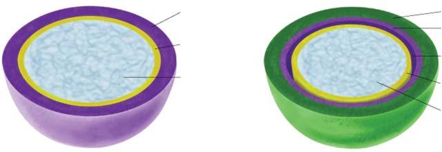
Prokaryotic cells are surrounded by an envelope (3.10.1, Fig. 2.2-2). It not only has an enclosing and protective function. Rather, a number of metabolic reactions take place at transmembrane proteins (e.g., respiration and ATP synthesis) or at membrane associated proteins. In bacteria, the sequence of membrane components from the interior outwards is:
• The plasma membrane, a lipid bilayer with embedded proteins
• The rigid cell wall, which in the case of bacteria consists of either multiple layers (Gram positive bacteria) or a single layer (Gram negative bacteria) of peptidoglycans (murein).
• An additional outer membrane (only in Gram negative bacteria).
• An additional gelatinous capsule is frequently superimposed on the cell wall. It consists mainly of polysaccharides (polymerized glucose, rhamnose, uronic acids etc.). There may also be mucus layers.
Extensions of the cell envelope are pili and flagella, which provide for cellular contact, conjugation, propulsion, etc.
The composition of an E. coli cell by weight is H2O about 70 %, protein 15 %, DNA 1 %, RNA 6 %, polysaccharides 3 %, lipids 2 % (both are mainly present in the envelope), small organic molecules 1 %, inorganic molecules 1 %.
Mycoplasms are a group of bacteria which lack a cell wall. Among them are the smallest self-reproducing organisms (0.10 … 0.25 μm diameter).
Archaea differ from bacteria by
• A different composition and arrangement of rRNAs
• Differences in the RNA polymerase and in the translation mechanism (Table 2.1-1)
• Different composition of the cellular envelope. e.g., murein (3.10.1) is absent, acylglycerols are replaced by branched chain glycerol ethers (3.4.3.3)
• Unusual pathways of metabolism and habitats (methanogens, 3.10.6.2, halobacteria, 3.12.1, thermophiles, etc.)
Figure 2.2-1: General Structure of a Bacterial Cell The colors are for easy differentiation only. After Campbell, N.A.: Biology 4th Ed. Benjamin/Cummings 1996.
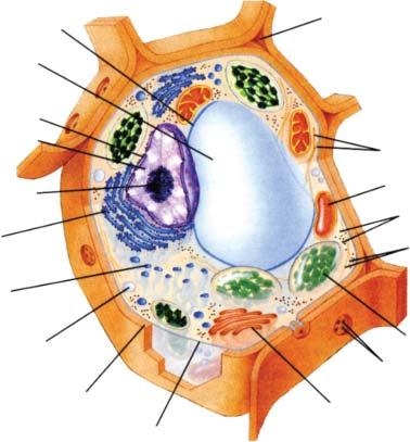
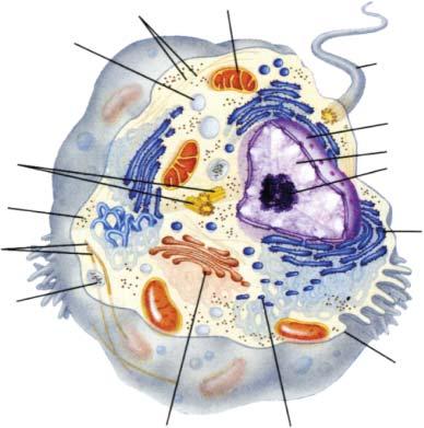
2.2.2 General Characteristics of Eukaryotic Cells
(Fig. 2.2-3)
Compared to prokaryotic cells, eukaryotic cells exhibit a much more complicated structure. Inside the plasma membrane there are the nucleus and the cytoplasm, which encompass the fluid cytosol and many organelles. These are compartments enclosed by individual membranes, which are devoted to specific functions.
Nucleus: All eukaryotic cells show the presence of a separate nucleus, which contains the major portion of the genetic material of the cell. (The rest is present in mitochondria and chloroplasts, see below.) The nuclear DNA is organized in a number of chromosomes. Each double helix of chromosomal DNA (2.6.3) can be present once (in haploid organisms) or twice (in diploid organisms). During cell division (4.3), the condensed chromosomes arrange themselves separately. Otherwise they are combined with proteins as a ball of chromatin with an elaborate fine structure (2.6.4).
The number of chromosomes present in the various species differs widely (from 4 to > 500; humans have 46 in the diploid set). While bacterial genomes contain < 106 … 5 * 107 bp, the diploid set of eukaryotic DNA varies between ca. 107 bp (some fungi) and > 1011 bp (lungfish, some algae). The diploid human genome contains ca. 5.8 * 109 bp (2 * 2,900,000 bp).
In addition to the DNA the nucleus also contains the nuclear matrix, which is composed of the enzymes and factors required for DNA replication, DNA repair, transcription, and processing of the transcription products (3.9, Chapter 4).
The nucleus is surrounded by a double membrane of lipid bilayers with integrated proteins. Nuclear pores (4.5.3, ca. 125 nm diameter) span the nuclear membrane and enable the transport of proteins, rRNA etc. The inner surface of the nuclear membrane is covered by nuclear lamina, a net of protein fibers which stabilizes the structure
(a) Gram-positive bacteria(b) Gram-negative bacteria
Peptidoglycan (cell wall)
Plasma mambrane
Cytoplasm
Outer membrane
Peptidoglycan (cell wall)
Periplasmic space
Plasma mambrane
Cytoplasm
Figure 2.2-2. Structure of the Bacterial Envelope After Voet, D. and J.G, Pratt, C.W.: Fundamentals of Biochemistry. John Wiley & Sons 2002, Figure 8-14.
and provides attachment points for the chromatin (2.6.4). During cell division, the nuclear membrane dissolves.
Cytosol: Although in eukarya many cytosolic functions have been taken over by specific organelles (see below), a large number of metabolic reactions still take place here. This includes glycolysis (3.1.1) and the synthesis of cytosolic proteins (4.2) and fatty acids (3.4.1.1).
Endoplasmic reticulum (ER, Fig. 2.2-4, Section 4.4.1): This is a labyrinth of membranes, which frequently encompasses half of the total amount of membranes. It consists of a system of sac- and tube-like structures, which locally expand into cisterns. Its internal lumen is connected with the intermembrane space of the nuclear membrane.
Part of the ER is studded on the outside with ribosomes (rough ER), which take part in protein synthesis (4.2). The proteins thus formed enter the ER lumen and are processed mainly by glycosylation (4.4.2). They leave the ER via vesicles (4.5.2). The rough ER is also the site of
2.2-3. General Structure of a Plant Cell (Top) and an Animal Cell (Below)
The colors are for easy differentiation only. After Campbell, N.A.: Biology 4th Ed. Benjamin/Cummings 1996.
membrane-phospholipid synthesis, thus providing membrane material for the departing vesicles (3.4.3, 4.4.1).
The other part of the ER is free of ribosomes (smooth ER). Enzymes of the smooth ER are involved in the synthesis of fatty acids (desaturation, 3.4.1.3), phospholipids (3.4.3), steroids, especially steroid hormones (3.5.4), and other lipids. They are located on the outside of this organelle. The smooth ER plays a role in detoxification by hydroxylation reactions. Its equivalent in muscles (sarcoplasmic reticulum) stores Ca2+ ions and is thus involved in the many reactions regulated by this ion (7.4.4). In liver, the smooth ER is also the storage site for glycogen (3.1.2.2).
Golgi apparatus (4.5.2): This organelle consists of stacks of flattened membrane sacs, which are especially numerous in plants (dictyosomes). The properties on both sides of an individual sac and the sacs of the whole stack differ (details in 4.4.2). Their main function is the further processing and sorting of proteins and their export to the final targets. In most cases, these are secreted or membrane proteins. In addition, the Golgi apparatus also produces polysaccharides (e.g., hyaluronic acid, 2.9.2), glycosphingolipids (3.4.4.1) etc.
Lysosomes: Lysosomes are vesicles of 0.1 … 0.8 μm diameter, which are enclosed by a lipid bilayer. They are formed by budding from the trans side of the Golgi apparatus (4.4.2). These organelles are filled with many enzymes for polysaccharide, lipid, protein and nucleic acid degradation. They fuse with endosomes containing internalized lipoproteins (6.2.4) or phagocytosed nutrients (6.1.5) and hydrolyze these compounds (Fig. 2.2-4). They act also on intracellular material to be removed and even contribute to the apoptosis (programmed cell death, 7.6, 8.2.5) of their own cell. Lysosomes of special cells (e.g., macrophages) destroy, in the same way, bacteria or viruses as a defense mechanism (8.2.8).
The degradative enzymes of lysosomes exhibit an activity optimum at pH = 5, identical to the pH of the lysosome lumen. It is kept constant by continuous proton pumping into the lysosome lumen. The pH difference to the usual cytosolic pH of 7.0 is a safety measure, since after accidental leakage of some lysosomes, the released enzymes are almost inactive at cytosolic pH. Only a cumulated release from many lysosomes is deleterious to the cell.
Insufficient activity of lysosomal enzymes is the reason for many diseases. Examples are mucopolysaccharidoses (2.9.2), gangliosidoses (3.4.4) and glycogen storage diseases (e.g., Pompe’s disease, 3.1.2.5).
Peroxisomes: This is another example of how enclosure into an organelle allows reactions to take place which would otherwise be deleterious to the rest of the cell. Peroxisomes (ca. 0.5 μm diameter) are surrounded by a single membrane. They are generated from components of the cytosol and do not bud from other membranes. The main task of these organelles is the performance of monooxygenase (hydroxylase) or oxidase reactions, which produce hydrogen peroxide (H2O2).
Figure
Figure 2.2-4. Interrelationship of Rough Endoplasmic Reticulum, Golgi Apparatus, Lysosomes, Endosomes and Transport Vesicles
This dangerous compound is immediately destroyed by catalase within this enclosed space:
X-H + O2 + H2O = X-OH + H2O2 or X-CH2OH + O2 = X-CHO + H2O2, followed by 2 H2O2 = 2 H2O + O2.
Reactions of this kind are the oxidative degradation of fatty acids (3.4.1.5), of alcohols etc. In glyoxisomes of plants, the reactions of the glyoxylate cycle (3.1.9.1) take place.
Mitochondria (Fig. 2.2-5): In a typical eukaryotic cell, there are in the order of 2000 of these organelles, which are often of ellipsoidal shape (length ca. 1 … 10 μm). They have a smooth outer membrane and a highly folded inner membrane with numerous invaginations (cristae), which contain most of the membrane-bound enzymes of mitochondrial metabolism. The internal area contains the mitochondrial matrix, while the intermembrane space is the narrow area between both membranes. Since protons are permanently pumped out through the inner membrane, the matrix is more alkaline than the intermembrane space and the cytoplasm (pH 8 vs. ª pH 7).
Mitochondria are the site of respiration and ATP synthesis (3.11.4.5), but also of many other central reactions of metabolism, e.g., citrate cycle (3.1.8), fatty acid oxidation (3.4.1.5), glutamine formation (3.2.2.1), and part of the pathway leading to steroid hormones (3.5.5.1, 3.5.8.1). The latter sequence, as well as the initiation of gluconeogenesis (3.1.3.5), are examples of how the site of reactions frequently changes from one organelle to another within a single pathway.
Besides chloroplasts (2.2.3), mitochondria are the only organelles which are equipped with their own (circular) DNA, RNA and ribosomes and thus can perform their own protein synthesis. The components and the mechanism resemble more the bacterial than the eukaryotic system. However, less than 10 % of the mitochondrial proteins are generated by this means, the rest is encoded by nuclear DNA and imported (4.5). Mitochondria reproduce by binary scission similar to bacteria. Their membranes do not exchange material with the rest of the cellular membrane system; the membrane proteins are produced on internal or cytosolic ribosomes. The membranes contain many transport systems resembling those of bacteria. These and other arguments are the base of the endosymbiont theory, which assumes that mitochondria have originated from ingested bacteria (e.g., aerobi-
Figure 2.2-5. General Structure of Mitochondria (Top) and Chloroplasts (Below)
After Lehninger A.L., Nelson, D.L., Cox, M.M.: Principles of Biochemistry 2nd Ed. Worth (1993).
cally living heterotrophs). They provide effective energy production, but are dependent on the host in many aspects.
Cytoskeleton: The internal cytoskeleton is a general component of eukaryotic cells, but is of special importance in animal cells. Here it is the primary factor for maintenance of the external structure, which is achieved in plant cells by means of the rigid cell wall (2.2.3). The cytoskeleton also provides anchoring points for the organelles and for some enzymes and even contributes to movements either by its own formation and degradation or by motor proteins moving along the filaments.
The cytoskeleton is composed of microtubules, actin filaments and intermediary filaments (Table 2.2-1). More details on their composition, formation and degradation are presented in 6.1.6.
Table 2.2-1. Components of the Cytoskeleton
MicrotubulesMicrofilamentsIntermediary Filaments
Components a/b-tubulinactinkeratin, vimetin, desmin etc.
Shapehollow tube of 13 tubulin sequences 2 strings of actin monomers, coiled variable, frequently coiled coil of a helices
Diameter25 nm7 nm8 … 12 nm
Examples of function chromosome separation (4.3.5), axonal transport (7.2.6), other movements
muscle contraction (7.4.5), movement of the cytoplasm, cell division (furrow ingression, 4.3.5)
structure stabilization, anchoring of organelles
The hollow microtubules (Fig. 6.1-3) frequently originate from the centrosome, which is located close to the nucleus (4.3.5). The moving parts of cilia and flagella are circular arrangements of tubulin. Attached dynein molecules cause movements of the tubules relative to each other and thus curvature of these cellular annexes (6.1.6).
Actin filaments associate with myosin. The heads of the myosin molecules can ‘walk along’ the string of actin monomers. The best known action of actin filaments is the muscle contraction (7.4.5), but they are also involved in many other, mostly movement, functions. E.g., the cytoplasmic movement in some algae is caused by the transport of organelles along actin filaments by myosin.
Intermediary filaments are composed of variable units, depending on the particular cell type. In general they have a structural function. They are more long-lasting than the other components of the cytoskeleton, but are also subject to rearrangements. Vimetin occurs in, e.g., endothelial cells and adipocytes and anchors the nucleus and the fat droplets. Desmin filaments keep the Z disks of muscle cells in place (7.4.5). Neurofilaments reinforce the long axons of neuronal cells. The nuclear lamina (see above) consists likewise of intermediary filaments.
2.2.3 Special Structures of Plant Cells (Fig. 2.2-3)
Plant cell walls: Cell walls are an essential factor distinguishing plant and animal cells. They provide stability and prevent an expansion of the cell beyond its fixed size. These secondary walls consist mainly of cellulose (3.1.2.2), hemicelluloses (3.1.6.3) and lignin (3.13.1.2). During an early phase of formation, the primary walls contain considerable amounts of pectate (3.1.5.6) and glycoproteins (4.4.1). On the inside, the cell contents are enclosed by the usual lipid bilayer membrane. Plasmodesmata interconnect neighboring cells and enable the transport of water and metabolites.

Chloroplasts (Fig. 2.2-5): These are lens-shaped organelles of about 1 … 5 μm diameter, which occur only in photosynthesizing green plants. They are enclosed by a double membrane with a thin intermembrane space in between. The interior contains the fluid stroma and a third membrane system surrounding the thylakoid space. This system has the shape of interconnected flat disks, which, in most cases, are stacked on top of each other (grana). The thylakoid membranes are the site of photosynthesis (3.12.1). In photosynthesizing bacteria, their role is taken over by the cytoplasmic membrane. Due to the permanent pumping of protons into the thylakoid space, its interior is much more acidic than the stroma (pH = 5 vs. pH = 8).
While some chloroplast proteins originate from their own protein synthesis system, the majority are nuclear encoded and are imported by a special mechanism into the stroma and the thylakoid space (4.5).
Analogous to mitochondria (2.2.2), chloroplasts resemble bacteria in several aspects (protein synthesis, membrane structure, reproduction etc.). Likewise the endosymbiont theory refers to them. Possible precursors of chloroplasts could be photoautotrophic cyanobacteria.
Vacuoles: Although vacuoles also occur in other kingdoms (e.g., protists), they are most prominent in plants. Young plant cells contain several vacuoles, which originate in vesicles released from the endoplasmic reticulum and the Golgi apparatus. In mature plant cells they combine to form a single central vacuole, which is enclosed by a membrane (tonoplast).
The central vacuole can occupy up to 90 % of the total cell volume and is a storage space for inorganic salts, saccharose, proteins, pigments and waste. The accumulation of these compounds causes the inflow of water by osmosis and keeps up the internal pressure (turgor).
2.2.4 Special Structures of Animal Cells
Animal cell membrane and extracellular matrix (Fig. 2.2-6): The actual cell membrane consists only of the lipid bilayer. However, it is covered by a complex, gelatinous extracellular matrix, which is formed by glycoproteins secreted from the cell. Its main component is collagen (2.3.1). The collagen fibers are enclosed by a network of carbohydrate rich proteoglycans (2.9.1.2). Tasks of the extracellular
matrix are the interconnection and anchoring of the cells, the support of the structure and to a certain extent also the localization of cells (e.g., during embryonic development).
Depending on their organ-specific structure, they allow the passage of fluids and dissolved material at varying degrees (e.g., filtration effects in the glomerulus of the kidney). The components of the extracellular matrix are connected via linker proteins (mostly fibronectin) with membrane-spanning proteins (integrins). The integrins, in turn, are associated at the interior side of the membrane with microfilaments of the cytoskeleton.
Interconnection between cells (Fig. 2.2-7): Neighboring cells (especially epithelial cells) are interconnected in several different ways. Tight junctions firmly attach the cells to each other and prevent any fluid passage between them. Point desmosomes (also frequently occurring in plants) are firm interconnections of neighboring cells via a network of glycoprotein filaments. They are reinforced on the inside of the cells by filaments of the cytoskeleton. Small channels known as gap junctions pass through both membranes of neighboring cells. They allow the exchange of small molecules and ions (7.2.3) and represent an equivalent of plant plasmodesmata (2.2.3). Pores, channels, transporters and receptors involved in material and signal passage through individual membranes are described elsewhere (7.1.2, 7.2.3, 6.1).
Literature:
de Duve, C. A Guided Tour of the Living Cell. Scientific American Books, 1984.
Textbooks of biology, e.g., Campbell, N.A. Biology 4th Ed. The Benjamin/Cummings Publ. Co, 1996.
2.3 Protein Structure and Function
While the two previous sections dealt with the cellular structures, the rest of this chapter describes properties of the two most important classes of macromolecules: proteins and nucleic acids.
Proteins and peptides are linear polymers which are built from the 22 naturally occurring proteinogenic L-amino acids (1.3 and 3.2, including selenocysteine and pyrrolysine, 4.1.3.4) and are linked by peptide bonds (1.3.2, Fig. 2.3-1). Peptides containing D-amino acids
Figure 2.3-1. Amino Acid Composition of Proteins (gray = aliphatic, red = acidic, green = small hydroxy, blue = basic, black = aromatic, white = amide, yellow = sulfur). After UniProtKB/Swiss-Prot protein knowledgebase (see references).
Figure 2.2-7. Types of Contact between Cells
Figure 2.2-6. Structure Elements of the Extracellular Matrix
are less frequent. They occur mostly in microorganisms. Chains of up to about 50 amino acid residues are usually named ‘oligo-(or poly-) peptides’, larger ones are termed ‘proteins’. However, the expression ‘peptides’ is frequently also used for the whole class of compounds containing peptide bonds. The average protein sequence length listed in UniProtKB (Chapter 10) is 352 amino acids, the longest sequence has 35,213 amino acids.
The average amino acid distribution in proteins is far from even, some amino acids occur much more frequently than others. Leucine is found nine times as often as tryptophan (Fig. 2.3-1).
All polypeptides and proteins are synthesized by ribosomal synthesis, using mRNA as the source of information for the sequence of amino acids (4.1.2, 4.2.2). However, a number of small peptides are formed by a non-ribosomal sequence of specific enzyme reactions, e.g., glutathione (3.2.5.7), the penicillin precursor peptide (3.10.8) and many peptide antibiotics.
After formation of the peptide chain, additional post-ribosomal modifications of the amino acid components may take place, e.g., hydroxylation (3.2.3, 3.2.5.2), carboxylation (3.2.2.2, 3.7.13), methylation (e.g., of glutamate during chemotactic mechanisms), acylation (acetylation, myristoylation, palmitoylation, 4.4), phosphorylation (2.5.2, 7.5), glycosylation (4.4.2) and formation of disulfide bonds between the -SH groups of cysteine (4.5.1).
In a number of cases, a long peptide is cleaved after synthesis, e.g., to be activated for a special function. Examples are hormones (7.1.3, 7.1.5), digestive enzymes (2.5.1) and blood coagulation factors (9.2 … 5).
2.3.1
Levels of Organization
The amino acid sequence contains all the information necessary to determine the three-dimensional structure of the protein which is assumed to be the thermodynamically most stable one among the kinetically possible ways of folding (4.5). Several levels of structural organization can be distinguished:
Primary structure (Fig. 2.3-2): This is the amino acid sequence. The CO–NH bond is fairly rigid and usually assumes the trans configuration. To a limited extent, rotations are possible around both other bonds of the peptide backbone (torsion angles F and Y). Steric restrictions are caused by the side chains of the amino acids and the oxygen atom of the CO group.
looking along the axis of the helix from the N to the C terminus, the chain turns clockwise). One turn is formed by 3.6 residues, the pitch amounts to 0.54 nm. a-Helices occur both in globular and in fibrous proteins and are found in almost all transmembrane sections.
The b-pleated sheet forms slightly twisted planes. This structure is also stabilized by hydrogen bonds, however, between different strands. Between 2 … 15 strands may arrange themselves in parallel or antiparallel directions. The side chains of consecutive amino acids extend sideways in opposite directions.
Tertiary structure is a term which refers to larger arrangements, describing the location (x,y,z coordinates) of all atoms in space. Additional levels are frequently used:
Supersecondary structures are combinations of secondary structure elements, such as the coiled-coil a-helices (e.g., in many fibrillar proteins) or barrels (in globular proteins) or nucleotide-binding structures. A number of structural motifs occur in these superfolds.
Two or more a-helices can associate into an antiparallel arrangement, which allows interactions of their side chains (aa motif). This is an essential element of fibrous proteins (e.g., in a-keratin, Fig. 2.3-7 and in the segments of spectrin, which is present in erythrocytes), but does also occur in globular proteins (e.g., in hemoglobin, Fig. 2.3-6).
A number of common motifs occurring in globular proteins are caused by the turns of the peptide backbone at the end of b-pleated sheets (Fig. 2.3-4): Antiparallel b-pleated sheets frequently form b-loops (also called hairpin loops), which are stabilized by hydrogen bonds between a CO group and the third NH group of the sequence. A series of such turns can form a b-meander. Another arrangement of turns is the so-called ‘Greek key’. Parallel b-pleated sheets require crossover connections, which often assume right-handed helix structures (bab motif). These loops and crossover connections frequently occur on the outside of globular proteins.
Figure 2.3-2. Primary Structure of a Polypeptide Chain in Extended Conformation
The distances (in nm = 10 Å) and the bond angles are shown. The green quadrangles indicate the rigid structure of the peptide bonds. Y and F are the angles characterizing the rotation around the Ca-CO and N-Ca bonds (in this figure Y = F = 180°). The formation of hydrogen bonds leading to a helices is indicated.
Secondary structure (Fig. 2.3-3): These are regular arrangements of the backbone of the polypeptide chain, which are stabilized by hydrogen bonds between amide and carboxyl groups of the peptide and by sterically favorable backbone torsion angles. Frequently occurring secondary structures are the a-helix and the b-pleated sheet (Fig. 2.3-3). In globular proteins, a variable portion (often below 40 %, sometimes up to 100 %) is arranged in less regular loop structures, which are frequently found on the outside of water-soluble proteins.
The a-helix is a rod-like structure, which is formed by hydrogen bonds between a CO group and the fourth following NH group of the sequence (shown in Fig. 2.3-2). Helices formed by hydrogen bonds between other CO–NH pairs are rare. There are also van der Waals bonds across the helix. The side chains of the amino acids point to the outside. All a helices occurring in nature are right-handed (i.e., when
Figure 2.3-3. `-Helix (Top) and Antiparallel a-Pleated Sheet Structure (Bottom) C = black, O = red, N = blue, H = white, side chains = grey.