American University of Antigua College of Medicine Antigua, West Indies
Nihal Apaydın, MD, PhD Professor, Department of Anatomy Faculty of Medicine
Vice Director, Brain Research Center Ankara University Ankara, Turkey
Keith E. Baynes, MD
Section Chief, MSK and General Radiology
Associate Professor of Radiology
Medical College of Wisconsin Milwaukee, Wisconsin
Francisco J. Caycedo, MD
Specialist in Foot and Ankle Surgery
Sports Medicine and Musculoskeletal Ultrasound OrthoSports Associates–St. Vincent’s Birmingham Birmingham, Alabama
William E. Cullinan, PhD Professor, Department of Biomedical Sciences Director, Integrative Neuroscience Research Center
Dean, College of Health Sciences Marquette University Milwaukee, Wisconsin
Joe Iwanaga, DDS, PhD
Assistant Professor Division of Gross and Clinical Anatomy
Department of Anatomy Kurume University School of Medicine Kurume, Japan
Christopher R. Kelly, MD
Clinical Fellow
Division of Cardiology
Columbia University Medical Center New York, New York
Robert Louis, MD
Director, Skull Base and Pituitary Tumor Program
Minimally Invasive Brain and Spine Surgery
Hoag Neurosciences Institute Newport Beach, California
Virginia T. Lyons, PhD
Associate Professor of Medical
Education
Associate Dean of Preclinical Year 1
Geisel School of Medicine at Dartmouth Hanover, New Hampshire
Thazhumpal Chacko Mathew, PhD Professor and Vice Dean for Research, Training, and Consultation
Faculty of Allied Health Sciences Health Sciences Centre
Kuwait University
Kuwait City, Kuwait
Paul E. Neumann, MD
Professor, Department of Anatomy and Neurobiology
Faculty of Medicine
Dalhousie University
Halifax, Nova Scotia, Canada
Eduardo Cotecchia Ribeiro, PhD
Associate Professor of Descriptive and Topographic Anatomy Department of Morphology and Genetics School of Medicine
Federal University of São Paulo São Paulo, Brazil
Danielle F. Royer, PhD Associate Professor
Cell and Developmental Biology
University of Colorado, Anschutz Medical Campus Aurora, Colorado
Jonathan Spratt, MB, BChir Clinical Director of Radiology
Sunderland City Hospitals
Sunderland, United Kingdom
Former Examiner in Anatomy
Royal College of Radiologists and Royal College of Surgeons of England
Visiting Professor of Anatomy
St. George’s University
Grenada, West Indies
Susan Standring, MBE, PhD, DSc Professor Emeritus of Anatomy
Department of Anatomy
King’s College London
London, United Kingdom
Mark E. Sturgill, DO
Pediatric and Neuroradiologist
Radiology Partners
Hopkinsville, Kentucky
William J. Swartz, PhD Professor of Cell Biology and Anatomy
Louisiana State University Health Sciences Center
New Orleans, Louisiana
Kimberly Topp, PT, PhD Professor and Chair
Department of Physical Therapy and Rehabilitation Science
Department of Anatomy
University of California, San Francisco
San Francisco, California
Ivan Varga, PhD
Professor of Anatomy, Histology, and Embryology
Faculty of Medicine
Comenius University
Bratislava, Slovak Republic
Peter J. Ward, PhD
Associate Professor of Anatomy
West Virginia School of Osteopathic Medicine
Lewisburg, West Virginia
Robert J. Ward, MD Chief, Musculoskeletal Imaging and Intervention Director, Bone Densitometry
Department of Radiology
Tufts Medical Center
Director, Undergraduate Radiology
Education
Assistant Professor of Radiology and Orthopedics
Tufts University School of Medicine
Boston, Massachusetts
Kristy A. Weir, PhD
School of Biomedical Sciences
The University of Queensland
St Lucia, Queensland, Australia
NEW TO THIS EDITION
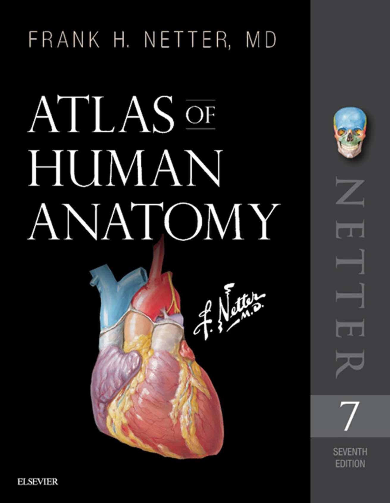
With your copy of the Frank H. Netter, MD, Atlas of Human Anatomy, you own a collection of some of the most wellknown depictions of human anatomy in medicine and healthcare. In addition to the famous work of Dr. Netter, with your copy of this 7th edition, you also have access to nearly 100 painted pieces by Carlos A. G. Machado, MD, one of the foremost medical illustrators working today. Dr. Machado’s contributions to the Atlas highlight important views of anatomy that have become more clinically relevant in recent years— anatomic views that have resulted from improved dissection techniques and modern imaging. In addition, you have access to more than 50 carefully selected radiologic images that help bridge the idealized illustrated anatomy with living anatomy viewed in the clinic.
While numerous updates have been made to the illustrated plates and tables to make them easier to learn from, the most significant changes to this edition include:
Introductory Section
To fulfill the requests from many students and fans of Netter’s Atlas, we have added a new opening section containing several overview plates. These plates provide the very first head-to-toe views in the Atlas of Human Anatomy!
Clinical Tables
The Atlas of Human Anatomy is the only anatomy atlas illustrated by physicians. Dr. Netter was a surgeon and Dr. Machado is a cardiologist. The views of anatomy in this atlas have always reflected a clinical perspective. In line with this clinical focus, and in congruence with integrated curricula in health and medicine, tables at the end of each regional section highlight the most commonly injured structures, as well as other structures with high clinical significance and commonly covered in anatomy courses. The tables provide students with quick summaries, organized by body system, and note where to best view these key structures in the illustrated plates.
New Art Plates by Dr. Machado
For this edition alone, over 25 new illustrations have been painted by Dr. Machado. Suggestions for new plates of additional anatomic views and concepts are submitted by students, faculty, anatomists, physicians, and others. Sometimes suggestions are solicited at major anatomy conferences with a “What Should Carlos Paint Next?” idea box. Decisions around which new plates are prioritized and given space in a new edition come from discussions among consulting editors. The new plates for this edition are largely those that portray structures with clinical significance (Fascial Columns of the Neck, Deep Veins of the Leg, Hip Bursae, and Vasculature of the Prostate) or those that are difficult to visualize (Infratemporal Fossa)—and, of course, the new additions created for the introductory section.
Terminology Updates
The Atlas of Human Anatomy uses terminology accepted (in Göttingen, Germany, on September 24, 2016) by the Federative International Programme on Anatomical Terminologies and published as updates to the 1998 Terminologia Anatomica. Numerous updates to terminology have been made, so in select cases, former terminology has been included within parentheses to assist with the transition.
New Radiologic Images
Over 50 radiologic images—some completely new views and others replacing existing views using newer imaging tools—are included in this edition. Images have been selected based on their utility to students studying gross anatomy.
Your Atlas of Human Anatomy content has been updated, created, and overseen by a team of dedicated and passionate consulting editors, with the help of a stellar international advisory board, and guided by the feedback of many students, educators, anatomists, and clinicians that love Netter’s Atlas. Please feel free to comment on the Netter Images Facebook page or Twitter feeds or email us directly with your thoughts, suggestions, or questions at NetterAppFeedback@Elsevier.com
PREFACE TO THE FIRST EDITION
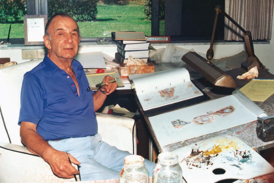
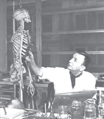
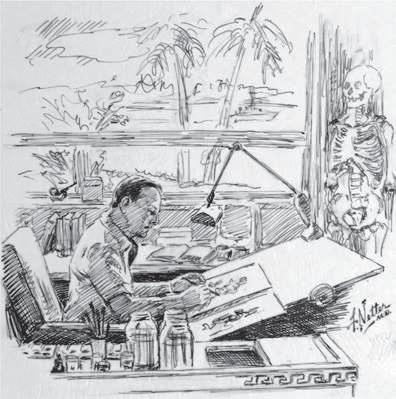
I have often said that my career as a medical artist for almost 50 years has been a sort of “command performance” in the sense that it has grown in response to the desires and requests of the medical profession. Over these many years, I have produced almost 4,000 illustrations, mostly for The CIBA (now Netter) Collection of Medical Illustrations but also for Clinical Symposia. These pictures have been concerned with the varied subdivisions of medical knowledge such as gross anatomy, histology, embryology, physiology, pathology, diagnostic modalities, surgical and therapeutic techniques, and clinical manifestations of a multitude of diseases. As the years went by, however, there were more and more requests from physicians and students for me to produce an atlas purely of gross anatomy. Thus, this atlas has come about, not through any inspiration on my part but rather, like most of my previous works, as a fulfillment of the desires of the medical profession. It involved going back over all the illustrations I had made over so many years, selecting those pertinent to gross anatomy, classifying them and organizing them by system and region, adapting them to page size and space, and arranging them in logical sequence. Anatomy of course does not change, but our understanding of anatomy and its clinical significance does change, as do anatomical terminology and nomenclature. This therefore required much updating of many of the older pictures and even
FRANK H. NETTER, MD
Frank H. Netter was born in New York City in 1906. He studied art at the Art Students League and the National Academy of Design before entering medical school at New York University, where he received his Doctor of Medicine degree in 1931. During his student years, Dr. Netter’s notebook sketches attracted the attention of the medical faculty and other physicians, allowing him to augment his income by illustrating articles and textbooks. He continued illustrating as a sideline after establishing a surgical practice in 1933, but he ultimately opted to give up his practice in favor of a full-time commitment to art. After service in the United States Army during World War II, Dr. Netter began his long collaboration with the CIBA Pharmaceutical Company (now Novartis Pharmaceuticals). This 45-year partnership resulted in the production of the extraordinary collection of medical art so familiar to physicians and other medical professionals worldwide.
Icon Learning Systems acquired the Netter Collection in July 2000 and continued to update Dr. Netter’s original paintings and to add newly commissioned paintings by artists trained in the style of Dr. Netter. In 2005, Elsevier Inc. purchased the Netter Collection and all publications from Icon Learning Systems. There are now over 50 publications featuring the art of Dr. Netter available through Elsevier Inc.
revision of a number of them in order to make them more pertinent to today’s ever-expanding scope of medical and surgical practice. In addition, I found that there were gaps in the portrayal of medical knowledge as pictorialized in the illustrations I had previously done, and this necessitated my making a number of new pictures that are included in this volume.
In creating an atlas such as this, it is important to achieve a happy medium between complexity and simplification. If the pictures are too complex, they may be difficult and confusing to read; if oversimplified, they may not be adequately definitive or may even be misleading. I have therefore striven for a middle course of realism without the clutter of confusing minutiae. I hope that the students and members of the medical and allied professions will find the illustrations readily understandable, yet instructive and useful.
At one point, the publisher and I thought it might be nice to include a foreword by a truly outstanding and renowned anatomist, but there are so many in that category that we could not make a choice. We did think of men like Vesalius, Leonardo da Vinci, William Hunter, and Henry Gray, who of course are unfortunately unavailable, but I do wonder what their comments might have been about this atlas.
Frank H. Netter, MD (1906–1991)
Dr. Netter’s works are among the finest examples of the use of illustration in the teaching of medical concepts. The 13-book Netter Collection of Medical Illustrations, which includes the greater part of the more than 20,000 paintings created by Dr. Netter, became and remains one of the most famous medical works ever published. The Netter Atlas of Human Anatomy, first published in 1989, presents the anatomic paintings from the Netter Collection. Now translated into 16 languages, it is the anatomy atlas of choice among medical and health professions students the world over.
The Netter illustrations are appreciated not only for their aesthetic qualities, but, more importantly, for their intellectual content. As Dr. Netter wrote in 1949 “clarification of a subject is the aim and goal of illustration. No matter how beautifully painted, how delicately and subtly rendered a subject may be, it is of little value as a medical illustration if it does not serve to make clear some medical point.” Dr. Netter’s planning, conception, point of view, and approach are what inform his paintings and what make them so intellectually valuable.
Frank H. Netter, MD, physician and artist, died in 1991.
ACKNOWLEDGMENTS
Carlos A. G. Machado, MD
I struck luck when joining this golden team of consulting editors exceedingly knowledgeable in the fields of clinical anatomy and medical education. It has been a great honor to work with and be under their guidance, as well as under the highly competent coordination of Elyse O’Grady and Marybeth Thiel, Elsevier’s Executive Content Strategist and Senior Content Development Specialist, respectively.
This unique book would not exist without the genius of its creator, Dr. Frank Netter, to whom I owe special thanks, also in the name of generations of students and health professionals who, like myself, have learned so much from his incommensurable body of work.
I dedicate my work and express my most sincere thanks to my beloved parents, Carlos and Neide, who provided me with the foundation of my education; to my patient wife, Adriana, and talented daughter, Beatriz, for their love and support; to the students, teachers, and health professionals who rely on my work to learn and teach; to all the body donors and living friends that have respectively been the subjects of my studies and models of most of the illustrations I have created for the Atlas; and to my teachers Eugênio Cavalcante, Mário Fortes, and Paulo Carneiro for taking my interest in human/clinical anatomy much further.
John T. Hansen, PhD
At Elsevier I would like to thank Marybeth Thiel, Senior Content Development Specialist, Elyse O’Grady, Executive Content Strategist, John Casey, Senior Project Manager, Patricia Tannian, Publishing Services Manager, Julia Dummitt, Design Manager, Karen Giacomucci, Illustration Buyer, and Madelene Hyde, Publishing Director, for their continuous support and meticulous attention to detail during the development of this seventh edition of the Atlas of Human Anatomy. They, along with the entire editorial, production, design, and marketing teams at Elsevier have been a delight to work with and to know. I also wish to thank my consulting editors for their insightful and constructive suggestions as we strive to make every new edition of the Atlas better. I am also indebted to Carlos Machado for his superb artistic skill in producing and updating a number of plates that appear in this latest edition of the Atlas. His renderings of human anatomy are the perfect complement to the Netter images. In addition to my fellow editors, I wish to express my thanks to my faculty
colleagues at Rochester and to all my past and present students who have provided generous and constructive feedback and have enriched my life. Finally, I am indebted to my entire family for their continued support and especially to my wife, Paula. Their love and encouragement sustains me and is the source of all the happiness and joy I know.
Brion Benninger, MD, MSc
Every day I am thankful for my wife, Alison, and son, Jack, for the laughs we have as a family, often from my follies, which is such a tonic. I thank Elsevier, especially Marybeth Thiel, Elyse O’Grady, and Madelene Hyde for their professionalism and guidance, enabling John Hansen, Carlos Machado, and my fellow coeditors to work in a unique and dynamic environment. I thank those clinicians who trained me, especially my early gifted surgeon/anatomist/ teacher mentors, Drs. Gerald Tressidor and Harold Ellis CBE (Cambridge & Guy’s Hospital); Dr. S. Standring, who embodies professionalism and displays fortitude; Drs. P. Crone and J. Heatherington, and the University Board for their stellar support; my past and future students and patients; and clinical colleagues from all corners of the world who keep anatomy dynamic, fresh, and wanting more. Special thanks to Jim Diegel and Erik Szeto, friends, mentors and fellow visionaries who also see “outside the box,” challenging status quo. A heartfelt tribute to my late mentors, friends, and sister, Jim McDaniel, Bill Bryan, and Gail Hendricks, all who represent what is good in teaching, caring, and healing. They made this world a wee bit better. Lastly, I thank my mother for her love of education and equality and my father for his inquisitive and creative mind.
Jennifer Brueckner-Collins, PhD
Many thanks to the Elsevier team, particularly Marybeth Thiel and Elyse O’Grady, for their guidance and expertise during our preparation of the seventh edition. It is always an honor to work with Carlos Machado, whose passion for and mastery of the art of clinical anatomy and medicine never cease to amaze me. I am forever indebted to Brian MacPherson, who has served as a teacher, mentor, and friend to me for more than 20 years….you showed me what it means to be a true educator and I have been so fortunate to have the opportunity to build a career based on those principles. To Kurt and Lincoln, you are my inspiration….my world…my life and I love you to the snow moon and back.
Todd M. Hoagland, PhD
It is a privilege to teach clinical human anatomy and I am eternally grateful to all the body donors and their families for enabling healthcare professionals to train in the dissection laboratory. It is my honor to work with outstanding medical students and colleagues at the Medical College of Wisconsin. I am grateful to John Hansen and the professionals of the Elsevier team for the opportunity to be a steward of the incomparable Netter’s Atlas. Marybeth Thiel and Elyse O’Grady were especially helpful and a pleasure to work with. It was an honor to collaborate with the brilliant Carlos Machado and all the consulting editors. I thank Bill Swartz and Mark Moss for being outstanding mentors, and I thank all of the graduate students I’ve worked with, especially Rebecca Lufler. I am deeply appreciative of Stan Hillman and Jack O’Malley for inspiring me with masterful teaching and rigorous expectations. I am indebted to Gary Kolesari and Richard Hoyt Jr. for
helping me become a competent clinical anatomist, and to Rob Bouchie for his camaraderie. I am most grateful to my brother, Bill, for his unwavering optimism and gregarious nature. I thank my mother, Liz, for her dedication and love and for instilling a strong work ethic. Finally, I am humbled by my two awesome children, Ella and Caleb, for helping me redefine love, wonder, and joy.
R. Shane Tubbs, MS, PA-C, PhD
Elsevier and the Netter team have once again been a joy to work with. I thank Elyse O’Grady, Marybeth Thiel, and John Casey for their tremendous work on this edition. In addition, Carlos Machado has again added his expertise to bringing his anatomical images to life. As always, my work is inspired by my beautiful wife, Susan, and son, Isaiah. Lastly, I am indebted to my parents, Richard and Karon Tubbs, who supported me in my career to better understand the human body.