One Minute
Ophthalmology


Collaboration in Research and Beyond
In ancient times, India was a centre of great learning. As a nation of almost 140 crore people, with the largest number of English speaking PhDs being produced, we still have a great potential. The Indian innovation ranks right on top and we do it cheaper than any other English speaking nation in the world. The Mars Orbiter Mission or Mangalyan travelled about 650 million km distance at Rs 7 per kilometre incurred cost in covering the to Mars by the unmanned spacecraft. It was cheaper than the cost of autorickshaw. We are today a major global research and development hub for automobiles
Medicine could be the next sunshine area. The time has come for us to claim our place at the high table of research, knowledge and innovation. But this is not going to be an easy task. It will require hard work and quality output by all of us.
Let us support each other in our research and skillshare endeavours in Ophthalmology. Let us work together to improve sustainable medical development and ensure that the dreams of our children are fulfilled. "Teamwork is the ability to work together toward a common vision. The ability to direct individual accomplishments toward organizational objectives. It is the fuel that allows common people to attain uncommon results " Andrew Carnegie
Dr Jatinder Bali Editor
"Teamwork is the ability to work together toward a common vision. The ability to direct individual accomplishments toward organizational objectives. It is the fuel that allows common people to attain uncommon results." --Andrew Carnegie
Forced duction test (FDT) in ocular motility disorder post scleral buckling- How conclusive?

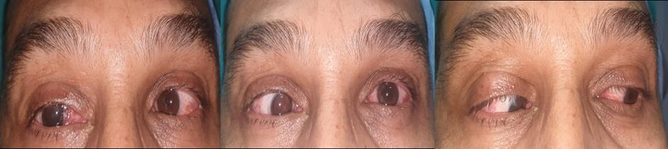
Case: Patient presented with diplopia post buckle surgery which was done 2 weeks back. On examination esotropia of 30 Prism dioptre was noted along with limitation in abduction (Figure 1). Fundus examination showed retinal detachment. Literature has documented strabismus in upto 50% of patients who undergo scleral buckling procedure for retinal detachment.[1 3] Incidence of squint further increases with repeated surgeries. We here in present a case of esotropia post buckle surgery.
Figure 1
A) Iatrogenic restrictive component
medial rectus (MR) B) Iatrogenic disinsertion of lateral rectus (LR) C) Sixth
D) Presence of
to Buckle
Next Step:
in
Nerve Paresis
Squint prior
surgery
Author
Dr. Anita Ganger
Findings:
FDT was found to be positive for Medial rectus indicative of restrictive etiology. Active force generation test (AFGT) was positive for LR which ruled out possibility of disinsertion of Lateral rectus. Exploration of extra ocular muscles and pars plana vitrectomy for redetachment was planned. During intraoperative exploration MR was found to attached near limbus with lot of adhesions. All the adhesions have been released along with reattachment of medial rectus at its original insertion (5.5 mm from limbus).
Diagnosis: Iatrogenic Esotropia post Buckle surgery due to restrictive etiology.
Correct Answer: A) Esotropia due to Iatrogenic restrictive component in medial rectus (MR)

Discussion:
The possibility of inadvertent disinsertion of MR and reinsertion at wrong anatomical position was considered after doing FDT and AFGT. In this case diagnosis and management was decided as per the FDT results. Postoperatively diplopia got resolved and optimal eye alignment was noted along with attached retina (Figure 2). Extraocular muscles should be handled with utmost care during scleral buckle surgeries and FDT is very conclusive in differentiating various etiologies.
References:
1. Sharifi M, Ansari Astaneh M, R: Forced Duction Test: Is It Necessary after the Scleral Buckling Procedure? Case Rep Ophthalmol 2020;11:282 286.
2. Seaber JH, Buckley EG. Strabismus after retinal detachment surgery: etiology, diagnosis, and treatment. Semin Ophthalmol. 1995 Mar;10(1):61–73.
3. Wu TE, Rosenbaum AL, Demer JL. Severe strabismus after scleral buckling: multiple mechanisms revealed by high resolution magnetic resonance imaging. Ophthalmology. 2005 Feb;112(2):327–36.
Authors:
1. Dr. Anita Ganger, Consultant Ophthalmology, Squint specialist, Associate Professor (GRIPMER), Sir Ganga Ram Hospital, New Delhi

2. Dr. Kirti Rani, FNB trainee Vitreo retinal surgery, Sir Ganga Ram Hospital, New Delhi
3. Dr. S.N. Jha, Senior Consultant Vitreo-retinal surgery Ophthalmology, Sir Ganga Ram Hospital, New Delhi
Dr. Anita Ganger Author
For more details please check out on
Amazon.in
“ A dream is not what you see when you sleep. A dream is something that it does not let you sleep.” – Dr APJ Abdul Kalam