Cambridge International AS Level Biology
used as the storage carbohydrate. This is called glycogen. Glycogen, like amylopectin, is made of chains of 1,4 linked α-glucose with 1,6 linkages forming branches (Figure 2.7b). Glycogen molecules tend to be even more branched than amylopectin molecules. Glycogen molecules clump together to form granules, which are visible in liver cells and muscle cells, where they form an energy reserve. QUESTION 2.4 List five ways in which the molecular structures of
glycogen and amylopectin are similar.
34
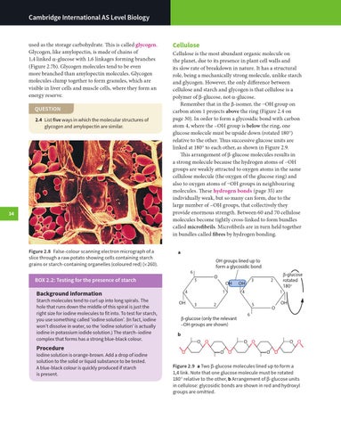
Figure 2.8 False-colour scanning electron micrograph of a slice through a raw potato showing cells containing starch grains or starch-containing organelles (coloured red) (× 260).
Cellulose
Cellulose is the most abundant organic molecule on the planet, due to its presence in plant cell walls and its slow rate of breakdown in nature. It has a structural role, being a mechanically strong molecule, unlike starch and glycogen. However, the only difference between cellulose and starch and glycogen is that cellulose is a polymer of β-glucose, not α-glucose. Remember that in the β-isomer, the –OH group on carbon atom 1 projects above the ring (Figure 2.4 on page 30). In order to form a glycosidic bond with carbon atom 4, where the –OH group is below the ring, one glucose molecule must be upside down (rotated 180°) relative to the other. Thus successive glucose units are linked at 180° to each other, as shown in Figure 2.9. This arrangement of β-glucose molecules results in a strong molecule because the hydrogen atoms of –OH groups are weakly attracted to oxygen atoms in the same cellulose molecule (the oxygen of the glucose ring) and also to oxygen atoms of –OH groups in neighbouring molecules. These hydrogen bonds (page 35) are individually weak, but so many can form, due to the large number of –OH groups, that collectively they provide enormous strength. Between 60 and 70 cellulose molecules become tightly cross-linked to form bundles called microfibrils. Microfibrils are in turn held together in bundles called fibres by hydrogen bonding. a OH groups lined up to form a glycosidic bond 6
BOX 2.2: Testing for the presence of starch
Procedure
Iodine solution is orange-brown. Add a drop of iodine solution to the solid or liquid substance to be tested. A blue-black colour is quickly produced if starch is present.
OH
4
Background information
Starch molecules tend to curl up into long spirals. The hole that runs down the middle of this spiral is just the right size for iodine molecules to fit into. To test for starch, you use something called ‘iodine solution’. (In fact, iodine won’t dissolve in water, so the ‘iodine solution’ is actually iodine in potassium iodide solution.) The starch–iodine complex that forms has a strong blue-black colour.
O
5
OH
1 3
3
OH 4
1
2
5
β-glucose (only the relevant –OH groups are shown)
β-glucose rotated 180°
2
O
OH
6
b O O O
O O O O
O O O O
Figure 2.9 a Two β-glucose molecules lined up to form a 1,4 link. Note that one glucose molecule must be rotated 180° relative to the other, b Arrangement of β-glucose units in cellulose: glycosidic bonds are shown in red and hydroxyl groups are omitted.