S
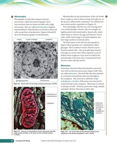
Mitochondria Photographs of cells taken using an electron microscope, called electronmicrographs, show tiny structures that are almost invisible with a light microscope. They are called mitochondria (singular: mitochondrion). Mitochondria are found in almost all cells, except those of prokaryotes. Figures 2.8 and 2.9 show electronmicrographs of mitochondria. nucleus
nuclear envelope
cytoplasm
Mitochondria are the powerhouses of the cell. Inside them, oxygen is used to release energy from glucose, in the process called aerobic respiration. You will find out more about aerobic respiration in Chapter 11. Not surprisingly, cells that use a lot of energy have a lot of mitochondria. Muscle cells, for example, are tightly packed with mitochondria. Sperm cells, which need energy to swim to the egg, and neurones (nerve cells), which need energy to transmit impulses, also have large numbers of mitochondria. The black spots in the electron micrograph in Figure 2.8 are granules of a carbohydrate called glycogen. This is similar to starch. (Starch is never found in animal cells – they store glycogen instead.) Glycogen is a reserve fuel. When required, it can be broken down to glucose, to be used as a fuel by the mitochondria in the liver cell, or transported in the blood to other cells that need it.
Ribosomes
glycogen granules
mitochondria
Figure 2.8 Part of a liver cell seen using an electron microscope (× 20 000).
Even tinier structures than mitochondria can just be seen with an electron microscope (Figure 2.10). They are called ribosomes. They look like tiny dots attached to a network of membranes that runs throughout the cytoplasm. This network is called the rough endoplasmic reticulum. Ribosomes may also just be scattered freely in the cytoplasm. Ribosomes are found in all types of cells – bacteria, protoctists, fungi, animals and plants all have ribosomes in their cells.
membranes of mitochondrion
ribosomes attached to membranes of rough endoplasmic reticulum
cytoplasm
Figure 2.9 Close-up of a mitochondrion. Electron microscopes only show images in black and white, so this photo has been artificially coloured (× 30 000).
22
Cambridge IGCSE Biology
Figure 2.10 You can just make out tiny ribosomes attached to the membranes in this electron micrograph of a cell (× 30 000).
Original material © Cambridge University Press 2014
S