AI
WHERE IT LANDS TODAY AND HOW IT MIGHT IMPACT
WHERE IT LANDS TODAY AND HOW IT MIGHT IMPACT
By Bryan Kay and Jocelyn Hudson
The speed with which artificial intelligence (AI) is beginning to envelope aspects of the human experience strikes many as jarring. Its entrance into the world of vascular surgery, too, has picked up a pace in recent years, with models and applications beginning to pop up in papers probing aspects of AI uses in care across the vasculature. But what is the extent of its abilities in vascular practice and where might AI lead the field in the future?
See page 4
2 Guest editorial Rebuttal: Vikram Kashyap, MD, makes the case for the benefits of a heart and vascular center
5 VAM 2026
Vascular Annual Meeting chairs discuss pathways to the podium, posters
10 Carotid disease
Not all carotid revascularization procedures are equal
18 Quality Vascular Verification Program opens doors for early engagement
www.vascularspecialistonline.com

By Bryan Kay
NEWS THAT U.S. PRESIDENT
Donald J. Trump has a vascular condition may have simultaneously revealed that a substantial seven-figure vascular surgery branding campaign was starting to yield tangible results, and provided a singular opportunity to platform the specialty as the go-to vascular experts.
The Society for Vascular Surgery (SVS) launched a multipronged branding effort last year in an effort to help vascular surgery become an instantly recognizable specialty inside and outside of the hospital. The Society and vascular surgeons at large have long sought avenues to broaden awareness of what a vascular surgeon does, and the comprehensive and longitudinal nature of the care they provide after a patient is diagnosed with a vascular condition.
Well, opportunity knocks: when it was revealed to the world in mid-July that the president had been diagnosed with chronic venous insufficiency (CVI), vascular disease was thrown into the media grinder and the press corps began dissecting what CVI is, what it means and what the prognosis might be. Step forward vascular surgery. In the immediate aftermath of the news breaking, vascular surgeons began populating the pages and airwaves of the world’s media. They laid out the facts of CVI and what Trump might—or might not—be looking at down the road.
Vascular surgery, vascular surgeons and the SVS were name-checked across the news media, from international broadcast platforms like the BBC, to domestic newspaper powerhouses such as the New York Times. That broad footprint, says William Shutze, MD, who led the SVS branding
See page 4
Medical Editor Malachi Sheahan III, MD
Associate Medical Editors
Bernadette Aulivola, MD | O. William Brown, MD | Elliot L. Chaikof, MD, PhD
| Carlo Dall’Olmo, MD | Alan M. Dietzek MD, RPVI, FACS | John F. Eidt, MD | Robert Fitridge, MD | Dennis R. Gable, MD | Linda Harris, MD | Krishna Jain, MD | Larry Kraiss, MD | Joann Lohr, MD | James McKinsey, MD | Joseph Mills, MD | Erica L. Mitchell, MD, MEd, FACS | Leila Mureebe, MD | Frank Pomposelli, MD | David Rigberg, MD | Clifford Sales, MD | Bhagwan Satiani, MD | Larry Scher, MD | Marc Schermerhorn, MD | Murray L. Shames, MD | Niten Singh, MD | Frank J. Veith, MD | Robert Eugene Zierler, MD
Resident/Fellow Editor
Saranya Sundaram, MD
Executive Director SVS
Kenneth M. Slaw, PhD
Senior Director for Public Affairs and Advocacy
Megan Marcinko, MPS
Communications Specialist
Marlén Gomez
Published by BIBA News, which is a subsidiary of BIBA Medical Ltd.
Publisher Stephen Greenhalgh
Managing Editor Bryan Kay bryan@bibamedical.com
Editorial contribution Jocelyn Hudson, Will Date, Jamie Bell and Éva Malpass Design Josh Lyon and Terry Hawes
Advertising Nicole Schmitz nicole@bibamedical.com
Letters to the editor vascularspecialist@vascularsociety.org
BIBA Medical, Europe 526 Fulham Road, London SW6 5NR, United Kingdom
BIBA Medical, North America 155 North Wacker Drive – Suite 4250, Chicago, IL 60606, USA
Vascular Specialist is the official newspaper of the Society for Vascular Surgery and provides the vascular specialist with timely and relevant news and commentary about clinical developments and about the impact of healthcare policy. Content for Vascular Specialist is provided by BIBA News. Content for the news from SVS is provided by the Society for Vascular Surgery. | The ideas and opinions expressed in Vascular Specialist do not necessarily reflect those of the Society or the Publisher. The Society for Vascular Surgery and BIBA News will not assume responsibility for damages, loss, or claims of any kind arising from or related to the information contained in this publication, including any claims related to the products, drugs, or services, or the quality or endorsement of advertised products or services, mentioned herein. | The Society for Vascular Surgery headquarters is located at 9400 W. Higgins Road, Suite 315, Rosemont, IL 60018. | POSTMASTER: Send changes of address (with old mailing label) to Vascular Specialist, Subscription Services, 9400 W. Higgins Road, Suite 315, Rosemont, IL 60018. |
RECIPIENT: To change your address, e-mail subscriptions@bibamedical.com | For missing issue claims, e-mail subscriptions@bibamedical. com. Vascular Specialist (ISSN 1558-0148) is published monthly for the Society for Vascular Surgery by BIBA News. Printed by Ironmark |
©Copyright 2025 by the Society for Vascular Surgery
Vikram S. Kashyap, MD, pushes back against a recent editorial from Vascular Specialist medical editor Malachi Sheahan III, MD, in which the latter called into question the value of heart and vascular centers to the vascular surgical specialty.
Iam risking professional harm debating Dr. Mal Sheahan. After witnessing his wit, charm and persuasiveness firsthand, I know I may never recover from writing this commentary. However, as the chair of the Frederik Meijer Heart and Vascular Institute (HVI), I feel obliged to respond to Dr. Sheahan’s piercing commentary published in Vascular Specialist in May 2025. He argues vehemently how HVIs are bad for vascular surgeons. I will try to convince you that is not uniformly the case. I have worked at three institutions with HVIs—Cleveland Clinic, University Hospitals/Case Western Reserve University and Corewell Health—and each has been different in structure, strategy and operational effectiveness. There are multiple reasons that HVIs can function well and will likely be the dominant organizational structure going forward.
First, Dr. Sheahan focused on the vascular surgeon and how HVIs negatively affect us as a group. If you look at HVIs from a patient’s viewpoint, there are many positives. Healthcare can be confusing, even for those of us in medicine. Patients must navigate a complex matrix to find the right doctor and get the right care. Coordinating and simplifying the delivery of care is accomplished when cardiovascular care is under one roof. Patients are not subjected to disparate, poor quality and inappropriate care when all cardiovascular specialists (cardiology, cardiothoracic surgery, vascular surgery and affiliated areas) are aligned to the same strategic goals. Aligning goals and ensuring the highest quality care is more feasible in an institute or service line model, and this benefits patients.
Organizational structure in a HVI is critical.1 We have more than 1,000 team members in our HVI, and we strive to collaborate and not compete. Clinical and operational leaders work together and focus on strategic growth in multiple clinical areas (coronary, structural heart, electrophysiology, aortic, limb salvage, wound care, etc.). Growing the pie becomes more important than fighting over the pie.


and 95,000 cardiovascular imaging studies, leading to over $1 billion in gross revenue. This fiscal impact allows us to provide resources that otherwise would not be available. For instance, we opened our fourth hybrid operating room (OR) last year. We have started or completed 61 heart and vascular clinical trials in the last five years, coupled with significant coordinator and statistical support. We have six training programs, including both a vascular surgery residency and fellowship. Recently, we lobbied and were approved to expand the residency to two spots per year. Many of these developments would not have occurred without the fiscal heft of an HVI. In my experience, a department of surgery may not support many of these priorities and a small independent department of vascular surgery would not be able to accomplish these initiatives. Dr. Sheahan states that the real reason for an HVI (or service lines) is to capture downstream revenue, especially from ordering tests. In fact, we are piloting different value-based care models that will decrease the cost of care, increase appropriateness of care and ensure seamless quality. We are working with our payors to formalize this for patients with certain conditions, including heart failure, atrial fibrillation and claudication.

An HVI provides fiscal stability and additional options for our surgeons. We offer competitive salaries and can mitigate the downward reimbursement pressure due to our size. We are opening an ambulatory surgery center (ASC) as a joint venture between Corewell Health and our physicians. This will lead to performing endovascular procedures in an efficient, less costly venue, while also providing additional revenue for vascular surgeons who are investors.
Aligning goals and ensuring the highest quality care is more feasible in an institute or service line model, and this benefits patients
An HVI can provide a home for all cardiovascular specialists. Vascular surgeons have more in common with other specialists in aortic disease, peripheral arterial disease (PAD), complex venous care, prevention, etc. What are the other options for vascular? Historically, we have been sections/divisions in a general surgery department—has that gotten us very far? Alternatively, we can lobby for a separate vascular surgery department. In most institutions, this would be a small area with limited clinical and fiscal impact. In 2024, the Frederik Meijer Heart and Vascular Institute performed more than 4,000 surgical procedures, 10,000 catheterizations
I do agree with Dr. Sheahan on some of his suggestions. Most importantly, we need to continue to track outcomes across specialties and ensure seamless quality. Vascular surgeons specializing in an area allows focus on clinical improvement, standardized protocols, and leading multidisciplinary conferences, whether it be in PAD, cerebrovascular disease, aortic disease or other areas. Of course, if you have seen one HVI, you have only seen one HVI. Local circumstances (and local leaders) may impact the landscape—for better or worse—for vascular surgeons. Still, I believe an HVI provides the best structure for both patients and their doctors. Vascular surgeons should actively participate in an HVI and should lead or co-lead the entity. This will be the best way to ensure highquality care for our patients and our longterm professional growth.
Mansour MA, Kashyap VS. The business of healthcare vascular care in a heart and vascular institute. Journal of Vascular Surgery-Vascular Insights 2025; 3:100242
VIKRAM S. KASHYAP, MD, is the endowed chair at the Frederik Meijer Heart and Vascular Institute, vice president of Cardiovascular Health at Corewell Health and professor of surgery at Michigan State University College of Human Medicine in Grand Rapids, Michigan.
‘CRITICAL’
By Jocelyn Hudson
TEN-YEAR DATA HIGHLIGHT THE long-term efficacy and durability of the Endurant stent graft (Medtronic) in abdominal aortic aneurysm (AAA) patients who survived beyond five years post-enrollment in the ENGAGE registry. However, researchers also draw attention to the incidence of late events in this extended follow-up cohort, which they say underscores the need for lifelong surveillance. These findings were recently published online in the European Journal of Vascular and Endovascular Surgery (EJVES).
ENGAGE is a Medtronic-sponsored, observational, multicenter, non-randomized, prospective global registry designed to shed light on the long-term outcomes of endovascular aneurysm repair (EVAR).
with the Endurant stent graft. Verhagen and colleagues share that inclusion criteria for the registry were “minimal” and allowed for the incorporation of patients who fell outside the instructions for use guidance.
Exclusion criteria, meanwhile, were “high probability of non-adherence to follow-up requirements, or concurrent participation in another trial that could confound results.”
In their EJVES paper, Hence Verhagen, MD, professor and chief of vascular surgery at Erasmus University Medical Centre Rotterdam in Rotterdam, the Netherlands, and a team of co-investigators from across Europe, the U.S. and Canada note that the registry will be the first to report long-term outcomes of real-world, global AAA patients
The authors note that clinical and imaging data were continuously collected to evaluate treatment efficacy through 10 years of follow-up.
Of the 1,263 patients enrolled in the ENGAGE registry, Verhagen and colleagues state that 390 reconsented for follow-up from more than five through 10 years, constituting an extended follow-up cohort. The remaining 873 patients made up a non-extended follow-up cohort.
In EJVES, the authors report the continued long-term efficacy and durability of the Endurant stent graft. They share that freedom from site-reported all-cause mortality and clinical event committee (CEC)-adjudicated
aneurysm-related mortality for the extended follow-up cohort was 75.7% and 97.3% through 10 years, respectively.
Furthermore, through 10 years, Verhagen et al note that each rate for freedom from aneurysm-related rupture (96.2%) and aneurysm-related interventions (71.4%) was comparable with the respective rate through the first five years.
Among several other datapoints, the authors detail that late re-interventions (n=72) were associated with type Ia endoleaks (18/72), type II endoleaks (18/72), and type Ib endoleaks, adding that, at 10 years, 64.1% of patients exhibited sac regression, 19.2% were sac stable, and 16.8% had sac expansion.
Verhagen and colleagues do highlight some limitations of their paper. “Potential bias was introduced as a subset of patients and sites did not reconsent from the original ENGAGE population when the protocol was amended and thus the data in the extended follow-up cohort (more than five to 10 years) do not represent the full ENGAGE cohort,” they write, for example.
However, the authors state that event rates in the extended and non-extended follow-up cohorts were similar from zero to five years and that the team provided baseline differences in the extended follow-up cohort “to allow readers to put outcomes in context and be transparent with any potential bias of the patient population.”
Nonetheless, Verhagen and colleagues also draw attention to the fact that “this study was the first to demonstrate long-term performance and durability of the Endurant stent graft for aortic aneurysm treatment.”
THE SVS EXECUTIVE BOARD HAS elected not to pursue an independent Vascular Surgery Board (VSB) following two years of evaluation by a dedicated task force and a ballot of the Society membership.
The VSB currently functions as a federated board under the American Board of Surgery (ABS). SVS members were balloted in a survey over whether the VSB should remain a constituent board of the ABS or become a free-standing, independent VSB. The results, delivered at the 2025 Vascular Annual Meeting (VAM) in New Orleans (June 4–7), showed a near even split among those who cast a vote.
SVS President Keith Calligaro, MD, delivered news of the SVS decision in a recent member communique, telling members: “After two years of thorough evaluation by a dedicated task force, the SVS Executive Board has decided not to pursue the creation of a free-standing Vascular Surgery Board at this time.
“Citing the lack of a clear mandate from membership and insufficient financial backing, the [SVS] board has chosen to maintain the current federated [VSB] of the American Board of Surgery. A sub-committee will be formed to prepare for any future developments should circumstances change.”—Bryan Kay
There are challenges. But there are also opportunities. That was the argument from Amun G. Hofmann, MD, of Klinik Ottakring in Vienna, Austria, when he spoke about integrating AI in vascular surgery in a recent letter to the editor of the European Journal of Vascular and Endovascular Surgery (EJVES).
In another letter, this time to the editor of the Journal of Vascular Surgery (JVS) entitled “Artificial intelligence and legal implications,” Antonio V. Sterpetti, MD, and colleagues from Sapienza University in Rome, Italy, wrote that, “Recommendations made by AI should remain simple suggestions, which cannot substitute for the opinion of surgeons in a complex clinical analysis, to prevent legal controversies and to preserve the dignity of patients and vascular surgeons.”
Sterpetti and colleagues were responding to a study by Joachim S. Skovbo, MD, of Odense University Hospital in Odense, Denmark, and colleagues, who in JVS had previously outlined the successful development of the SHAPFire AI tool to identify abdominal aortic aneurysms (AAAs) at increased risk of rupture with “significantly higher” accuracy than diameter alone.
AI papers are now myriad. At the 2025 Vascular Annual Meeting (VAM) in New Orleans earlier this summer, a full half of a plenary session was dedicated to emerging AI uses in vascular surgery. At the Eastern Vascular Society (EVS) annual meeting in Nashville, Tennessee, next month, data scientist Yelena Yesha, PhD, is set to speak on the “brave new world” of AI and data science in the field.
So what does that brave new world look like right now?
One of the papers at VAM 2025 looked at the use of a large language model—in common parlance the ChatGPTs of the world—to accurately extract aortic information from abdominal imaging reports in a large, real-world, multicenter database in San Francisco, California. Robert Chang, MD, assistant chair of vascular surgery at Kaiser Permanente Northern California, and Colleen Flanagan, MD, chief resident in the UCSF Division of Vascular and Endovascular Surgery, who led the study, found that an open source or “off-the-shelf” large language model was able to extract critical information about the aorta from 16,000 AAA imaging reports from across 16 years. This “was consistent” across multiple imaging modalities, they noted. “The accuracy of the model, LLaMa 3.3, was over 90% overall and across a
continued from page 1
campaign from its earliest stages, seems to indicate vascular surgery is now firmly on the journalistic map. “I was excited to see the amount of engagement of the press with actual vascular surgeons compared to what we have seen in the past, where these types of vascular situations like the president’s CVI diagnosis come up and they may not even get anybody from the cardiovascular space,” he says. “That has still happened with this event,” Shutze points out, but
number of these subcategories,” Chang and Flanagan told Vascular Specialist. “We think this could support our ability to closely track AAAs.”
The same VAM 2025 session saw Justin Bader, MD, present a paper outlining the creation of a prediction model for safe contrast volume thresholds to prevent post-contrast acute kidney injury (PC-AKI) after endovascular aneurysm repair (EVAR). The research team is now using AI to harness the model’s future potential. Bader, a general surgery resident at Yale School of Medicine in New Haven, Connecticut, explains that the team used data from 49,417 patients in the Vascular Quality Initiative (VQI) database to create a “calculator” that allows physicians to generate a recommended contrast volume to minimize PC-AKI risk by inputting 13 patient-specific variables. “It serves as a guideline for surgeons when they’re operating,” he says. The research team—led by Cassius Iyad Ochoa Chaar, MD, associate professor of surgery at Yale School of Medicine—reported that the model is “working extremely well,” Bader tells Vascular Specialist The team is presently collaborating with statistics experts at
“It is going to be difficult for AI to have enough data to deal with different variables unless there is a human to guide it”
PREM CHAND GUPTA
Yale on advanced AI and machine learning techniques to get to “higher order relationships between variables to make it an even more accurate calculator.”
Ben Li, MD, a resident in the Division of Vascular Surgery at the University of Toronto, Canada, and colleagues had two AI papers at VAM 2025, one in the plenary session, the other a poster. The plenary paper looked at developing an AI model to predict one-year mortality after major lower extremity amputation, the poster to predict one-year successful clinical use of an arteriovenous access for hemodialysis. Using the VQI for both, they found that in both cases their machine learning models could “very accurately predict” outcomes
vascular surgeons were prominent and not absent or overlooked for other specialists in the vascular space, like interventional radiologists or cardiologists, as has been the case in the past. “To me, this seemed to be transformative based on the amount of interview requests for which vascular surgeons were sought out.”
While Shutze can’t say for certain that the thick of vascular surgical voices among the voices contextualizing President Trump’s CVI diagnosis owed much to the SVS branding efforts, “it is either a great coincidence or this is the first tangible moment where we can say, ‘Yes, it’s working.’”
The Plano, Texas-based vascular surgeon counted no fewer than 20 vascular surgeons who were quoted across 49 different media placements amid the initial blitz after news of Trump’s condition broke. A few days on, after the initial furor had settled, Shutze says content that contained a vascular surgeon as the resource had amassed 1.1
continued from page 1
and performed better than logistic regression. Li has tracked the development of AI uses in vascular surgery over the years in a review and found that though the number of papers on the topic has been increasing and the quality improving over the years, they remain “suboptimal.” A few AI applications are starting to be routinely implemented into clinical practice, Li observes, but stresses the importance of “developing the models in a robust way that follow guidelines.”
Meanwhile, the VAM session also saw data from Prem Chand Gupta, MD, head of vascular surgery at CARE Hospitals in Hyderabad, India, and colleagues that looked at the correlation of imaging characteristics of carotid plaque with clinical and histopathological features, and the application of AI. “We found that routinely performed ultrasound by us was better than CT [computed tomography] angiography and MR [magnetic resonance] angiography in deciding whether the plaque was vulnerable or not,” he tells Vascular Specialist. “Since ultrasound is very subjective, we applied AI to make the study more objective. So far, we have found the machine learning can recognize the carotid artery, and if there is a more than 50% narrowing, there is 96% chance it picks up that narrowing. We still have a long way to go because the machine learning will still require a lot more images.”
So where is AI in vascular surgery headed? Chang and Flanagan cite programs such as that highlighted in their use case: streamlining the running of an AAA surveillance program. “Other areas potentially include understanding the basis of vascular disease such as computer vision to assess aneurysm or plaque characteristics, cell and molecular modeling for drug discovery, and more advanced predictive analytics to help inform operative risk,” they say.
For Gupta, the idea that “not only could AI replace the diagnostics” but that it could be combined with robotics and “actually replace surgery too” is impossible to predict from the vantage point of today. “As of now, what we know is, even when we look at current evidence based on which we try to treat patients, there is always a physician modifier. I may not go by the evidence because my gut tells me, which AI is not going to be able to pick up. Similarly for surgery, there are just too many variables which come into surgery too. It is going to be very difficult for AI to have enough data to deal with different variables unless there is a human to guide it.”
million impressions. “I think we had 23 primary placements in global, national and some regional media outlets. Of those, there were 22 outlets that picked these up secondarily, repurposing them.” Among others, Shutze listed BBC News, The Star of Kenya, Hindustan Times, National Geographic, NBC News, ABC News and Newsweek
“One I was really excited to see was AARP [formerly the American Association of Retired Persons]: If we are on AARP’s radar now, that is great. That is the geriatric population, those over 55, the heart and soul of the age group we treat. They have a wide circulation of the media content they produce for their members. It was great to have a vascular surgeon represented as the expert on a vascular condition for the
president among that population.” The vascular surgeon in question was Mikel Sadek, MD, the vascular surgery program director at NYU Langone Health in New York City, who told AARP that anyone who has a known vein issue and any symptoms of CVI should see a vein specialist.
“It’s worthwhile getting checked out,” Sadek was quoted as saying, adding, “People always think of varicose veins [and CVI] as a purely cosmetic or aesthetic issue, but it’s often far more than that.”
Shutze encouraged all vascular surgeons to consider joining the branding effort by engaging with their local media: “Is the juice worth the squeeze? I would say that, yes, this is our first tangible evidence [that the branding campaign] is working.”
How likely are your surgical patients to have disruptive bleeding?
By Marlén Gomez
WHETHER YOU’RE A SEASONED VASCULAR surgeon or a first-time attendee, SVS Program Committee Chair Jason Lee, MD, and Postgraduate Education Committee (PGEC) Chair Claudie Sheahan, MD, want you to know there’s a place—and a pathway—for everyone at the Vascular Annual Meeting (VAM).
The SVS invites all aspiring presenters, including clinicians, researchers, students and international colleagues to participate at VAM 2026, scheduled for June 10–13 in Boston.
“Each of these pathways offers a different way to submit to participate in VAM. All of the submissions and ideas are valuable to the education of our members,” Lee said. “We’re always looking for more volunteerism related to that.”
Education sessions, which constitute nearly 50% of daily programming, are ideal for those interested in both clinical and nonclinical topics. These sessions cater to diverse practice settings through various formats, including interactive discussions, case-based learning, didactic lectures and hands-on workshops. VAM typically features 18–22 education sessions held concurrently during breakfast and afternoon slots from the Wednesday to Friday of VAM.
The new submission process, as proposed by the PGEC, will include sessions that require a full speaker and presentation list as part of proposals in addition to the previous submission fields.
The education sessions prioritize five core topics important to the specialty every year: cerebrovascular disease, aortic disease, dialysis access, peripheral vascular disease and venous disease. Sheahan explains how they prioritize other topics that haven’t been featured, usually on a threeyear cycle. “What we want to do is offer expert content to our entire membership and expert content delivered by diverse experts; we don’t want our presenters always to be the same,” she said.
Sheahan broke down the numbers for this past VAM, where over 60% of the presenters were headed to the VAM podium for the first time.
Submission for the education sessions opened July 16 and will close Aug. 20. New for 2025 and continuing into 2026 is the “Hot Topics” format, where individual lectures
EVS set to host its second annual day of service in conjunction with the American Venous Forum (AVF).
By Bryan Kay
grouped into a single session highlight emerging issues in vascular surgery. “The hot topics will include single-topic presentations that are current and highly relevant. These presentations are important to present, even if there isn’t a need for a full 90-minute session,” Sheahan explained.
Sheahan uses recent trial results as an example for a “Hot Topic” session. “These are trials that have just been completed and weren’t included in our educational materials. However, we believe it is important to discuss these findings and present them in an educational format,” she said.
Submissions for the “Hot Topics” sessions will open Sept. 17 and close Oct. 15.
SVS section sessions and resident and student programs do not require formal submissions; interested parties should coordinate directly with SVS staff liaisons. Lee explains how different sections and new members can further participate in the VAM planning, such as the Young Surgeons Section, which assists in creating the visual abstracts for accepted plenary papers.
For those with original research or clinical studies, the plenary, international, Vascular and Endovascular Surgery Society (VESS) and poster tracks offer excellent visibility and competitive opportunities. Plenary sessions feature either rapid-paced or full-length presentations, with a full manuscript required for consideration by the Journal of Vascular Surgery (JVS).
“The acceptance of a plenary paper allows for a slightly expedited review, enabling simultaneous publication at the VAM. Last year, over two-thirds of our plenary papers were published simultaneously; as you listen to a talk, you can see that it was immediately published, allowing you to access the details right away. It also provides insights into more granular data that can help shape your interpretation of the paper and influence your practice,” Lee said.
Young Surgeons Competition offer a global platform for emerging voices in vascular surgery.
Posters are categorized into three types: interactive, competition and international. The winners from the Poster Competition and the International Poster Competition advance to the Poster Championship on Saturday morning, allowing for podium presentations. The submission window for plenaries, VESS sessions, international, posters and competitions will be from Nov. 12–Jan. 7, 2026.

Late-breaking abstracts—returning for the 2026 meeting—will showcase cutting-edge data and innovative studies. “With late-breaking and Hot Topic abstracts, we will explore the submissions and determine how to organize a session around them. Our goal is to provide opportunities for those who may not have a complete session idea but still want to present. This way, everyone has a chance to share their work,” Sheahan said.

Late-breaking abstract submissions will run from Feb. 4–March 2, 2026.
In addition to submitted content, VAM 2026 will feature invited sessions selected by the SVS Executive Board (EB) and the Program Committee, with suggestions from the SVS membership. These include the James S.T. Yao Resident Research Award, W. Stanley Crawford Critical Issues Forum, Roy Greenberg Distinguished Lecture on Innovation, Frank J. Veith Distinguished Lecture on Advances in PAD and Limb Salvage, the awards ceremony and SVS Presidential Address.
“The acceptance of a plenary paper allows for a slightly expedited review, enabling simultaneous publication at the VAM” JASON LEE
With its scientific sessions, VESS continues its partnership with the SVS and VAM, with its content taking place on the first day of the meeting. To submit an abstract for the VESS sessions, either the submitting author or a co-author must be a VESS member.
“While many abstracts may not be chosen for the plenary sessions, there are still numerous excellent scientific contributions that will be considered for the VESS scientific sessions on that Wednesday,” he added. “I greatly appreciate the contribution of VESS to VAM and recognize their significant support.”
The International Plenary Session and the International
The glut of early fall regional vascular society annual meetings is just around the corner, with no fewer than five slated for the month of September. Several have recently made announcements about standout events taking place at their conferences.
Early in the month is the annual meeting of the Eastern Vascular Society (EVS), hosting its 36th gathering in Nashville, Tennessee (Sept. 4–7), with a second annual day of service one of the highlights of this year’s program. The
Meanwhile, the SVS Keynote Series will enter its third year at VAM. Lee discusses how various groups have collaborated to incorporate feedback from previous years, allowing them to develop a keynote series that is impactful for the membership.
Regarding the distinguished lectures, Lee highlights opportunities for the SVS leadership to focus on current ideas, concepts and innovations, allowing SVS members to engage directly with the EB and elected officers to share their suggestions.
“At the end of the day, it’s the SVS members who attend VAM. The Program Committee and the EB want to hear from you, the members, about your feedback on these invited sessions and any suggestions for improvement. I’m always encouraged by the ideas our members have to make VAM even better,” said Lee.
For more information on VAM 2026, visit vascular.org/VAM
event is set to take place on Friday, Sept. 5, from 6:30–8:30 p.m. at Jefferson Street Missionary Baptist Church in the Music City.
Taking the form of a community health fair, it will include free health screenings, food and beverages, and vascular health educational sessions.
The Midwestern Vascular Surgical Society (MVSS) will host its 49th annual meeting in Cincinnati, Ohio (Sept. 18–20), with organizers putting out a recent call for fellow and senior vascular surgery
residents to apply for its Technology & Techniques Vascular Simulation Program, which is slated to take place Thursday, Sept 18, from 7:30 a.m.–12 p.m. Around 30 Midwest trainees will be selected to take part on a first-comefirst-served basis, with priority given to those in their final year of training. The winner will be invited to participate in next year’s Society for Clinical Vascular Surgery (SCVS) annual meeting Top Gun Program. For more information, email mvss@administrare.com

As part of J&J MedTech, we’ll continue to deliver our same unmatched commitment to innovation, customer service and improving patient outcomes — with the added energy of the global leader in healthcare.
Optimizing team-based care: A call for physicians and APPs to work together to address complex needs of vascular patients

As members of the Society of Vascular Nursing (SVN) Board of Directors, the Society for Vascular Surgery (SVS) Advanced Practice Provider (APP) Section, and representatives of our broader membership, we feel compelled to respond to Dr. Arthur Palamara’s recent opinion piece published April 29. While Dr. Palamara rightly identifies concerns about physician shortages and systemic challenges in healthcare, his characterization of advanced practice providers (APPs) warrants a more nuanced discussion based on current evidence.
The value of APPs in vascular care extends far beyond the mere “substitution” of physicians. Multiple studies demonstrate that APPs improve healthcare delivery through collaborative practice models. A systematic review published in the Journal for Nurse Practitioners found consistent evidence supporting the quality of care provided by nurse practitioners across various settings.1 This directly contradicts the assertion that APPs provide “negligible success” in healthcare delivery. Comprehensive analysis shows that when properly integrated into care teams, APPs contribute significantly to positive patient outcomes, enhanced access to care and cost-effectiveness.2 The statement by Dr. Palamara that APPs are “utterly unqualified to diagnose and treat patients” misrepresents our training and expertise. In a 2023 study, APP utilization in a single specialty surgical practice was found to reduce complications, decrease postoperative readmissions, and improve physician accessibility to see new and more complex patients.3 Another recent study published in the Journal of Trauma Surgery and Acute Care showed that APP utilization can improve healthcare costs, length of stay, time to consultation and treatment, overall mortality, and patient satisfaction.4
Regarding the Hattiesburg Clinic study referenced by Dr. Palamara, it’s important to note this study evaluated APPs functioning with separate patient panels rather than in collaborative team-based models that characterize modern vascular care. This distinction is crucial, as research shows
optimal results occur when APPs practice within their scope as part of integrated care teams. A systematic review and meta-analysis demonstrated that nurse practitioner-led care in patients with cardiovascular disease was associated with positive outcomes comparable to physician-led care.5 Additionally, a comprehensive review concluded that outcomes for patients under the care of nurse practitioners were comparable and, in some ways, superior to those of physicians alone.6
Our collective experience in vascular care demonstrates that APPs facilitate smooth transitions across care settings, enhance patient education, improve adherence to follow-up protocols and allow surgeons to focus on complex surgical cases where their specialized training is most needed. Multiple studies have documented that APPs in surgical specialties contribute to shorter hospital stays, reduced readmission rates, improved patient satisfaction and more efficient use of healthcare resources. Vascular surgery hospitalist programs have demonstrated improved surgeon satisfaction, resource utilization, timeliness of patient care, and enhanced communication among referring physicians and staff.7 Research specific to vascular surgery has shown that integrating nurse practitioners into outpatient vascular clinics significantly increased patient satisfaction scores, with substantial improvements in metrics such as “likelihood to recommend” and perceptions of staff collaboration.8
The future of vascular care depends on collaborative models that leverage the complementary skills of all team
The future of vascular care depends on collaborative models that leverage the complementary skills of all team
By Saranya Sundaram, MD
THE VASCULAR SURGERY FIELD
received a rare moment in the public spotlight recently as President Donald J. Trump was diagnosed with chronic venous insufficiency (CVI). The White House press secretary and appointed physician to the president even made statements on this condition, noting it as a “benign” cause of swelling/bruising in the legs and “common in people over 70.” Suddenly, articles addressing the causes and symptoms of chronic venous insufficiency appeared on every major

news outlet, including CNBC, CNN and the New York Times
The American Heart Association publicly addressed this issue on its website in an effort to bring reliable, evidence-based perspectives on CVI to the forefront.
This is not the first-time vascular pathologies have had a public spotlight. In 2016, actor Alan Thicke unfortunately passed away from complications related to an acute aortic dissection. Many of the articles reporting his death linked to the John Ritter Foundation for Aortic
members. Rather than promoting divisive rhetoric, we should focus on optimizing team-based care where physicians and APPs work together to address the complex needs of vascular patients. The evidence overwhelmingly supports that APPs enhance, not diminish, the quality of care when properly integrated into specialty practices. As we face growing demands for vascular services amid physician shortages, we must move beyond outdated paradigms to embrace collaborative solutions that serve our patients’ best interests while supporting the well-being of all healthcare providers.
Respectfully submitted,
The SVN Board of Directors and the SVS APP Section
References
1. Stanik-Hutt J et al (2013). The quality and effectiveness of care provided by nurse practitioners. The Journal for Nurse Practitioners, 9(8), 492–500. https://doi.org/10.1016/j. nurpra.2013.07.004
2. Swan M et al (2015). Quality of primary care by advanced practice nurses: A systematic review. International Journal for Quality in Health Care, 27(5), 396–404. https://doi. org/10.1093/intqhc/mzv054
3. Hollenbeck BK et al. Effects of advanced practice providers on single-specialty surgical practice. Ann Surg. 2023 Jan 1;277(1):e40-e45. doi: 10.1097/SLA.0000000000004846. Epub 2021 Mar 3. PMID: 33914476; PMCID: PMC8989058
4. Lasinski AM et al. Advancing the practice of trauma: utilizing advanced practice providers to improve patient outcomes through a collaborative team approach. Trauma Surg Acute Care Open. 2024 Aug 21;9(1):e001281. doi: 10.1136/tsaco-2023-001281. PMID: 39175840; PMCID: PMC11340716
5. Smigorowsky MJ et al (2020). Outcomes of nurse practitioner-led care in patients with cardiovascular disease: A systematic review and meta-analysis. Journal of Advanced Nursing, 76(1), 81–95. https://doi.org/10.1111/jan.14229
6. Newhouse, RP et al (2011). Advanced practice nurse outcomes 1990-2008: A systematic review. Nursing Economics, 29(5), 230–250.
7. Cull, DL et al (2013). The influence of a vascular surgery hospitalist program on physician and patient satisfaction, resident education, and resource utilization. Journal of Vascular Surgery, 58(4), 1123–1128. https://doi. org/10.1016/j.jvs.2013.04.052
8. Dunlap E, Fitzpatrick S, Nagarsheth K (2022). Collaboration with advanced practice registered nurses to improve patient satisfaction in outpatient clinic. The Journal for Nurse Practitioners, 18(9), 1009–1012. https://doi. org/10.1016/j.nurpra.2022.06.012
Health, founded in honor of another actor (John Ritter) who passed away from a thoracic aortic dissection in 2003. In cases such as these, conscious media efforts to highlight serious medical conditions (which are often directed by family/friends of the affected high-profile figure) can offer productive public discussions that may be able to reach an affected patient who would be otherwise unaware of their medical risks.
Actress Phylicia Rashad (otherwise known as Claire Huxtable from The Cosby Show) has been outspoken about her diagnosis of peripheral arterial disease (PAD). She has even taken part in a campaign co-sponsored by the PAD Coalition as a part of National PAD
Awareness Month. Her efforts go above just simply raising awareness for those who consume popular culture media; her high-profile status also brings engagement with a nationally recognized, non-profit organization.
There can be a downside to
the “media frenzy” that accompanies the spotlight
for celebrity diagnoses
By Keith Calligaro, MD
DURING THE UPCOMING YEAR, I
plan to provide a monthly summary of critical issues the SVS is facing to keep you better informed. There are more than 100 projects SVS committees, councils and task forces are working on, so I will focus on three or four issues each month. I want to thank the many SVS members who devoted their time and efforts on behalf of our society and thank the SVS staff for their invaluable assistance.
SVS dues and finances
There have been several recent inquiries from members relating to senior dues and SVS finances. Tom Forbes, MD, the SVS treasurer, at the request of the Executive Board, prepared a comprehensive response, which was posted on SVSConnect on July 2.
Board status
SVS Past President Joseph Mills, MD initiated a Task Force two years ago to examine the pros and cons of a freestanding (“independent”) Vascular Surgery Board (VSB) vs. maintaining
the current federated VSB (part of the American Board of Surgery). Past President Mike Dalsing, MD, served as chair of the task force, which was composed of a balance of members for and against this proposal. Past President Matthew Eagleton, MD, continued the project, and the task force met monthly for more than a year, including a two-day live meeting at SVS HQ.
The SVS hired an external financial expert to analyze the costs of working towards a freestanding board and an external polling agency to help develop a non-binding questionnaire. The survey was emailed to all board-certified SVS members in the spring, along with a summary of the task force findings. The response rate was very high (30%) and yielded statistically valid results. The responses were 49% in favor of a free-standing board and 51% favoring the federated board. The SVS bylaws and American Board of Medical Specialties (ABMS) rules state that the Executive
By Dylan Lopez
CONGRESS ENTERED SUMMER RECESS AFTER passing the One Big Beautiful Bill Act (OBBBA), which was signed into law by President Donald J. Trump on July 4. This law has various healthcare provisions that will affect Medicare and Medicaid, provider taxes and federal student loans. However, there remain policy areas to be addressed in the annual budget that Congress and the President must put forward before the government funding deadline of Sept. 30. It is worth noting that there is a possibility that Congress will decide to pass a continuing resolution, which would mean no changes in budgetary policy from the previous fiscal year. However, should that not be the case, let’s take a look at a few decisions facing our lawmakers in Washington.
Medicare and Medicaid healthcare extenders will be considered, as their funding expires on Sept. 30. This encompasses a set of policies that largely touch rural health
Board can only recommend a course of action but cannot be directly involved in operating a free-standing board. And after two years of thorough evaluation by a dedicated task force, the SVS Executive Board has decided not to pursue the creation of a free-standing VSB at this time (see news story in p. 3)
The SVS and ACS embarked on a Vascular Verification Program several years ago with the goal of certifying high-quality hospitals and vascular ambulatory care sites as vascular centers of excellence based on SVS standards and linkage to a registry such as the SVS Vascular Quality Initiative (VQI) database. The SVS Clinical Practice Council and many SVS members contributed a great deal of time and effort to this project, but the applications have been fewer than desired. To lead by example, improve the application rate, and demonstrate we are committed to this project, we requested our SVS leadership, namely the 30 members of the SVS Strategic Board of Directors (SBOD), apply immediately to the program. I will establish deadlines to track progress of this initiative. Although the application process is rigorous, those centers that have completed the process

systems and those that see a disproportionate share of Medicaid payments and telehealth visits. Affordable Care Act (ACA) subsidies would lose funding on Dec. 31 absent Congressional action. These subsidies, which come in the form of a tax credit, benefit the majority of the 24-plus million people who get their insurance through the ACA marketplace.
Discussions regarding policy proposals for longer-term Medicare payment reform are likely to continue. However, because the OBBBA provided for a positive payment adjustment for CY 2026 there is less urgency to advance any other payment-related provisions in the latter half of the year.
The SVS advocacy team continues to monitor these issues, and others. The September government funding deadline sets up a well-defined deadline for action on Capitol Hill, which is something that lawmakers are generally responsive to.
DYLAN
LOPEZ is the advocacy and public affairs manager for the SVS.
found doing so greatly improved their quality of care. The cost of the program to the hospital or center is approximately $20,000, but we should approach hospital administrators enthusiastically and emphasize the value of vascular surgeons to the centers and to our patients. Since the Vascular Annual Meeting (VAM), I am glad to report that 21 new applications have been submitted.
The SVS has grown exponentially in the last 10 years and has provided many more additional services with the creation of new and exciting committees and sections. As a result, the expenses incurred by the SVS increased significantly. I recommended a comprehensive evaluation of all services to see where we can contain costs and not burden our members with increased dues and VAM registration fees nor count on increased industry sponsorship. I will lead a search committee to hire an outside firm to objectively review the strategic alignment of priorities and resources as well as to seek new sources of revenue. If you have a particular issue or question you would like addressed, please email me at governance@ vascularsociety.org
KEITH CALLIGARO is the 2025–26 SVS president.
THE VASCULAR SURGERY Merit-based Incentive Payment System (MIPS) Value Pathway (MVP) has been included in the Centers for Medicare & Medicaid Services (CMS) Proposed Rule for 2026. Developed by the SVS Quality and Performance Measures Committee (QPMC), this vascularbased MVP encompasses measures that focuses on scope of vascular care while streamlining reporting and supporting quality. Once the final rule is approved, the vascular surgery MVP will be available for use in 2026. Educational material will then be available.
Comparative analysis looks at stroke-related disability and mortality after three modes of carotid revascularization.
By Bryan Kay
Strokes after transfemoral carotid artery stenting (TFCAS) were “the most disabling and lethal” when compared with transcarotid artery revascularization (TCAR) and carotid endarterectomy (CEA) in a multicenter, retrospective analysis of Vascular Quality Initiative (VQI) data using modified Rankin Scale (mRS).
The study, recently published in press in the Journal of Vascular Surgery (JVS), Mohammed Hamouda, MD, a postdoctoral fellow at UC San Diego in San Diego, California), and Mahmoud B. Malas, MD, the Joan Klein Jacobs endowed chair in surgery and chief of the Division of Vascular and Endovascular Surgery at UC San Diego, and initially presented as a plenary presentation during a Vascular and Endovascular Surgery Society (VESS) session that took place during the 2025 Vascular Annual Meeting (VAM) in New Orleans (June 4–7).
The VQI database was queried from September 2016–August 2024 for patients
who suffered a postoperative stroke after their carotid revascularization procedure, with the magnitude of the event quantified using mRS 0–6, where 0 is a stroke with no symptoms and 6 is a stroke leading to death. A severe stroke was defined as one having mRS>4.
Hamouda and colleagues found a total of 2,752 patients suffered a postoperative stroke after all three procedures. Overall, 22.5% of the postoperative strokes had mRS>4, the researchers discovered. When stratifying by procedure type, TFCAS had the highest rate of severe strokes—CEA 21.2% vs. TCAR 19.1% vs. TFCAS 30.7% (p<0.001). After adjusting for confounding variables, “there was no significant difference between TCAR and CEA in terms of odds of severe stroke, odds of a lethal stroke (mRS=6/dead) even after stratifying by symptomatic status (p>0.05),” they report. However, TFCAS-related strokes had 75% (adjusted odds ratio [aOR] 1.75, confidence interval [CI] 1.26–2.43), p<0.001)
and 45% (aOR 1.45, CI 1.05–2.00, p=0.024) increased odds of being severe compared to CEA and TCAR, respectively.
“After stratifying by symptomatic status, there was no significant difference observed in asymptomatic patients, however, symptomatic TFCAS patients had more than double the odds (aOR 2.21, CI 1.47–3.33, p<0.001) of severe strokes compared to CEA and 72% increased odds of severe stroke (aOR 1.72, CI 1.12–2.67, p=0.014) compared to TCAR patients,” Hamouda and colleagues write in JVS
Likewise, risk of dying following postoperative stroke was the highest with TFCAS—“the odds being 79% and 47% higher compared to CEA and TCAR, respectively.” In terms of one-year outcomes, patients who underwent TFCAS complicated by a stroke were at higher risk of recurrence and mortality compared to those getting a stroke after CEA, the investigators continue.
Similarly, TFCAS-related strokes were associated with “higher hazard of death during the first year of follow-up” compared to the TCAR group, but there was no difference in ipsilateral stroke recurrence. “Finally, there was no significant difference between TCAR and CEA in any of the one-year outcomes,” they add, with strokes after CEA and TCAR in the study “of a very similar magnitude in terms of postoperative functional outcomes as well how the index stroke impacts oneyear recurrence or mortality.” Concluding, Hamouda and colleagues note, “CEA exhibited the highest unadjusted stroke-free survival rate” among the three procedures.
By Bryan Kay
A POST-HOC COMPARISON OF THE BEST-CLI AND PREVENT III multicenter prospective randomized controlled trials (RCTs) aimed at evaluating outcomes of vein bypass in chronic limb threatening ischemia (CLTI) patients over the past two decades found that they have “significantly improved” over the near 20-year study period, “even amidst concerns about declining case volume and training.”
That was the headline finding of a paper presented during a VESS) session at VAM 2025, delivered by presenting author Mohamad A. Hussain, MD, an assistant professor of surgery at Harvard Medical School in Boston.
With BEST-CLI demonstrating superiority of vein bypass compared with endovascular treatment for patients with CLTI and the rise in endovascular techniques as vein bypass procedures decline, the authors sought to elucidate concerns over whether open surgery can still be safely and effectively carried out. The study included 2,114 patients who underwent vein bypass for CLTI—710 in BEST-CLI (2014–2019 and 1,404 in PREVENT III (2001–2003), with the study findings revealing that the primary outcome measure of one-year major adverse limb event (MALE) or death was lower in BEST-CLI at 21% compared with PREVENT III at 38%
(adjusted hazard ration [HR], 0.50; 95% confidence interval [CI], 0.40-0.62; p<0.0001). This was an observation found to be consistent across subgroup analyses, which included comparisons between patients in which single-segment great saphenous vein was available for use and those where it was not; those who underwent a femoropopliteal bypass and those who did not; and those aged below 65 and those equal to or above 65.
Meanwhile, major reinterventions—new bypass, surgical revision, thrombectomy or endovascular intervention for graft occlusion—were also lower in BEST-CLI (7.2% vs. 18.4%; adjusted HR, 0.40; 95% CI, 0.28-0.57; p<0.0001) at one year, although rates of any reinterventions were similar (25.8% vs. 29.3%; adjusted HR, 0.90; 95% CI, 0.72-1.14; p=0.39), Hussain and colleagues discovered.
CELEBRITY-PROMPTED AWARENESS IN VASCULAR SURGERY
continued from page 8
There can be a downside to the “media frenzy” that accompanies the spotlight for celebrity diagnoses. For example, a private vein clinic website has an article published to their website reporting “celebrities who have suffered or died from varicose veins & DVT [deep vein thrombosis].” But not a single celebrity listed died from complications from their varicose veins. Predatory practices can take advantage of the public’s inability to distinguish specific conditions that fall under a similar umbrella pathology (i.e. DVT versus varicose veins) to promote their interventional practice. If a celebrity almost died from this condition, shouldn’t I get my varicose veins fixed? Though this sounds absurd between vascular colleagues, it can undermine the efforts of our field to provide appropriate care.
In short, the surge in CVI interest from President Trump’s recent statements offers an opportunity for the public to educate themselves on a medical condition and can, in turn, profit the providers that evaluate/ manage these vascular conditions (i.e. vascular surgeons).
However, it is important for vascular surgeons and non-profit medical societies to guide this awareness to evidence-based literature, best practice advisories or non-profit foundations that do not gain from increased treatment interest.
Without conscious signaling from vascular experts on the most appropriate pathways to manage these conditions, the public can fall victim to those engaged in predatory practices that use “surges in awareness” to capture newly concerned but poorly informed patients.
References:
1. What Is Chronic Venous Insufficiency? Trump’s Diagnosis, Explained. Time time.com/7303280/what-is-chronicvenous-insufficiency-trump/. 17 July 2025
2. What Is Chronic Venous Insufficiency? American Heart Association. newsroom. heart.org/news/what-is-chronic-venousinsufficiency. 17 July 2025
3. Experts: Uncommon condition killed Alan Thicke. USA Today. usatoday. com/story/life/nation-now/2016/12/23/ experts-uncommon-condition-killedalan-thicke/95807532. Dec 23 2016
Mohamad A. Hussain
The gains detected in the comparison between the two trials “are likely driven by better risk factor optimization, refined surgical techniques and advancements in perioperative care,” Hussain told VESS, acknowledging that the need for graft surveillance and maintenance was underscored by data showing that the overall burden of reinterventions was consistent between BEST-CLI and PREVENT III.
4. Cosby Star Tackles a Silent Heart Threat. ABC News. abcnews. go.com/Health/Healthday/ story?id=4508727&page=1. March 24, 2008
SARANYA SUNDARAM is Vascular Specialist’s resident/fellow editor. She is a vascular surgery resident at Medical University of South Carolina in Charleston.

Unique Design1
Easy deployment. FreestyleTM technology stabilizer for controlled, accurate stent deployment. Closed-cell design for exact placement, crush resistance, and minimized plaque protrusion.
Clinical Data1-5
Backed by the largest and most stringent clinical data support in the industry. You can trust the reliability and effectiveness of our stent.
Proven Success1-5
More than 10,000 patients have received the XACTTM stent.
Proven low stroke rates through 5 years.
1.Data on file at Abbott.

2. Gruberg L. SECURITY: Multicenter Evaluation of Carotid Stenting with a Distal protection Filter. Nov 2003. https://www.medscape.com/ viewarticle/462170#vp_1
3. Matsumura, J., et al. Results of carotid artery stenting with distal embolic protection with improved systems: Protected Carotid Artery Stenting in Patients at High Risk for Carotid Endarterectomy (PROTECT) trial. JVS 2012.
4. Rosenfield K, et al. Randomized Trial of Stent vs. Surgery for Asymptomatic Carotid Stenosis. New England Journal of Medicine. 2016; 374:1011-1120
5. Gray, W.A. Thirty-Day Outcomes for Carotid Artery Stenting in 6320 Patients from 2 Prospective, Multicenter, High-Surgical Risk Registries. Circ Cardiovasc intervention 2009;2;159-166.
INDICATIONS
The XACT™ Transcarotid Stent System (XACT™ TSS), used in conjunction with the ENROUTE‡ Transcarotid Neuroprotection System, is indicated for the improvement of the lumen diameter of carotid arteries in patients considered at high risk for adverse events from carotid endarterectomy who require percutaneous carotid angioplasty and stenting for occlusive artery disease and meet the criteria outlined below:
•Patients with carotid artery stenosis (≥ 50% for symptomatic patients by ultrasound or angiography or ≥ 80% for asymptomatic patients by ultrasound or angiography), located between the origin of the common carotid artery and the intra-cranial segment of the internal carotid artery AND
• Patients must have a reference vessel diameter ranging between 4.8 mm and 9.1 mm at the target lesion.
Information contained herein for DISTRIBUTION in the U.S. ONLY.
INDICATIONS
The XACT™ Transcarotid Stent System (XACT™ TSS), used in conjunction with the ENROUTE‡ Transcarotid Neuroprotection System, is indicated for the improvement of the lumen diameter of carotid arteries in patients considered at high risk for adverse events from carotid endarterectomy who require percutaneous carotid angioplasty and stenting for occlusive artery disease and meet the criteria outlined below:
• Patients with carotid artery stenosis (≥ 50% for symptomatic patients by ultrasound or angiography or ≥ 80% for asymptomatic patients by ultrasound or angiography), located between the origin of the common carotid artery and the intra-cranial segment of the internal carotid artery AND
• Patients must have a reference vessel diameter ranging between 4.8 mm and 9.1 mm at the target lesion.
CONTRAINDICATIONS
Contraindications associated with angioplasty must be considered when using the XACT™ Transcarotid Stent System. These include, but are not limited to:
• Patients in whom anticoagulant and / or antiplatelet therapy is contraindicated.
• Patients with a known allergy or hypersensitivity to stent materials (nickel-titanium alloy) or contrast medium, who cannot be adequately premedicated.
• Patients with uncorrected bleeding disorders.
• Patients in whom carotid bifurcation is less than 5 cm above the clavicle (ENROUTE‡ Transcarotid Neuroprotection System only).
WARNINGS
Use of the device should be restricted to physicians or allied healthcare professionals under the direction of such physicians trained to the specifics of the device and to the Instructions for Use. Users must be knowledgeable of the current medical literature and appropriately trained on the principles, clinical applications, complications, side effects, and hazards commonly associated with carotid interventional procedures.
GENERAL
This device is designed and intended for single use only. Do not reuse, reprocess or resterilize. Reuse, reprocessing or resterilization may compromise the structural integrity of the device and / or delivery system and / or lead to device failure, which may result in patient injury, illness or death. Reuse, reprocessing or resterilization may also create a risk of contamination of the device and / or cause patient infection or cross-infection, including, but not limited to, the transmission of infectious diseases(s) from one patient to another. Contamination of the device and / or delivery system may lead to injury, illness or death of the patient. DO NOT USE if the sterile pouch is damaged. Keep dry. Keep away from sunlight. Do not expose to organic solvents or ionizing radiation.
Carefully inspect device components prior to use to verify that they have not been damaged and that the size, shape, and condition are suitable for the procedure for which they are to be used. A device or access device which is kinked or damaged in any way should not be used. Do not use the product after the “Use by” Date specified on the label.
Refer to instructions supplied with all interventional devices to be used with the XACT™ Transcarotid Stent System for their intended uses, contraindications, and potential complications.
Confirm the compatibility of the XACT™ Transcarotid Stent Delivery System with the other interventional devices intended for use in the procedure. (Refer to the Instructions for Use for Materials Required)
The safety and efficacy of the XACT™ Transcarotid Stent System has not been demonstrated with embolic protection systems other than ENROUTE‡ Transcarotid Neuroprotection System. Refer to the Instructions for Use document for the embolic protection system that will be used for specific device instructions.
As with any vascular implant, infection secondary to contamination of the stent may lead to thrombosis, pseudoaneurysm, or rupture.
Stenting across a major bifurcation may hinder or prevent future diagnostic or therapeutic procedures.
Appropriate anti-platelet, anticoagulant, and, if necessary, vasodilator therapy must be used during the procedure. Anticoagulant therapy sufficient to maintain an Activated Clotting Time of at least 250 seconds for the duration of the procedure is recommended.
The appropriate antiplatelet and anticoagulation therapy should be administered pre- and post-procedure as suggested in these instructions. Special consideration should be given to those patients with recently active gastritis or peptic ulcer disease.
In patients requiring antacids and / or H2-antagonists before or immediately after stent placement, oral absorption of antiplatelet agents (e.g., aspirin) may be adversely affected.
When multiple stents are required, stent materials should be of similar composition.
The safety and effectiveness of the XACT™ Transcarotid Stent System has NOT yet been established in patients with the characteristics noted below:
• Low to moderate risk for adverse events from carotid endarterectomy.
• Previously placed stent in target artery.
• Total occlusion of the target lesion.
• Angiographically visible thrombus.
• Carotid string sign (a tiny, long segment of contrast in the true lumen of the artery).
• Other abnormal angiographic findings that indicate the patient is at risk of a stroke due to a problem other than that of the target lesion, such as: ipsilateral arterial stenosis greater in severity than the target lesion, cerebral aneurysm, or arteriovenous malformation of the cerebral vasculature.
• Vessel anatomy precluding the use of the stent system or appropriate positioning of the embolic protection system.
• Presence of carotid artery dissection prior to initiation of the procedure.
• Evidence of a stroke within the previous 30 days.
• History of ipsilateral stroke with fluctuating neurologic symptoms within 1 year.
• History of intracranial hemorrhage within the past 3 months.
• Any condition that precluded proper angiographic assessment or made percutaneous arterial access unsafe (e.g., morbid obesity, sustained systolic blood pressure > 180 mmHg).
• History or current indication of bleeding diathesis or coagulopathy, including thrombocytopenia or an inability to receive heparin in amounts sufficient to maintain an activated clot time at > 250 seconds.
• Hemoglobin (Hgb) < 8 gm/dl (unless on dialysis), platelet count < 50,000, INR > 1.5 (irreversible), or heparinassociated thrombocytopenia.
• Known cardiac sources of emboli.
• Atherosclerotic disease involving adjoining vessels (i.e., the aortic arch or ostial common carotid artery) precluding safe placement of the guiding catheter or sheath.
• Severe dementia.
• Pregnant patients or patients under the age of 18.
• Patients in whom transcarotid access is not possible.
• Patients with aneurysmal dilation immediately proximal or distal to the lesion.
The safety and effectiveness of concurrent treatment of lesions in patients with bilateral carotid artery disease have not been established.
Overstretching of the artery may result in rupture and lifethreatening bleeding.
Maintain a snug seal between the device and the hemostatic valve during insertion. Failure to observe this may result in air being drawn into the access device through the hemostatic valve. Device insertion should be performed slowly to minimize the risk of air entrainment. During the insertion of Rapid Exchange catheters through guide catheters or sheaths, careful handling is required to ensure that air is not drawn into the hemostatic valve. Therefore, flushing of contrast media (or other fluids) is recommended before or after insertion of the catheter, but not while the catheter is within the hemostatic valve.
Do not advance any component of the XACT™ Transcarotid Stent Delivery System against significant resistance. The cause of any resistance should be determined via fluoroscopy, and remedial action should be taken.
Maintain continuous flush while removing and reinserting devices on the guide wire. Perform all exchanges slowly to prevent air embolism or trauma to the artery.
Implanting a stent may lead to dissection of the vessel distal and / or proximal to the stent. It may cause acute vessel closure requiring additional intervention (carotid endarterectomy, further dilatation, or placement of additional stents).
The stent may cause thrombus, distal embolization, or may migrate from the site of the implant down the arterial lumen. Appropriate sizing of the stent to the vessel is required to reduce the possibility of stent migration. In the event of thrombosis of the expanded stent, thrombolysis and percutaneous transluminal angioplasty (PTA) should be attempted. (Refer to the Instructions for Use for stent sizing) In the event of complications such as infection, pseudoaneurysm, or fistulization, surgical removal of the stent may be required.
Caution should be used if pre-dilating the lesion without embolic protection as this may increase the risk of an adverse outcome.
Ensure optimal positioning of the stent prior to deployment. Once deployment is initiated, the stent cannot be repositioned or recaptured. Stent retrieval methods (use of additional wire, snares, and / or forceps) may result in additional trauma to the carotid vasculature and / or the vascular access site. Complications may include death, stroke, bleeding, hematoma, or pseudoaneurysm.
To reduce the potential of emboli during lesion crossing, the device should be carefully manipulated and not advanced against resistance.
Do not attempt to reposition the delivery system once the stent has made contact with the vessel wall.
Do not torque the XACT™ Transcarotid Stent System. If resistance is met during delivery system introduction, the system should be withdrawn and another system used.
Should unusual resistance be felt at any time during removal of the Delivery System post-stent implantation, the entire system should be removed together with the transcarotid arterial sheath as a single unit.
Persons with known history of allergies to any of the components of the XACT™ Transcarotid Stent System listed below may suffer an allergic reaction to this device. Prior to use on the patient, the patient should be counselled on the materials contained in the device, and a thorough history of allergies must be discussed. The delivery system contains stainless steel, polyether block amide, polytetrafluoroethylene, polyamide, silicone, polyethylene, polyetheretherketone, polyimide, tungsten, barium sulphate, and platinum-iridium alloy.
PRECAUTIONS
Precautions to prevent or reduce clotting should be taken when any interventional device is used. Flush or rinse all devices entering the vascular system with sterile isotonic heparinized saline prior to use.
The device must only be flushed using the 3 ml syringe and flushing tip provided.
Do not remove the stent from its delivery system, as removal may damage the stent. The stent and delivery system are intended to be used in tandem. If removed, the stent cannot be put back on the delivery system.
The delivery system should not be used in conjunction with other stents.
Femoral venous access should be available during carotid stenting to manage potential hemodynamic instability (i.e., bradycardia and / or hypotension) by either pharmaceutical intervention or placement of a temporary pacemaker, if needed.
For use via the transcarotid artery revascularization (TCAR) approach, the XACT™ Transcarotid Stent System must be used with a guiding catheter or transcarotid arterial sheath to main adequate support of the 0.014” (0.36mm) guide wire throughout the procedure.
The outside diameter of the Outer Sheath is 5.7 Fr. An appropriately sized sheath / guiding catheter should be selected based on this diameter.
Do not use if the stent is partially deployed within the stent delivery system.
If, after preparation, a gap between the catheter tip and the outer sheath exists, rotate the Deployment Actuator in an counter-clockwise direction until the gap is closed.
Advancement and deployment of the XACT™ Transcarotid Stent System should only be performed under fluoroscopic observation.
If more than one stent is required to cover the lesion, or if there are multiple lesions, the most distal lesion should be stented first, followed by the stenting of the proximal lesion. Stenting in this order obviates the need to cross the proximal stent for placement of the distal stent and reduces the chance of dislodging stents that have already been placed.
If an overlap of sequential stents is necessary, the amount of overlap should be kept to a minimum. In no instance, should more than 2 stents ever overlap.
Do not expose the delivery system to organic solvents (e.g., alcohol) as the device’s structural integrity and / or function may be impaired.
Care must be exercised when crossing a newly deployed stent with other interventional devices to avoid disrupting the stent geometry and placement of the stent.
The deployment actuator should not be rotated before the undeployed stent within the RX delivery system has been positioned at its intended deployment location.
Do not attempt to pull a partially expanded stent back through the guiding catheter or sheath; dislodgment of the stent from the delivery system may occur.

MR Conditional
MRI Safety Information
Nonclinical testing has demonstrated that the XACT Carotid Stent is MR Conditional for single and overlapping lengths up to 75 mm in length. A person with the XACT Carotid Stent may be safely scanned under the following conditions. Failure to follow these conditions may result in injury.
Device Name XACTTM Carotid Stent
Static Magnetic Field Strength (B0)
1.5 or 3 Tesla
Maximum Spatial Field Gradient 3000 gauss/cm (30 T/m)
Maximum Gradient Slew Rate 200 T/m/s per axis
RF Excitation Circularly Polarized (CP)
RF Trasnmit Coil Type There are no Transmit Coil restrictions
Operating Mode Normal Operating Mode
Maximum MR System reported whole-body-averaged specific absorption rate (SAR)
2.0 W/kg (normal operating mode)
Scan Duration 60 minutes of continuous RF scanning with 2 W/kg wholebody average SAR
MR Image Artifact The presence of this implant may produce an image artifact
The XACT™ Carotid Stent should not migrate in this MRI environment. Nonclinical testing at field strengths greater than 3 Tesla has not been performed to evaluate stent migration or heating. MRI at 1.5 or 3 Tesla may be performed immediately following the implantation of the XACT Carotid Stent.
POTENTIAL ADVERSE EVENTS
As reported in the literature, the following adverse events are potentially associated with carotid stents and embolic protection systems:
• Allergic reaction or hypersensitivity to contrast agent, anesthesia, stent materials (nitinol, nickel, titanium), and drug reactions to anticoagulation, or antiplatelet drugs
• Vascular access complications which may require transfusion or vessel repair, including:
o Catheter site reactions
o Bleeding (ecchymosis, oozing, hematoma, hemorrhage, retroperitoneal hemorrhage)
o Arteriovenous fistula, pseudoaneurysm, aneurysm, dissection, perforation / rupture, and laceration
o Embolism (air, tissue, plaque, thrombotic material, or device)
o Thrombophlebitis
• Target artery complications which may require additional intervention, including:
o Total occlusion or abrupt closure
o Arteriovenous fistula, pseudoaneurysm, aneurysm, dissection, perforation / rupture
o Embolism (air, tissue, plaque, thrombotic material or device)
o Artery, stent, or filter thrombosis / occlusion thrombosis
o Stenosis or restenosis
o Vessel spasm or recoil
• Cardiac arrhythmias (including conduction disorders, atrial and ventricular arrhythmias)
• Cardiac ischemic conditions (including myocardial ischemia, myocardial infarction, and unstable or stable angina pectoris)
• Stroke / cerebrovascular accident (CVA) and transient ischemic attack (TIA)
• System organ failure:
o Cardio-respiratory arrest
o Cardiac failure
o Cardiopulmonary failure (including pulmonary edema)
o Renal failure / insufficiency
o Shock
• Bleeding
• Blood cell disorders, including heparin-induced thrombocytopenia (HIT) and other coagulopathy
• Hypotension / hypertension
• Peripheral nerve injury
• Other ischemic conditions / infarct
• Infection – local and systemic (including postprocedural)
• Nausea and vomiting
• Chest pain
• Dyspnea
• Edema / cerebral edema and fluid overload
• Fever
• Pain, including headache
• Hyperperfusion syndrome
• Other neurologic and systemic complications
o Seizure
• Cerebral hemorrhage
• Death
• Device-related complications which may require additional intervention, including:
o Detachment and / or implantation of a component of the system
o Stent /filter entanglement / damage
o Stent malposition
o Stent migration / embolization
• Emergent or urgent endarterectomy surgery (CEA)
CAUTION: This product is intended for use by or under the direction of a physician. Prior to use, reference the Instructions for Use, inside the product carton (when available) or at manuals.eifu.abbott for more detailed information on Indications, Contraindications, Warnings, Precautions and Adverse Events. This material is intended for use with healthcare professionals only.
Illustrations are artist’s representations only and should not be considered as engineering drawings or photographs. Photos on file at Abbott.
Abbott
3200 Lakeside Dr., Santa Clara, CA 95054 USA, Tel: 1.800.227.9902
™ Indicates a trademark of the Abbott Group of Companies. www.cardiovascular.abbott
©2025 Abbott. All rights reserved. MAT-2501262 v1.0
By Bryan Kay
A RECENT FRENCH STUDY SUGGESTS SMOKING cessation in patients with peripheral arterial disease (PAD) is achievable with intensive and appropriate management, with the prescription of a pharmacologic smoking cessation treatment and follow-up with smoking cessation services both identified as important elements that increase abstinence.
The research team behind the findings—led by Anne-Laurence Le Faou, MD, a public health expert at Université Paris Cité in Paris, France—probed data on 3,656 smokers with PAD who were followed in smoking cessation services. They found that 48% smoked more than 20 cigarettes per day, with 65% being “high-nicotine dependent”—among whom 46% reached one month of abstinence. Factors favoring not smoking included equal-to-or-greater-than one previous quit attempt, high confidence in quitting, increasing number of follow-up visits, and taking smoking cessation medications, Le Faou and colleagues report.


The study, which was published in the August edition of the Journal of Vascular Surgery (JVS), comes as the SVS Vascular Quality Initiative (VQI) continues a monthly webinar series on smoking cessation throughout 2025. The educational content consists of a 12-month program—the audience for which includes physicians, advanced practice providers (APPs), nurses and patients—and draws on insights gained by Betsy Wymer, DNP, SVS director of quality, during her training for tobacco specialist certification.
The data in the French study were drawn from a national, multicenter database of 266 nationwide institutions—mostly hospital-based—and cover an 18-year time period. It included current smokers over the age of 18 who reported smoking “every day” or “some days” at the time of their first visit who had PAD and completed at least 28 days of follow-up in a smoking cessation service. Among the near quarter million patients recorded in the national CDTnet database, 12,655 had PAD and 6,984 (55%) visited a smoking cessation service at least once after their first visit. Among retained smokers, 29% were followed for at least 28 days.
Le Faou et al found that most smokers were prescribed a smoking cessation pharmacologic treatment (87%), and more than one-half benefited from two to three follow-up visits and 45% from more than four visits after the first contact. Some 43% of women and 47% of men quit smoking, the researchers discovered. Those who achieved absti-
rates were female smokers (43%); smokers between 35 and 54 years old; smokers without diplomas (42%); those who were unemployed (35%); smokers with comorbidities, especially diabetes (43%), myocardial infarction and angina (43%), chronic bronchitis and/or chronic obstructive pulmonary disease (COPD) (43%); and the smokers with psychiatric disorders (42%).
Factors negatively associated with quitting were being unemployed, having diabetes, taking antidepressants, presenting with moderate and high nicotine dependence, having consumed cannabis in the previous 30 days, and being prescribed an oral nicotine form solely at the first visit. The findings show that smoking cessation pharmacologic treatment and psychosocial support “are underutilized, hampering the chances of quitting in smokers with PAD,” Le Faou et al comment. “For hospitalized patients, an individual approach with several visits, starting during hospitalization with motivational interviewing techniques, explanations on tobacco addiction, and personalized treatment with subsequent tailored follow-up visits after hospital discharge would be helpful to quit smoking,” they add.
“For hospitalized patients, an individual approach with several visits … and personalized

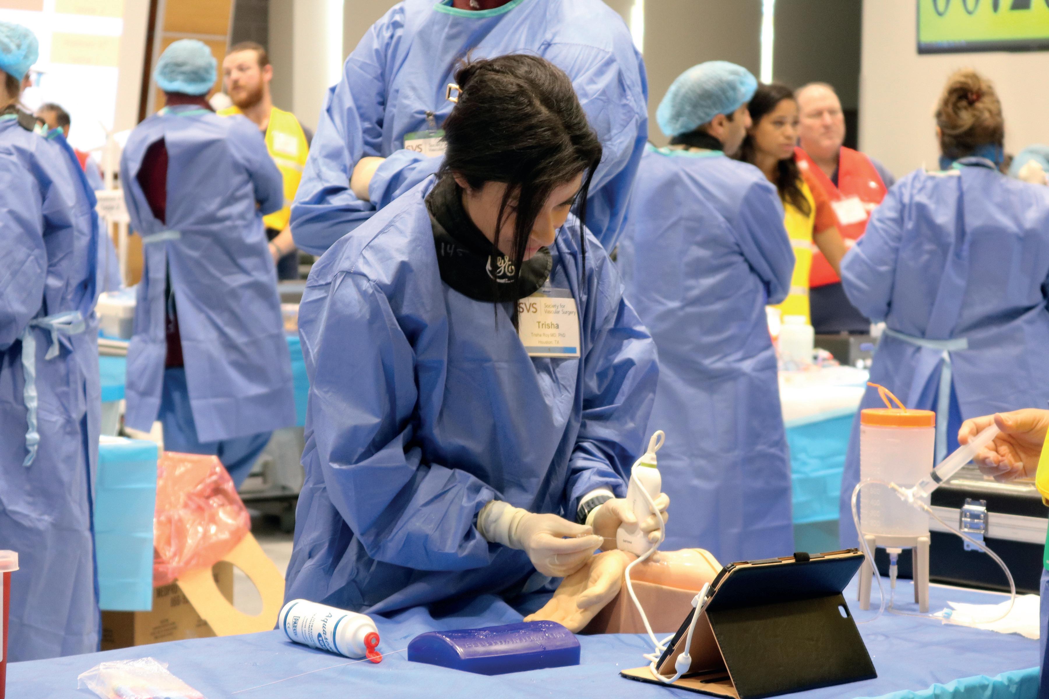

By Marlén Gomez
THE SVS FOUNDATION IS PLACING a spotlight on its “Legacy Teams”—those who have gone the extra mile, literally!—to make the annual Vascular Health Step Challenge a success year after year.
This year will see the return of two standout legacy teams: The Heart Hospital Pulse Patrol and Team FAB, who have played a role in the challenge’s growth and success. Their continued leadership is helping to build early momentum for this year’s campaign.
“I look forward to the Vascular Step Challenge every year. It has become a fixture for me as summer transitions into fall. The motivation to walk every day, the friendly competition with my vascular colleagues and the money raised to support the foundation’s mission are win, win, win in my book,” said William Shutze, MD, a member of the Heart Hospital Pulse Patrol, based out of Plano, Texas.
The challenge officially begins Sept. 1, coinciding with Peripheral Arterial Disease (PAD) Awareness Month. Participants are encouraged to walk 60 miles throughout the month to symbolize the 60,000 miles of blood vessels in the human body. The challenge is open to both individuals and teams, with registration having opened on Aug. 1.
The Heart Hospital Pulse Patrol and Team FAB have consistently demonstrated a strong
MULTIDISCIPLINARY CARE
commitment to promoting vascular health and inspiring their communities to engage in physical activity through their participation in the challenge.
Team FAB has been a part of the Step Challenge since 2022 and has walked more than 9,000 miles while raising more than $19,000. Last year, they won the title of the top team in the challenge, clocking a total of 3,188 miles.
“Team FAB is proud to unite vascular surgeons from across the country in support of the Step Challenge. We’re preparing with team-based step goals, weekly check-
ins and fitness tracking to stay motivated,” said Leigh Ann O’Banion, MD, a member of the group, from the University of California, San Francisco-Fresno (UCSF Fresno).
“The challenge not only promotes wellness among our colleagues, but also deepens our commitment to raising awareness of vascular health and encouraging patients to take simple, impactful steps, literally, toward preventing PAD. It’s a meaningful way for us to lead by example and stay connected as a national team.”
The Heart Hospital Pulse Patrol has changed their strategy this year to ensure their placement in the leaderboard is unchallenged.
“I’m thrilled to be partnering with Dr. Shutze and the SVS for this year’s annual Vascular Health Step Challenge,” said Vanessa Ferris, RN. “We’re aiming to expand our hospital’s participation and inspire even
60 miles
$60,000 60,000 miles of blood vessels
By Marlén Gomez
HEALTHCARE PROFESSIONALS FROM MULTIPLE disciplines are embracing the Wound Care Curriculum, a flexible online learning experience tailored to meet the evolving needs of vascular care teams.
Developed collaboratively by the SVS, including the former Physician Assistant (PA) Section (now part of the Advanced Practice Provider [APP] Section), the Society for Vascular Nursing (SVN), and the American Podiatric Medical Association (APMA), the curriculum offers a blended learning model that combines the convenience of online education with the depth of hands-on experience.
“The SVS wound course was an invaluable and enriching educational experience,” said Jessica Neff, FNP-C, a nurse practitioner from Albuquerque, New Mexico. “With my background in wound care, I found the curriculum to be both an excellent refresher and a source of new, evidence-based strategies that have deepened my understanding of complex wound management. The expert-led discussions and practical insights gained will greatly enhance my clinical skills as a vascular nurse practitioner and directly improve the quality of care I provide to patients.”
Available now, the curriculum features 27 short, on-demand videos that participants can access anytime, anywhere. This format allows busy healthcare professionals to absorb key concepts at their own pace, revisit material as needed and
“As a physician assistant in vascular surgery, the wound care curriculum provided a comprehensive, evidence-based framework for managing complex wounds” RACHEL BREAULT
more people to join us in promoting vascular health and wellness for a great cause.”
Other legacy teams returning to the walk—and looking to take the top spot— include IU Health Vascular Surgery, Team Bowling Green, KY Happy Feet and the Piedmont Triphasics. Together they have covered more than 9,000 miles and raised more than $7,000 in support of the challenge.
“The Step Challenge is not only an inspiring way to raise awareness and critical funds for the vascular community, but also a fun and meaningful opportunity to ignite friendly competition among SVS members. We are thrilled to welcome back these incredible legacy teams. Their participation brings renewed energy and excitement to the event. We hope their example motivates even more walkers to step up and get involved,” said Catherine Lampi, SVS Foundation director. Every step taken during the challenge helps raise awareness of PAD and supports the SVS Foundation’s goal of raising $60,000 for vascular research, education and outreach. Participants are encouraged to invite friends, family and colleagues to join their teams and walk together in support of better vascular health.
While these teams may be legacy steppers, the path to the top of the leader board is wide open. The Step Challenge welcomes everyone, whether first-time walkers or challenge veterans, to compete for first place while inspiring others to act and make a difference, one step at a time.
Visit vascular.org/StepChallenge for more information.
integrate new insights into their clinical practice.
“As a physician assistant in vascular surgery, the wound care curriculum provided a comprehensive, evidence-based framework for managing complex wounds,” said Rachel Breault, PA-C, a physician assistant from Palos Heights, Illinois. “It deepened my understanding of adjunctive therapies and reinforced a practical, multidisciplinary approach I can apply directly to patient care.”
Each module includes pre- and post-tests to assess knowledge acquisition, helping participants track their progress and identify areas for improvement.
In addition to the online content, the curriculum features hands-on workshops held in conjunction with the Vascular Annual Meeting (VAM) in 2026, 2027 and 2028, offering opportunities for in-person skill development and peer collaboration.
The Complex Wound Care Masterclass workshops, in-person at VAM, feature short didactics followed by complex cases and a panel discussion with an interprofessional team of vascular surgeons, PAs, vascular nurses and a podiatrist. The confirmed themes for 2026 are methods of debridement and dressing management, and oxygen therapy.
The SVS designates the online curriculum for a maximum of 6.25 AMA PRA Category 1 Credits™. The curriculum also offers continuing medical education (CME), continuing education units (CEUs) and continuing education (CE) credits in podiatric medicine, ensuring that professionals across specialties benefit from the program.
To learn more or to purchase access to the modules, visit vascular.org/WoundCare.
By Jocelyn Hudson
A FIVE-YEAR PROSPECTIVE COHORT STUDY— using longitudinal registry data from two population-based screening trials—has found that high-dose statin use was associated with decreased abdominal aortic aneurysm (AAA) growth rates and lowered risk of undergoing repair, rupture and death. Researchers therefore suggest high-dose statins should be “strongly considered” for patients with small AAAs.
Writing in Circulation, authors Joachim S. Skovbo, MD, from Odense University Hospital, in Odense, Denmark, and colleagues state that AAAs present with high morbidity and mortality when they occasionally rupture. Multiple meta-analyses have identified both metformin and statins as potential treatments, the authors go on to note, but neither has successfully been proven to reduce AAA growth. The present study, therefore, aimed to investigate a relationship between statin use and AAA growth rates and risk of undergoing repair, rupture, or death.
Skovbo and colleagues detail that the study population included all men with screening-detected AAAs—ranging in

Joseph V. Lombardi, MD, MBA, FACS Chief Physician Executive
AtlantiCare Regional Medical Center Chair, The Heart and Vascular Institute, Atlantic City, NJ
Joseph J. Ricotta, MD, MS, FACS
National Medical Director, Vascular Surgery and Endovascular Therapy, Tenet Healthcare Corporation
Professor of Surgery, Charles E. Schmidt College of Medicine
Program Director, Vascular Surgery Fellowship
Florida Atlantic University, Boca Raton, FL
Ali Azizzadeh, MD Cedars-Sinai Medical Center, Los Angeles, CA
James H. Black, III, MD The Johns Hopkins University School of Medicine, Baltimore, MD
G. Chad Hughes, MD, Duke University Medical Center, Durham, NC
James F. McKinsey, MD, Jacobson Aortic Center Mount Sinai Medical System, New York, NY
Darren B. Schneider, MD The Hospital of the University of Pennsylvania, Philadelphia, PA
diameter from 30–55mm—from two large, population-based screening trials: the Viborg Vascular Screening trial and the Danish Cardiovascular Screening trial, which included patients in the years 2008–2011 and 2014–2018, respectively.
The authors add that the clinical database was supplemented with data from the nationwide Danish Healthcare Registries, including prescription and outcome data. In order to evaluate the risk of repair, patients were followed from inclusion until surgery, rupture, death, five-year follow-up or Dec. 31, 2021.
The researchers included a total of 998 aneurysmal men with a mean age of 69.5 years and a median AAA diameter of 35.4mm in the study. They report in Circulation that statin use was “significantly associated with reduced AAA growth rate,” sharing that an increase of one defined daily dose statin per day was associated with an adjusted change in growth rate of -0.22mm per year.
Additionally, Skovbo et al relay that the five-year adjusted hazard ratio for undergoing repair per doubling of statin dose presented a “significantly reduced adjusted hazard ratio” of 0.82, which they note was significant after 2.5 years.
“Statin use was associated with a significantly lower risk of the composite outcome (surgery, rupture, and death) in a dose-dependent manner,” the authors continue, citing an adjusted hazard ratio of 0.83 per doubling of statin dose.
Skovbo and colleagues write that their findings were “robust” in several sensitivity analyses and conclude that high-dose statin use was associated with a dose-dependent reduction in AAA growth rates and a lower risk of undergoing repair, rupture and death during a five-year
“These
follow-up period. “These findings suggest that high-dose statins may offer direct protection against AAA progression, beyond cardiovascular risk reduction alone,” Skovbo et al write. Considering the clinical implications of their work, the authors advise that, “Because of their proven cardiovascular benefits, safety profile, and cost-effectiveness, high-dose statins should be strongly considered for patients with small AAAs, particularly those without contraindications.”
“Although randomized trials may be ethically challenging,” the authors continue, “further high-quality observational studies are needed to validate these findings and inform clinical guidelines.”
In their Circulation paper, Skovbo and colleagues highlight certain limitations that curb the generalizability of their findings. For example, they note that the study included only male patients, reducing the generalizability of the results to the female population.
Furthermore, the authors recognize that the study cohort primarily consists of White individuals from a Western society between 60 and 74 years of age, going on to note that “caution is warranted when extrapolating these findings to other racial and ethnic groups, age groups, or different societal contexts.”
JOACHIM S. SKOVBO
Recognizing the difficulties of performing randomized controlled trials (RCTs) to assess the treatment options for patients with AAA, Skovbo and colleagues instead “advocate for observational studies with a similar design, emphasizing precise exposure measurement and detailed dose-response outcome, to replicate and further validate our findings especially in women and ethnic groups.”


Compiled by Jocelyn Hudson and Bryan Kay
First patient treated with InterVene’s Recana thrombectomy catheter system
INTERVENE HAS ANNOUNCED THAT the first patient has been treated with its Recana thrombectomy catheter system for venous in-stent restenosis (ISR).
The company shares in a press release that Recana is the first fully integrated mechanical thrombectomy system designed specifically to address the long-term complications of venous disease, including ISR and native vein obstructions. The case was performed at University Hospital Galway in Galway, Ireland, by interventional radiologist Gerry O’Sullivan, MBBCh. “The long-term sequelae of venous obstructions, whether in-stent or native vessel, are a devastating consequence of deep venous thrombotic disease. There remains a considerable gap in effective interventional treatments for physicians and their patients living with persistent symptoms related to venous outflow obstructions,” said O’Sullivan. “I had previously tried and failed to recanalize this patient’s vessels on four separate occasions—Recana succeeded in just one. This technology offers real promise for patients with limited treatment options.”
The case was a result of collaboration between InterVene and the University of Galway Institute of Clinical Trials.


Getinge’s iCast covered stent system approved as bridging stent in US
GETINGE RECENTLY REVEALED
that it has received Food and Drug Administration (FDA) premarket approval (PMA) for the iCast covered stent system. The device is now available as a bridging stent for the treatment of patients with aneurysmal disease.
Getinge states that this new indication enables physicians to preserve blood flow between a branch vessel and an endovascular graft that is approved for use with the iCast covered stent.
In March 2024, Getinge and Cook Medical entered into a distribution agreement under which Cook assumed sales, marketing and distribution responsibilities for the iCast covered stent system in the U.S.
The product, now with its expanded bridging stent indication, will continue to be distributed by Cook Medical.
iCast covered stent system

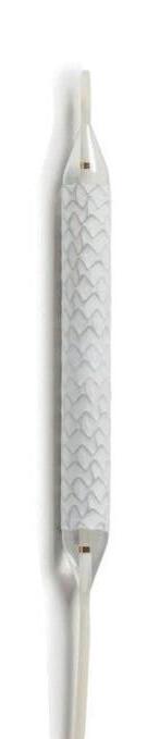
Cook announces US launch of Zenith iliac branch device
COOK MEDICAL HAS announced that its Zenith iliac branch device (ZBIS) is now commercially available in the U.S. with Food and Drug Administration (FDA) approval as an endovascular treatment option for aortoiliac or iliac aneurysmal disease.
The company has also shared that first implantations of the ZBIS were performed at several centers in the U.S.
According to Cook, the ZBIS—when used with the necessary additional components (Zenith AAA and a covered bridging stent)—is indicated for the endovascular treatment of patients with an aortoiliac or iliac aneurysm to preserve internal iliac arterial blood flow when the distal sealing site in the common iliac artery is insufficient for the abdominal aortic aneurysm (AAA) device alone and when the vessel morphology is suitable for repair.
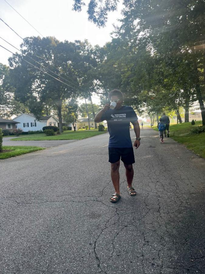
Medtronic enrolls first patient in Onyx liquid embolic IDE clinical study
THE FIRST PATIENT HAS BEEN ENROLLED IN THE Peripheral Onyx liquid embolic (PELE) clinical trial, which will evaluate the safety and effectiveness of the Onyx liquid embolic system (LES) for embolization of arterial hemorrhage in the peripheral vasculature.
The procedure was performed by Christopher Stark, MD, a vascular and interventional radiologist at Albany Medical Center in Albany, New York, Medtronic said in a press release
Data from the PELE investigational device exemption (IDE) clinical study is intended to evaluate the safety and effectiveness of Onyx LES in the treatment of patients with active arterial bleeding in the peripheral vasculature who are deemed to be suitable for embolization treatment.
“Embolic agents are an important tool to address hemorrhage. In this case, the patient experienced bleeding due to a ruptured blood vessel following a routine medical procedure. Onyx LES was administered into the target vessel to successfully facilitate embolization,” said Stark.
This pivotal, prospective, multicenter, non-randomized, single-arm study will enroll up to 119 patients from up to 25 sites in the U.S. The primary safety and efficacy endpoints will be evaluated through 30 days following treatment with Onyx LES.
Medtronic advises that, in the U.S., the use of Onyx LES for embolization of arterial hemorrhage in the peripheral vasculature is investigational only and has not been approved or cleared by the Food and Drug Administration (FDA).
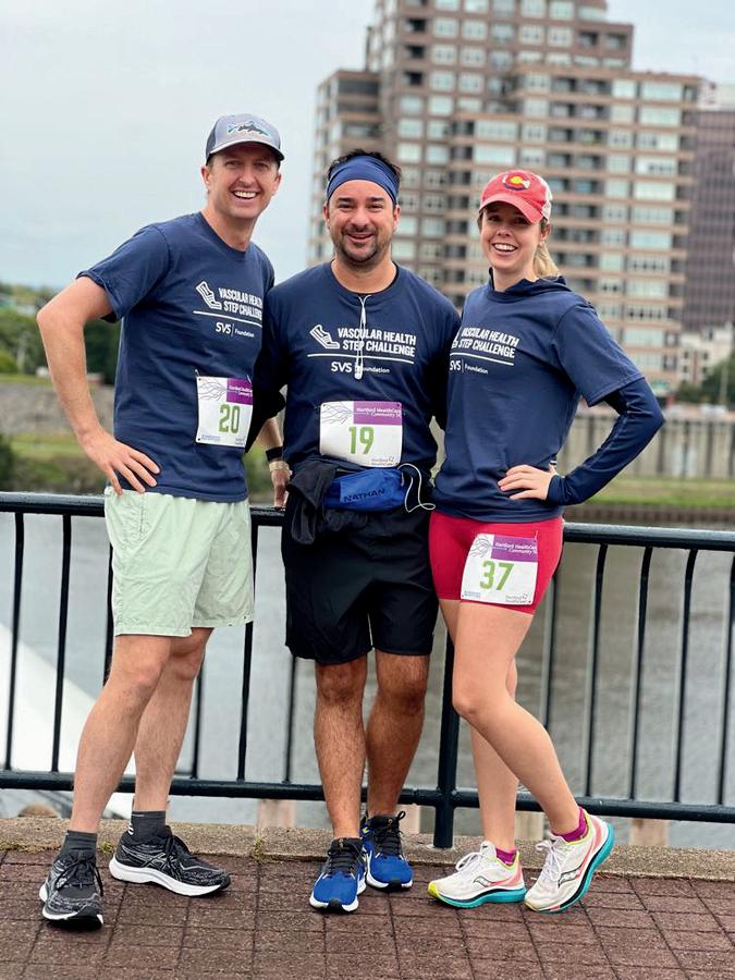
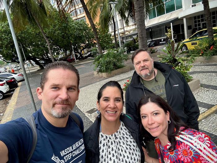

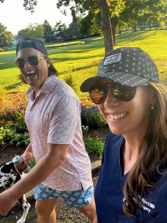
STEP UP TO THE CHALLENGE
Sept. 1 – 30

THERE’S NO PLACE LIKE HOME
within the newly established SVS Advanced Practice Provider (APP) Section, where vascular advanced practice registered nurses (APRNs) and physician assistants (PAs) aim to provide a “professional home” for their colleagues working in vascular care settings.
The groups finalized the change in May 2025 with the integration of the former PA Section into the APP section. This collaborative effort includes the SVS, the Society for Vascular Nursing (SVN) and the old PA Section.
Chris Owen, MS, and Erin Hanlon, PAC, will co-chair the new section to ensure proper representation.
Owen, a past president of the SVN and a practicing nurse practitioner, has seen firsthand the impact of a unified, collaborative voice in advancing the profession and improving patient outcomes.
“Team-based care is essential to the future of healthcare—ensuring that all providers, regardless of background, are respected for their contributions,” Owen said. “I’m honored to be part of this
“ Team-based care is essential to the future of healthcare— ensuring that all providers, regardless of background, are respected for their contributions”
CHRIS OWEN
collaborative inaugural SVS APP Section and grateful for the recognition of the vital role we all play in caring for vascular patients.”
The APP Section offers numerous benefits, including opportunities for professional networking and peer mentoring, as well as leadership development through engagement with SVS committees.
The SVN Annual Conference will feature education specifically tailored to APPs. Additional perks include discounts on SVS events and subscriptions to the Journal of Vascular Surgery (JVS), Vascular Specialist and the weekly Pulse e-newsletter.
Members will also gain access to the APP Section on the SVSConnect online community.
For more membership information, visit vascular.org/APPSection.
Compiled by Marlén Gomez
YOU DON’T HAVE TO BE TOO FAR DOWN THE PATH TO GET STARTED.
That’s the message William Shutze, MD, chair of the SVS-American College of Surgeons (ACS) Vascular Verification Program (VVP), wants vascular specialists across the country to grasp as they take the first step toward participating in the VVP, even if they’ve only just downloaded the standards.
The SVS, in collaboration with the ACS, launched the VVP as a national quality verification initiative designed to enhance the care and treatment of patients undergoing vascular surgical and interventional procedures in both inpatient and outpatient settings. The program offers two levels of participation, enabling institutions to engage based on their current capabilities and guiding them toward ongoing quality improvement.
“Even if you’re just beginning to explore the standards, we want to hear from you,” said Shutze. “The ACS quality team is eager to support you early in your journey and help you move toward a successful site review.”
The VVP organizes its framework around 12 domains that cover the five phases of care: preoperative, immediate preoperative, intraoperative, postoperative and follow-up. These domains ensure that institutions not only have standardized protocols in place but also actively collect and analyze data to drive improvements.
A recent systematic review published in the Journal of the American College of Surgeons (JACS) analyzed over 150 studies and found that programs like the VVP led to:
● A 1.9-day reduction in average hospital stay
● A $1,763 decrease in cost per admission
● Fewer complications across the board
● Empowering conversations with the C-suite
One of the barriers to adoption, according to Shutze, is that many vascular specialists don’t feel equipped to make the case for the program to their hospital leadership. To address this, the SVS is developing a toolkit that includes a brochure outlining the value of vascular surgery, return on investment (ROI) data and executive testimonial videos featuring hospital leaders who have seen the benefits firsthand. “We want our members to feel empowered when they walk into those meetings,” said Shutze. “This package will help them speak the language of the C-suite.”
As of this summer, eight programs have already been approved, with several more in the pipeline. Seven additional institutions are scheduling site visits, and a few are completing the final steps for certification. Shutze, of Texas Vascular Associates, also extended thanks to R. Clement Darling III, MD, Anton Sidawy, MD, and Anil Hingorani, MD, for their leadership, as well as the Vascular Quality Program Task Force. For institutions considering participation, the first step is straightforward: visit the ACS Quality Programs website at facs.org/vascular and start the process.
THE JOURNAL OF VASCULAR Surgery (JVS) has appointed Audra Duncan, MD, of Western University in London, Ontario, Canada, as its new editor-in-chief.
Duncan will lead the journal for a fouryear term, renewable for two more years, following the decision of the Publications Committee and the approval of the SVS Executive Board.
She succeeds Thomas Forbes, MD, of the University of Toronto, Canada, who concluded his three-year tenure at this summer’s Vascular Annual Meeting (VAM 2025). Under Forbes’ editorial leadership, JVS continued to grow as a leading platform for cutting-edge vascular research.
Previously serving as an associate editor for the journal, Duncan has been a key contributor to its editorial direction and
peer-review standards. Her appointment underscores a commitment to fostering high-quality scholarship.
In her new role, Duncan will champion initiatives that enhance the journal’s reach, promote diversity in research and seek to uphold rigorous scientific standards.
“I am excited for this opportunity to further build the journal that meets the needs of our SVS community and pushes boundaries in innovation and collaboration,” said Duncan. “We are such a strong vascular group; we have the ability to use our journal to showcase the great work we do, and what vascular surgery looks like in 2025.”
Countdown to Capitol Hill: SVS Advocacy and Leadership Conference nears

THE SVS IS GEARING UP FOR ITS inaugural Advocacy and Leadership Conference, taking place Sept. 14–16, in Washington, D.C.
With just weeks to go, the SVS is encouraging members at all career stages to register and take part in this landmark event designed to empower vascular professionals to shape the future of their specialty.
Over the course of three days, attendees will have the opportunity to participate in a series of dynamic workshops, keynote presentations, and panel discussions. Additionally, attendees can visit Capitol Hill and directly engage with lawmakers and their staff.
This first-of-its-kind conference will unite vascular surgeons, trainees and other key stakeholders to engage in the policymaking process. Attendees will gain insights into the most pressing legislative and regulatory issues affecting vascular care.
“Vascular surgeons across the country are feeling the strain on our healthcare system and demanding change,” said Megan Tracci, MD, JD, chair of the SVS Advocacy Council.
“During the inaugural Advocacy and Leadership Conference coming up, the SVS is offering our surgeons a chance to participate in driving that change.
“This is a unique opportunity to examine the pressing issues facing the vascular surgery specialty, meet leaders and policymakers, learn how to be an effective advocate and participate— personally—in transforming the healthcare landscape through visits with members of Congress and policy staff on Capitol Hill.”
Space is limited and interest high. The SVS encourages members to register today at vascular.org/ advocacyconference25
Esprit™ BTK Everolimus Eluting Resorbable Scaffold System
INDICATIONS
The Esprit™ BTK Everolimus Eluting Resorbable Scaffold System is indicated for improving luminal diameter in infrapopliteal lesions in patients with chronic limb-threatening ischemia (CLTI) and total scaffolding length up to 170 mm with a reference vessel diameter of ≥ 2.5 mm and ≤ 4.00 mm.
CONTRAINDICATIONS
The Esprit™ BTK Everolimus Eluting Resorbable Scaffold System is contraindicated for use in:
• Patients who cannot tolerate, including allergy or hypersensitivity to, procedural anticoagulation or the post-procedural antiplatelet regimen.
• Patients with hypersensitivity or contraindication to everolimus or structurally related compounds or known hypersensitivity to scaffold components poly(L-lactide), poly(D, L-lactide), and platinum.
WARNINGS
• Th is device is intended for single use only. Do not reuse, reprocess, or re-sterilize. Note the product “Use-by” date on the package. Reuse, reprocessing, or re-sterilization may compromise the structural integrity of the device and / or delivery system and / or lead to device failure, which may result in patient injury, illness, or death. Reuse, reprocessing, or resterilization may also create a risk of contamination of the device and / or cause patient infection or cross-infection, including, but not limited to, the transmission of infectious disease(s) from one patient to another. Contamination of the device and / or delivery system may lead to injury, illness, or death of the patient.
• The Esprit™ BTK System is intended to perform as a system. The scaffold should not be removed for use with other dilatation catheters.
• The Esprit™ BTK System should not be used in conjunction with other non-everolimus drug eluting devices in the same vessel as the Esprit™ BTK Scaffold.
• It is not recommended to use this scaffold to treat lesions located at any joint or other hinge points, such as the knee or ankle. The recommended region for below-the-knee (BTK) treatment with the Esprit™ BTK Scaffold is the infrapopliteal arteries at a location ≥ 10 cm above the proximal margin of the ankle mortise. The Esprit™ BTK Scaffold has not been tested for use outside the recommended implant locations.
• Th is product should not be used in patients with aneurysms immediately adjacent to the scaffold implantation site.
• Insertion of the Esprit™ BTK System and implantation of the scaffold should be performed only under fluoroscopic observation with radiographic equipment providing high resolution images.
• Quantitative imaging is strongly recommended to accurately measure and confirm appropriate vessel sizing (reference vessel diameter ≥ 2.5 mm). If quantitative imaging determines a vessel size < 2.5 mm, do not implant the Esprit™ BTK Scaffold.
• Adequate lesion preparation prior to scaffold implantation is required to ensure safe delivery of the scaffold across the target lesion. It is not recommended to treat patients having a lesion that prevents complete inflation of an angioplasty balloon.
• Successful pre-dilatation with residual diameter stenosis of < 30% by visual estimation is required for treatment of the target lesion; < 20% by visual estimation is preferred.
• En sure the scaffold is not post-dilated beyond the allowable expansion limits.
• Use of appropriate anticoagulant and / or antiplatelet therapy per standard of care is recommended for use of this scaffold system.
• Th is product should not be used in patients who are not likely to comply with the recommended antiplatelet therapy.
• Judicious selection of patients is necessary, since the use of this device carries the associated risk of
scaffold thrombosis, vascular complications, and / or bleeding events.
PRECAUTIONS
• Scaffold placement should not be performed in patients with known allergies to contrast agent that cannot be medically managed.
• It is not recommended to treat patients having a lesion with excessive tortuosity proximal to or within the lesion.
• When multiple scaffolds are required, only combinations of Esprit™ BTK Scaffolds must be used. Any potential interaction with other drugeluting or coated devices has not been evaluated.
• The delivery system is intended for deployment of the scaffold only and should not be used to dilate other locations.
• Implantation of the scaffold should be performed only by physicians who have received appropriate training.
• As w ith all catheter-based procedures, scaffold placement should be performed at facilities where patient can be prepared for necessary intervention and / or surgical removal of the device and vessel repair as per facility protocol.
• Pre-dilatation should be performed with an angioplasty balloon. Cutting or scoring balloons can be used per physician discretion, if the lesion appears to be mildly calcified.
• Fa ilure to pre-dilate the vessel may impair nominal / optimal scaffold delivery.
• Implanting a scaffold may lead to dissection of the vessel distal and / or proximal to the scaffold, requiring additional intervention.
Note: In cases of bailouts, bailout treatment of the target lesion can be done using the Esprit™ BTK Scaffold of the appropriate length. If an appropriate length Esprit™ BTK Scaffold is not available, physicians should use standard of care.
• An u nexpanded scaffold may be retracted into the introducer sheath one time only. An unexpanded scaffold should not be reintroduced into the artery once it has been pulled back into the introducer sheath.
• Post-dilatation is strongly recommended for optimal scaffold apposition. When performed, postdilatation should be performed at high pressure (> 16 atm) with a non-compliant balloon up to 0.5 mm larger than the nominal scaffold diameter.
• Use an appropriately sized non-drug coated balloon to pre-dilate the lesion. When treating a long lesion, scaffold the distal portion of the lesion prior to scaffolding the proximal portion of the lesion.
• En sure that the scaffolded area covers the entire lesion / dissection site and that no gaps exist between scaffolds.
• The extent of the patient’s exposure to drug and polymer is directly related to the number of scaffolds implanted. The safety of everolimus, polymer, and polymer breakdown products was evaluated in preclinical studies and the biocompatibility assessment of the Esprit™ BTK Scaffold.
• The safety and effectiveness of the Esprit™ BTK Scaffold in patients with prior brachytherapy of the target lesion or the use of brachytherapy for treatedsite restenosis in the Esprit™ BTK Scaffold have not been established. Both vascular brachytherapy and the Esprit™ BTK Scaffold alter arterial modeling. The potential combined effect on arterial remodeling by these two treatments is not known.
• The safety and effectiveness of the Esprit™ BTK System have not been established in clinical trials with the use of either mechanical atherectomy devices (directional atherectomy catheters, rotational atherectomy catheters) or laser atherectomy catheters.
• Formal drug interaction studies have not been performed with the Esprit™ BTK Scaffold because of limited exposure to everolimus eluted from the scaffold.
• Everolimus, the Esprit™ BTK Scaffold’s active pharmaceutical ingredient, is an immunosuppressive agent. Therefore, consideration should be given to patients taking other immunosuppressive agents or who are at risk for immune suppression.
• Oral everolimus use in renal transplant and advanced renal cell carcinoma patients was associated with increased serum cholesterol and triglyceride levels, which in some cases required treatment.
• Non-clinical testing has demonstrated the Esprit™ BTK Scaffold is MR Conditional. A person with the Esprit™ BTK Scaffold may be safely scanned under the following conditions. Failure to follow these conditions may result in injury.
• Static magnetic field strength of 7 Tesla or less
• The Esprit™ BTK Scaffold should not migrate in this MRI environment. MRI at 7 Tesla or less may be performed immediately following the implantation of the Esprit™ BTK Scaffold.
POTENTIAL ADVERSE EVENTS
Potential adverse events include, but are not limited to: Allergic reaction or hypersensitivity to contrast agent, anesthesia, scaffold materials (poly[L-lactide] [PLLA], poly[D, L-lactide] [PDLLA], platinum, or everolimus), and drug reactions to anticoagulation or antiplatelet drugs
• Va scular access complications which may require transfusion or vessel repair, including:
• Catheter site reactions
• Bleeding (ecchymosis, oozing, hematoma, hemorrhage, retroperitoneal hemorrhage)
• Arteriovenous fistula, pseudoaneurysm, aneurysm, dissection, perforation / rupture, and laceration
• Embolism (air, tissue, plaque, thrombotic material, or device)
• Peripheral ischemia
• Ta rget artery complications which may require additional intervention, including:
• Total occlusion or abrupt closure
• Arteriovenous fistula, pseudoaneurysm, aneurysm, dissection, perforation / rupture
• Embolism (air, tissue, plaque, thrombotic material, or device)
• Artery or scaffold thrombosis
• Stenosis or restenosis
• Vasospasm
• Tissue prolapse / plaque shift
• Bleeding (non-access site)
• Additional surgery such as peripheral artery bypass graft surgery or amputation
• Per ipheral nerve injury, neuropathy
• Compartment syndrome
• Tissue necrosis, gangrene, ulcer and acute limb ischemia
• Reperfusion injury
• New or worsening pain
• Intervention due to
• Damaged scaffolds
• Partial scaffold deployment
• Scaffold migration / unintentional placement of scaffold
• Ot her general surgical risks, including:
• Cardiac arrhythmias (including conduction disorders, atrial and ventricular arrhythmias, and blocks)
• Stroke / cerebrovascular accident (CVA) and transient ischemic attack (TIA)
• Venous thromboembolism (including pulmonary embolism)
• Nausea and vomiting
• Hypotension / hypertension
• Infection – local and systemic (including post-procedural)
• Fever
• Blood cell disorders including heparininduced thrombocytopenia (HIT) and other coagulopathy
• Death
• System organ failures:
• Cardiac Failure
• Cardio-respiratory arrest (including pulmonary edema)
CAUTION: This product is intended for use by or under the direction of a physician. Prior to use, reference the Instructions for Use, inside the product carton (when available) or at manuals.eifu.abbott for more detailed information on Indications, Contraindications, Warnings, Precautions and Adverse Events. This material is intended for use with healthcare professionals only.
Illustrations are artist’s representations only and should not be considered as engineering drawings or photographs. Photo on file at Abbott.
Information contained herein for DISTRIBUTION in the U.S. ONLY.
Abbott
3200 Lakeside Dr., Santa Clara, CA 95054 USA, Tel: 1.800.227.9902
™ Indicates a trademark of the Abbott Group of Companies. www.cardiovascular.abbott
©2024 Abbott. All rights reserved. MAT-2408411 v3.0
• Respiratory failure
• Renal failure
• Shock
The risks described below include the anticipated adverse events referenced in the contraindications, warnings, and precautions sections of the everolimus labels / SmPCs and / or observed at incidences ≥ 10% in clinical trials with oral everolimus for different indications. Refer to the drug SmPCs and labels for more detailed information and less frequent adverse events.
• Abdominal pain
• Anemia
• Angioedema (increased risk with concomitant angiotensin-converting enzyme [ACE] inhibitor use)
• Ar terial thrombotic events
• Bleeding and coagulopathy (including hemolytic uremic syndrome [HUS], thrombotic thrombocytopenic purpura [TTP], and thrombotic microangiopathy; increased risk with concomitant cyclosporine use)
• Constipation
• Cough
• Diabetes mellitus
• Diarrhea
• Dyspnea
• Embryo-fetal toxicity
• Er ythema
• Er ythroderma
• Headache
• Hepatic artery thrombosis (HAT)
• Hepatic disorders (including hepatitis and jaundice)
• Hy persensitivity to everolimus active substance, or to other rapamycin derivates
• Hy pertension
• In fections (bacterial, viral, fungal, or protozoan infections, including infections with opportunistic pathogens). Polyoma virus-associated nephropathy (PVAN), JC virus-associated progressive multiple leukoencephalopathy (PML), fatal infections and sepsis have been reported in patients treated with oral everolimus.
• Kidney arterial and venous thrombosis
• Laboratory test alterations (elevations of serum creatinine, proteinuria, hypokalemia, hyperkalemia; hyperglycemia, dyslipidemia including hypercholesterolemia and hypertriglyceridemia; abnormal liver function tests; decreases in hemoglobin, lymphocytes, neutrophils, and platelets)
• Ly mphoma and skin cancer
• Ma le infertility
• Menstrual irregularities
• Nau sea
• Nephrotoxicity (in combination with cyclosporine)
• Non-infectious pneumonitis (including interstitial lung disease)
• Oral ulcerations
• Pai n
• Pa ncreatitis
• Pericardial effusion
• Per ipheral edema
• Pleural effusion
• Pneumonia
• Py rexia
• Ra sh
• Renal failure
• Upper respiratory tract infection
• Ur inary tract infection
• Venous thromboembolism
• Vom iting
• Wound healing complications (including wound infections and lymphocele)
There may be other potential adverse events that are unforeseen at this time.

48% fewer patients require reinterventions through 2 years.1,2 Go above expectations with the only device that does it all for CLTI.*

*All: drug, scaffold, resorbable excluding platinum markers. 1. Reintervention defined as CD-TLR. 2. Brian G. DeRubertis et al., Two-Year Outcomes of the LIFE-BTK Randomized Controlled Now Proven With 2-Year RCT Data Trial Evaluating the Esprit™ BTK Drug-eluting Resorbable Scaffold for Treatment of Infrapopliteal Lesions, VIVA 2024
TAKE THE FIRST STEP. LEARN MORE TODAY!
INDICATIONS The Esprit™ BTK Everolimus Eluting Resorbable Scaffold System is indicated for improving luminal diameter in infrapopliteal lesions in patients with chronic limb-threatening ischemia (CLTI) and total scaffolding length up to 170 mm with a reference vessel diameter of ≥ 2.5 mm and ≤ 4.00 mm. See Important Safety Information referenced on the previous page. Information contained herein for DISTRIBUTION in the U.S. ONLY. ©2024 Abbott. All rights reserved. MAT-2408411 v3.0