


Go Mobile and Thrive State-of-the-Art Imaging Technology

Partner with Shared Imaging exceptional patient care saving costs and meeting demand for imaging

Technology & Coaches
Take


Partner with Shared Imaging exceptional patient care saving costs and meeting demand for imaging
Take

First FDA-approved, cost-effective generic that is fully substitutable to Visipaque®.*


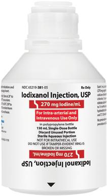
WARNING: NOT FOR INTRATHECAL USE
Inadvertent intrathecal administration may cause death, convulsions/seizures, cerebral hemorrhage, coma, paralysis, arachnoiditis, acute renal failure, cardiac arrest, rhabdomyolysis, hyperthermia, and brain edema.
Please see Important Safety Information on next page.
• AP Rated
• Iso-Osmolar
• Dimeric
• Preservative Free
• Polymer Bottle LEARN MORE
Explore Available Contrast Agents GenericContrastAgents.com
WARNING: NOT FOR INTRATHECAL USE
Inadvertent intrathecal administration may cause death, convulsions/seizures, cerebral hemorrhage, coma, paralysis, arachnoiditis, acute renal failure, cardiac arrest, rhabdomyolysis, hyperthermia, and brain edema.
Contraindications: Iodixanol Injection is contraindicated for intrathecal use.
Warnings and Precautions:
Hypersensitivity Reactions: Life-threatening or fatal reactions can occur. Most severe reactions develop shortly after the start of the injection, but reactions can occur up to hours later. Always have emergency equipment and trained personnel available.
Contrast-Induced Acute Kidney Injury: Acute injury including renal failure can occur. Minimize dose and maintain adequate hydration to minimize risk.
Cardiovascular Adverse Reactions: Life-threatening or fatal cardiovascular reactions, including hypotension, shock, and cardiac arrest have occurred with the use of Iodixanol. Most deaths occur during injection or five to ten minutes later, with cardiovascular disease as the main aggravating factor. Use the lowest necessary dose of Iodixanol in patients with congestive heart failure.
Thromboembolic Events: Serious, rarely fatal, thromboembolic events causing myocardial infarction and stroke can occur during angiocardiography procedures with both ionic and nonionic contrast agents.
Extravasation and Injection Site Reactions: Extravasation of Iodixanol injection may cause tissue necrosis and/or compartment syndrome, particularly in patients with severe arterial or venous disease. Ensure intravascular placement of catheters prior to injection.
Thyroid Storm in Patients with Hyperthyroidism: Thyroid storm has occurred after the intravascular use of iodinated contrast agents in patients with hyperthyroidism, or with an autonomously functioning thyroid nodule.
Thyroid Dysfunction in Pediatric Patients 0 to 3 Years of Age: Thyroid dysfunction characterized by hypothyroidism or transient thyroid suppression has been reported after both single exposure and multiple exposures to iodinated contrast media in patients 0 to 3 years of age. After exposure to iodinated contrast media, individualize thyroid function monitoring based on underlying risk factors, especially in term and preterm neonates.
Hypertensive Crisis in Patients with Pheochromocytoma: Hypertensive crisis has occurred after the use of iodinated contrast agents in patients with pheochromocytoma. Inject the minimum amount of contrast necessary, assess the blood pressure throughout the procedure, and have measures for treatment of a hypertensive crisis readily available.
Sickle Cell Crisis in Patients with Sickle Cell Disease: Iodinated contrast agents when administered intravascularly may promote sickling in individuals who are homozygous for sickle cell disease.
Severe Cutaneous Adverse Reactions: Severe cutaneous adverse reactions (SCAR) may develop from one hour to several weeks after intravascular contrast agent administration. These reactions include Stevens-Johnson syndrome and toxic epidermal necrolysis (SJS/TEN), acute generalized exanthematous pustulosis (AGEP), and drug reaction with eosinophilia and systemic symptoms (DRESS). Avoid administering iodixanol to patients with a history of a severe cutaneous adverse reaction to iodixanol.
Adverse Events: Serious, life-threatening, and fatal reactions, mostly of cardiovascular origin, have been associated with the administration of iodine-containing contrast agents, including Iodixanol Injection. Most common adverse reactions (incidence greater than 0.5%) in adult patients after iodixanol injection: Discomfort, warmth, pain; Cardiovascular: angina. Gastrointestinal: diarrhea, nausea, vomiting. Nervous System: agitation, anxiety, insomnia, nervousness, dizziness, headache, migraine, unusual skin sensations, sensory disturbance, fainting, sensation of spinning. Skin: itchy rash, severe itching, hives. Special Senses: Smell, taste, and vision alteration. Pediatric patients experienced similar adverse reactions.
To report SUSPECTED ADVERSE REACTIONS, contact Fresenius Kabi USA, LLC at 1-800-551-7176, option 5, or FDA at 1-800-FDA-1088 or www.fda.gov/medwatch.
Lactation: A lactating woman may consider interrupting breastfeeding and pumping and discarding breast milk for 10 hours after iodixanol administration in order to minimize drug exposure to a breast fed infant.
Pediatric Use: Pediatric patients at high risk of adverse reactions during and after administration of contrast agents include those with asthma, hypersensitivity to other medication and/or allergens, cyanotic and acyanotic heart disease, chronic heart failure, or a serum creatinine >1.5 mg/dL. Patients with immature renal function or dehydration may be at increased risk due to prolonged elimination of iodinated contrast agents.
Geriatric Use: Dose selection for an elderly patient should be cautious usually starting at the low end of the dosing range, reflecting the greater frequency of decreased hepatic, renal or cardiac function, and of concomitant disease or other drug therapy.
INDICATIONS AND USAGE
Iodixanol injection is a radiographic contrast agent indicated for the following:
Intra-arterial Procedures
Adults and pediatric patients 12 years of age and over
• Intra-arterial digital subtraction angiography (270 mg Iodine/mL and 320 mg Iodine/mL).
• Angiocardiography (left ventriculography and selective coronary arteriography), peripheral arteriography, visceral arteriography, and cerebral arteriography (320 mg Iodine/mL).
Pediatric patients less than 12 years of age
• Angiocardiography, cerebral arteriography, and visceral arteriography (320 mg Iodine/mL).
Intravenous Procedures
Adults and pediatric patients 12 years of age and over
• Computed tomography (CT) imaging head and body (270 mg Iodine/mL and 320 mg Iodine/mL).
• Excretory urography (270 mg Iodine/mL and 320 mg Iodine/mL).
• Peripheral venography (270 mg Iodine/mL).
• Coronary computed tomography angiography (CCTA) to assist diagnostic evaluation of patients with suspected coronary artery disease (320 mg Iodine/mL).
Pediatric patients less than 12 years of age
• CT imaging of the head and body (270 mg Iodine/mL).
• Excretory urography (270 mg Iodine/mL).
This Important Safety Information does not include all the information needed to use Iodixanol Injection, USP safely and effectively. Please see full prescribing information, including BOXED WARNING, for Iodixanol Injection, USP. Full prescribing information is also available at at www.fresenius-kabi.com/us
FDA-approved, cost-effective generic gadolinium-based contrast agent (GBCA) is bioequivalent and fully substitutable to Dotarem®.*
Explore Available Contrast Agents GenericContrastAgents.com • AP Rated • Macrocyclic 1,2 • Ionic 1,2 • Preservative Free 1,2
LEARN MORE


WARNING: RISK ASSOCIATED WITH INTRATHECAL USE and NEPHROGENIC SYSTEMIC FIBROSIS
Risk Associated with Intrathecal Use
Intrathecal administration of gadolinium-based contrast agents (GBCAs) can cause serious adverse reactions including death, coma, encephalopathy, and seizures. Gadoterate Meglumine Injection is not approved for intrathecal use.
Nephrogenic Systemic Fibrosis
GBCAs increase the risk for nephrogenic systemic fibrosis (NSF) among patients with impaired elimination of the drugs. Avoid use of Gadoterate Meglumine Injection in these patients unless the diagnostic information is essential and not available with non-contrasted MRI or other modalities. NSF may result in fatal or debilitating fibrosis affecting the skin, muscle and internal organs.
• The risk for NSF appears highest among patients with:
- Chronic, severe kidney disease (GFR < 30 mL/min/1.73m2), or
- Acute kidney injury.
• Screen patients for acute kidney injury and other conditions that may reduce renal function. For patients at risk for chronically reduced renal function (e.g. age > 60 years, hypertension, diabetes), estimate the glomerular filtration rate (GFR) through laboratory testing.
• For patients at highest risk for NSF, do not exceed the recommended Gadoterate Meglumine Injection dose and allow a sufficient period of time for elimination of the drug from the body prior to any re-administration.
1. Gadoterate Meglumine Injection, USP Single Dose Vial Package Insert, August 2024
2. Gadoterate Meglumine Injection, USP Pharmacy Bulk Package Insert, August 2024
*Dotarem® is a registered trademark of Guerbet.
Please see Important Safety Information on next page.
WARNING: RISK ASSOCIATED WITH INTRATHECAL USE and NEPHROGENIC SYSTEMIC FIBROSIS
Risk Associated with Intrathecal Use
Intrathecal administration of gadolinium-based contrast agents (GBCAs) can cause serious adverse reactions including death, coma, encephalopathy, and seizures. Gadoterate Meglumine Injection is not approved for intrathecal use.
Nephrogenic Systemic Fibrosis
GBCAs increase the risk for nephrogenic systemic fibrosis (NSF) among patients with impaired elimination of the drugs. Avoid use of Gadoterate Meglumine Injection in these patients unless the diagnostic information is essential and not available with non-contrasted MRI or other modalities. NSF may result in fatal or debilitating fibrosis affecting the skin, muscle and internal organs.
• The risk for NSF appears highest among patients with:
- Chronic, severe kidney disease (GFR < 30 mL/min/1.73m2), or
- Acute kidney injury.
• Screen patients for acute kidney injury and other conditions that may reduce renal function. For patients at risk for chronically reduced renal function (e.g. age > 60 years, hypertension, diabetes), estimate the glomerular filtration rate (GFR) through laboratory testing.
• For patients at highest risk for NSF, do not exceed the recommended Gadoterate Meglumine Injection dose and allow a sufficient period of time for elimination of the drug from the body prior to any re-administration.
Contraindications
Gadoterate Meglumine Injection is contraindicated in patients with history of clinically important hypersensitivity reactions to Gadoterate Meglumine Injection.
Warning and Precautions
Risk Associated with Intrathecal Use: Intrathecal administration of GBCAs can cause serious adverse reactions including death, coma, encephalopathy, and seizures. The safety and effectiveness of Gadoterate Meglumine Injection have not been established with intrathecal use. Gadoterate Meglumine Injection is not approved for intrathecal use.
Nephrogenic Systemic Fibrosis: GBCAs increase the risk for NSF among patients with impaired elimination of the drugs. Avoid use of Gadoterate Meglumine Injection among these patients unless the diagnostic information is essential and not available with non-contrast MRI or other modalities.
Hypersensitivity Reactions: Anaphylactic and anaphylactoid reactions have been reported with Gadoterate Meglumine Injection, involving cardiovascular, respiratory, and/or cutaneous manifestations. Some patients experienced circulatory collapse and died. Monitor patients closely for need of emergency cardiorespiratory support.
Before Gadoterate Meglumine Injection administration, assess all patients for any history of a reaction to contrast media, bronchial asthma and/or allergic disorders. These patients may have an increased risk for a hypersensitivity reaction to Gadoterate Meglumine Injection.
Gadolinium Retention: Gadolinium is retained for months or years in several organs. The highest concentrations have been identified in the bone, followed by other organs (e.g. brain, skin, kidney, liver and spleen). While clinical consequences of gadolinium retention have not been established
in patients with normal renal function, certain patients might be at higher risk. These include patients requiring multiple lifetime doses, pregnant and pediatric patients, and patients with inflammatory conditions Minimize repetitive GBCA imaging studies, particularly closely spaced studies when possible.
Acute Kidney Injury: In patients with chronically reduced renal function, acute kidney injury requiring dialysis has occurred with the use of GBCAs. The risk of acute kidney injury may increase with increasing dose of the contrast agent; administer the lowest dose necessary for adequate imaging. Extravasation and Injection Site Reactions: Ensure catheter and venous patency before the injection of Gadoterate Meglumine Injection. Extravasation into tissues during Gadoterate Meglumine Injection administration may result in tissue irritation.
Adverse Reactions
The most frequent (>_ 0.2%) adverse reactions in clinical studies were nausea, headache, injection site pain, injection site coldness, and rash. Serious adverse reactions in the Postmarketing experience have been reported with Gadoterate Meglumine Injection. These serious adverse reactions include but are not limited to arrhythmia, cardiac arrest, respiratory arrest, pharyngeal edema, laryngospasm, bronchospasm, coma, convulsion, and acute pancreatitis with onset within 48 hours after GBCA administration.
To report SUSPECTED ADVERSE REACTIONS, contact Fresenius Kabi USA, LLC at 1-800-551-7176, option 5, or FDA at 1-800-FDA-1088 or www.fda.gov/medwatch.
Use in Specific Populations
Pregnancy: GBCAs cross the human placenta and result in fetal exposure and gadolinium retention. Use only if imaging is essential during pregnancy and cannot be delayed.
Lactation: There are no data on the presence of gadoterate in human milk, the effects on the breastfed infant, or the effects on milk production. However, published lactation data on other GBCAs indicate that 0.01 to 0.04% of the maternal gadolinium dose is present in breast milk.
Pediatric Use: The safety of Gadoterate Meglumine Injection has not been established in preterm neonates. No dosage adjustment according to age is necessary in pediatric patients.
Geriatric Use: Use of Gadoterate Meglumine Injection in elderly patients should be cautious, reflecting the greater frequency of impaired renal function and concomitant disease or other drug therapy. No age-related dosage adjustment is necessary.
Renal Impairment: No dosage adjustment is recommended for patients with renal impairment
INDICATIONS AND USAGE
Gadoterate Meglumine Injection is a prescription gadolinium-based contrast agent indicated for intravenous use with magnetic resonance imaging (MRI) in brain (intracranial), spine and associated tissues in adult and pediatric patients (including term neonates) to detect and visualize areas with disruption of the blood brain barrier (BBB) and/or abnormal vascularity.
This Important Safety Information does not include all the information needed to use Gadoterate Meglumine Injection, USP safely and effectively. Please see full prescribing information, including BOXED WARNING, for Gadoterate Meglumine Injection, USP Single Dose Vials and Pharmacy Bulk Package Vials at www.fresenius-kabi.com/us.


Providing a cost-effective MR contrast option that’s fully substitutable to Gadavist ® . *
• FDA-approved, AP Rated
• High Relaxivity 1,2
• Macrocyclic Bond 1,2
• High Concentration GBCA 1,2
• Preservative Free 1,2
• The container closure is not made with natural rubber latex
• Bioequivalent and fully substitutable to Gadavist ®*
Explore Available Contrast Agents GenericContrastAgents.com LEARN MORE

WARNING: RISK ASSOCIATED WITH INTRATHECAL USE and NEPHROGENIC
Risk Associated with Intrathecal Use
Intrathecal administration of gadolinium-based contrast agents (GBCAs) can cause serious adverse reactions including death, coma, encephalopathy, and seizures. Gadobutrol injection is not approved for intrathecal use.
Nephrogenic Systemic Fibrosis
GBCAs increase the risk for nephrogenic systemic fibrosis (NSF) among patients with impaired elimination of the drugs. Avoid use of gadobutrol injection in these patients unless the diagnostic information is essential and not available with non-contrasted MRI or other modalities. NSF may result in fatal or debilitating fibrosis affecting the skin, muscle and internal organs.
• The risk for NSF appears highest among patients with:
- Chronic, severe kidney disease (GFR < 30 mL/min/1.73m2), or
- Acute kidney injury.
• Screen patients for acute kidney injury and other conditions that may reduce renal function. For patients at risk for chronically reduced renal function (for example, age > 60 years, hypertension or diabetes), estimate the glomerular filtration rate (GFR) through laboratory testing.
• For patients at highest risk for NSF, do not exceed the recommended gadobutrol injection dose and allow a sufficient period of time for elimination of the drug from the body prior to any re-administration.
WARNING: RISK ASSOCIATED WITH INTRATHECAL USE and NEPHROGENIC SYSTEMIC FIBROSIS
Risk Associated with Intrathecal Use
Intrathecal administration of gadolinium-based contrast agents (GBCAs) can cause serious adverse reactions including death, coma, encephalopathy, and seizures. Gadobutrol injection is not approved for intrathecal use.
Nephrogenic Systemic Fibrosis
GBCAs increase the risk for nephrogenic systemic fibrosis (NSF) among patients with impaired elimination of the drugs. Avoid use of gadobutrol injection in these patients unless the diagnostic information is essential and not available with non-contrasted MRI or other modalities. NSF may result in fatal or debilitating fibrosis affecting the skin, muscle and internal organs.
• The risk for NSF appears highest among patients with:
- Chronic, severe kidney disease (GFR < 30 mL/min/1.73m2), or
- Acute kidney injury.
• Screen patients for acute kidney injury and other conditions that may reduce renal function. For patients at risk for chronically reduced renal function (for example, age > 60 years, hypertension or diabetes), estimate the glomerular filtration rate (GFR) through laboratory testing.
• For patients at highest risk for NSF, do not exceed the recommended gadobutrol injection dose and allow a sufficient period of time for elimination of the drug from the body prior to any re-administration
Contraindications: Gadobutrol Injection is contraindicated in patients with history of severe hypersensitivity reaction to Gadobutrol Injection.
Hypersensitivity Reactions: Anaphylactic and other hypersensitivity reactions with cardiovascular, respiratory, or cutaneous manifestations, ranging from mild to severe, including death, have uncommonly occurred following Gadobutrol Injection administration. Before Gadobutrol Injection administration, assess all patients for any history of a reaction to contrast media, bronchial asthma and/or allergic disorders. These patients may have an increased risk for a hypersensitivity reaction to Gadobutrol Injection. Administer Gadobutrol Injection only in situations where trained personnel and therapies are promptly available for the treatment of hypersensitivity reactions, including personnel trained in resuscitation.
Gadolinium Retention: Gadolinium is retained for months or years in brain, bone, and other organs. Linear GBCAs cause more retention than macrocyclic GBCAs. At equivalent doses, retention varies among the linear agents. Retention is lowest and similar among the macrocyclic GBCAs. Consequences of gadolinium retention in the brain have not been established, but they have been established in the skin and other organs in patients with impaired renal function. While clinical consequences of gadolinium retention have not been established in patients with normal renal function, certain patients might be at higher risk. These include patients requiring multiple lifetime doses, pregnant and pediatric patients, and patients with inflammatory conditions. Consider the retention characteristics of the agent and minimize repetitive GBCA studies, when possible.
Acute Kidney Injury: In patients with chronic renal impairment, acute kidney injury sometimes requiring dialysis has been observed with the use of GBCAs. Do not exceed the recommended dose; the risk of acute kidney injury may increase with higher than recommended doses.
Extravasation and Injection Site Reactions: Ensure catheter and venous patency before the injection of Gadobutrol Injection. Extravasation into tissues during Gadobutrol Injection administration may result in moderate irritation.
Overestimation of Extent of Malignant Disease in MRI of the Breast: Gadobutrol Injection MRI of the breast overestimated the histologically confirmed extent of malignancy in the diseased breast in up to 50% of the patients.
Low Sensitivity for Significant Arterial Stenosis: The performance of Gadobutrol Injection MRA for detecting arterial segments with significant stenosis (>50% renal, >70% supra-aortic) has not been shown to exceed 55%. Therefore, a negative MRA study alone should not be used to rule out significant stenosis.
Adverse Events: The most frequent (>_ 0.5%) adverse reactions associated with Gadobutrol Injection in clinical studies were headache (1.7%), nausea (1.2%) and dizziness (0.5%).
The following additional adverse reactions have been reported during postmarketing use of Gadobutrol Injection: Cardiac Arrest; Nephrogenic Systemic Fibrosis (NSF); Hypersensitivity reactions (anaphylactic shock, circulatory collapse, respiratory arrest, pulmonary edema, bronchospasm, cyanosis, oropharyngeal swelling, laryngeal edema, blood pressure increased, chest pain, angioedema, conjunctivitis, hyperhidrosis, cough, sneezing, burning sensation, and pallor); General Disorders and Administration Site Conditions: fatigue, asthenia, pain syndromes, and heterogeneous clusters of symptoms in the neurological, cutaneous, and musculoskeletal systems; Skin: Gadolinium associated plaques, Gastrointestinal Disorders: Acute pancreatitis with onset within 48 hours after GBCA administration.
To report SUSPECTED ADVERSE REACTIONS, contact Fresenius Kabi USA, LLC at 1-800-551-7176, option 5, or FDA at 1-800-FDA-1088 or www.fda.gov/medwatch.
Pregnancy: GBCAs cross the placenta and result in fetal exposure and gadolinium retention. Because of the potential risks of gadolinium to the fetus, use Gadobutrol Injection only if imaging is essential during pregnancy and cannot be delayed.
INDICATIONS AND USAGE
Gadobutrol Injection is a gadolinium-based contrast agent indicated for use with magnetic resonance imaging (MRI):
• To detect and visualize areas with disrupted blood brain barrier and/or abnormal vascularity of the central nervous system in adult and pediatric patients, including term neonates.
• To assess the presence and extent of malignant breast disease in adult patients.
• To assess myocardial perfusion (stress, rest) and late gadolinium enhancement in adult patients with known or suspected coronary artery disease (CAD).
Gadobutrol Injection is indicated for use in magnetic resonance angiography (MRA):
• To evaluate known or suspected supra-aortic or renal artery disease in adult and pediatric patients, including term neonates.
This Important Safety Information does not include all the information needed to use Gadobutrol Injection safely and effectively. Please see full prescribing information, including BOXED WARNING, for Gadobutrol Injection Single Dose Vials and Imaging Bulk Package at www.fresenius-kabi.com/us .

Fresenius Kabi has expanded access to affordable generic contrast agents, allowing the radiology community the opportunity to diversify manufacturers. It’s good to know you have another choice.
By partnering with Fresenius Kabi, you will have access to the same safe and effective contrast agents you have always relied on – at a lower cost. That’s how Fresenius Kabi brings confidence within reach. Now you have a choice! Explore Available Contrast Agents.

CONGRATULATIONS TO ALL THE WINNERS!

Most Influential Radiology Researcher
Erik H. Middlebrooks, MD, Mayo Clinic, Jacksonville, FL

Most Effective Radiology Educator
Elliot K. Fishman, MD, Johns Hopkins Medicine

Most Effective Radiologic Sciences Educator
Kori Stewart, PhD, Quinnipiac University, Hamden, CT

Most Effective Radiology Administrator/Manager
Dmitry Beyder, CNMT, Barnes-Jewish Hospital / Washington University, St. Louis, MO

Best Radiologist Training Program
Emory University Hospital, Atlanta, GA

Best Radiologic Sciences Program
Thomas Jefferson University, Philadelphia, PA

Most Significant News Event in Radiology
Researchers explore radiology applications for large language models

Best New Radiology Vendor
Radpair

Biggest Threat to Radiology
Radiology workforce shortages

Hottest Clinical Procedure
Theranostics

Best New Radiology Device
Aquilion One/Insight Edition CT scanner, Canon Medical Systems

Best New Radiology Software
PowerScribe Smart Impression, Microsoft

MRI Made Easy... well almost, BestApps (iOS) Best Educational Mobile App


Scientific Paper of the Year
Radiologist Workforce Attrition from 2019 to 2024: A National Medicare Analysis. Rosenkrantz, et al, Radiology, July 23, 2024.
Diffusion tensor imaging (3T, 32 encoding directions, b=800 s/mm, 2mm isotropic resolution) reveals that high levels of soccer heading over two years is associated with changes in brain microstructure similar to findings seen in mild traumatic brain injury. Image from Michael Lipton, MD, PhD, of Columbia University, et al.

While COVID and radiology salaries hit the top of AuntMinnie.com’s Top 10 most-read posts in 2023, they rounded out the bottom of this year’s list.
Instead, the AuntMinnie community gravitated more toward articles describing how advanced image analysis -- including AI-based assessment of body composition on CT scans -- can track treatment results and predict patient outcomes.
MRI safety also remains a hot-button issue. Among our most popular stories, an incident in North Hollywood, California, demonstrated what can happen when a SWAT team raids an imaging center.
Most-read articles rarely leave out the medicolegal, such as when results of an imaging study are not shared and not addressed, or money matters such as radiology reimbursement.
We make it easy for you to read AuntMinnie’s hottest stories of 2024. Read more in the snapshots below.
. CT shows body composition changes in patients treated with Ozempic
Our most-viewed article focused on using CTbased AI tools to visualize biomarkers of body composition changes in patients using Ozempic (semaglutide) for diabetes type 2 treatment or obesity.
The research suggested favorable shifts in body composition measures related to cardiometabolic risk. On the downside, however, there appeared to be diminished muscle quality, presumably related to accumulation of intramuscular fat.
Researchers from the University of Wisconsin applied a suite of CT-based AI body composition tools to both pre-semaglutide and postsemaglutide scans. The study findings added to current research regarding Ozempic’s efficacy, primarily in relation to frailty and mortality risk.


A Wake Forest University study showed a concerning trend in women who underwent ovary removal by premenopausal bilateral oophorectomy (PBO) and used conjugated equine estrogens.
Having both ovaries removed before natural menopause can cause sudden endocrine dysfunction, which in turn raises the risk of cognitive impairment and dementia. However, few neuroimaging studies serve to better understand underlying factors, according to the researchers.
Diffusion tensor imaging (DTI) - MRI found that women who underwent PBO before age 40 had significantly lower fractional anisotropy compared with other women in the study. Researchers saw no differences in fractional anisotropy or mean diffusivity white matter integrity for women who had the procedure between the ages of 40 and 44. Women who underwent PBO between the ages of 45 and 49 had lower frontal anisotropy.
There were other findings as well.

#3. Overdue baby dies after scan results sent ‘to nowhere’
In New Zealand, the results of a scan of an overdue baby with medical concerns were not sent to the mother’s midwife. The baby died.
This was a case of the radiology practice miscoding an ultrasound exam conducted on the pregnant patient at 41 weeks that showed low amniotic fluid -- which resulted in the scan being sent “to nowhere,” rather than to the mother’s midwife.
The New Zealand Herald broke the news, reporting that a complaint was filed with New Zealand’s Health and Disciplinary Commission. Pacific Radiology and the midwife were found to have breached the woman’s right to care by not sending the scan through and not following up on it.
#4. Near-miss MRI incident in California prompts oversight questions
A “freak accident” at a California healthcare facility in April highlighted a lack of standard reporting mechanisms and oversight in MRI safety incidents, this AuntMinnie brief noted.
A trainee reportedly brought a non-MRI-safe wheelchair into a healthcare facility’s MRI environment causing a dangerous reaction with the magnets. The wheelchair was sucked across the room, attaching itself sideways to the MRI scanner door, narrowly missing the patient, according to accounts.
Agencies seemed to be playing “pass the buck.”
#5. Thyroid medication linked to bone loss
In perhaps a surprise development, levothyroxine -- a common medication taken for hypothyroidism -- may be associated over time with bone loss, AuntMinnie.com reported in November. Approximately 23 million Americans take levothyroxine daily.
The study noted that people taking the medication six years or more had greater loss of total body bone mass and bone density compared with nonusers, even in participants whose thyroid-stimulating hormone (TSH) levels were within the normal range.
Co-senior author Jennifer Mammen, MD, PhD, from Johns Hopkins Medicine in Baltimore, MD, recommended a risk-benefit assessment be conducted, weighing the strength of the indications for treatment against the potential adverse effects.

A radiologist died when his plane destined for São Paulo, Brazil, crashed. Leonel Ferreira, MD, a full member of the Brazilian College of Radiology and Diagnostic Imaging, was one of eight doctors and 62 others who died August 9 in the crash.
Ferreira had reportedly retired three months prior to the trip. He was regarded as a receptive person who helped other doctors in the discussion of cases to reach diagnoses.
News accounts credited Ferreira with opening the Dr. Leonel Ferreira Imaging Center in 1994 in Cascavel, about 930 km (578 miles) west of São Paulo, and installing the region’s first digital x-ray and introducing fully digital radiology reports.
In September, AuntMinnie published results of its annual compensation and benefits SalaryScan survey.
Of radiologists, top billing went to musculoskeletal specialists, who had the highest average base salary of $501,000, while mammography specialists were at the bottom of the list, with an average base salary of $415,000. Radiologists in general reported an average base salary of $473,000 in 2023, up from $439,944 the year before.
Our geographic analysis noted that the West North Central region was the highest paying for radiologists, with an average base salary of $584,000. The South Atlantic region was next, with an average base salary of $507,000. The New England region was the lowest, with an average base salary of $369,000.
We also noted that radiologic technologists (RTs) had an average base salary of $92,000, up from 2022’s $85,238. Those who specialized -particularly in CT, MRI, neuroimaging, and nuclear medicine/PET -- earned more.

#8. AuntMinnie 2021: COVID-19 vaccine affects imaging results, researchers warn
Earning a top spot when it was originally published in 2021, this article captured attention once again as part of AuntMinnie’s 25-for-25 25th Anniversary activities.
Researchers warned that the COVID-19 vaccine was manifesting on imaging in ways that appeared to be disease. Imaging closely after COVID vaccination showed axillary lymphadenopathy -- examples included a neck mass, swollen axillary lymph nodes on screening breast MRI, clusters of swollen lymph nodes in a patient with a history of liposarcoma, swollen lymph nodes in a patient with a history of triple-negative ductal carcinoma in situ.
Recommendations followed.
#9. LAPD officer’s gun pulled against an MRI machine during cannabis raid
In an example of MRI risks in the process of law enforcement, a cannabis raid became even more dangerous when a Los Angeles police officer reportedly entered an MRI room with a rifle.
The raid happened in October 2023 at NoHo Diagnostic Center in North Hollywood, but the owner of the outpatient MR imaging center filed a lawsuit in U.S. District Court in September 2024, naming the LAPD, the city of Los Angeles, and individual police officers. Law360. com first reported the case.
Police suspected the outpatient imaging center was a front for an illegal cannabis cultivation facility, according to news reports
#10. Medicare proposed rule again cuts radiology reimbursement in 2025
From bad news of across-the-board radiology reimbursement cuts to good news about Medicare coverage of CT colonography, Sandy Coffta’s drilldown of the proposed 2025 Medicare Physician Fee Schedule also highlighted another bit of potentially good news -- that Merit-based Incentive Payment System scoring could have a positive effect on radiology practices in 2025.


The presidential election may have ended last week in the U.S., but votes were still being counted for the 2024 edition of the Minnies.
With 100% of precincts now reporting, we’re finally ready to declare this year’s winners in our annual awards program recognizing excellence in radiology. Once again, we had a top-notch group of finalists, presenting our expert voting panel with some very difficult decisions. For the second year in a row, the voting results were very close in nearly every one of the 15 Minnies categories.
Generative AI was a common theme that resonated with our expert panel, figuring in three different Minnies this year. The voters also zeroed in on the ongoing shortage of radiologists as the Biggest Threat to Radiology.
This year’s awardees include a few previous trophy winners, as well as several first-time recipients. As was the case last year, we’ve also invited each of the Minnies winners to say a few words about their Minnies award. We hope you enjoy our article as well as the opportunity to hear directly from the recipients.
Congratulations to all the winners of the 2024 Minnies awards, and also to the runners-up and the hundreds of candidates who made the semifinalist list and finalist list.
Interested in the history of the Minnies? You can view our comprehensive list of all the Minnies winners over the past 25 years.
Minnies 2024 Winner: Erik H. Middlebrooks, MD, Mayo Clinic, Jacksonville, FL
This year’s Minnies winner for the category Most Influential Radiology Researcher is Erik H. Middlebrooks, MD, of the Mayo Clinic in Jacksonville, FL. Middlebrooks’ research interest consists of using ultrahigh-field, 7-tesla MRI to plot brain microstructure and develop surgical treatment of brain tumors, epilepsy, and neurodegenerative and movement disorders such as Parkinson’s disease, essential tremor, and dystonia.

Erik H. Middlebrooks, MD.
Middlebrooks is associate chair of the Mayo Clinic’s research and innovation division in its department of radiology, as well as senior editor of the American Journal of Neuroradiology and president of the Southeastern Neuroradiological Society. He earned his medical degree at the University of Alabama School of Medicine and completed a residency in diagnostic radiology at the University of Florida.
He is particularly excited about the growth of ultrahigh field MRI in neuroimaging -- especially since he reads neurodegenerative and movement disorder cases every day, he told AuntMinnie.com.
“We are now witnessing the payoff of decades of dedicated research and development that went into building [ultrahigh-field MRI],” he said. “As it becomes increasingly accessible to patients, we are learning daily about new applications and ways to improve patient outcomes. While the benefits have been clear in areas such as epilepsy and multiple sclerosis, these only hint at the broader potential of 7T MRI. Importantly, we are gaining deeper insights into disease processes themselves, which enhances our ability to diagnose and potentially treat conditions that were previously beyond our reach.”
His lab team is currently focused on “implementing advanced techniques, such as parallel transmit, to achieve more consistent image quality and fully harness the power of 7T for every patient,” Middlebrooks said.
“Given the extensive array of sequences and techniques employed in clinical imaging, we must address the inherent challenges associated with each,” he noted. “Avoiding certain sequences due to difficulties at 7T is not always practical. Over the past few years, we have succeeded in developing robust, high-quality imaging protocols that are effective in approximately 85% of patients, but we strive to raise this to nearly 100%. We also recognize the potential of AI in image acquisition and reconstruction
at conventional MRI field strengths, and we believe these techniques can substantially benefit 7T MRI, helping us overcome existing challenges.”

Runner up: Perry Pickhardt, MD, University of Wisconsin -- Madison
Most Effective Radiology Educator Minnies 2024 Winner: Elliot K. Fishman, MD, Johns Hopkins Medicine
Elliot K. Fishman, MD, has been around the proverbial Minnies block many times. In fact, this year is his record fifth win in the Most Effective Radiology Educator category. He’s also been awarded a Minnie five times for Best Educational Mobile App, once for Most Influential Radiology Researcher, and once for Best Radiology Image.
Fishman joined the faculty at Johns Hopkins University in Baltimore in 1981 and is currently a professor of radiology and radiological science, oncology, surgery, and urology as well as director of body CT, principal investigator of the Felix 2.0 AI Project, and a member of the Johns Hopkins Kimmel Cancer Center. Throughout the 1980s, he worked on 3D imaging with Pixar Animation Studios and continues this work with firms such as Microsoft and Nvidia. He founded and runs an educational website called Ctisus (which produces the apps that garnered his five Minnies in the Best Educational Mobile App category). In 2018, Fishman received an endowed professorship from Johns Hopkins.
His research interests span a gamut of topics, from 3D medical visualization, applications of AI in the early detection of cancer, and multide-

Elliot K. Fishman, MD.
tector computed tomography/CT angiography development to oncologic imaging and Webbased education and training.
But it’s the opportunities to contribute to radiology education that Fishman appreciates, noting that for him, what makes being an educator exciting is that educational themes and goals are dynamic rather than static.
“Education is always changing,” he told AuntMinnie.com. “In a short time, we’ve gone from textbooks to the Web and we need to continue to take advantage of these new ways of teaching.”
Fishman’s current educational foci include the Ctisus website, which offers free, up-to-date in-
formation on CT imaging to members in more than 210 countries, and AI in radiology.
“I do believe that AI is the future and through lectures and other content I use it to share my experience with early cancer detection,” he told AuntMinnie.com. “I’ve also been focusing on how ChatGPT can be used for a more personal teaching experience -- it’s a unique approach to engaging with our trainees.”

Runner up: Francis Deng, MD, Johns Hopkins Medicine
Fishman concedes that there are barriers to educational pursuits in the radiology sphere -- not the least of which is pressure on radiologists to generate high relative value units. But these barriers aren’t deal-breakers.
“New strategies will be needed to train the next generation of radiologists, and I’m very excited about these new opportunities,” he said. “Projects like our work with Microsoft’s AI for Good initiative help us learn from others what works in different fields and then translate those learnings to radiology.”
Minnies 2024 Winner: Kori Stewart, PhD, Quinnipiac University, Hamden, CT
Kori Stewart, PhD, never set out to become an educator, but she surely has soared as one, winning this year’s Minnies award for Most Effective Radiologic Sciences Educator.
A dedicated teacher and radiologic technologist since 2009, Stewart currently serves as associate professor of diagnostic imaging, and
program director of radiologic sciences at Quinnipiac University in Connecticut. Speaking with AuntMinnie, she credited one of her mentors, Professor William Hennessy, for his impact in her life, specifically, suggesting one day that she teach a couple of lab classes.
“I thought, ‘I’m not a teacher. I’m a radiographer“ Stewart recalled.
But Hennessy encouraged her.
“Obviously, I accepted his offer, and I taught a couple of [radiologic sciences] labs as an adjunct, and one of the first labs I was asked to teach was a patient care lab,” she said. “It was an immediate sense of joy.”

Kori Stewart, PhD.
She added that she is glad to now be giving back in the classroom in a dedicated way.AI in the early detection of cancer, and multide
“I have an opportunity to impact those students, the future of our profession,” she said.
A majority of Stewart’s students are typical undergraduates who have just graduated from high school. The program at Quinnipiac is rigorous, Stewart said, essentially a four-year degree’s worth of classes condensed into three years.
With students’ well-being in mind, Stewart’s classes usually begin with a wellness check, “a thumbs up, thumbs in the middle, thumbs down kind of thing,” to see how students are doing to help determine the day’s lesson approach.
“This is also an opportunity to help students learn about themselves and how to navigate life’s challenges,” Stewart said. “I’m trying to help them understand that you cannot show up in a clinical environment and give your very best to a patient if you have not first taken care of yourself.”
Stewart was appointed to her current teaching role in August 2022 but says achieving her PhD in health informatics and working later at the Society for Imaging Informatics in Medicine (SIIM) has brought her career full circle, so far. Her research focuses on predictive modeling in AI, clinical decision support, medical image processing, clinical problem-solving, and decision-making.
“My aha moment started in the fall of 2015 when I realized that informatics played such a huge role in medical imaging,” Stewart said. “I never saw this as a student, but I realized how much crossover there was and so I sprinkle informatics into RS classes that you wouldn’t necessarily expect.” For example, in patient care courses there is a natural crossover.
To those in the radiological sciences, Stewart encourages them to embrace informatics and AI.
“If we don’t embrace informatics, embrace AI, and start getting more involved in that research, ourselves as subject matter experts, we’re going to end up sitting on the sideline because AI is here,” Stewart added. “It’s going to start really being embedded in more things that we do in our workflows and radiology.”
Quinnipiac University’s radiologic sciences program requires a research project. It’s a perfect fit for Stewart who calls herself a “research nerd” and encourages her students to grow their imaging knowledge through research projects that truly interest them.
“A student with a passion for brain injuries ... when they have an opportunity to dive into that more, it also gets them to learn about other areas of imaging,” Stewart said.
Stewart’s work has led to her current tenure-track appointment.

Runner-up: Colleen Dempsey, EdD, Thomas Jefferson University, Philadelphia, PA

Minnies 2024 Winner: Dmitry Beyder, CNMT, Barnes-Jewish Hospital / Washington University, St. Louis, MO
pharmaceutical therapy (RPT) comprehensive center of excellence at WU, BJH, and Siteman Cancer Center last year. Looking back, he said a family promise led him into healthcare, and whether intentional about it or not, he’s been ambitious to better healthcare since coming to the U.S. from Ukraine.
A recent trip to Korle-Bu Teaching Hospital in Ghana has Beyder thinking about what the future holds for the international cancer center. With training as a technologist, clinical provider, and administrative leader, Beyder called the project eye-opening and said he is looking forward to working with the grant provider, the U.S. Department of Energy and project leader, the Society of Nuclear Medicine and Molecular Imaging, on the way forward to “really help the people in West Africa.”
Back at home, though, Beyder is rounding through clinical departments, patient transport, and with clinical leaders and application specialists for the different modalities. Above all, “I come to work to take care of our staff and take care of our patients,” he said, adding, “It’s important to see your patients, to understand what they’re going through.”
This year’s Most Effective Radiology Administrator/Manager wears multiple hats at Barnes-Jewish Hospital (BJH) and Washington University (WU) in St. Louis, MO.
In his roles as theranostics practice administrator, radiology program manager, and patient transport program manager, “what has been awesome is surrounding myself with really good people and understanding how to work with them and understanding how to see other people’s strengths and passion,” he shared with AuntMinnie.
Beyder played a key role in launching the radioDmitry Beyder.
Looking ahead, Beyder said he is now involved in the larger health system work of BJC Health System of St. Louis. The organization which is home to BJH has embarked on a 14-hospital project with the goal to standardize processes, such as accepting patients into the imaging facility.
“I want the patient to walk into a radiology suite, and I want us to be able to scan their armband and for everything to come up automatically in the medical record system, for their name to come up automatically on the scanner, for the contrast I need to inject, and fully automated with some machine learning and AI, to make sure we’re doing the right protocol,” Beyder said. “We’ve been working on that.”
Another project has Beyder working with health system leaders in radiology, IT, safety, and policy. Workflows, policies, and procedures created will be especially useful as BJC Health merged earlier this year with St. Luke’s Health System of Kansas City toward an integrated, academic, and patient-centric Missouri-based health system.
“We’re more than 20 hospitals now,” Beyder said. “I now have the opportunity on the east side of our system to work with amazing people to make our operations better. A lot of the dreams and goals that I have is I want to make sure we always do the right patient, want to make sure we always do the right protocol, and our technologists work to the top of their scope to provide extraordinary patient care.”
His advice to the up-and-coming is to “push,” and “don’t be uncomfortable moving the cheese, that is integral to make the improvements we need to better healthcare.” He added, “I give people the feedback that has been instrumental to me.”

Runner-up: Ashley Darby, University of Mississippi Medical Center and Children’s of Mississippi, Jackson, MS

- Best Customer Satisfaction: Hologic
- Best System Performance: Hologic
- Best Service: Hologic
- Best Customer Satisfaction: Siemens Healthineers
- Best System Performance: Tie between Canon and Carestream
- Best Service: Canon

