2 minute read
Skull Series Films

SKULL SERIES FILMS
Plain film x-rays can be taken of the skull for various purposes. These types of x-rays don’t normally get used for evaluation of the brain but of the bony skull itself. There are 8 cranial bones in the skull and 14 facial bones. The base of the skull is complicated and is made of the ethmoid and sphenoid bones as well as the paired temporal bones. There are many overlapping shadows that make up the skull film. The positioning of the patient depends greatly on the bones that one is specifically looking at.
Advertisement
The patient can have skull imaging while recumbent or erect. The erect film is most useful for detecting air fluid levels in the sinuses or cranium. Respiration is held back during the taking of the x-ray in order to avoid breathing artifact.
There are several planes that are used for imaging the skull. There is the median sagittal plane, which is perpendicular to the ground and separates the skull into its left half and right half equally. The coronal or auricular plane is also vertical and divides the skull into its anterior and posterior portions. The transverse or anthropological plane is horizontal and divides the skull into upper and lower halves.
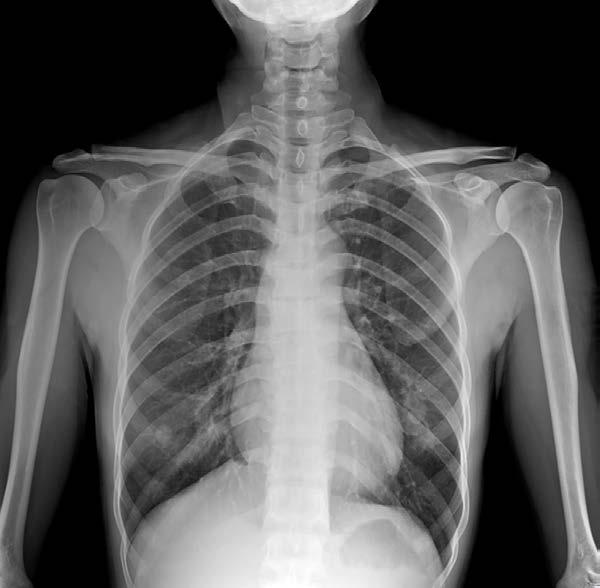
The PA occipital-frontal skull film is done to assess the patient for skull fractures. The patient lies prone with a vertical beam set at 20 degrees from upright. It cannot be used in patients with facial bone fractures or in the unconscious patient because of the prone position used. The patient’s nose and forehead rest against the table. Figure 1 depicts this type of x-ray:
Figure 1.
A PA axial Caldwell film is angled caudally so that the frontal sinuses and other paranasal sinuses are best evaluated. It can also detect certain skull fractures. The patient is placed erect with the tube set to angle at 15 degrees caudally. It is centered at the nose with the midsagittal plane perpendicular to the tube.
The PA axial Haas skull film is done in order to see the sella where the pituitary gland rests. It will show clearly the foramen magnum, occipital bone, and petrous pyramids. The patient lies prone with the x-rays directed at a 25-degree angle.
An AP fronto-occipital skull film is used to get an antero-posterior image of the skull. The patient lies supine. This view is particularly used for patients who are unable to lie prone for a PA view.
The AP axial Towne method is used specifically to look for fractures of the occipital bone. The patient lies supine and the beam is angled at 30 degrees from the vertical. The sella turcica will be projected through the patient’s foramen magnum. Figure 2 depicts this special view of the occipital bone:

Figure 2.
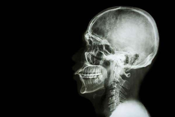
A lateral skull film can be done on a supine patient. It can be done from the left or right side and will reveal mainly skull fractures. Often, both the left and right side can be done, one after the other. The details closest to the x-ray beam are visualized the best. The sella turcica can also be seen. A lateral skull film can be done on the erect patient as well. Figure 3 shows a lateral skull film: