6 minute read
Preface

This course introduces the healthcare practitioner to the different areas involved in the study of radiology. Over the last several decades, radiology has come from the diagnostics involved in plain film x-rays and fluoroscopy to many more modern techniques, such as ultrasound, computed tomography scanning or CT scanning, and magnetic resonance imaging or MRI. Radiology has also advanced from diagnostic studies only to interventional radiology, in which radiology techniques are used to do therapeutic procedures, and therapeutic radiology, with the use of x-rays in order to treat disease, mainly cancer, specifically.
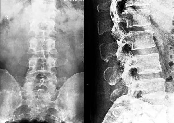
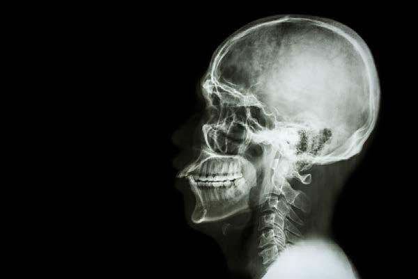
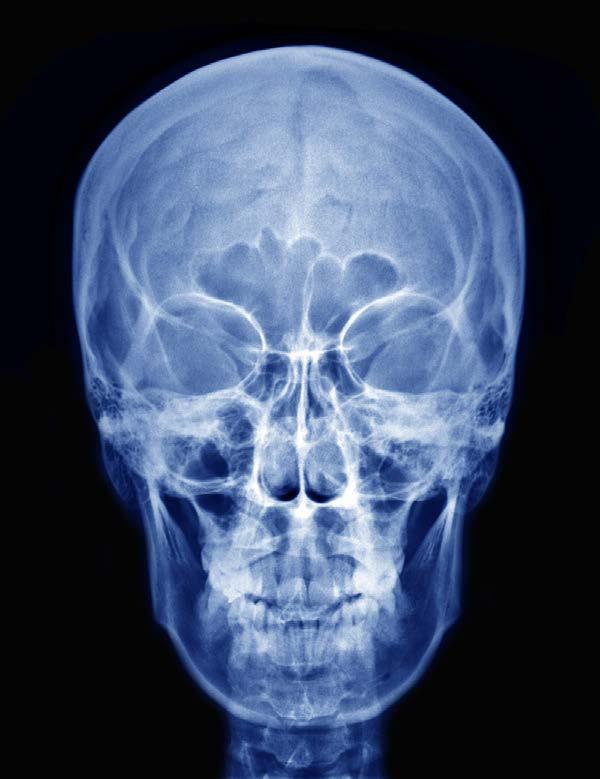
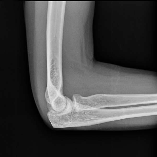
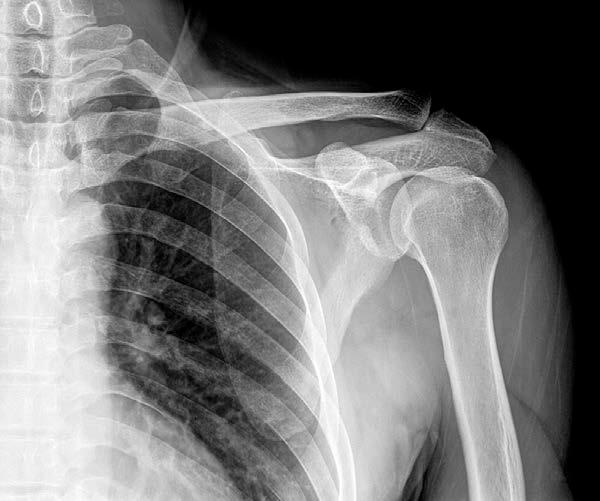
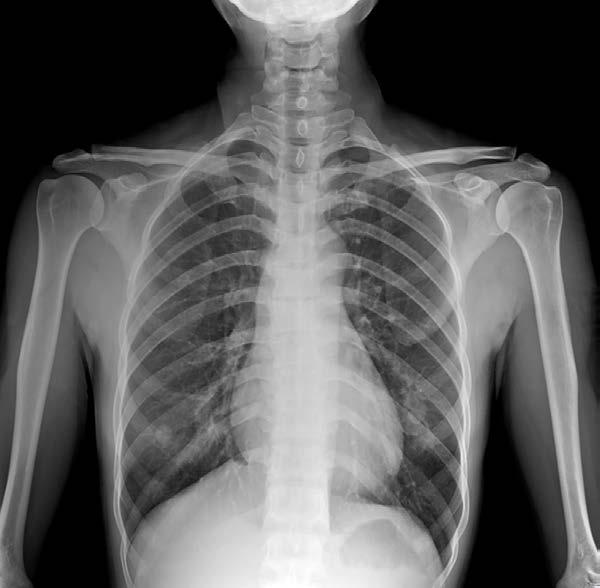
The focus of chapter one in the course is diagnostic plain film radiology. This is the original radiological technique used in medicine. The history and practice of plain film radiology is discussed as well as the techniques used when x-rays are obtained of the head and neck area, chest, abdomen, and extremities. In addition, the expected results of normal and abnormal plain film x-rays are covered in this chapter. You will come to see the limitations of these types of films in the field of radiology.
Advertisement
The major topics of chapter two are the principles and practice of fluoroscopy, which is real-time imaging using x-ray technology rather than still pictures, and radiocontrast materials. There are many procedures that involve fluoroscopic techniques. The chapter also covers the different substances used in radiology—including those for plain film xrays, fluoroscopy, CT scanning, and MRI scanning. The different risks of giving and receiving radiocontrast material is also discussed in this chapter.
Chapter three in the course talks about ultrasound technology and how it is used in a medical setting. Ultrasonography is one method in the field of radiology that does not make use of any sort of ionizing radiation but uses high-frequency sound waves to create images from within the human body. In the chapter, we talk about how ultrasound machines work and about the different kinds of ultrasounds used in medicine. In addition, we discuss the different medical specialties and how they make use of different
kinds of ultrasound techniques in order to do various diagnostic tests and procedures within the field of medicine.
The major discussion in chapter four is computed tomography or CT scanning, formally called a computerized axial tomography or CAT scan. The basics of how CT scanning is performed will be covered as well as the different ways CT scanning is used in various medical applications. There are related studies in radiology called the PET scan and the SPECT scan, which involve radionuclides introduced into the body that are picked up by alternate cameras and combined with CT scanning.
The topic of chapter five in the course is magnetic resonance imaging or MRI scanning. This is one of the newest methods of detection in radiology and can look mainly at the soft tissues of the body. It does not rely on the use of ionizing radiation as in many other radiological techniques but is more costly and sometimes more difficult to do than CT scanning or ultrasonography. The uses of MRI in medicine are covered in this chapter as well as the study of the brain called functional MRI, which is not yet in common usage.
There are several gastrointestinal x-ray studies looked at in chapter six. Evaluation of the upper GI tract can be done with a barium swallow, which may involve a small bowel follow-through to evaluate GI motility. There are also specialized motility tests that make use of markers that help determine gastrointestinal motility overall. The barium enema is a colonic study using contrast dye. There are specialized radiological studies that evaluate the hepatobiliary system, which are also covered in this chapter.
The focus of chapter seven in the course is breast imaging, which generally means imaging the breasts for breast cancer. The standard of breast imaging in radiology is the mammogram, which uses x-ray technology in order to visualize the breasts in a special mammography device. More recently, magnetic resonance imaging has been done in order to see breast tissue without using ionizing radiation. In lieu of an open breast biopsy, imaging techniques are being employed in order to obtain a minimally-invasive breast biopsy.
Chapter eight involves the techniques required to adequately image the heart. The standard test used for coronary arterial imaging is coronary angiography. This is usually
done using conventional x-ray imaging and contrast dye that outlines the various coronary arteries. In addition to conventional x-rays, CT scanning can be combined with contrast techniques in order to evaluate the coronary arteries. Ultrasound is the basic technology used for echocardiography of the heart, which evaluates the blood flow and internal structures of the heart. This can be done through transesophageal or transthoracic means.
The purpose of chapter nine in the course is to cover the basics of cardiovascular imaging or the tests involved in looking at the blood vessels of the body. There are many different modalities for evaluating peripheral vascular disease, which primarily involves the lower extremities. There are similar studies that can be used to look at the veins in the lower extremities, such as in cases of deep vein thromboses of the legs. The techniques involved in carotid artery evaluation are also discussed as well as the imaging necessary to look for abdominal aortic aneurysms.
Chapter ten covers imaging in the field of obstetrics. The standard test for the evaluation of the gravid uterus is the basic obstetric ultrasound, which can evaluate many aspects of fetal anatomy, structure of the placenta, and dating of the pregnancy. Ultrasound can also be used in certain situations in order to evaluate the blood flow through the umbilical vessels. A more targeted approach to obstetric imaging is the biophysical profile, which evaluates the viability and characteristics of the fetus near term. Ultrasound is also used to guide the needle in an amniocentesis and chorionic villus sampling.
The topic of chapter eleven in the course is gynecological imaging. The most common gynecological imaging test is the pelvic ultrasound, which is noninvasive and can evaluate all of the pelvic structures in the evaluation of pelvic pain, abnormal bleeding, and pelvic trauma. Another valuable imaging study is the hysterosalpingogram. This is a contrast dye study that is primarily used in the evaluation of infertility but has other valuable applications in the study of the uterus and fallopian tubes.
Chapter twelve looks into the various techniques used in central nervous system imaging. Plain film technology, CT scanning, MRI scanning, and ultrasound can be used to image the nervous system. Brain imaging usually involves magnetic resonance
imaging, with special techniques used to evaluate patients with multiple sclerosis and intracranial aneurysms. Imaging of the spinal cord is also covered, including the techniques used in myelography and in identifying spinal stenosis. Intracranial ultrasound has specific indications, which is discussed in this chapter.
The focus of chapter thirteen in the course is the imaging of the urinary tract. There are special techniques involved in imaging the kidneys, ureters, and bladder, with separate techniques employed to evaluate the patient with possible renal artery stenosis. The prostate gland in men is evaluated with a transrectal ultrasound. There are a variety of techniques used to look for adrenal adenomas and adenocarcinoma. Imaging techniques also described in this chapter include those used to identify and describe nephrocalcinosis and kidney stones.
Chapter fourteen covers the different types of radiological imaging studies used for looking at the musculoskeletal system. The study of the joints can be done with arthrography or magnetic resonance imaging of the joints. The MRI scan is especially good for identifying soft tissue anomalies so this type of scanning becomes particularly important to studying these areas. The DXA scan is used to look at bone density in the evaluation of osteoporosis. There are various techniques that can be used to look for bony metastases, which is discussed as part of this chapter.
Chapter fifteen in the course looks at the different techniques used in nuclear medicine studies. The thyroid scan involves radioactive iodine uptake that is specific to the thyroid gland. In gastrointestinal medicine, the gastric emptying study involves eating a radioactive meal with the amount of radioactivity measured in the stomach over time. The bone scan uses technetium-99 to look for bone metastases and other bony abnormalities. Finally, the ventilation-perfusion scan looks at the lungs and their ability to combine ventilation and perfusion in the act of breathing.