2 minute read
Chest X-ray
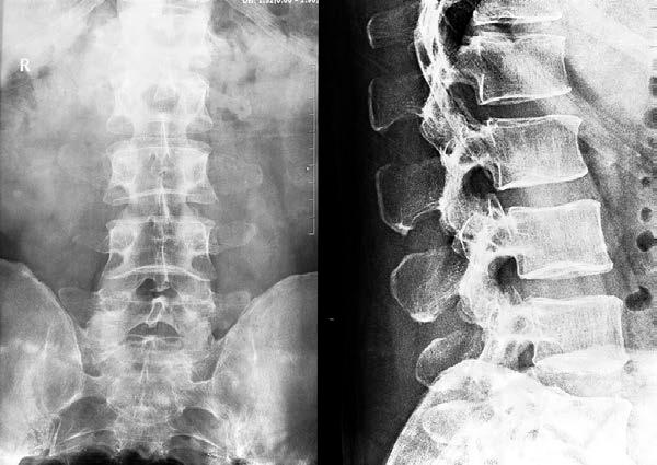
to decrease lumbar lordosis. An oblique view can be obtained in order to better see the articular processes or lumbosacral processes. The patient is turned to 45 degrees to get this oblique view.
The sacrum is best evaluated through an AP view. The x-ray evaluates the sacroiliac joints and the lumbosacral junction. The patient is supine with a vertical beam that is angled at 15 degrees. It cannot be used in children. The bladder needs to be emptied if possible. A lateral view can also be taken of the lumbosacral junction. The patient lies on their side in order to get this view.
Advertisement
CHEST X-RAY
The patient can have plain x-rays of the chest, which is particularly helpful in detecting heart, lung, chest wall, and diaphragmatic injuries. This is the initial film used to detect lung abnormalities. Unlike many films, this is a PA film and not an AP film, because the goal is to minimize x-ray scatter. Oblique or lordotic views can be taken of the chest in order to evaluate the patient for nodules or to see things that are overlapping. A lateral decubitus film can look at pleural effusions to see if there are loculations or free-flowing liquid in the pleural space. An end-expiratory view can detect small pneumothoraxes in the patient. Portable views are not very helpful unless the patient is too sick for a regular chest x-ray.
The plain film can detect problems in the chest in the patient who has persistent cough, shortness of breath, fever, chest injury or chest pain. It does not show as much as a CT scan of the chest but is rapid and easy to do for situations of pneumonia, COPD, and certain lung cancers. It is the most commonly done x-ray test in radiology. Usually, both the lateral and PA views are taken of the chest. The patient is usually standing upright for this film.
The radiologist looking at a chest x-ray should remember the mnemonic ABCDEF when looking at a standard chest film. This stands for airways, bones, cardiac outline, diaphragm, expansion of the lungs, and foreign bodies.
In taking this film, the patient should stand with the shoulders pressed forward to get the PA view. The top of the lungs and the diaphragm should be completely visible. The
patient should take a deep breath and hold it for the taking of the x-ray. Figure 6 shows a good chest x-ray film:
Figure 6.
On the lateral film, the patient is also erect and usually the left lateral is taken. The patient should lean slightly forward during the taking of the film. The arms are above the head or locked behind the back in order to rotate the shoulders in the posterior direction. Again, the patient should have expanded lungs and should hold their breath. The top and bottom of the lungs should be visualized.
A PA and lateral of the chest can be done on the patient while sitting erect if they are unable to stand. The tests are otherwise identical to the standing image. The entire lungs should be visualized, including the top of the lungs, the lower parts of the diaphragm, and the costophrenic angles on both sides.
The AP chest is only done on the patient who is unable to sit or stand. It is done on the supine patient. The apical or lordotic AP film is done to get an added view of the upper

