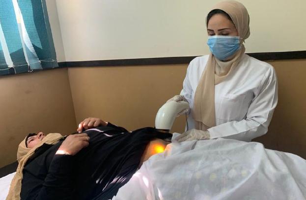
BIOPTRON HYPERLIGHT THERAPY IN WOUND HEALING

2022 Bioptron
Medical Team
Properties of Bioptron

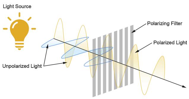
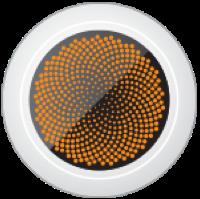
Polarized: 95% of polarization, ensures optimal penetration of tissues to stimulate.
Polychromatic: Contains the visible light and a part of the infrared spectrum (350 to 3400nm). Does not include UV.


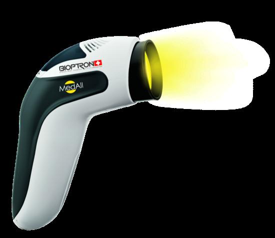
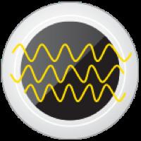


Incoherent: Dynamic penetration without the risk of damaging the tissue as coherent light.
Low energy: Consistent intensity of 2.4J /cm2 per minute safe and precise dose.
Hyperpolarized: Fullerene (C60) coated filter.
Therapeutic
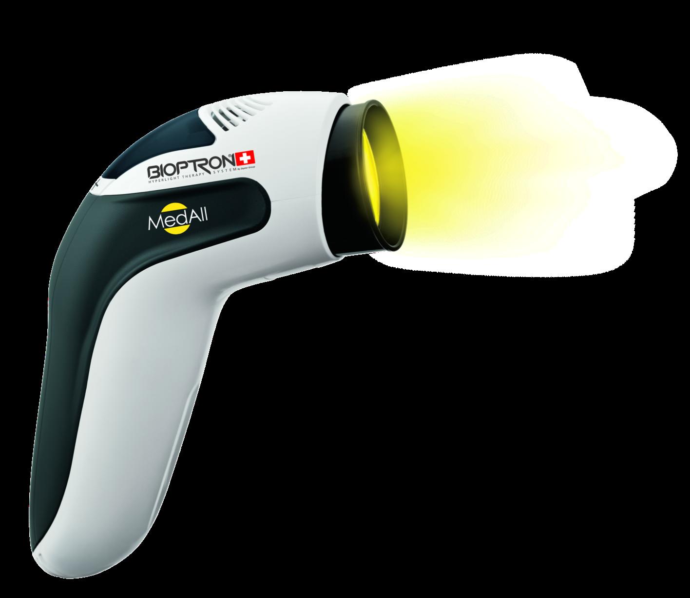
MedAll Diameter:
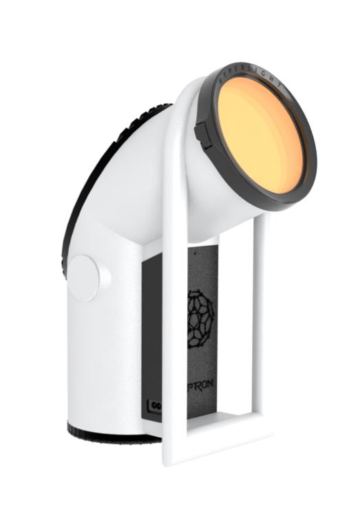
BPro1
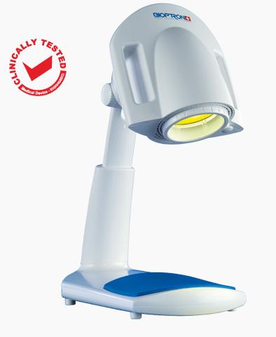
B2
Figure: Feehan J et. al.,
applications of polarized light: Tissue healing and immunomodulatory effects, Maturitas, Volume 116, 2018, Pages 11-17, ISSN 0378-5122
5cm
Diameter: 11cm
Diameter: 15cm
300 350 400 450 500 550 600 700 650 750 800 1’000 3’400
UV light visible light infrared light BIOPTRON light wavelength [λ] nm
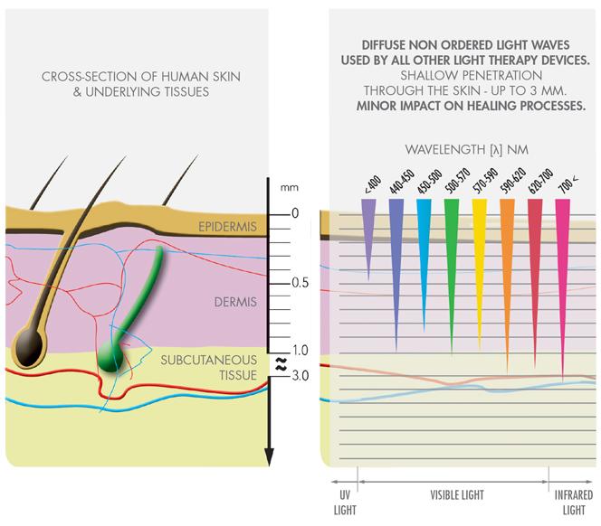
of action Cross section of skin Polarized light penetration Cellular metabolism↑ Changes in the lipid bilayer, membrane surface charges, lipid-protein interaction1 Mitochondria stimulation, augmented cell energy and nucleic acid production2 Microcirculation↑ VEGF production↑3, neoangiogenesis↑5 Production of specific cells and proteins↑ Fibroblast stimulation, expression of type1 procollagen mRNA↑4 Kerotinocyte proliferation↑6 Growth factors↑11, collagen synthesis↑6 Regulation of inflammation↑ Th lymphocyte proliferation↑7 IgM and IgA↑9 Anti-inflammatory cytokine↑ Pro-inflammatory cytokine↓8 Nociceptive signals↓ Pain receptor activation↓ NO synthesis↑10 1. Kertesz I, Fenyö M, Mester E, Bathory G. Hypothetical physical model for laser biostimulation Opt Laser Technol. 1982;14(1):31 32. 2. Tunèr J, Hode L. Laser therapy: clinical practice and scientific background. In Chapt 1 Some basic laser physics Prima Books AB, 2002; 1 44. 3 Akilbekova D, Boddupalli A, Bratlie KM. The effect of polarized light on the organization of collagen secreted by fibroblasts. Lasers Med Sci. 2018;33(3):539 547. 4. Tada K., Ikeda K., and Tomita, K. Effect of polarized light emitting diode irradiation on wound healing. J. Trauma. 2009;67(5):1073 1079. 5. Iordanou P. Effect of visible and infrared polarized light on the healing process of full-thickness skin wounds: an experimental study. Photomed Laser Surg. 2009
6. Samoilova KI. Enhancement of growth promoting activity of human blood on keratinocytes after its irradiation in vivo (transcutaneously) and in vitro with visible and infrared polarized light. Tsitologiia. 2003;45(6):596 605.
Mechanism
Apr;27(2):261-7.
Bioptron

Hyperlight Therapy is clinically tested and medically certified in the treatment of wounds. Injuries and traumas Post operational wounds Burn wounds Venous leg ulcers (stasis ulcer) Pressure ulcers (decubitus) Diabetic foot ulcers*
Therapy is not medically certified in diabetic foot ulcers. Clinical evidence in this area will be presented.
the
each
to navigate
the document. EN ISO 13485:2016 by TÜV 93/42/EEC-Annex II by TÜV Certificate assuring the quality management Certificate assuring the compliance with EU directive for manufacturers of medical products
Hyperlight Therapy in Wound Healing Bioptron
*Bioptron Hyperlight
Click on
link on
section
in
Venous Leg Ulcer
of Venous Leg Ulcers Medenica L, Lens M. The use of polarised polychromatic non coherent light alone as a therapy for venous leg ulceration. J Wound Care. 2003 Jan;12(1):37 40. Inclusion Criteria • Age: >40 • Ulcer: >1, >1cm2 • No arterial disease Outcomes • Wound surface area • Photographs and qualitative assessment • Histological evaluation (n=11) Epithelization, cellular content, granulation tissue, collagen deposition, neovascularisation Definition of healing: Intact epidermis and no exudate Aim • To assess the effectiveness of polarized, polychromatic, noncoherent light therapy in the treatment of venous leg ulcers Treatment • Bioptron: Once a day for 8 minutes • No additional treatment is permitted Exclusion Criteria • Other ulcers: infectious, pressure, post-operative • Cancer • Concomitant diseases and drugs (n=25) Week 0 Week 4 Bioptron (Once a day) Wound assessment +Biopsy Week 3 Week 1 Week 2 Wound assessment +Biopsy Wound assessment Wound assessment Wound assessment
Treatment
of Venous Leg Ulcers Medenica L, Lens M. The use of polarised polychromatic non coherent light alone as a therapy for venous leg ulceration. J Wound Care. 2003 Jan;12(1):37 40. * p<0.001 Clinical results Out of 25 patients and 73 total ulcers: • 72 ulcers (99%) had a positive change • 22 ulcers (30%) healed completely 26.45 12.79 0 5 10 15 20 25 30 Ulcer Surface Area (cm 2 ) Before Treatment After treatment * Histological Findings Before Treatment: • Complete epidermal & dermal necrosis • Inflammatory infiltration↑ • Poor granulation ↓ After Treatment: • Re-epithelialisation • Proliferation of the granulation tissue • New collagen depositions • Neo-angiogenesis
Treatment
Treatment of
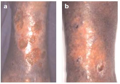
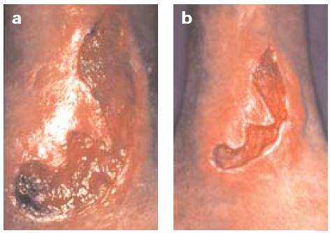
L, Lens M. The use of polarised polychromatic non coherent light alone as a therapy for venous leg ulceration. J Wound Care. 2003 Jan;12(1):37 40. Qualitative Assessment Figure 1: Change in ulcer surface area 20.9 cm2 →14.5 cm2 (a→b) Figure 2: Change in ulcer surface area 35.6 cm2 →5.1 cm2 (a→b) Bioptron Hyperlight Therapy applied as a monotherapy was associated with positive healing rates in patients with venous leg ulcers.
Venous Leg Ulcers Medenica
of Venous Leg Ulcers
Aim • To establish the effects of electroionizing radiation and polarized light on venous ulcer healing, as well as the degree of correlation with controls. Treatment • EL: Once a day for 10 minutes after cleaning • PL: Once a day for 10 minutes after cleaning • C: Standard Standard treatment: Appropriate care, wash and bandage, topical ab or specific bandages when necessary (n=15) Week 0 Week 7 Standard treatment + Electroionizing radiation (EL) Wound assessment Week 5 Week 3 Wound assessment Wound assessment Wound assessment (n=15) Standard treatment + Bioptron (PL) (n=15) Standard treatment (C) Week 1 Wound assessment
Treatment
Aleksandar, Jankovic & Milenko, Stanojevic & Binic, Ivana. (2005). PHYSICAL THERAPY OF VENOUS ULCERS: EFFECTS OF ELECTROIONOTHERAPY AND POLARIZED LIGHT. Acta Facultatis Medicae Naissensis.
Treatment of Venous Leg Ulcers Aleksandar, Jankovic & Milenko, Stanojevic & Binic, Ivana. (2005). PHYSICAL THERAPY OF VENOUS ULCERS: EFFECTS OF ELECTROIONOTHERAPY AND POLARIZED LIGHT. Acta Facultatis Medicae Naissensis. (n=15) Week 0 Week 7 Standard treatment + Electroionizing radiation (EL) Wound assessment Week 5 Week 3 Wound assessment Wound assessment Wound assessment (n=15) Standard treatment + Bioptron (PL) (n=15) Standard treatment (C) Week 1 Wound assessment Outcomes • Ulcer (0-12) • Fibrin deposits (0-3) • Exudation (0-3) • Granulation (0-3) • Epithelization (0-3) Number, surface, depth, volume, borders • Ulcer vicinity (0-15) • Erythema (0-3) • Edema (0-3) • Maceration (0-3) • Desquamation (0-3) • Scleroatrophy (0-3) • Associated symptoms (0-6) • Pain (0-3) • Pruritus (0-3) 0: prominent 1: moderate 2: slight 3: absent
Treatment of
Venous
Leg Ulcers
EFFECTS
ELECTROIONOTHERAPY
POLARIZED
80.03 76.31 60.18 0 10 20 30 40 50 60 70 80 90 Change in total score (%) Reduction of the ulcer vicinity parameters Bioptron Electroionizing radiation Control 60.87 85.72 92.42 68.48 93.17 96.89 46.85 70.66 91.6 0 10 20 30 40 50 60 70 80 90 100 Surface Depth Volume Change in total score (%) Reduction of the ulcer parameters Bioptron Electroionizing radiation Control
Aleksandar, Jankovic & Milenko, Stanojevic & Binic, Ivana. (2005).
PHYSICAL THERAPY OF VENOUS ULCERS:
OF
AND
LIGHT. Acta Facultatis Medicae Naissensis.
Treatment
90.39 87.62 67.17 0 10 20 30 40 50 60 70 80 90 100 Change in total score (%) Total reduction of the ulcer parameters Bioptron Electroionizing radiation Control
Jankovic & Milenko, Stanojevic & Binic, Ivana. (2005). PHYSICAL THERAPY OF VENOUS ULCERS: EFFECTS OF ELECTROIONOTHERAPY AND POLARIZED LIGHT. Acta Facultatis Medicae Naissensis. 96.13 92.1 62.5 0 20 40 60 80 100 120 Change in total score (%) Reduction of the symptoms: Pain & Pruritus Bioptron Electroionizing radiation Control Bioptron Hyperlight Therapy accelerates the healing of venous leg ulcers.
of Venous Leg Ulcers
Aleksandar,
Diabetic Foot Ulcer
*Bioptron Hyperlight Therapy is not medically certified in diabetic foot ulcers. Clinical evidence in this area will be presented.
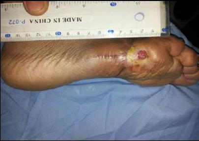
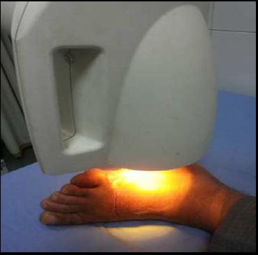
of Diabetic Foot Ulcer El Deen, H. B., Fahmy, S. E. H. A. M., Ali, S. A., & El Sayed, W. M. (2014). Polarized light versus light emitting diode on healing of chronic diabetic foot ulcer. Romanian Journal of Biophysics, 24(2), 1 15. Inclusion Criteria • Age: 48-72 • Grade-2 Ulcer Outcomes • Ulcer Surface area • Ulcer Depth Aim • To compare the influence of polarized light therapy versus LED therapy on enhancement of healing of chronic • diabetic foot ulcer and to determine which one was better and faster. Exclusion Criteria • Other diseases that might cause ulcers Study • Double-Blind RCT Follow-up Final assessment Intermediary assessment Day 0 Month 3 Initial assessment Month 2 Month 1 (n=15) Group PL: Bioptron Lamp+standard (n=15) Group LED: LED Device+standard 8 minutes, 3 times per week, 2 months 8 minutes, 3 times per week, 2 months Bioptron Hyperlight Therapy is not medically certified in diabetic foot ulcers. Clinical evidence in this area will be presented.
Treatment
Treatment of Diabetic Foot Ulcer

Results
8 patients from the Bioptron group and 5 patients from the LED group achieved a complete healing of their ulcers in 2 months.
Bioptron Hyperlight Therapy is not medically certified in diabetic foot ulcers. Clinical evidence in this area will be presented.
Both Bioptron and LED therapies can be considered as valuable for the treatment of various wounds and wound healing disorders.
El Deen, H. B., Fahmy, S. E. H. A. M., Ali, S. A., & El Sayed, W. M. (2014). Polarized light versus light emitting diode on healing of chronic diabetic foot ulcer. Romanian Journal of Biophysics,
24(2), 1 15.
Application Before treatment with Bioptron 2 months after treatment with Bioptron
of Diabetic Foot Ulcer 59.3 77.3 0 10 20 30 40 50 60 70 80 90 Change in ulcer Surface Area (%) LED Group Bipoptron Group * Parameters Bioptron LED Significance First month PCUSA (%) 55.05±23.45 34.04±0.98 p<0.05 PCUD (%) 56.7±21.46 42.96±12.8 p<0.05 Second month PCUSA (%) 77.3±21.46 59.28±12.8 p<0.05 PCUD (%) 82.08±18.31 76.35±11.58 p>0.05 Bioptron light therapy seems to be more effective in accelerating the healing rate and shortening hospitalization time than LED therapy. PCUSA: percentage of change (%) of ulcer surface area PCUD: percentage change (%) of ulcer depth Percentage of changes (%) of ulcer surface area after two months Percentage of change (%) of ulcer surface area and ulcer depth in first and second months El Deen, H. B., Fahmy, S. E. H. A. M., Ali, S. A., & El Sayed, W. M. (2014). Polarized light versus light emitting diode on healing of chronic diabetic foot ulcer. Romanian Journal of Biophysics, 24(2), 1 15. *= p<0.05 Bioptron Hyperlight Therapy is not medically certified in diabetic foot ulcers. Clinical evidence in this area will be presented.
Treatment
Treatment of Diabetic Foot Ulcer Mohamed, M. H., Selem, M. N., Mohamed, M. S., & Abd EL Ghaffaar, H. A. (2019). Interleukin 6 response to shock wave therapy versus polarized light therapy in the treatment of chronic diabetic foot ulcers. Drug Invention Today, 11(11). Inclusion Criteria • Age: 55-65 • Grade-2, Ulcer surface>1cm2 • >2 months Outcomes • Ulcer Surface area • IL-6 levels Aim • To investigate the response of interleukin 6 (IL-6) and ulcer surface area to shock wave therapy versus polarized light therapy in chronic diabetic foot ulcer. Exclusion Criteria • Other comorbidities • Grade-4, Ulcer surface>10cm2 Initial assessment Study • Single-blind, parallel-group, activecontrol, RCT 1 session every week, with 500 pulses per 1 cm2 with 0.1 mJ/mm2 density, 3 minutes, 2 months 8 minutes, 3 times per week, 2 months (n=15) Week 0 Week 8 Standard treatment + Shockwave therapy (n=15) Standard treatment + Bioptron (n=15) Standard treatment Final assessment Bioptron Hyperlight Therapy is not medically certified in diabetic foot ulcers. Clinical evidence in this area will be presented.
Treatment of
outcome
Ulcer
Standard treatment + Shock wave Standard treatment + Bioptron Standard treatment Before treatment After treatment P-value Before treatment After treatment P-value Before treatment After treatment P-value
level 184.46±35.11 62.26±15.08 0.001 180.26±34.04 33.06±17.36 0.001 177.86±35.34 177.73±35.53 0.98 USA 7.17±1.5 2.67±0.56 0.001 7.3±1.9 1.54±0.98 0.001 7.04±1.83 7.02±1.76 0.97
Diabetic Foot Ulcer Both Bioptron and shock wave therapies showed a significant change in
measures in 2 months. IL-6 levels and
Surface Area measurements before and after the treatments of each group Variables
IL-6
Mohamed, M. H., Selem, M. N., Mohamed, M. S., & Abd EL Ghaffaar, H. A. (2019). Interleukin 6 response to shock wave therapy versus polarized light therapy in the treatment of chronic diabetic foot ulcers. Drug Invention Today, 11(11). USA: Ulcer surface area
Treatment of Diabetic Foot Ulcer
33.06 62.2 177.73 0 20 40 60 80 100 120 140 160 180 200 IL6 levels (concentration) IL-6 levels after 2 months of treatments Bioptron therapy Shock wave therapy
1.54 2.67 7.02 0 1 2 3 4 5 6 7 8 Ulcer Surface area (cm 2 ) Ulcer Surface Area after treatments
*
Control
Bioptron therapy Shock wave therapy Control
Bioptron is progressively more successful in accelerating the healing of diabetic foot ulcer and reduction of IL-6 level than shock wave therapy.
* § α
Mohamed, M. H., Selem, M. N., Mohamed, M. S., & Abd EL Ghaffaar, H. A. (2019). Interleukin 6 response to shock wave therapy versus polarized light therapy in the treatment of chronic diabetic foot ulcers. Drug Invention Today, 11(11). *: p= 0.001 §: p= 0.044 α: p=0.006
Bioptron Hyperlight Therapy is not medically certified in diabetic foot ulcers. Clinical evidence in this area will be presented.
Pressure Ulcer
Treatment of Pressure Ulcer Đurović, A., Marić, D., Brdareski, Z., Jevtić, M., & Đurđević, S. (2008). The effects of polarized light therapy in pressure ulcer healing. Vojnosanitetski pregled, 65(12), 906 912.. Inclusion Criteria • Stage I–III ulcer • Absence of relative contraindications for using of polarized light Outcomes • The Pressure Ulcer Scale for Healing (PUSH) • Ulcer Surface area • Exudate amount • Tissue type (appearance) • Rank of ulcers Aim • To establish the effects of polarized light therapy in the healing process of pressure ulcer. Exclusion Criteria • Previous study participation • Poor nutrition • Infections • Other treatments that might effect Study • Single-Blind RCT Final assessment Week 0 Week 4 Initial assessment (n=20) Experimental group: Bioptron + Standard (n=20) Control group: Standard wound care From 10 cm, 6 minutes, 5 times per week
Pressure Ulcer Đurović, A., Marić, D., Brdareski, Z., Jevtić, M., & Đurđević, S. (2008). The effects of polarized light therapy in pressure ulcer healing. Vojnosanitetski pregled, 65(12), 906 912.. Characteristics of the pressure ulcers at the start and the end of treatment Variables Standard treatment + Bioptron Standard treatment Before treatment After treatment P-value Before treatment After treatment P-value Surface of the ulcer 15.10 ± 17.61 10.80 ± 19.18 0.01 19.15 ± 22.73 22.97 ± 15.69 0.001 Rank of the ulcer 7.40 ± 1.96 5.95 ± 2.48 0.0004 8.2 ± 1.51 8.6 ± 1.05 0.01 Total PUSH score 10.65 ± 2.25 7.35 ± 3.17 0.0001 10.45 ± 2.74 11.85 ± 2.35 0.003 PUSH: The Pressure Ulcer Scale for Healing Healing of a sacral ulcer in 4 weeks Before treatment After treatment
Treatment of
Treatment of Pressure Ulcer
15.1 7.4 10.6 10.8 5.95 7.35 0 5 10 15 20 25 Surface of ulcer (cm2) Rank of ulcer Total PUSH score Changes in the ulcer parameters Before treatment After Treatment
Đurović, A., Marić, D., Brdareski, Z., Jevtić, M., & Đurđević, S. (2008). The effects of polarized light therapy in pressure ulcer healing. Vojnosanitetski pregled, 65(12), 906 912.. PUSH: The Pressure Ulcer Scale for Healing 19.1 8.2 10.4 22.9 8.6 11.8 0 5 10 15 20 25 Surface of ulcer
Rank of ulcer Total PUSH score Changes in the ulcer parameters
treatment After Treatment Standard treatment + Bioptron Standard treatment
(cm2)
Before
15.1 7.4 10.6 10.8 5.9 7.3 0 5 10 15 20 25 Surface of ulcer (cm2) Rank of ulcer Total PUSH score Changes in the ulcer parameters Before treatment After Treatment Treatment
Pressure Ulcer Đurović, A., Marić, D., Brdareski, Z., Jevtić, M., & Đurđević, S. (2008). The effects of polarized light therapy in pressure ulcer healing. Vojnosanitetski pregled, 65(12), 906 912.. PUSH: The Pressure Ulcer Scale for Healing *: p=0.0005, **: p=0.00003 19.1 8.2 10.4 22.9 8.6 11.8 0 5 10 15 20 25 Surface of ulcer (cm2) Rank of ulcer Total PUSH score Changes in the ulcer parameters Before treatment After Treatment * * ** The effects of Bioptron polarized light as an adjuvant therapy for pressure ulcers were satisfactory.
of
Treatment of Pressure Ulcer
Al
Hassan,
G.
Efficacy of polarized light in treatment of pressure ulcers. JMSCR, 3, 5800 5809. Inclusion Criteria • Grade II–III sacral ulcer Outcomes • Ulcer surface area • Ulcer volume Aim • To evaluate the efficacy of the polarized light therapy in accelerating pressure ulcers healing Study • RCT Final assessment Week 0 Week 4 Initial assessment (n=15) Experimental group: Bioptron + Standard (n=15) Control group: Standard wound care From 10 cm, 10 minutes, twice a day
Abd
kader, A. M.,
M. A., & Elsayed, H.
(2015).
Treatment of Pressure Ulcer Abd Al kader, A. M., Hassan, M. A., & Elsayed, H. G. (2015). Efficacy of polarized light in treatment of pressure ulcers. JMSCR, 3, 5800 5809. Variables Standard treatment + Bioptron Standard treatment Before treatment After treatment P-value Before treatment After treatment P-value Surface of the ulcer 15.77 ± 5.85 8.45 ± 3.58 0.0001 15.76 ± 5.87 15.75 ± 5.81 0.996 Volume of the ulcer 19.12 ± 8.15 8.66 ± 4.93 0.0001 19.21 ± 8.23 19.20 ± 8.17 0.997 15.7 19.1 8.4 8.6 15.7 19.2 15.7 19.2 0 5 10 15 20 25 Surface of ulcer (cm2) Volume of ulcer (ml) Changes in the ulcer parameters Test group, before Test group, after Control group, before Control group, after * * Bioptron was significantly effective in enhancing healing of the pressure ulcers.
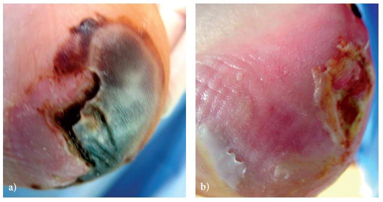
Treatment of Pressure Ulcer Iordanou, P., Baltopoulos, G., Giannakopoulou, M., Bellou, P., & Ktenas, E. (2002). Effect of polarized light in the healing process of pressure ulcers. International journal of nursing practice, 8(1), 49 55. Inclusion Criteria • Stage I–III ulcer • 2 ulcer (bigger one picked as experimental) Outcomes • Ulcer Surface Area • Epithelial tissue • Color • The amount of exudate Aim • To examine the effect of polarized light on pressure ulcers Exclusion Criteria • Necrosis • Poor nutrition • Surgery of the ulcer Study • Self control case series Final assessment Week 0 Week 2 Initial assessment (n=20) Experimental ulcer: Bioptron + Standard (n=20) Same patients Distance?, 5 minutes, 5 times per week Control ulcer: Standard
Treatment of Pressure Ulcer
Of the 55 subjects, 23 of the subjects’ ulcers responded satisfactorily to Bioptron and healed at the end of the first week, and excluded from the study.
Mean values of ‘none and minimal exudate’ of the experimental ulcers were significantly increased in the first week and in the second week compared to the control ulcers.
1st
7th
15th
1st
7th
15th
Iordanou, P., Baltopoulos, G., Giannakopoulou, M., Bellou, P., & Ktenas, E. (2002). Effect of polarized light in the healing process of pressure ulcers. International journal of nursing practice, 8(1), 49 55. Quantity of exudate Experimental ulcers Control ulcers
day (n=55)
day (n=55)
day (n=32)
day (n=55)
day (n=55)
day (n=32) None (%) 1 (1.8) 16 (29.1) 12 (37.5) 1 (6.8) 4 (7.3) 3 (9.3) Minimal (%) 11 (20) 19 (34.5) 9 (28.1) 11 (20) 9 (16.3) 8 (25) Moderate (%) 22 (40) 10 (18.2) 9 (28.1) 22 (40) 18 (32.7) 5 (15.7) Severe (%) 21 (38.1) 10 (18.2) 2 (6.3) 21 (38.1) ) 24 (43.7) 16 (50)
Treatment of Pressure Ulcer
the healing
pressure ulcers.
journal
nursing
8(1), 49 55. 2.84 2.54 2.26 0 0.5 1 1.5 2 2.5 3 3.5 4 4.5 5 Ulcer Surface Area (cm 2 ) 1st day 7th day 15th day 2.1 2.08 2.04 0 0.5 1 1.5 2 2.5 3 3.5 4 4.5 5 Ulcer Surface Area (cm 2 ) 1st day 7th day 15th day
Iordanou,
P., Baltopoulos, G., Giannakopoulou, M., Bellou, P., & Ktenas, E. (2002). Effect of polarized light in
process of
International
of
practice,
Experimental ulcers Control ulcers With Bioptron rapid changes in appearance and size with complete healing in half of the cases and accelerated partial healing in the remaining cases were observed.
Treatment of Pressure Ulcer
(114)
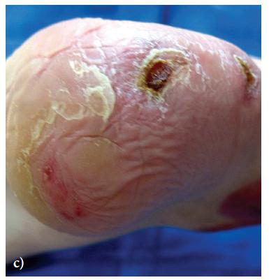
Białożyt J., Materniak K., Kawecki M. 2018 Use of polarise lighting in support of treatment of pressure ulcers among patients after burns. Preliminary report. Dermatologia Estetyczna. vol.20, 1
Inclusion Criteria • Grade II–IV ulcer Outcomes
• Ulcer surface area
days
• Ulcer healing Aim • To present and initially assess the results of the application of the therapy which supports the healing of decubitus ulcers. Study • Case series Final assessment Day 0 Day 15 Initial assessment (n=10) Bioptron + Standard treatment Twice a day, 8 minutes, for at least 15
Treatment of Pressure Ulcer
Bedsores healed completely in four cases, while significant progress in healing and a marked decrease in the size of pressure ulcers were observed in the remaining six cases.
Before,
Before,
(114)
Białożyt J., Materniak K., Kawecki M. 2018 Use of polarise lighting in support of treatment of pressure ulcers among patients after burns. Preliminary report. Dermatologia Estetyczna. vol.20, 1
Bioptron is a good method supporting the treatment of pressure ulcers, especially of the second and third degree.
grade IV 15 sessions 30 sessions
Before, grade III 15 sessions
grade III 10 sessions 22 sessions
Chronic Ulcers
Treatment of Chronic Ulcerative Lesions
Outcomes • Exudation • Signs of infection • Pain (VAS)
• First
the evaluation of skin lesion healing treated
polarized light
months, with changes in pain symptoms, repair and regeneration process.
• Case series (n=30) Week 0 Week 12 Bioptron (20 minutes, Twice a week) Wound assessment Wound assessment Week 4 Wound assessment Combined with wound care Ulcer type Number of patients Venous/vascular origin 24 Autoimmune/connective tissue diseases 4 Dehiscence of abdominal wound 2
Aragona, S. E., Grassi, F. R., Nardi, G., Lotti, J., Mereghetti, G., Canavesi, E., ... & Lotti, T. (2017). Photobiomodulation with polarized light in the treatment of cutaneous and mucosal ulcerative lesions. Journal of Biological Regulators and Homeostatic Agents, 31(2 Suppl. 2), 213 218.
Aim
aim was
with
at 6
Study
Treatment of Chronic Ulcerative Lesions
Outcome 1 month 3 months Reduction of exudation 16 patients (53%) 25 patients (83%) Regressed infection 30 patients (100%) Decreased pain 21 patients (70%) 30 patients (100%) Results
use
Aragona, S. E., Grassi, F. R., Nardi, G., Lotti, J., Mereghetti, G., Canavesi, E., ... & Lotti, T. (2017). Photobiomodulation with polarized light in the treatment of
cutaneous
and mucosal ulcerative lesions. Journal of Biological Regulators and Homeostatic Agents, 31(2 Suppl. 2), 213 218.
The
of Bioptron with a wound care protocol for the chronic and mucosal skin lesions, improved considerably the repair and the regeneration processes.
Treatment of Chronic Wounds INTSAR S. WAKED, Ph.D., R.M., & ASHRAF E.M. ELSEBAIE, M.D., M.B. (2021). Effect of Negative Pressure Therapy versus Polarized Light Therapy on Chronic Wound Healing. Inclusion Criteria • Grade II–III chronic wounds Outcomes • Ulcer surface area • Ulcer volume Aim • To investigate the difference in the effect between negative pressure therapy and polarized light therapy on chronic wound healing. Study • RCT Final assessment Week 0 Week 6 Initial assessment (n=15) Experimental group: Bioptron + Standard (n=15) Control group: Negative Pressure + Standard From 10 cm, 10 minutes, 3 times a week Exclusion Criteria • Acute wounds • Burns • Malignancy Ulcer type Experimental Control Diabetic 7 (47%) 6 (40%) Pressure 3 (20%) 4 (27%) Venous 5 (33%) 5 (33%) Mid assessment Week 3 Bioptron Hyperlight Therapy is not medically certified in diabetic foot ulcers. Clinical evidence in this area is presented.
Treatment of Chronic Wounds INTSAR S. WAKED, Ph.D., R.M., & ASHRAF E.M. ELSEBAIE, M.D., M.B. (2021). Effect of Negative Pressure Therapy versus Polarized Light Therapy on Chronic Wound Healing. Before treatment (Mean± SD) 3 weeks after treatment (Mean± SD) 6 weeks after treatment (Mean± SD) P-value Before vs 3 weeks Before vs 6 weeks 3 weeks vs 6 weeks Wound Surface Area (cm2) Negative Pressure 17.11±6.35 11.18±3.46 5±1.76 0.001 0.001 0.001 Bioptron 16.46±6.52 12.25±5.6 7.53±3.75 0.001 0.001 0.001 Significance p=0.78 p=0.53 p=0.02 Wound Volume Negative Pressure 8.26±3.07 5.1±1.91 2.34±0.89 0.001 0.001 0.001 Bioptron 7.83±2.37 5.46±2 3.26±1.03 0.001 0.001 0.001 Significance p=0.66 p=0.61 p=0.01 Bioptron Hyperlight Therapy is not medically certified in diabetic foot ulcers. Clinical evidence in this area is presented.
of Chronic Wounds INTSAR S. WAKED, Ph.D., R.M., & ASHRAF E.M. ELSEBAIE, M.D., M.B. (2021). Effect of Negative Pressure Therapy versus Polarized Light Therapy on Chronic Wound Healing. *: p=0.02, **: p=0.01 7.8 8.2 5.4 5.1 3.2 2.3 0 1 2 3 4 5 6 7 8 9 10 Ulcer Volume (cm 3 ) Before 3weeks6weeks Before 3weeks6weeks Bioptron group Negative pressure group Ulcer Volume 16.5 17.1 12.2 11.1 7.5 5 0 2 4 6 8 10 12 14 16 18 20 Ulcer Surface Area (cm 2 ) Before 3weeks6weeks Before 3weeks6weeks Bioptron group Negative pressure group Ulcer Surface Area Wound surface area and volume are significantly decreased after using both Bioptron or negative pressure. * ** Bioptron Hyperlight Therapy is not medically certified in diabetic foot ulcers. Clinical evidence in this area is presented.
Treatment
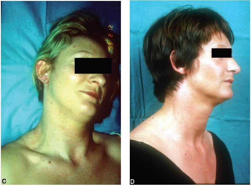
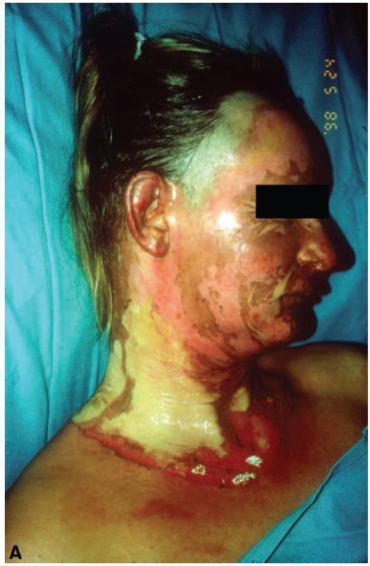
Post Operational Wounds
Treatment
Simic, A., Stojakov, D., Sabljak, P., Jekic, I., Bjelovic, M., & Pesko, P. (1999). Piler Light Therapy Effect on Wound Healing in Esophagogastric Surgery. EUROPEAN SURGICAL RESEARCH, 31(1), 225 225. Final assessment Day 0 Initial assessment (n=72) Experimental group: Bioptron + Standard (n=36) Control group: Standard Bioptron significantly depresses the possibility of seroma and infection and significantly shortens the postoperative hospitalization 80.56 12.5 5.56 1.38 Healing of the wound Excellent Satisfactory Unsatisfactory Bad 28.89 36.11 19.44 5.56 Healing of the wound Excellent Satisfactory Unsatisfactory Bad Experimental Group Control Group Wound healing: Excellent: Healed without seroma or infection, Sutures removed at 8th day Satisfactory: Small amount of seroma, no infection, Sutures removed at 8th day Unsatisfactory: Seroma and infection, Suture removal is delayed Bad: Suture removal is highly delayed, Secondary sutures due to infection Bioptron should be applied to the standard treatment in almost all surgical wounds. Day 8
of Post Operational Wounds: Esophagogastric Surgery
Treatment of Post Operational Wounds: Esophagogastric Surgery
Simic, A. (2001, May). Importance of Bioptron light therapy in the treatment of surgical incisions. In Second Balkan Congress for PRAS and Bioptron Satellite Symposium, Belgrade, May (pp. 24 26). Final assessment Day 0 Initial assessment (n=168) Experimental group: Bioptron + Standard (n=120) Control group: Standard Bioptron significantly decreases the incidence of seroma and infection and significantly shortens the postoperative hospitalization 79.8 14.65 4.54 1.01 Healing of the wound Excellent Satisfactory Unsatisfactory Bad 54 26.66 14.66 4.66 Healing of the wound Excellent Satisfactory Unsatisfactory Bad Experimental Group Control Group Wound healing: Excellent: Healed without seroma or infection, Sutures removed in time Satisfactory: Small amount of seroma, no infection, Sutures removed in time Unsatisfactory: Large amount of seroma and infection, Sutures removed in time Bad: Large amount of seroma and infection, Suture removal is highly delayed Bioptron should be applied to the standard treatment in almost all surgical wounds in patients operated due to stomach/esophageal diseases. Day 7
Post Operational
Simic, A., Pesco, P., Bjelovic, M., STOJAKOV, D., TODOROVIC, M., TODOROVIC, V., ... & KOTARAK, M. (2001). Bioptron light therapy and thoracophrenolaparotomy wound healing in patients operated due to cardiac carcinoma, paper presented at the 4th International Gastric Cancer Congress Final assessment Day 0 Initial assessment (n=26) Experimental group: Bioptron + Standard (n=26) Control group: Standard Bioptron significantly decreases the incidence of seroma and infection and significantly shortens the postoperative hospitalization 80.77 19.23 0 0 Healing of the wound Excellent Satisfactory Unsatisfactory Bad 53.85 26.92 15.38 3.85 Healing of the wound Excellent Satisfactory Unsatisfactory Bad Experimental Group Control Group Wound healing: Excellent: Healed without seroma or infection, Sutures removed in time Satisfactory: Small amount of seroma, no infection, Sutures removed in time Unsatisfactory: Large amount of seroma and infection, Suture removal is delayed Bad: Large infection, fascia dehiscence Bioptron should be applied to the standard treatment in almost all surgical wounds. Day 10 Patient group is defined as elderly/compromised patients with chronic diseases.
Treatment of
Wounds: Cardiac Carcinoma
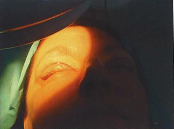
Colić, M. M., Vidojković, N., Jovanović, M., & Lazović, G. (2004). The use of polarized light in aesthetic surgery. Aesthetic plastic surgery, 28(5), 324 327. Patients • 45 Face-lifts • 67 Blepharoplasties • 350 Facial ancillary procedures (local lifts, rejuvenation, fat transfers) Outcomes • Descriptive and photographic documentation (day 0, 1, 3, 7) Study • Self control case series Final assessment Week 0 Week 1 Initial assessment (n=462) Experimental Side: Bioptron Same patients 15 cm, 10 minutes, 3 times a day in the first day, once a day for the next 6 days Control side: None Treatment of Post Operational Wounds: Aesthetic operations Treatment side Control side
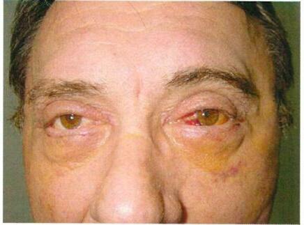
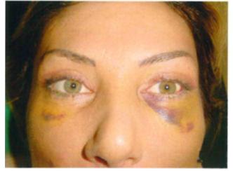
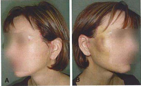
Treatment of Post Operational Wounds: Aesthetic operations
M. M., Vidojković, N., Jovanović, M., & Lazović, G. (2004). The use of polarized light in aesthetic surgery. Aesthetic plastic surgery, 28(5), 324 327. Patient groups Difference between treated and control sides Significant Moderate None Face-lift group 58% 18% 24% Blepharoplasty group 72% 19% 9% Facial ancillary group 47% 17% 36% Bioptron facilitates resolution of the typical postoperative symptoms and relieves immediate post-operative pain Treatment side Control side Treatment side Control side
Colić,
Outcomes • Complicated healing Study • RCT
Post Operational
(n=748) As soon as possible after the delivery Laser Therapy (n=581) Bioptron (n=715) Control Group A Group B Group C Group K Bioptron+Pulsed magnetic field (n=592) 670 nm, power 20 mW, with continuous alternations of frequencies 10 Hz, 25 Hz, and 50 Hz, 2J/cm2 During the hospitalization (3-4 days) 400-2,000 nm wavelength in an interval of power 20 mW and frequency 100
Combined regime No physical or pharmacological treatment Wound healing
•
• Keloids Aim • To provide objective consideration of possible benefits of phototherapy implemented with therapeutic laser or possibly polarized light in treating episiotomy wounds.
Contribution of phototherapy to the treatment of episiotomies. J Clin Laser Med
35-39
Treatment of
Wounds: Episiotomy Wounds
Hz, 5J/cm2
complications (reopenings) which might lead to:
Contractions
Kymplová J, Navátil L, Knizek J (2003)
Surg 2003 Feb; 21(1): pp
Contribution of phototherapy to the treatment of episiotomies. J Clin Laser Med Surg 2003 Feb; 21(1): pp 35-39 *: P<0.01 Group Number of complications Number of patients Number of complications (control) Number of patients (control) Statistical significance Group A (Laser Therapy) 2 748 58 592 <0.01 Group B (Bioptron Therapy) 3 581 <0.01 Group C (Pulsed magnetic field+Bioptron) 8 715 <0.01 Both laser therapy and Bioptron therapy are found effective in treatment of episiotomy wounds. 0.27 0.52 1.1 9.8 0 2 4 6 8 10 12 Complications (%) Laser Therapy Bioptron Therapy Pulsed magnetic field+Bioptron Control * % of complications in patient groups Treatment of Post Operational Wounds: Episiotomy Wounds
Kymplová J, Navátil L, Knizek J (2003)
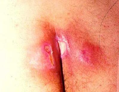
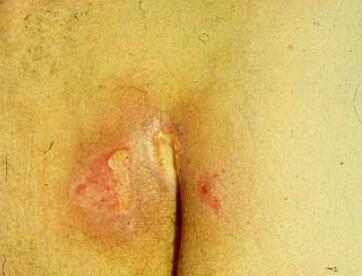
After treatment Ahmed, A. A. E. S. A., Abdel Aziz, K. S., Ahmed, M., Awad, M., Mahmoud, A. H. A., & Ahmed, A. A. Effect Of Polarized Light Therapy On Incisional Pain After Cesarean Section. European Journal of Molecular & Clinical Medicine, 7(10), 2020. Inclusion Criteria • 25-35 years • 1st 24 hours after CS Outcomes • VAS (0-100mm) • Electronic algometer Aim • To investigate the effect of polarized light therapy on incisional pain after caesarean section. Exclusion Criteria • History of any abdominal operations • History of any radiotherapy or chemotherapy, diabetes, psychological problems, acute infection in the area treated and skin disease Day 0 Before treatment (n=20) Group A: Standard treatment (n=20) Group B: Standard + Bioptron From 10cm, 15 minutes once a day Study • RCT Day 5 Treatment of Post Operational Wounds: Cesarean Section Wounds
Ahmed, A. A. E. S. A., Abdel Aziz, K. S., Ahmed, M., Awad, M., Mahmoud, A. H. A., & Ahmed, A. A. Effect Of Polarized Light Therapy On Incisional Pain After Cesarean Section. European Journal of Molecular & Clinical Medicine, 7(10), 2020. 8 8.14 1.63 2.85 0 1 2 3 4 5 6 7 8 9 10 VAS (0100) * Day 0 Test group Day 5 Day 0 Control group Day 5 Changes in VAS between day 0 to day 5 * * 0.41 0.45 1.49 0.91 0 0.5 1 1.5 2 2.5 3 Pressure Algometer Day 0 Test group Day 5 Day 0 Control group Day 5 Changes in Pressure algometer between day 0 to day 5 ↑ 100% * ↑ 261% Polarized light therapy for 5 consecutive days post Cesarean section is an effective adjuvant therapy in treatment of incisional pain. Treatment of Post Operational Wounds: Cesarean Section Wounds
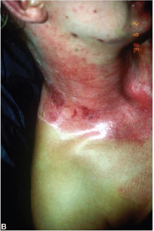
Treatment of Graft Donor Site Wounds
(n=20) Wounds treated with Bioptron Same patients From 10 cm, 10 minutes, 3 times a week Aim • To compare the wound healing process in pairs of standardized wounds (with and without phototherapy) in one single patient. Post-op day 1
assessment Study • Single blind RCT Outcomes
The
• The
• The
• The
•
•
•
Each
these
variables was scored on a 1-5 scale Wounds not treated with Bioptron Day 12 Final assessment Wounds • The skin grafts were always taken with an identical thickness of 0.3mm • Donor sites were located on an identical area of the body
Monstrey, S., Hoeksema, H., Depuydt, K., Van Maele, G. E. O. R. G. E. S., Van Landuyt, K., & Blondeel, P. (2002). The effect of polarized light on wound healing. European Journal of Plastic Surgery, 24(8), 377-382.
Initial
•
degree of epithelialization
quality of granulation tissue
degree of inflammation
degree of infection
Blister formation
The formation of early scar tissue
The subjective feeling of the patient.
of
outcome
Treatment of Graft Donor Site Wounds
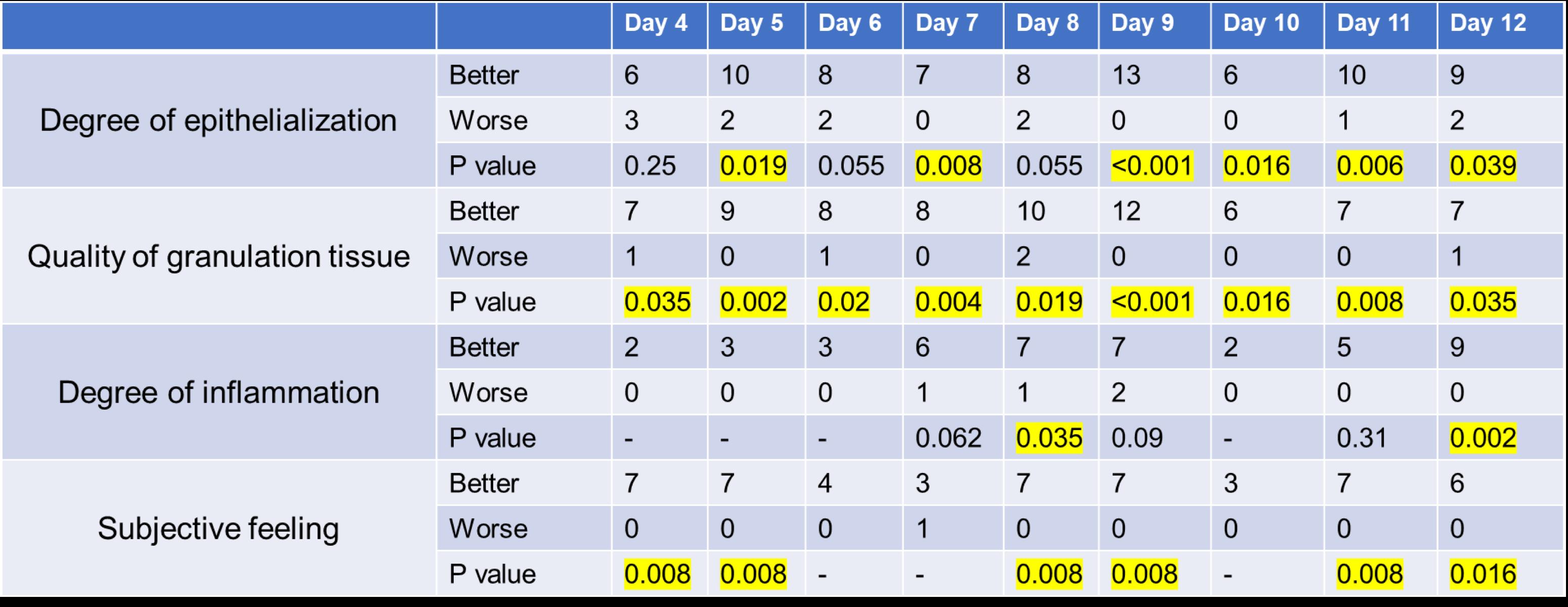
Polarized light therapy yields significantly better wound healing with faster epithelialization and an improved quality of early scar tissue formation.
Monstrey, S., Hoeksema, H., Depuydt, K., Van Maele, G. E. O. R. G. E. S., Van Landuyt, K., & Blondeel, P. (2002). The effect of polarized light on wound healing. European Journal of Plastic Surgery, 24(8), 377-382.
Trauma
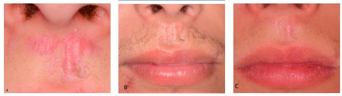
of Traumatic Wounds: Cracked nipples Abdou WEM et al, Effect of Bioptron in Treating Cracked Nipples in Breast Feeding Women: A Randomized Controlled Trial, World Journal of Medical Sciences 16 (1): 35-40, 2019 ISSN 1817-3055 (n=15) Lanolin cream (n=15) Lanolin cream + Bioptron Group A Group B Cream applied once a day 2 weeks Cream applied once a day Bioptron is used for 10 minutes for each affected area, 5 times a week, from 10cm. Aim • To evaluate the effect of polarized polychromatic non- coherent light therapy in treatment of cracked nipples in breast feeding women. Day 0 Final assessment Initial assessment Study • RCT Outcomes • VAS score • McGill pain questionnaire • Storr scale for wound healing+photographing
Treatment
VAS Before treatment After treatment Significance Group A (cream only) 7.6±1.93 4.9±1.86 p=0.0001 Group B (cream+Bioptron) 8.55±1.63 1.25±1.68 p=0.0001 MPQ Before treatment After treatment Significance Group A (cream only) 26 (4) 19 (6) p=0.0001 Group B (cream+Bioptron) 26.5 (5.5) 1 (3) p=0.0001 35.5 85.38 0 10 20 30 40 50 60 70 80 90 100 Improvement in VAS (%) Group A Group B * * * p=0.0001 Changes in VAS and MPQ scores in patient groups, before and after treatment Abdou WEM et al, Effect of Bioptron in Treating Cracked Nipples in Breast Feeding Women: A Randomized Controlled Trial, World Journal of Medical Sciences 16 (1): 35 40, 2019 ISSN 1817 3055 Treatment of Traumatic Wounds: Cracked nipples
Group A (cream only) Normal color Slightly reddened Reddened Developing fissure Fissure Before treatment 0 (0%) 0 (0%) 4 (20%) 8 (40%) 8 (40%) After treatment 0 (0%) 9 (45%) 7 (35%) 4 (20%) 0 (0%) Group B (cream+Bioptron) Normal color Slightly reddened Reddened Developing fissure Fissure Before treatment 0 (0%) 0 (0%) 0 (0%) 9 (45%) 11 (55%) After treatment 8 (40%) 10 (50%) 2 (10%) 0 (0%) 0 (0%) * * * p=0.0001 * Bioptron therapy is an effective and safe way to alleviate pain and enhance healing of cracked nipples in breast feeding women. Changes in Storr scale in patient groups, before and after treatment Abdou WEM et al, Effect of Bioptron in Treating Cracked Nipples in Breast Feeding Women: A Randomized Controlled Trial, World Journal of Medical Sciences 16 (1): 35 40, 2019 ISSN 1817 3055 Treatment of Traumatic Wounds: Cracked nipples
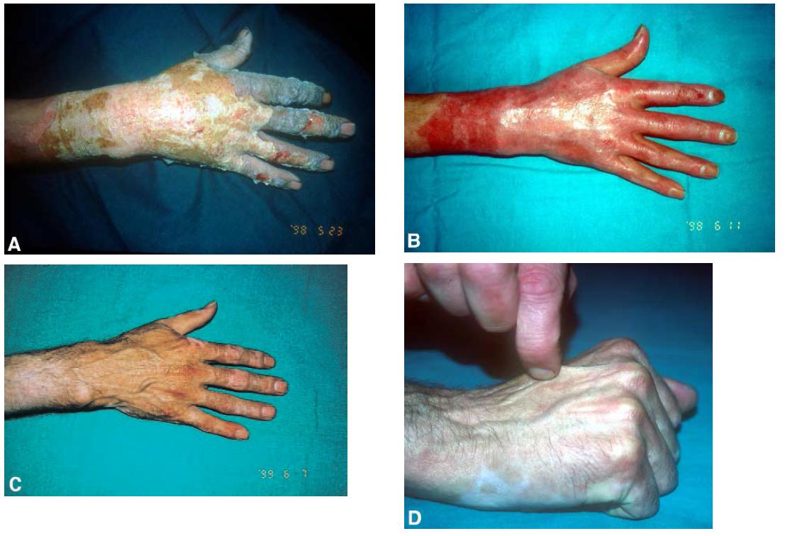
Treatment of Traumatic Wounds: Case Reports
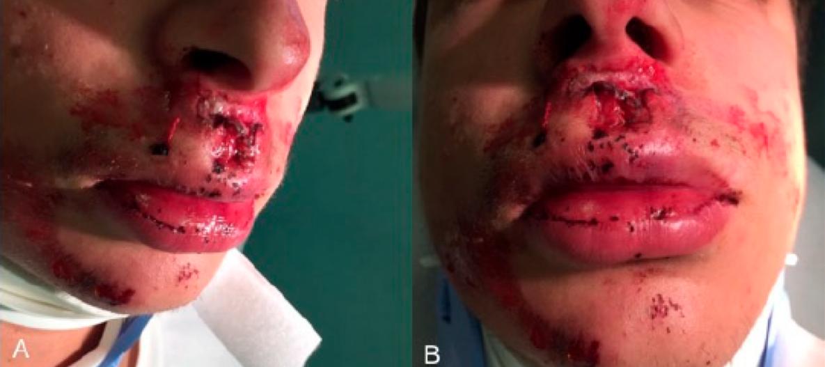
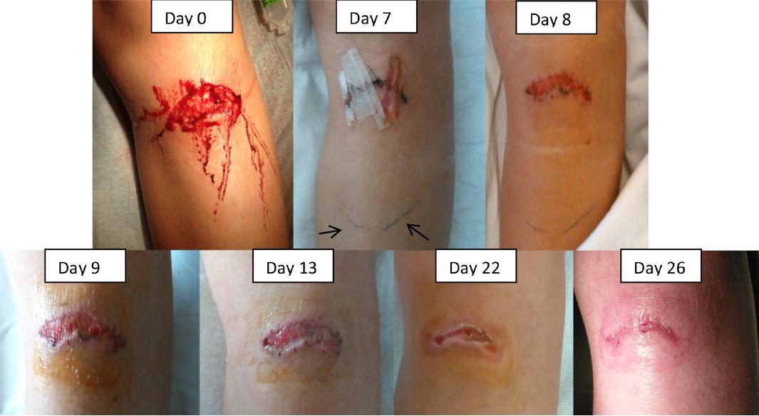
Case-1
After accident
1. Rutteman B, Aeromonas wound infection in a healthy boy, and wound healing with polarized light. JMM Case Rep. 2017 Oct 16;4(10):e005118.
2. Nardi GM, Phototherapy and Tailored Brushing Method. Personalized Oral Care in Patients with Facial and Dental Trauma. A Report of a Case. Healthcare (Basel). 2021 May 11;9(5):561.
12 year old child, infected wound due to falling Bioptron is started on 8th day, 9 min twice a day
Case-2 18 year old man, face trauma due to traffic accident 5 minutes from a distance of 5 cm for the first 5 days then twice a week for 3 months
1st
2nd
3rd
month
month
month
Burn wounds
Treatment of Burns
Week 0
Bioptron + standard wound care
Wound assessment
Aim
• To investigate whether polarized-light therapy could accelerate wound closure in deep dermal burn wounds, thus reducing the need for surgery, without increasing hypertrophic scarring and contractures.
Study
• Case series
Patients
• Deep dermal burns • Not eligible for surgery
Until the wound is healed completely (mean: 3.2 weeks)
(n=22)
Wound assessment
Questionnaires
6 minutes, from 10 cm, every day
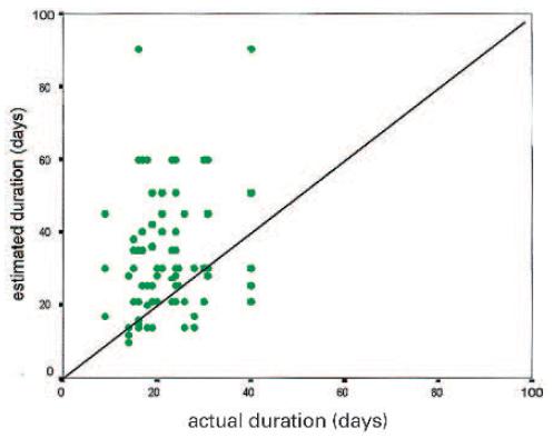
4 experienced surgeons were given 3 questionnaires:
First: 30 cases (22 study, 8 controls) • Necessity of surgery • How long would it take to treat conservatively? • Would you expect hypertrophic scars
Second: 22 cases (before, during and after treatment) • Estimate the time interval • Would you expect hypertrophic scars
Third: 22 cases (After long term follow up)
• Score the result as much worse, worse, comparable, better or much better than the result they would have expected after surgical or conventional conservative therapy
Monstrey, S. J., Hoeksema, H., Saelens, H., Depuydt, K., Hamdi, M., Van Landuyt, K., & Blondeel, P. N. (2002). A conservative approach for deep dermal burn wounds using polarised light therapy. British journal of plastic surgery, 55(5), 420 426.
Treatment of Burns
Necessity of surgery
• %59 of the cases were assessed as “surgery is needed” by the doctors
Time of treatment
• The estimated time for healing (41 days) was significantly longer* than the actual healing time (22 days).
Estimated/Actual time of treatment
Hypertrophic scarring
• 15.8 cases were thought to healed with hypertrophic scarring. This complication was observed only in 1 case* .
Compared to surgical treatment
• 73.8% of the results were comparable/better/much better
Compared to conservative treatment
• 97.6% of the results were comparable/better/much better
Evaluation of the results compared to surgery
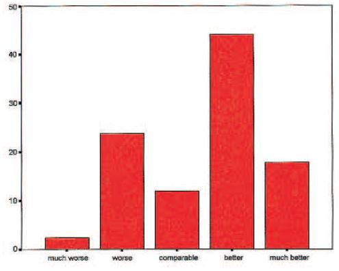
Monstrey, S. J., Hoeksema, H., Saelens, H., Depuydt, K., Hamdi, M., Van Landuyt, K., & Blondeel, P. N. (2002). A conservative approach for deep dermal burn wounds using polarised light therapy. British journal of plastic surgery, 55(5), 420 426.
Treatment of
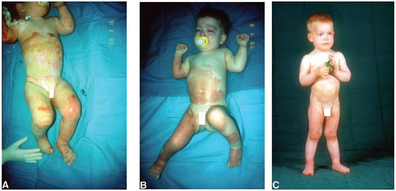
1 year old child with hot oil burn 8 days after the accident 1 month 1 year follow-up 2 days after the accident 12 days Young
with flame burn Young woman with flame burn 2 days after the accident 19 days 11 months With Bioptron • Wound healing is accelarated • No extension of hospital stay is needed
Burns Monstrey Depuydt Blondeel, P. N. (2002). A conservative approach for deep dermal burn wounds using polarised light therapy. British journal of plastic surgery, 55(5), 420
woman
Treatment of Burns
days
21
19
Monstrey, S. J., Hoeksema, H., Saelens, H., Depuydt, K., Hamdi, M., Van Landuyt, K., & Blondeel, P. N. (2002). A conservative approach for deep dermal burn wounds using polarised light therapy. British journal of plastic surgery, 55(5), 420 426. With Bioptron Need of surgery was reduced; • Physiotherapy was started immediately without a period of immobilization. • It allowed early start of pressure therapy. With better functional and aesthetic results, Bioptron is a therapy of choice for deep dermal burns.
A young man
with flame burn 2
after the accident
days
months Optimal results
Treatment of Burn Scars
Hamada,
Effect of orange polarized light on post burn pediatric
(n=15) Control group: Standard scar management (n=15) Treatment group: Standard + Bioptron 4 weeks From 10 cm, 15 minutes medical filter + 15 minutes orange filter, 3 times a week Aim • To evaluate the effect of orange filtered polarized polychromatic light on post burn pediatric scar. Day 0 Final assessment Initial assessment Study • Single-blind RCT Outcomes Vancouver Scar Scoring (0-13) • Vascularity • Pigmentation, • Height/thickness • Pliability Inclusion Criteria • Children 3-7 years old • Hypertrophic burn scar (≥2 months post healing) Exclusion Criteria • Chronic skin conditions and keloid history
Abd Elrashid, N. A., Sanad, D. A., Mahmoud, N. F.,
H. A., Abdelmoety, A. M., & Kenawy, A. M. (2018).
scar: a single blind randomized clinical trial. Journal of Physical Therapy Science, 30(10), 1227-1231.
of Burn Scars
orange polarized
VSS Before treatment After treatment Significance Group A (standard only) 10.66 ± 2.01 8.8 ± 1.61 p<0.05 Group B (standard+Bioptron) 8.92 ± 2.36 4.93 ± 1.86 p<0.05 * 17.44 44.73 0 10 20 30 40 50 60 Improvement in VSS (%) Group A Group B Bioptron hyperlight therapy and orange filter showed significantly better results in the treatment of burn scars in children.
Treatment
Abd Elrashid, N. A., Sanad, D. A., Mahmoud, N. F., Hamada, H. A., Abdelmoety, A. M., & Kenawy, A. M. (2018). Effect of
light on post burn pediatric scar: a single blind randomized clinical trial. Journal of Physical Therapy Science, 30(10), 1227-1231.*: p<0.05
Treatment of Burn Scars: Range of Motion
Flexion ROM Before treatment After treatment Significance Group A (standard only) 59.27±7.04 63.6±5.87 p=0.001 Group B (standard+Bioptron) 60.87±5.96 67.67±4.24 p=0.001 Extension ROM Before treatment After treatment Significance Group A (standard only) 20.47±3.25 22.07±3.24 p=0.001 Group B (standard+Bioptron) 22.73±3.20
p=0.001 *
*
Abd El-Rashid NA, Sanad DA, Ayoub HS, Elhenawy AN. Effect of orange polarized light on Metacarpophalyngeal range of motion in pediatric hand burn: a single blind randomized trial. Bioscience Res. 2019;16(3):2417–22..*: p<0.05
26.27±3.22
Orange filtered polarized light has a special and beneficial effect on improving the range of motion in children with hand burn.
Treatment of Burns
Wound Surface Area and Colony Count of Various Modes of
(n=15) Group A: Standard + Bioptron (n=15) Group B: Standard + LLLT 4 weeks From 10 cm, 10 minutes, 3 times a week Aim • To evaluate the wound healing efficacy of polarized light therapy against LLLT to determine which is more efficient and successful at speeding burn healing. Day 0 Final assessment Initial assessment Study • RCT Outcomes • Wound surface area • Bacterial colony count Inclusion Criteria • Dermal burns in forearms with TBSA of (15-25%). • Thermal injury Exclusion Criteria • Chronic conditions, pregnancy Ga-As laser, 850 nm, 200 mV, 1.2 J/cm2 and 2.4 J/cm2
Emam Mowafy, Z. M., Mostafa Ibrahim, I. S., Ibrahim, M. B., & Mohamed Mokhtar Elshahawy, A. M. (2021).
Phototherapy. Egyptian Journal of Hospital Medicine, 85(2)
Burns
Treatment of
Surface Area and Colony Count of Various Modes
Outcome Group A Group B Significance WSA (cm2) Before treatment 6.17 ± 1.55 6.33 ± 1.2 p=0.75 After treatment 2.77 ± 0.9 3.93 ± 0.74 p=0.001 Significance p=0.001 p=0.001 Colony count (x1010) Before treatment 13.46 ± 2.85 13.66 ± 2.61 p=0.84 After treatment 8.26 ± 2.37 11.53 ± 2.5 p=0.001 Significance p=0.001 p=0.001 55.1 37.9 0 10 20 30 40 50 60 Improvement in WSA (%) Group A Group B Polarized light treatment appears more efficient than low-level laser therapy in speeding the process of recovery and reducing hospital stay duration.
Emam Mowafy, Z. M., Mostafa Ibrahim, I. S., Ibrahim, M. B., & Mohamed Mokhtar Elshahawy, A. M. (2021).
Wound
of Phototherapy. Egyptian Journal of Hospital Medicine, 85(2)
Mowafy,
Journal of Health Sciences, 6(S2), 13053–13063. https://doi.org/10.53730/ijhs.v6nS2.8444
of Foot Burns
Treatment
light therapy
Z. M. E., Abdelrahman, S. M. E., Ali, K. M., & Ali, A. M. A. (2022). Low level laser therapy versus polarized
on healing of foot burn. International
(n=20) Group A: Standard + LLLT (n=20) Group B: Standard + Bioptron 4 weeks From 10 cm, 10 minutes, 3 times a week Aim • To evaluate the efficacy of the low level laser therapy (LLLT) versus polarized light therapy (BLT) on healing of foot burns. Day 0 Final assessment Initial assessment Study • RCT Outcomes • Wound surface area • Bacterial colony count Inclusion Criteria • Dermal burns in foots with TBSA of (1-5%). • Thermal injury Ga-As laser, 850 nm, 200 mV, 1.2 J/cm2 and 2.4 J/cm2
Treatment of Burns
Journal of Health Sciences, 6(S2), 13053–13063. https://doi.org/10.53730/ijhs.v6nS2.8444
Outcome Group A Group B
WSA (cm2) Before treatment 9.51 ± 0.42 9.51 ± 0.4 After treatment 3.44 ± 0.31 4.2 ± 0.2 Significance p=0.001 p=0.001 Results of this study support the expectation that application of both polarized light therapy and LLLT had a valuable healing effect by enhancing healing of burns, as manifested by the highly decreases BSA.
Mowafy, Z. M. E., Abdelrahman, S. M. E., Ali, K. M., & Ali, A. M. A. (2022). Low level laser therapy versus polarized light therapy on healing of foot burn. International
9.51 9.51
0 1 2 3 4 5 6 7 8 9 10 Wound
Before
Wound Surface Area * *
4.2 3.44
Surface area (cm 2 )
After Before After Bioptron group LLLT group
Reviews
Bioptron in reviews
Overall however, the evidence is largely favorable of PLT as a therapy in a range of conditions, with a strong safety profile, and unanimously beneficial effects reported.
PLT can be used as an adjunctive therapy for treatment of different types of wounds, i.e Lower extremity wounds, Diabetic wounds and after burns for animals and humans.
The use of polarized light therapy has further questioned the need for early excision in deep second-degree bums. Polarized light allows for conservative treatment with better functional outcome due to continuous physical therapy and early application of pressure therapy and silicone.
1. Feehan J Therapeutic applications of polarized light: Tissue healing and immunomodulatory effects. Maturitas. 2018 Oct;116:11-17.

2. Monstrey S (2000) Wound Care. Hospital Healthcare Europe 2000; pp 55-57


Studies support the efficacy of polarized light to overcome musculoskeletal problems, but it is also important for its action on skin problems and burns.
3. M. Allam, N. (2022). Polarized Light Therapy in the Treatment of Wounds: A Review. The International Journal of Lower Extremity Wounds, 15347346221113991.
4. Nicolaou V, Stasinopoulos D, Lamnisos D. The Effectiveness of Polarized Light in Musculoskeletal, Skin Problems and Burns. 2020 - 10(2). AJBSR.MS.ID.001492. DOI: 10.34297/AJBSR.2020.10.001492.

Bibliography
Pressure ulcer
Abd Al-kader, A. M., Hassan, M. A., & Elsayed, H. G. (2015). Efficacy of polarized light in treatment of pressure ulcers. JMSCR, 3, 5800-5809. Białożyt J., Materniak K., Kawecki M. 2018 Use of polarise lighting in support of treatment of pressure ulcers among patients after burns. Preliminary report. Dermatologia Estetyczna. vol.20, 1 (114) Đurović, A., Marić, D., Brdareski, Z., Jevtić, M., & Đurđević, S. (2008). The effects of polarized light therapy in pressure ulcer healing. Vojnosanitetski pregled, 65(12), 906-912. Iordanou, P., Baltopoulos, G., Giannakopoulou, M., Bellou, P., & Ktenas, E. (2002). Effect of polarized light in the healing process of pressure ulcers. International journal of nursing practice, 8(1), 49 55.
Venous ulcer
Jankovi, A. (2005). Physical therapy of venous ulcers: effects of electroionotherapy and polarized light. vascular diseases, 4, 5. Medenica, L., & Lens, M. (2003). The use of polarised polychromatic non-coherent light alone as a therapy for venous leg ulceration. Journal of wound care, 12(1), 37-40.
Chronic Ulcer general
Aragona, S. E., Grassi, F. R., Nardi, G., Lotti, J., Mereghetti, G., Canavesi, E., ... & Lotti, T. (2017). Photobiomodulation with polarized light in the treatment of cutaneous and mucosal ulcerative lesions. Journal of Biological Regulators and Homeostatic Agents, 31(2 Suppl. 2), 213 218.
INTSAR S. WAKED, Ph.D., R.M., & ASHRAF E.M. ELSEBAIE, M.D., M.B. (2021). Effect of Negative Pressure Therapy versus Polarized Light Therapy on Chronic Wound Healing.
Diabetic foot ulcer
El-Deen, H. B., Fahmy, S. E. H. A. M., Ali, S. A., & El-Sayed, W. M. (2014). Polarized light versus light-emitting diode on healing of chronic diabetic foot ulcer. Romanian Journal of Biophysics, 24(2), 1-15. Mohamed, M. H., Selem, M. N., Mohamed, M. S., & Abd EL-Ghaffaar, H. A. (2019). Interleukin-6 response to shock wave therapy versus polarized light therapy in the treatment of chronic diabetic foot ulcers. Drug Invention Today, 11(11).
Post-operational
Simic, A. (1999) Effects of PILER light therapy on wound healing in patients operated due to stomach carcinoma. 3 rd International Gastric Cancer Congress April 27 – 30, 1999 Korea, SEUL. (Abstract) Simic, A., Stojakov, D., Sabljak, P., Jekic, I., Bjelovic, M., & Pesko, P. (1999). Piler Light Therapy-Effect on Wound Healing in Esophagogastric Surgery. EUROPEAN SURGICAL RESEARCH, 31(1), 225-225. (Abstract)
Simic, A. (2001, May). Importance of Bioptron light therapy in the treatment of surgical incisions. In Second Balkan Congress for PRAS and Bioptron Satellite Symposium, Belgrade, May (pp. 24 26). (Abstract)
Simic, A., Pesco, P., Bjelovic, M., STOJAKOV, D., TODOROVIC, M., TODOROVIC, V., ... & KOTARAK, M. (2001). Bioptron light therapy and thoracophrenolaparotomy wound healing in patients operated due to cardiac carcinoma, paper presented at the 4th International Gastric Cancer Congress. (Abstract) Colić, M. M., Vidojković, N., Jovanović, M., & Lazović, G. (2004). The use of polarized light in aesthetic surgery. Aesthetic plastic surgery, 28(5), 324 327. Kymplova, J., Navrátil, L., & Knížek, J. (2003). Contribution of phototherapy to the treatment of episiotomies. Journal of clinical laser medicine & surgery, 21(1), 35-39. Monstrey, S., Hoeksema, H., Depuydt, K., Van Maele, G. E. O. R. G. E. S., Van Landuyt, K., & Blondeel, P. (2002). The effect of polarized light on wound healing. European Journal of Plastic Surgery, 24(8), 377-382. (Burn graft donor sites)
Ahmed, A. A. E. S. A., Abdel Aziz, K. S., Ahmed, M., Awad, M., Mahmoud, A. H. A., & Ahmed, A. A. Effect Of Polarized Light Therapy On Incisional Pain After Cesarean Section. European Journal of Molecular & Clinical Medicine, 7(10), 2020.
Bibliography-2
Trauma/Incision
Abdou WEM et al, Effect of Bioptron in Treating Cracked Nipples in Breast Feeding Women: A Randomized Controlled Trial, World Journal of Medical Sciences 16 (1): 35 40, 2019 ISSN 1817 3055
Rutteman B, Borremans K, Beckers J, Devleeschouwer E, Lampmann S, Corthouts I, Verlinde P. Aeromonas wound infection in a healthy boy, and wound healing with polarized light. JMM Case Rep. 2017 Oct 16;4(10):e005118. doi: 10.1099/jmmcr.0.005118. PMID: 29188066; PMCID: PMC5692235.
Nardi GM, Guerra F, Ndokaj A, Corridore D, Straker MA, Sportelli P, Di Giorgio R, Grassi FR, Grassi R, Ottolenghi L. Phototherapy and Tailored Brushing Method. Personalized Oral Care in Patients with Facial and Dental Trauma. A Report of a Case. Healthcare (Basel). 2021 May 11;9(5):561. doi: 10.3390/healthcare9050561. PMID: 34064547; PMCID: PMC8150812.
Burns
Monstrey, S. J., Hoeksema, H., Saelens, H., Depuydt, K., Hamdi, M., Van Landuyt, K., & Blondeel, P. N. (2002). A conservative approach for deep dermal burn wounds using polarised light therapy. British journal of plastic surgery, 55(5), 420-426.
Emam Mowafy, Z. M., Mostafa Ibrahim, I. S., Ibrahim, M. B., & Mohamed Mokhtar Elshahawy, A. M. (2021). Wound Surface Area and Colony Count of Various Modes of Phototherapy. Egyptian Journal of Hospital Medicine, 85(2).
Mowafy, Z. M. E., Abdelrahman, S. M. E., Ali, K. M., & Ali, A. M. A. (2022). Low level laser therapy versus polarized light therapy on healing of foot burn. International Journal of Health Sciences, 6(S2), 13053 13063. https://doi.org/10.53730/ijhs.v6nS2.8444
Burn Scar
Abd Elrashid, N. A., Sanad, D. A., Mahmoud, N. F., Hamada, H. A., Abdelmoety, A. M., & Kenawy, A. M. (2018). Effect of orange polarized light on post burn pediatric scar: a single blind randomized clinical trial. Journal of Physical Therapy Science, 30(10), 1227 1231.
Abd El-Rashid NA, Sanad DA, Ayoub HS, Elhenawy AN. Effect of orange polarized light on Metacarpophalyngeal range of motion in pediatric hand burn: a single blind randomized trial. Bioscience Res. 2019;16(3):2417 22.
Reviews
Feehan J, Burrows SP, Cornelius L, Cook AM, Mikkelsen K, Apostolopoulos V, Husaric M, Kiatos D. Therapeutic applications of polarized light: Tissue healing and immunomodulatory effects. Maturitas. 2018 Oct;116:11-17. doi: 10.1016/j.maturitas.2018.07.009. Epub 2018 Jul 19. PMID: 30244771.
Monstrey S (2000) Wound Care. Hospital Healthcare Europe 2000; pp 55-57 M. Allam, N., Eladl, H. M., & Eid, M. M. (2022). Polarized Light Therapy in the Treatment of Wounds: A Review. The International Journal of Lower Extremity Wounds, 15347346221113991. Nicolaou V, Stasinopoulos D, Lamnisos D. The Effectiveness of Polarized Light in Musculoskeletal, Skin Problems and Burns. 2020 10(2). AJBSR.MS.ID.001492. DOI: 10.34297/AJBSR.2020.10.001492.
