
UCLA Department of Ophthalmology
Jules Stein Eye Institute and Doheny Eye Institute
UCLA Health


UCLA Department of Ophthalmology
Jules Stein Eye Institute and Doheny Eye Institute
UCLA Health
MAGAZINE
is a publication from the UCLA Department of Ophthalmology.
Jules Stein Eye Institute and Doheny Eye Institute are proud affiliates.
EDITOR-IN-CHIEF
Anne L. Coleman, MD, PhD
MANAGING EDITOR
Tina-Marie Gauthier
c/o UCLA Stein Eye Institute
100 Stein Plaza, UCLA Los Angeles, California 90095-7000 Tina@EyeCiteEditing.com
PUBLICATION COMMITTEE
Anthony Aldave, MD
Susan Lee DeRemer, CFRE
Deborah Ferrington, PhD
JoAnn Giaconi, MD
Marissa Goldberg
Kevin Miller, MD
Peter Quiros, MD
Roxana Radu, MD
Alfredo Sadun, MD, PhD
Molly Ann Woods, CFRE
WRITERS
Dan Gordon
Harlan Lebo
EDITORIAL SUPPORT
Stephanie Colucci
PHOTOGRAPHY
Reed Hutchinson
Rich Schmitt
Robin Weisz
DESIGN
Robin Weisz Design
© 2025 by The Regents of the University of California.
All rights reserved.
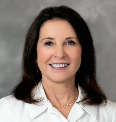
Dear Friends,
Traditionally the spring issue of EYE Magazine marks the excitement of a new year and the promise of all we have to look forward to. However, I want to acknowledge the immense toll the January wildfires have taken on families, first responders, and on everyone who has been affected. Many of you have been displaced, and countless lives have been disrupted.
The devastation caused by these fires has been overwhelming, and I know many of you are facing tremendous challenges as you grapple with the loss of homes, livelihoods, and the familiar landscapes we all hold dear. During times like these, it is important to remember that we are a resilient and compassionate community, and we will face these difficulties together.
As we focus on this issue of EYE, we celebrate the work of Dr. Sherwin Isenberg , who has achieved remarkable success in preventing blindness among premature infants in sub-Saharan Africa, a contribution that has earned him the 2025 Parks Medal for his lifetime achievements in pediatric ophthalmology.
We highlight groundbreaking technology from Dr. SriniVas Sadda and introduce you to five new faculty members—specialists in pediatric ophthalmology and strabismus, neuro-ophthalmology, cataract and refractive surgery, corneal diseases, and glaucoma.
I am also proud to share that for 35 consecutive years UCLA Health has been recognized on the U.S. News & World Report national honor roll of best hospitals, and Jules Stein Eye and Doheny Eye Institutes are ranked #1 in California and top five in the nation for ophthalmology. But with that said, our motto is “Never let it rest until good is better, and better is best!”
In closing, while we may not be able to control all that is happening around us, we can adjust how we respond and move forward together. As the quote by Charles R. Swindoll goes, “Life is 10 percent what happens to us and 90 percent how we react to it.”
My thoughts are with all of you, and I am confident that, together, we will overcome this challenging time and emerge stronger.
With warm regards,

Bradley R. Straatsma, MD, Endowed Chair in Ophthalmology Chair, UCLA Department of Ophthalmology Director, Jules Stein Eye Institute Affiliation Chair, Doheny Eye Institute

AI Model Facilitates Rapid, Accurate Analysis of 3D Biomedical Imaging Data Across Disciplines
UCLA researchers have created a deeplearning framework that can rapidly learn to analyze and diagnose 3D medical images with the same accuracy as clinical specialists, but in a fraction of the time. PAGE 8

FOCUS
A team headed by Dr. Sherwin Isenberg has achieved remarkable success in preventing blindness in premature babies born in sub-Saharan Africa. PAGE 2 On
UCLA DEPARTMENT OF OPHTHALMOLOGY
JULES STEIN EYE INSTITUTE AND DOHENY EYE INSTITUTE
INSTITUTE
2024 American Academy of Ophthalmology Meeting
Vision Scientists Receive Major Grant Funding Faculty Honors
10–16
Dr. Aldave Receives a First for an Investigator in the UCLA Department of Ophthalmology
Dr. Ip Joins the Prestigious American Ophthalmological Society
New Faculty Appointments
EDUCATION 17–18
Aesthetic Eyelid and Facial Rejuvenation Course
Women in Ophthalmology: Summer Symposium
Measures of Intraocular Inflammation for Use in Clinical Trials
6th Annual Doheny-UCLA International Glaucoma Symposium
Embracing and Exploiting Artificial Intelligence for Neuroscience Cataract Surgery Essentials Course Inaugural Doheny Oculomics Symposium Explores the Future of Ophthalmic Biomarkers
Doheny Distinguished Lecture Series
ALUMNI BULLETIN 19–20
A Successful Alumni Reception at the 2024 AAO Annual Meeting in Chicago Alumni Shine at APSOPRS 2024
COMMUNITY OUTREACH 20
UCLA Mobile Eye Clinic Gets New Look
INCLUSIVE EXCELLENCE 21
Visiting Medical Student Scholarship in Ophthalmology and Diversity in Vision Science Undergraduate Summer Research Program
Dr. Clémence Bonnet Awarded JAM Fellowship

A team led by UCLA
Jules Stein Eye Institute
ophthalmologist Dr. Sherwin Isenberg has produced extraordinary success in preventing blindness among premature babies born in sub-Saharan Africa— work that contributed to Dr. Isenberg receiving the 2025 Parks Medal for a lifetime of achievement in pediatric ophthalmology.
The baby was born six weeks early in King Faisal Hospital, a multispecialty medical center in the Rwandan capital of Kigali. At first identified only as “Baby Girl X,” (the “X” a placeholder for her parents’ name), she was tiny—only three pounds—and experienced the health complications typical of premature births, such as anemia, underdeveloped organs, low body temperature, and—in particular—underdeveloped lungs.
But thanks to recent progress in refining neonatal care in sub-Saharan Africa, Baby Girl X is one of the thousands who survive because of improvements in treatment for premature births on the African continent in the 21st century.
However, with that improved survival of premature babies has come a new challenge for their care after birth—treatment that can mean the difference between a life with vision or a life without the ability to see.
“We had started to receive reports from Africa about the increasing number of newborns who were going blind,” says Sherwin J. Isenberg, MD, distinguished professor of ophthalmology and pediatrics emeritus. “This was a problem that had not happened before in Africa and was a direct result of retinopathy of prematurity (ROP), the abnormal growth of blood vessels in the retina that can occur because of improper management of oxygen treatment in neonatal care.”
We had started to receive reports from Africa about the increasing number of newborns who were going blind. This was a problem that had not happened before in Africa and was a direct result of retinopathy of prematurity (ROP), the abnormal growth of blood vessels in the retina that can occur because of improper management of oxygen treatment in neonatal care.
Until recently, the challenges of blindness among newborns in Africa was less of a priority in pediatric care because of the low survival rate for premature infants. But with survival rates increasing dramatically in the first quarter of the 21st century, many countries in low-resource regions have refined their intensive care units for treatment of premature babies.
But one aspect of that lifesaving treatment—improving lung development with the application of oxygen—creates a new challenge: ensuring that oxygen therapy for premature babies with breathing problems does not lead to blindness triggered by the improper use of that oxygen.
Balancing the amount of oxygen used for premature infants is critical; use too little oxygen, and the threat increases for brain damage, cerebral palsy, and other risks. However, using too large a concentration of oxygen can generate other conditions, especially the irregular stimulation of blood vessel growth in the eye that can lead to vision loss or permanent blindness.
“There is a ‘sweet zone’ for oxygen use that is crucial,” says Dr. Isenberg. “Understanding that zone and treating it effectively is a complex medical procedure that requires extensive training and application in maternity wards.”
Without proper management of oxygen therapy, babies can develop ROP, which can result in severe vision loss or complete blindness. ROP is a leading cause of blindness in infants worldwide, especially in low-resource countries.
The issue of ROP has become especially acute in subSaharan Africa, where survival of premature babies has increased substantially in the past two decades. In Rwanda, for example, some 55,000 babies are born prematurely every year, with a rate of ROP as high as 15 percent—meaning some 5,000 children annually risk potential vision loss or preventable blindness.
A survey of oxygen management in African hospitals conducted by Dr. Isenberg’s group found that only 64 percent of neonatal units had any equipment to blend oxygen with room air, and only one unit had access to continuous monitoring for all the newborns receiving oxygen.
The problem of infant blindness is compounded by the acute shortage of ophthalmologists in Africa; for example, in Rwanda, with a population of more than 14 million, only 18 ophthalmologists serve the entire country, none of whom have advanced training in pediatric vision care.
The growing issue of preventable blindness among premature babies reached a transition point at a 2018 conference in Sydney, Australia, when a group of pediatric ophthalmologists discussed the issue as an impending epidemic.

Without proper management of oxygen therapy, babies can develop ROP, which can result in severe vision loss or complete blindness. ROP is a leading cause of blindness in infants worldwide, especially in low-resource countries.
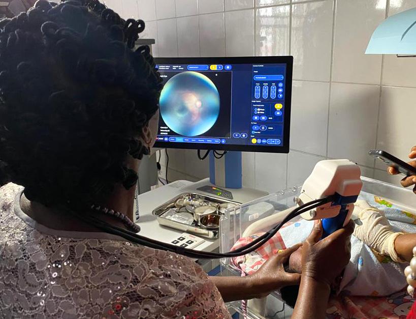
Below: As everyone focuses intently on the monitor, Dr. Sarah Rodriguez, Ugandan team leader, guides local doctors and nurses in using the retinal camera.
“We calculated that among babies with untreated ROP, 69 percent would be blind or develop severely impaired vision,” says Dr. Isenberg. “But we also knew that with increased training and advanced equipment, we could control ROP and reduce the occurrence of blindness in premature infants. We saw that ongoing success in improving survival rates for premature babies in Africa was going to continue to increase the problems of blindness caused by oxygen therapy—unless we did something about it.”
Beginning in 2018, Dr. Isenberg, as co-chair, began developing a program entitled Stop Infant Blindness in Africa (SIBA), a joint project of Children’s Eye Foundation, the American Association for Pediatric Ophthalmology and Strabismus, and the International Pediatric Ophthalmology and Strabismus Council.
Dr. Isenberg and his three co-chairs: ophthalmologist Scott Lambert, MD, from Stanford University; neonatologist Martha Mkony, MD, from Muhimbili National Hospital in Tanzania; and ophthalmologist Dupe Popoola, MD, from the University of Ilorin in Nigeria; along with other doctors and nurses in Africa, spent more than a year of planning and connection-building with King Faisal Hospital in Rwanda, Nsambya Hospital in Uganda, and Port Harcourt Hospital in Nigeria, to build new levels of training and expertise in identifying possible cases of ROP and treating the disease.

Right: This instrument is measuring the infant’s oxygen and heart rate, allowing the neonatology team to properly modulate oxygen levels to prevent retinopathy. This critical tool was sent to Rwanda by the SIBA project.
The result of the SIBA effort was a program to reduce the risk of ROP through enhanced training to blend oxygen with room air, and increase the monitoring of blood oxygen saturation levels that support breathing and lung development—but without increasing the risk of ROP.
“Our goal was to develop training and experience to reduce the epidemic of ROP while survival rates for premature infants continue to improve,” says Dr. Isenberg.
SIBA provided in-line oxygen blenders and pulse oximeters for oxygen therapy, as well as training in prevention, identification, and treatment of ROP for nursing and physician staff. The project received examination equipment (scleral depressors, lid specula, and an indirect ophthalmoscope), ICON III Phoenix Cameras from NeoLight, and indirect lasers donated by Alcon for use in treating some cases of ROP.
“This was a two-part program,” says Dr. Isenberg. “First, to train the neonatology teams in oxygen management; and second, to work with ophthalmologists to screen for ROP, and then to treat the babies who have ROP.”
The results of the SIBA program were immediate and impressive: in all three countries, the rate of ROP dropped in the first year. In Uganda, for instance, the rate of blindness was cut in half.
In addition, the number of infants with proper development of blood vessels in the eye increased from 16 percent to 44 percent.
“This was a tremendous improvement—and an excellent example of how advanced training and equipment for talented clinicians can make a huge difference in stopping preventable blindness,” says Dr. Isenberg.
Reducing blindness is not only a quality-of-life issue for thousands of babies, but it is also a potential relief from economic burden in financially strapped sub-Saharan Africa. A multiinstitute SIBA study showed that in Rwanda alone, the annual cost of new programs to reduce ROP—including training, screening, and treatments (about $1.9 million annually)—cost much less than the financial burden of blindness (additional medical care, lost economic productivity, and reduced income) of more than $5 million per year.
The SIBA program to address the issues of ROP is only a first step in new programs to reduce preventable blindness in sub-Saharan Africa. New phases of the program will include broader training and emerging technology to expand the benefits of the work.
“The ongoing challenge will be how we can continue to reduce blindness caused by ROP while the survival rate of premature babies continues to climb—all in countries with scarce resources for ophthalmology,” says Dr. Isenberg.

The results of the SIBA program were immediate and impressive: in all three countries, the rate of ROP dropped in the first year. In Uganda, for instance, the rate of blindness was cut in half.
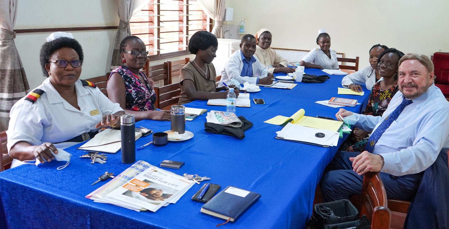
Dr. Isenberg envisions a growing network of care aided by establishing the three participant hospitals as teaching centers, where doctors and nurses from across sub-Saharan Africa can train in methods to prevent ROP.
In the meantime, the SIBA program has demonstrated that flexible new approaches to dealing with preventable blindness can be implemented with speed and powerful results.
“A 50 percent reduction in blindness is a gratifying result for the short term, but it is only a start,” says Dr. Isenberg. “We can continue to improve on that success as the program is refined and expanded.”
Dr. Isenberg’s work on the SIBA project caps a lifetime of achievement in reducing preventable blindness in children— accomplishments for which the American Association for Pediatric Ophthalmology and Strabismus awarded Dr. Isenberg with the 2025 Parks Silver Medal. The recognition, considered the Nobel Prize for pediatric ophthalmology, was given for Dr. Isenberg’s “remarkable contributions to the field of pediatric ophthalmology.”

This was a tremendous improvement—and an excellent example of how advanced training and equipment for talented clinicians can make a huge difference in stopping preventable blindness.

Thanks to the work of Dr. Isenberg (left) and Dr. Apt (center), the use of povidone-iodine has spared thousands of babies and adults from blindness caused by postsurgical infections, and it is now a globally adopted practice.
For more than 35 years, Dr. Sherwin Isenberg has investigated methods to reduce childhood blindness in low-resource countries—one he has addressed with extraordinary success.
Dr. Isenberg and late founding member of the Jules Stein Eye Institute, Dr. Leonard Apt , pioneered the use of povidone-iodine for prevention and treatment of potential blindness from infections at birth based on studies at UCLA and in Kenya. Because of their work, the application of povidone-iodine has prevented thousands of babies, as well as adults, from going blind from postoperative infections and is now used throughout the world.
“Povidone-iodine is not only effective, but it is also incredibly cheap, which makes it an especially important treatment in the developing world,” says Dr. Isenberg. “The cost of an application of povidone-iodine before and after surgery to prevent infection costs only pennies.” In studies conducted in India and the Philippines, Drs. Isenberg and Apt also proved low-cost povidone-iodine ophthalmic solution effective in treating conjunctivitis, also known as “pink eye,” caused by bacteria and chlamydia and potentially blinding corneal infections.
Povidone-iodine is now on the World Health Organization’s Model List of Essential Medicines—a medication deemed vital to addressing the most critical needs of the global health system.
Blindness is a major public health challenge tied to extreme poverty and shorter life expectancies, particularly in low-resource countries. In sub-Saharan Africa, where access to specialized healthcare is limited, the impact of untreated vision loss is profound, affecting individuals, families, and entire communities.
Bradley R. Straatsma, MD, JD, founding chair of the UCLA Department of Ophthalmology and founding director of the Jules Stein Eye Institute, has dedicated his life to advancing eye health and combating preventable blindness. A pivotal figure in this mission, Dr. Straatsma played a key role in the establishment of the Magrabi ICO Cameroon Eye Institute (MICEI) in Yaoundé, Cameroon.
MICEI is the first subspecialty and training eye hospital in Central Africa. Its core mission is to provide comprehensive eye care at an affordable cost, while simultaneously training the next generation of ophthalmologists and healthcare professionals—experts who are committed to becoming leaders in eye care across Cameroon and French-speaking Central West Africa. By focusing on both medical care and education, MICEI is changing the trajectory of eye health
in the region, ensuring that more people can see clearly and live healthier, longer lives.
The UCLA Department of Ophthalmology’s ongoing association with MICEI has continued to thrive under the leadership of Bartly J. Mondino, MD, immediate past chair and director, and Anne L. Coleman, MD, PhD, current chair and director, as Jules Stein Eye Institute faculty members have been involved in research collaboration and clinical training.
Since its official inauguration on March 29, 2017, MICEI has made a significant impact on the local community. In its first year, the institute provided vision care to 20,000 patients, a remarkable achievement for a newly established institution.
The Institute’s achievements were recognized in 2021 when MICEI won two prestigious awards: Best Hospital and Best Private Hospital in Cameroon.

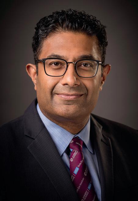
UCLA RESEARCHERS have developed a deep-learning framework capable of quickly teaching itself to analyze and diagnose 3D medical images with accuracy equal to that of clinical specialists, in a small fraction of the time. This advancement by the multidisciplinary research team—including SriniVas R. Sadda, MD, professor of ophthalmology at UCLA and director of artificial intelligence and imaging research at Doheny Eye Institute— could pave the way for diagnostic and treatment advances in ophthalmology and other biomedical disciplines, given the system’s wide adaptability across imaging modalities.
In medicine, artificial intelligence (AI) is used to teach computers how to recognize information through a process called training. Artificial neural networks train themselves through many repeated calculations and by incorporating relevant information from large datasets that have been examined and labeled by clinical experts. “To have a computer classify different types of disease, we have to show many examples of each type so that it can understand all of the variations,” Dr. Sadda explains. “In ophthalmology, we do this through images of the eye. But the need for huge numbers of these images is the limitation to being able to train AI models.”
In response to this limitation, many AI systems use transfer learning—incorporating other sources of data to train the most basic aspects of the model, while reserving the more specific but scarce data for fine-tuning. In their research, published in the October 1, 2024, issue of Nature Biomedical Engineering, Dr. Sadda’s group employed the transfer learning technique in developing an AI system that overcomes a major bottleneck for AI in medicine: the limited amount of 3D imaging data.
Most biomedical imaging data consists of 2D images, Dr. Sadda notes.
way that showed excellent performance in the model’s ability to predict that certain abnormalities were present inside that image.”
The AI model, Slice Integration by Vision Transformer (SLIViT), consists of a unique combination of two AI components and a novel learning approach that enables it to accurately identify disease risk factors from medical scans with moderately sized labeled datasets. Perhaps most importantly, Dr. Sadda says, unlike the few other AI models that analyze 3D images, SLIViT has wide adaptability across a variety of imaging modalities. Thus far, the researchers have studied it using OCT for disease-risk biomarkers, ultrasound videos for heart function, 3D MRI scans for liver-disease severity assessment, and 3D CT for chest nodule malignancy screening.
While 3D imaging technologies such as magnetic resonance imaging (MRI), computed tomography (CT), and optical coherence tomography (OCT) add considerable depth, these images take more time, skill, and attention for expert interpretation—for example, a 3D retinal imaging scan can be composed of more than 100 2D images, requiring several minutes of inspection by a highly trained clinical specialist to detect important disease biomarkers. Dr. Sadda and his colleagues have developed a novel AI foundational model that, using pretraining with transfer learning from 2D images, can accurately and rapidly assess disease risk from the 3D information. “The question was, how can we leverage the vast resources of 2D data to develop a model for 3D imaging devices,” Dr. Sadda explains. “That’s what we were able to come up with, in a
“In ophthalmology, we primarily use OCT as a 3D imaging technique, but in other fields it’s MRI, CT, or ultrasound,” Dr. Sadda notes. “And we showed that our model outperformed domainspecific state-of-the-art models across these different fields—achieving this high level of performance with relatively small amounts of the fine-tuning 3D data because of the pretraining with 2D data. SLIViT thrives with just hundreds—not thousands—of training samples for some tasks, giving it a substantial advantage over other standard 3D-based methods in almost every practical case related to 3D biomedical imaging annotation.”
Dr. Sadda believes SLIViT has the potential to lead to new insights into disease diagnosis and treatments while also accelerating the pace of research. Through the analysis of OCT images of the retina, it could improve the ability of ophthalmologists to quickly diagnose and follow up on ocular diseases such as age-related macular degeneration, delivering tailored interventions to delay disease progression.
Beyond expanding their studies to include additional diagnostic modalities, Dr. Sadda and his colleagues plan to investigate how SLIViT can be leveraged for predictive disease forecasting to enhance early diagnosis and treatment planning.
“We have tested SLIViT for certain things, but this is a foundational model with endless applications,” Dr. Sadda says. “Whatever the domain, the model comes pretrained with an architecture that allows it to perform very well with small amounts of additional data. If I am training a model to recognize a specific type of retinal problem and I need 1,000 examples, it might take 10 years to collect that many. But if I have a model that can learn and perform well based on 100 examples, I could collect those in a year and use it to help patients and other doctors nine years earlier.”

UCLA researchers have developed a deep-learning framework capable of quickly teaching itself to analyze and diagnose 3D medical images with accuracy equal to that of clinical specialists, in a small fraction of the time.
In the aftermath of the Soviet Union’s collapse in 1991, Cuba spiraled into an economic crisis known as the “Special Period.” The island nation faced widespread famine and fuel shortages, and its citizens struggled to survive. Not long thereafter, mysterious cases of blindness began spreading across the island, afflicting nearly 50,000 Cubans.
“Cuba’s Epidemic of Blindness: How Yankees Solved Castro’s Medical Mystery,” a book published in December 2024 by ophthalmologist Dr. M. Bruce Shields, tells the story of how, in a race against time, Dr. Alfredo A. Sadun led a team that faced political resistance, scientific rivalry, and a mounting national disaster. Through astute detective work, epidemiological methods, and a breakthrough discovery, Dr. Sadun and his team unraveled one of the most significant medical mysteries of the modern era and ended rampant blindness in Cuba. It was the early days of the epidemic and Fidel Castro was desperate for answers. He mobilized a team of 1,000 scientists to investigate the cause of the blindness and find a cure. After a year of research, Castro’s group concluded the epidemic was an optic neuropathy caused by a virus. But despite countless trials with antiviral treatments, the cases of blindness continued to increase.
Realizing that Cuba’s resources were insufficient, Castro turned to the United Nations (UN) and the outreach program Project Orbis for help. Enter Dr. Jim Martone, who recommended his colleague, Dr. Sadun, a world-renowned expert in diseases of the optic nerve. Drs. Sadun and Martone, along with Dr. Sadun’s technician, and a small team assembled by the UN, traveled to Havana. Dr. Sadun quickly cast doubt on the Cuban team’s diagnosis: the blindness was not caused by a virus. Tensions flared as the Cuban scientists resisted Dr. Sadun’s theory, forcing Castro to call in
a Nobel laureate in virology to settle the dispute. The expert confirmed Dr. Sadun’s conclusion: this was no viral epidemic.
As Dr. Sadun and his team dug deeper, they uncovered the cause of the blindness was a nutritional deficiency, particularly involving the vitamin folic acid. Dr. Sadun, however, contended there had to be an additional factor. His ongoing investigations revealed that the rise in blindness was exacerbated by the high methanol content in poorly aged Cuban rum. This methanol converts to formate, which poisons mitochondrial function if not detoxified by folic acid.

Fidel Castro (with beard) and the American team invited to investigate the Cuban epidemic of blindness: Dr. Jim Martone, Dr. Alfredo Sadun, and technician Lillie Reyes.
The book, which can be found on Amazon at: https://a.co/d/2Nxmyum, is a powerful testament to the ingenuity of scientists and the resilience of a country on the brink of collapse.
Castro accepted Dr. Sadun’s theory and instituted a nation-wide dietary supplement program of folic acid and other B vitamins that quickly eliminated the blindness epidemic, ending a major chapter in Cuba’s history.
Dr. Sadun is shown with Fidel Castro. Dr. Sadun is the Flora L. Thornton Endowed Chair in Vision Research, vice chair of Doheny Eye Centers UCLA, and professor of ophthalmology in the UCLA Department of Ophthalmology. He was a professor at the University of Southern California at the time of this event.

Aya Barzelay-Wollman, MD, PhD, and David Lozano Giral, MD, both health sciences clinical instructors, play an integral role in advancing the care of ocular trauma at UCLA. In addition to their clinical duties at the UCLA Stein Eye Institute, they serve as faculty for the ocular trauma service at Ronald Reagan UCLA Medical Center—a Level 1 designation trauma center with the highest level of trauma care capability.
The ocular trauma service at UCLA offers specialized expertise and supervision, providing essential clinical and surgical care for patients with ocular wounds and injuries. The service provides everything from initial assessment to advanced surgical intervention. With the collaboration of attending physicians, residents, and fellows, the team performs complex round-the-clock emergency surgeries, leveraging state-of-the-art infrastructure and cutting-edge diagnostic tools, including preoperative imaging.
This highly specialized service also facilitates patient access to a range of subspecialty services available through the Stein Eye Institute, the Doheny Eye Centers–UCLA, and other UCLA-affiliated county hospitals, ensuring that each patient receives the most advanced care available.
As a referral center for patients across Los Angeles County and California, the ocular trauma service at Ronald Reagan UCLA Medical Center is equipped to handle the full spectrum of eye injuries from initial diagnosis and surgery to visual rehabilitation and long-term care. This comprehensive approach ensures that every patient receives the highest level of care, no matter the severity of their injury.
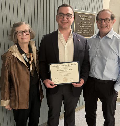
We are proud to congratulate Emile Vieta, MD, on completing the rigorous two-year board certification program in medical genetics through the UCLA Intercampus Medical Genetics Training Program. This highly demanding program covers all aspects of genetics and requires a deep understanding of genetics, internal medicine, and pediatrics.
Dr. Vieta excelled in his clinical training, earning praise from his mentors for his ability to perform effectively in all his clerkships. His achievement is noteworthy—Dr. Vieta is one of the few physicians in the country to be board certified in both medical genetics and ophthalmology.
We are excited to see how Dr. Vieta will apply his specialized knowledge in genetics to improve the diagnosis and management of inherited eye disorders, further enhancing the care we provide to our patients.

The Doheny Eye Center UCLA–Pasadena has relocated and moved to the Doheny Eye Institute campus. Centrally located, the new 15,000square-foot space serves as a vibrant hub of innovation for the treatment of vision-threatening eye conditions, cutting edge research, and education excellence.
HIGHLIGHTS INCLUDE:
f 36 fully equipped exam rooms
f 2 convenient in-office procedure rooms
f 3 advanced laser and injection rooms
f 3 certified clinical research rooms
f A state-of-the-art imaging center
f A dedicated pediatric ophthalmology suite
f A dedicated ocular plastics, cataract, and refractive suite.
150 N. Orange Grove Blvd. Suite 1400 Pasadena, CA 91103 (626) 817-4747
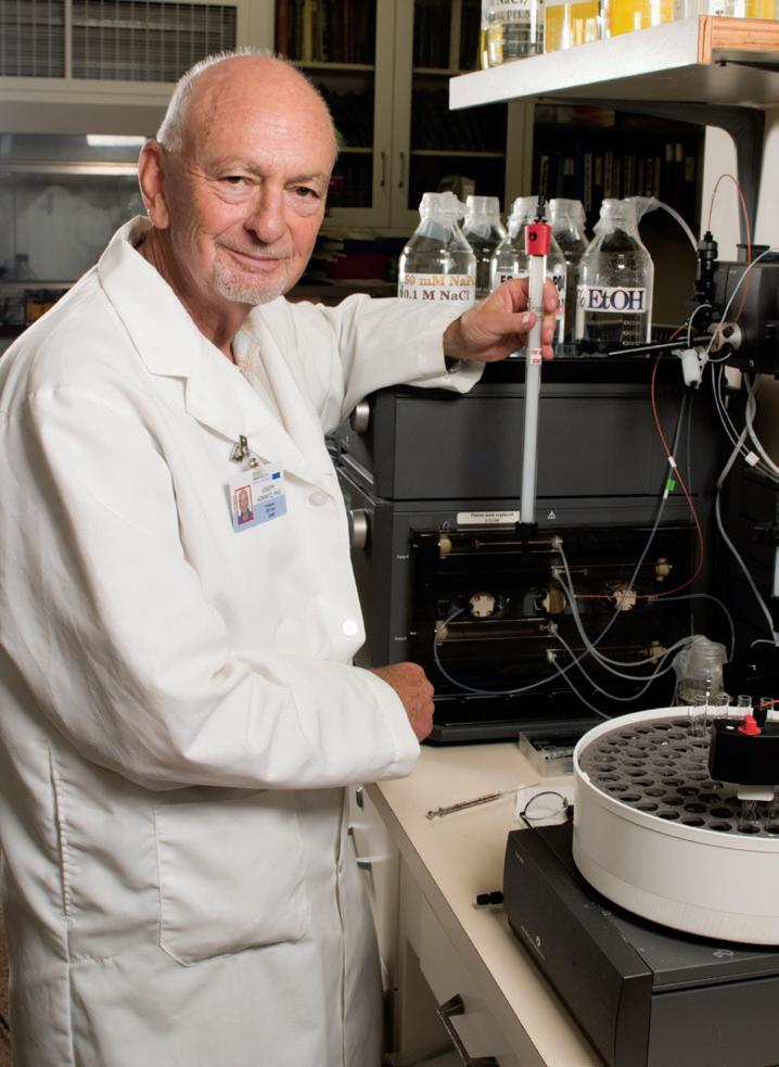
Dr. Joseph Horwitz, a distinguished researcher and beloved member of the UCLA Department of Ophthalmology, passed away on October 23, 2024, at the age of 88. Renowned for his groundbreaking work in vision science, Dr. Horwitz leaves behind a legacy of scientific innovation, mentorship, and warmth that profoundly impacted his colleagues and students.
A graduate of UCLA, Dr. Horwitz completed his undergraduate degree in physics, followed by a graduate degree in biophysics and a research fellowship at the Laboratory of Nuclear Medicine Radiation Biology. He joined the faculty of the Department of Ophthalmology in 1971, where he quickly became a pivotal figure, serving as the associate director of research from 1980 to 1994 and was founding co-chief of the Vision Science Division from 1981 to 1994.
Dr. Horwitz’s research led to significant advancements in understanding the biochemical and biophysical properties of normal and cataractous lens proteins. In 1992, he made a landmark discovery regarding alpha-crystallin, a key structural component of the lens, demonstrating its ability to slow protein deterioration. His collaborative efforts culminated in further insights into the molecular structure of alpha-crystallin in 1998, earning him the Senior Scientific Investigator Award from Research to Prevent Blindness.
Over his illustrious career, Dr. Horwitz, former Oppenheimer Brothers Chair and emeritus professor of ophthalmology, published 174 manuscripts and secured 14 National Institutes of Health (NIH) grants. He also served on several NIH committees, including chair of the Cataract Panel Vision Research National Plan.
Among his many accolades, Dr. Horwitz received the Proctor Medal from the Association for Research in Vision and Ophthalmology in 1992, the Alcon Award in 1984, and an NIH MERIT Award recognizing his laboratory’s impact on eye research.
Beyond his professional achievements, Dr. Horwitz was a devoted partner to his wife, Miriam, who was mother of his two children, Leora and Dan. He was also blessed in his marriage to Arlene and with his late-life partner, Hilda. He cherished his three grandsons, Elliott, Sawyer, and Max, fostering in them a love for travel and science.
Dr. Horwitz’s candor, humor, and passion for teaching made him a cherished mentor and friend. His contributions to vision science will be felt for generations, and the UCLA community mourns his passing while celebrating a life well-lived.
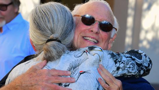
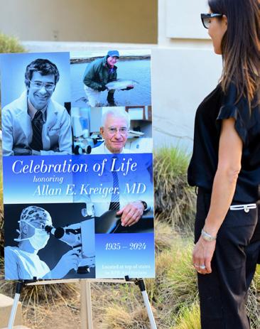
Faculty, friends, alumni, colleagues, and family gathered at the Jules Stein Eye Institute on September 4, 2024, to remember Dr. Allan “Buzz” Kreiger, the first faculty member of the Retina Division and its founding chief.
In-person speakers Drs. Anne L. Coleman, Gary N. Holland, Colin A. McCannel, Bartly J. Mondino, Pradeep S. Prasad , and Steven D. Schwartz, along with video tributes from countless others, spoke to the enormous impact Buzz had professionally on the field of ophthalmology and personally on his peers and those he mentored. The memorable evening touched on all those present, evoking both laughter and tears as stories and cherished memories celebrated the life and enduring spirit of a man whose life impacted so many.
If you would like to make a donation in memory of Dr. Kreiger, please go to giving.ucla.edu/ Kreiger or contact the Development Office at (310) 825-3381 or giving@jsei.ucla.edu. All donations will be directed to the Kreiger Retinal Support for Medically Underserved Populations Fund.
Our goal is to raise $1 million by June 30, 2025. If we reach this goal, the fund will be repurposed to establish the Allan E. Kreiger, M.D., Endowed Chair in Retinal Diseases. If the goal is not met, the Kreiger Retinal Support for Medically Underserved Populations Fund will remain an endowed fund, supporting the screening and treatment of retinal diseases in medically underserved populations. Please review UCLA and the UCLA Foundation’s Disclosure Statements for Prospective Donors at: www.uclafoundation.org/disclosures


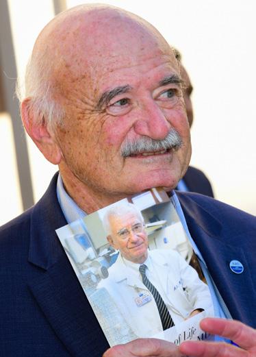

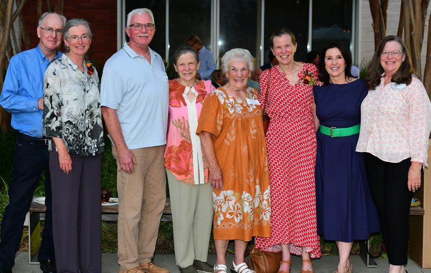
UCLA Department of Ophthalmology faculty took center stage at the annual American Academy of Ophthalmology (AAO) meeting, presenting over 100 educational events over the four-day meeting. The proceedings were also available virtually, on demand.
The AAO annual meeting—held October 18 to October 21, 2024, in Chicago, Illinois—is a premier event in ophthalmic education, drawing participants from across the globe. It includes lectures, instructional courses, and other presentations, showcasing the most recent developments in ophthalmology.
Congratulations to our faculty who were honored at the AAO annual meeting. Drs. Federico G. Velez and Stacy L. Pineles received Senior Achievement Awards. Dr. Simon K. Law was a Secretariat Awardee, and Dr. Robert A. Goldberg was the Wendell L. Hughes Lecturer.
Joseph L. Demer, MD, PhD, Arthur L. Rosenbaum, MD, Chair in Pediatric Ophthalmology, received a National Eye Institute (NEI) research project (R01) grant for his project, “Biomechanical Analysis in Strabismus Surgery.” This project, which has been funded continuously for 38 years, studies the role of the extraocular muscles that play complex roles in ocular rotation and alignment, and must mechanically balance against the eyeball, optic nerve, and suspensory tissues.

An R01 grant is the National Institutes of Health (NIH) most used grant program for independent research projects.
Yi-Rong Peng, PhD, assistant professor of ophthalmology and neurobiology, secured two prestigious research grants.
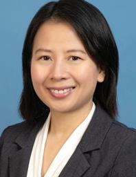
The first, an NEI R01 grant, will fund her project to elucidate control of neuronal position and connection in the retina. The results from this proposal will provide insight into understanding the general organization of the nervous system and shed light on therapeutic strategies to recover visual defects caused by errors in retinal circuit formation.
The second, a competitive seed grant received from the Brain Research Foundation, will fund a project to generate comprehensive measurements of the membrane proteins that construct the retinal circuit and those that change during retinal degeneration. These resources will facilitate the discovery of molecular mechanisms underlying circuit functions in visual processing and circuit remodeling in retinal degeneration.
Alapakkam P. Sampath, PhD, Grace and Walter Lantz Endowed Chair in Ophthalmology, received a four-year, multiprincipal investigator R01 grant with Kirill Martemyanov, PhD, from the Scripps Institute, University of Florida, to uncover the molecular mechanisms allowing photoreceptors to organize their synaptic contacts with downstream retinal neurons. Understanding how these connections are established will provide insights into strategies to treat blinding diseases.
Edmund Tsui, MD, MS, assistant professor of ophthalmology, received an NEI R13 grant to fund in part the second UCLA/American Uveitis Society International Workshop on Objective Measures of Intraocular Inflammation for Use in Clinical Trials held at UCLA on September 27–28, 2024. The funding provided 15 travel grants to trainees and early career researchers to attend the workshop.


Yuhua Zhang, PhD, professor of ophthalmology, is among a multidisciplinary group of scientists to receive a $4.7M annual three-year award from the NIH Common Fund Venture Program Oculomics Initiative. The award is to be used by the collaborating researchers to develop a noninvasive ocular imaging approach that can detect and characterize cerebral small vessel disease, a common nervous system disorder that causes failures of blood flow and contributes to vascular cognitive impairment and dementia.


Brian A. Francis, MD, MS, The Rupert and Gertrude I. Steiger Vision Research Chair, and John A. Irvine, MD, health sciences clinical professor, have been selected by unanimous vote of the DEC-UCLA faculty as the Doheny Eye Center UCLA Co-Medical Directors, effective January 2, 2025.
Kouros Nouri-Mahdavi, MD, MSc, Kay K. Pick Endowed Chair in Glaucoma Research, presented the KapetanskyAllergan lecture at the 46th Annual Meeting of the Midwest Glaucoma Society on November 9, 2024, in Louisville, Kentucky.
Pradeep S. Prasad, MD, MBA, health sciences associate clinical professor of ophthalmology, was inducted into the Retina Society on September 14, 2024, in Lisbon, Portugal.
Peter A. Quiros, MD, health sciences clinical professor of ophthalmology, was named assistant division chief of the Neuro-Ophthalmology Division, effective July 1, 2024.
SriniVas R. Sadda, MD, professor of ophthalmology, was named A. Ray Irvine, Jr., MD, Endowed Chair in Clinical Ophthalmology, effective August 7, 2024.
Gabriel H. Travis, MD, Charles Kenneth Feldman Chair in Ophthalmology, received
the Endre A. Balazs Prize on October 21 at the 2024 Biennial Meeting of the International Society for Eye Research (ISER) in Buenos Aires, Argentina, and he delivered the plenary lecture, “Photic mechanisms of visual pigment regeneration in vertebrates.” The award recognizes Dr. Travis’ outstanding contributions in the field of experimental eye research.
Federico G. Velez, MD, Leonard Apt Endowed Chair in Pediatric Ophthalmology, was the Pediatric Ophthalmology and Strabismus Network (POSN) Keynote Lecturer in Strabismus at the POSN meeting in Bengaluru, India, on September 21, 2024.
In addition, Dr. Velez presented the Emilio Campos Inaugural Lecture on September 28, 2024, during the Italian Association of Strabismus Meeting in Udine, Italy.
Dr. Velez was also the recipient of the Jules Stein Eye Institute’s Annual Golden Eye Award. The award recognizes tremendous surgical capabilities, as well as the ability to collaborate exceptionally with the operating room (OR) team. The award voting committee is comprised of OR nurses, scrub techs, and staff.
Dr. Aldave Receives a First for
Department of Ophthalmology
Anthony J. Aldave, MD, Bartly J. Mondino, MD, Endowed Chair in Ophthalmology, the principal investigator of a gene therapy program for congenital hereditary endothelial dystrophy (CHED), was granted both Orphan Drug Designation and Rare Pediatric Disease Designation status by the U.S. Food and Drug Administration (FDA), firsts for an investigator in the UCLA Department of Ophthalmology.
Dr. Aldave leads the Jules Stein Eye Institute Cornea Genetics Laboratory, which has been chosen to participate in the Foundation for the National Institutes of Health Accelerating Medicines Partnership Bespoke Gene Therapy consortium, and his laboratory received funding from the California Institute for Regenerative Medicine to study how gene therapy could treat CHED, a genetic condition that causes blindness in infants and children. The results of this research will be used to apply for approval to begin human clinical trials, taking the gene therapy closer to becoming a potential treatment for CHED.
Michael D. Ip, MD, Gavin S. Herbert Endowed Chair for Macular Degeneration, has been honored with membership in the American Ophthalmological Society (AOS), one of the most exclusive and prestigious societies in the field of ophthalmology.
Founded in 1864, the AOS is the second-oldest medical specialty society in the United States, and its membership is limited to 225 ophthalmologists. Candidates must be board certified by the American Board of Ophthalmology and demonstrate a proven track record of excellence in one or more areas: clinical practice, scientific research, education, administration, or public service.
Candidates must have published a minimum of six articles in major, peer-reviewed journals in the five years preceding their nomination. Once nominated, they are required to present a scholarly thesis to the society, which, if approved, is published in the American Journal of Ophthalmology. This process ensures that only ophthalmologists with a significant and sustained impact on the field are granted membership.
At the society’s spring session, Dr. Ip presented his original research on imaging findings and treatment outcomes following anti-VEGF therapy for macular edema due to retinal vein occlusion.
The UCLA Department of Ophthalmology boasts an impressive roster of AOS members, including Drs. Anthony C. Arnold, Joseph Caprioli, Anne L. Coleman, Robert A. Goldberg, Gary N. Holland, Allan E. Kreiger (deceased), Colin A. McCannel, Kouros Nouri-Mahdavi, SriniVas R. Sadda, Alfredo A. Sadun, David Sarraf, and Bradley R. Straatsma
Health Sciences
Assistant Clinical Professor
education, Dr. Fein enjoys teaching medical students, residents, and fellows.
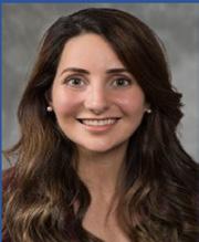
Dr. Fayad is a pediatric ophthalmologist and strabismus specialist. She completed her medical residency at New York Eye and Ear Infirmary of Mount Sinai followed by a pediatric ophthalmology and adult strabismus fellowship at the UCLA Stein Eye Institute. She specializes in the evaluation and management of strabismus and ocular muscle disorders, amblyopia, pediatric cataracts, pediatric glaucoma, lacrimal disorders, and more. She is chief of the Pediatric Ophthalmology Service at Harbor-UCLA Medical Center, where she teaches medical students, ophthalmology residents, and fellows. Academic interests include surgical outcomes in strabismus surgery and disparities in access to pediatric eye care.
LOCATIONS: Doheny Eye Center UCLA–Orange County, UCLA Stein Eye Institute in Westwood, and Harbor-UCLA Medical Center in Torrance.
Health Sciences
Assistant Clinical Professor
Dr. Fein is a faculty member in the NeuroOphthalmology Division and sees patients with a breadth of neuroophthalmic complaints.
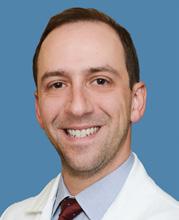
He earned his medical degree from SUNY Upstate Medical University in New York. After his internship at Mount Sinai in New York City, he completed his neurology residency at New York University (NYU), where he was a chief resident for education. Dr. Fein stayed on at NYU for his fellowship in neuro-ophthalmology. During his training, he became particularly interested in optic neuritis, optic neuropathies, and nystagmus. Dr. Fein is a member of the North American Neuro-Ophthalmology Society and serves on multiple professional committees. With a background as a science educator and with a passion for medical
LOCATIONS: UCLA Stein Eye Institute in Westwood and Doheny Eye Center UCLA–Pasadena.
Joan and Jerome Snyder Chair in Cornea Diseases
Professor of Ophthalmology
Dr. Malyugin specializes in cataract surgery and the treatment of corneal diseases.
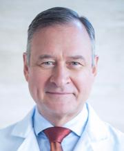
Born in Moscow, he was trained and worked at the Fyodorov Eye Microsurgery Institution. He has developed innovative surgical techniques and devices, including the “Malyugin Ring,” a device which temporarily stretches and expands the pupil during cataract surgery. He is highly regarded for development of complex cataract surgery techniques, secondary intraocular lens implantation, keratoplasty techniques, and ocular surface reconstruction.
His research has been featured in peer-reviewed journals, ophthalmic surgery textbooks, and atlases. He is an active member of leading ophthalmic organizations and serves on the editorial boards of several ophthalmology journals.
LOCATIONS: UCLA Stein Eye Institute in Westwood and Stein Eye Institute–Santa Monica.
Health Sciences
Associate Clinical Professor
Chief, Comprehensive Ophthalmology Division
Dr. Sand is the new chief of the Comprehensive Ophthalmology Division and practices a full scope of comprehensive ophthalmology and cornea/refractive surgery.
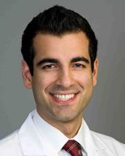
He attended medical school at Case Western Reserve University in Ohio and was elected into the Alpha Omega Alpha Honor Medical Society. His internship was
at Olive View-UCLA Medical Center and his residency at the Doheny Eye Institute/ University of Southern California. He did his cornea, external disease, and refractive surgery training as a Heed Ophthalmic Foundation Fellow at the University of Michigan, Kellogg Eye Center and completed an eye banking fellowship as the inaugural Eversight Eye Bank Fellow. He continues his involvement with the Eye Bank Association of America as a member of multiple national committees.
LOCATION: UCLA Stein Eye Institute in Westwood.
Health Sciences
Assistant Clinical Professor Dr. Qin was raised in New Jersey, where she graduated from Princeton University summa cum laude with a degree in molecular biology and a certificate in global health and health policy.

She earned her MD at Case Western Reserve University School of Medicine in Cleveland, Ohio, and graduated with AOA medical society honors. She spent her medical intern year at Lankenau Hospital in Philadelphia and conducted her ophthalmology residency at Scheie Eye Institute at the University of Pennsylvania. Dr. Qin completed a fellowship in glaucoma at University of California, San Francisco and is excited to be bringing her passion for glaucoma care and medical education to UCLA.
LOCATIONS: Doheny Eye Center UCLA locations in Pasadena and Arcadia.

The Aesthetic Eyelid and Facial Rejuvenation course, chaired by Robert A. Goldberg, MD, was held at the Jules Stein Eye Institute July 12–13, 2024. The course showcases the work of UCLA faculty, and presents safe, innovative, and minimally invasive techniques. With attendees from throughout the world, the laboratory dissection day concentrated on anatomic and surgical pearls of core aesthetic procedures and included a live audiovisual feed of course faculty performing high definition dissections. The second day featured lectures and discussions highlighting conceptual approaches, nuances of facial rejuvenation, and new surgical and nonsurgical techniques for facial rejuvenation. A highlight of the twoday event was Jocelyn Kohn, MD, presenting the Shorr Lecture. Course directors were Daniel B. Rootman, MD, and Jonathan A. Hoenig, MD
For information about upcoming orbital and ophthalmic plastic surgery courses, email Dr. Justin Karlin at jkarlin@ mednet.ucla.edu
The Second UCLA/American Uveitis Society International Workshop on Objective Measures of Intraocular Inflammation for Use in Clinical Trials was held at the Jules Stein Eye Institute on September 27–28, 2024.
The workshop had over 100 attendees, including uveitis specialists, clinical trialists, the FDA, and industry representatives. Michael F. Chiang, MD, the director of the National Eye Institute, delivered the keynote lecture, “Pushing Boundaries through Data Science, Standards, and Collaboration: Perspectives from the National Eye Institute.”
Throughout the two-day event participants presented their latest research findings, sharing novel methodologies and insights into quantification of intraocular inflammation. Attendees discussed clinically relevant objective outcome measures proposed for use in the evaluation of uveitis and explored their relevance to clinical trial design.
The workshop was co-organized by Gary N. Holland, MD, SriniVas R. Sadda, MD, and Edmund Tsui, MD, MS, and co-sponsored by the American Uveitis Society. Dr. Tsui was also awarded a National Eye Institute R13 grant, which provided 15 travel grants to trainees and early career researchers to attend the workshop.
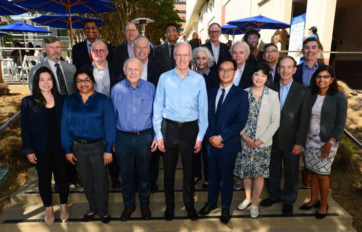
Top: The 2024 UCLA/American Uveitis Society International Workshop Organizing Committee, Scientific Program Committee, and featured speakers.
Bottom: Dr. Gary Holland (left) and Dr. Edmund Tsui.

The Annual Women in Ophthalmology (WIO) Meeting took place in Carlsbad, California, from August 22–25, 2024, and drew a record number of attendees from UCLA’s Department of Ophthalmology. Faculty, residents, medical students, alumni, and staff gathered for this dynamic event, which provided a valuable opportunity to engage in professional development and wellness activities outside the clinic.
The meeting attracted a diverse group of ophthalmologists from across the country, representing a wide range of subspecialties, academic institutions, and private practices. It offered a unique space for networking, sharing insights, and fostering collaboration among peers in the field.
UCLA faculty members played a prominent role throughout the event. Dr. Mitra Nejad chaired several stage sessions. Dr. Irena Tsui moderated engaging poster sessions, which featured a presentation by UCLA Resident Dr. Weinlin Song. In addition, Dr. Tara McCannel debuted a breakout session entitled, “Acupuncture and Ear Seeding.”
The meeting also supported inclusive excellence initiatives, which aim to advance gender equity and increase the representation of women in ophthalmology.
Overall, the 2024 WIO Meeting proved to be an enriching experience for all involved, reinforcing the importance of mentorship, wellness, and inclusivity in shaping the future of ophthalmology.
On Saturday, September 28, the 6th Annual Doheny-UCLA International Glaucoma Symposium drew more than 80 ophthalmic professionals and researchers to explore the latest advancements in glaucoma research and clinical care. Organized by Dr. Vikas Chopra and Dr. Brian Francis, the event featured a distinguished lineup of speakers, including Dr. Kuldev Singh from Stanford, who delivered the prestigious Donald Minckler Doheny Glaucoma Lecture, and Dr. Teresa C. Chen from Harvard, who joined as a special guest speaker. Dr. Donald Minckler, a renowned figure in glaucoma research and care, was also in attendance.
The symposium covered a wide range of critical topics, including the mechanisms of glaucoma progression, cutting-edge treatment modalities, innovative surgical techniques, and breakthroughs in diagnostic imaging.
A unique and forward-thinking workshop at Stanford University on October 12–13, 2024, brought together neuroscientists and artificial intelligence (AI) experts to explore how AI can revolutionize neuroscience research. The event was organized by Greg D. Field, PhD, Joan and Jerome Vision Science Chair, Jules Stein Eye Institute, and E.J. Chichilnisky, PhD (Stanford), and was led by Allison Duettmann, CEO of the Foresight Institute.
The workshop’s intent was to learn and discuss how to embrace AI ethically and creatively in science and education, and it offered a hands-on, collaborative environment for attendees to experiment with AI tools in real time. Rather than traditional talks, the workshop encouraged participants to work on practical scientific challenges using AI. Sessions included AI for Coding; AI for Literature and Networking; AI for Writing; and AI for Creativity, Thinking and (Experimental) Design.
The workshop highlighted the importance of interactive learning and ethical reflection in preparing neuroscientists for an AI-driven future, reinforcing the need for both innovation and responsibility in the integration of AI into scientific discovery.
Dr. Kevin M. Miller, chief of the Cataract and Refractive Surgery Division, presented the annual Cataract Surgery Essentials Course in conjunction with Bausch & Lomb on November 2, 2024, in Newport Beach, California.
The course featured labs and workstations where approximately 60 trainees from UCLA, USC, UCI, Loma Linda, UCSD, and the Naval Medical Center learned about essential surgical instruments and equipment used in cataract surgery. Dr. Miller’s next course covering advanced cataract surgery topics will be in Irvine, California, on April 5, 2025.
The Doheny Eye Institute hosted its inaugural Oculomics Symposium on November 23, 2024, a groundbreaking event organized by Yuhua Zhang, PhD, principal investigator at Doheny Eye Institute and professor of ophthalmology at the UCLA Department of Ophthalmology.
Oculomics is a rapidly evolving field that investigates how changes in the eye—specifically ophthalmic biomarkers—can reveal critical insights into overall health. Dr. Zhang brought together experts from institutions such as UCLA, Caltech, and Bascom Palmer Eye Institute to explore the cutting-edge intersection of ophthalmology, imaging technology, artificial intelligence, and systemic disease detection through ocular biomarkers.
The event featured a diverse range of topics, including advanced imaging techniques, retinal pathophysiology, and the role of ocular biomarkers in detecting cerebral small vessel disease. Distinguished speakers included Dr. Liang Gao and Dr. Tzung Haisi from UCLA Bioengineering, Dr. Michael Ip from Doheny-UCLA, and Dr. Jianhua Wang from Bascom Palmer.
Dr. Zhang emphasized the importance of future collaboration to propel oculomics research forward, advancing our understanding of systemic health and the potential for early disease detection through the eye. The inaugural symposium highlighted the promise of oculomics and set the stage for continued innovation and cooperation in this transformative field.
Doheny Eye Institute kicked off its 2024–25 Distinguished Lecture Series with a presentation from David Williams, PhD, from the University of Rochester, on December 13, 2024.
The Distinguished Lecture Series features presentations from renowned vision researchers sharing groundbreaking insights into visual science and eye health. Dr. Williams is a prominent scientist and professor, renowned for his groundbreaking work in the field of adaptive optics, a technology originally developed for astronomy that he applied to improve our understanding of the human eye and visual system.
The lecture series can be joined via Zoom. For information on how to attend and future speakers, email Yolanda Mercado at YMercado@doheny.org
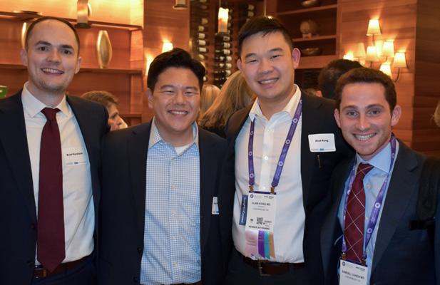
Alumni from the Jules Stein Eye Institute and the Doheny Eye Institute gathered for a lively social event at the 2024 American Academy of Ophthalmology Annual Meeting. It was a pleasure to reconnect with so many familiar faces and celebrate the achievements of our accomplished alumni. The event provided an invaluable opportunity for alumni and colleagues to share experiences, exchange ideas, and discuss advancements in the field.
A highlight of the evening was a special performance by DJ AJA, also known as Dr. Anthony Aldave, who added a fun and unique touch to the gathering.
Building on the success of last year’s collaboration, the Doheny Eye Institute and the Jules Stein Eye Institute once again joined forces to showcase our groundbreaking research and clinical innovations at a shared booth. The booth attracted more than 1,000 visitors, including prospective residents, fellows, and faculty members who had an opportunity to learn more about our programs. This engagement helps to spread our mission and vision, and fosters connections across the country and globe.
A heartfelt thank you to everyone who attended and made this event both memorable and impactful.


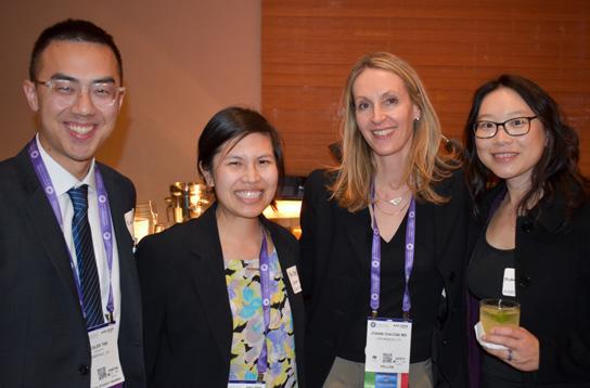

The Jules Stein Eye Institute family played a major role in the success of the Asia Pacific Society of Ophthalmic Plastic and Reconstructive Surgery (APSOPRS) meeting held November 29–20, 2024, in Seoul, Korea.
Highlights included Robert A. Goldberg, MD, Bert O. Levy Endowed Chair in Orbital and Ophthalmic Plastic Surgery, delivering the Keynote Lecture; fellow alumni Helen Lew, MD (’09), and Yoon-Duck Kim, MD, PhD (’94), serving as program chairs and local hosts; and international fellowship alumni Kam-Lung “Kelvin” Chong, MD (’10), Chee-Chew Yip, MD (’03), Milind Naik, MD (’07), Alice Goh, MD (’13), Bird Putthirangsiwong, MD, (’19), and Tomoyuki Kashima, MD (’16) presenting papers in the scientific session. Notably, in the elections to the board of APSOPRS, four fellow alumni were nominated to executive positions, including incoming president Dr. Kelvin Chong
International meetings allow Department of Ophthalmology alumni to spend time together as a family and emphasize the important role that our fellowship diaspora plays in guiding our discipline internationally.


The UCLA Mobile Eye Clinic (UMEC) is pleased to unveil a newly redesigned bus wrap, made possible by the generous support of The Douglas Foundation and the Wilbur May Foundation. In addition to the refreshed design, the Foundations are continuing their commitment to funding our efforts to provide vision screenings and examinations for children from preschool through high school. We are profoundly grateful to our donors, whose ongoing contributions are instrumental in advancing our mission to deliver vital eye care services to underserved communities throughout Los Angeles.
The 2024 Visiting Medical Student Scholarship in Ophthalmology was awarded to Chioma Amuzie. Chioma came to UCLA for three weeks in fall 2024 to complete her rotation in ophthalmology as part of the Visiting Student Learning Opportunity. During her time at UCLA, Chioma—a fourth-year medical student at the Miller School of Medicine at the University of Miami—explored how to best serve people whose access to ophthalmic services is limited.
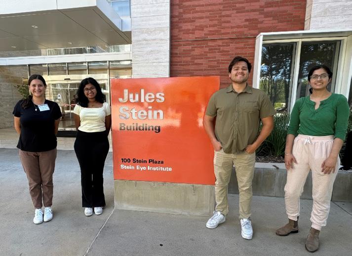
The Diversity in Vision Science Undergraduate 2024 Summer Research Program hosted four exceptional undergraduate students at Jules Stein Eye Institute research laboratories. Brianna Burns, a junior at the University of Alabama, joined Dr. Anthony J. Aldave’s laboratory. Kiara Abhayaratne, a junior at University of California, Berkeley, elected to conduct investigations in Dr. Greg D. Field’s laboratory. Dragui Salazar, a junior at the Utah Valley University, worked with Dr. Sophie Deng’s team, and Baani Sabharwal, a junior at the University of California, Berkeley, conducted research in Dr. Roxana Radu’s laboratory. The students’ research was presented at the end of the program during the UCLA Sponsored Projects for Undergraduate Research Showcase.
The students were selected out of 14 candidates by the Diversity Award Committee. The Diversity in Vision Science Undergraduate Summer Research Program aims to inspire and offer opportunities for students from disadvantaged or diverse backgrounds to explore ophthalmology and engage in research.
Clémence Bonnet, MD, PhD, health sciences assistant clinical professor, is the recipient of a one-year Justice, Equity, Diversity, and Inclusion “JAM” fellowship from the David Geffen School of Medicine at UCLA. The JAM fellowship has been specifically developed for faculty at the assistant professor level who demonstrate potential for careers in academic medicine and health leadership.
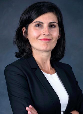
The JAM Fellowship provides comprehensive mentoring training, including extensive coaching, networking, and mentoring opportunities, with the goal of advancing the professional development and retention of diverse faculty in academic medicine.
Previous recipients of the JAM fellowship include Drs. Simon Fung and Edmund Tsui

405 Hilgard Avenue
Box 957000, 100 Stein Plaza
Los Angeles, California, 90095-7000
U.S.A.
Forwarding Service Requested
UCLA Stein Eye Institute, Westwood
100 Stein Plaza, UCLA Los Angeles, CA 90095
Referral Service: (310) 794-9770
Emergency Service: (310) 825-3090
After-Hours Emergency Service: (310) 825-2111 uclahealth.org/eye
Stein Eye Center–Calabasas 26585 W. Agoura Rd., Suite 330 Calabasas, CA 91302 (310) 825-5000
Stein Eye Center–Santa Monica 1807 Wilshire Blvd., Suite 203 Santa Monica, CA 90403 (310) 829-0160
Doheny Eye Center UCLA–Arcadia 622 W. Duarte Rd., Suite 101 Arcadia, CA 91007 (626) 254-9010
Doheny Eye Center UCLA–Orange County
Orange Coast Memorial Medical Center 18111 Brookhurst St., Suite 6400 Fountain Valley, CA 92708 (714) 963-1444
Doheny Eye Center UCLA–Pasadena
Doheny Eye Institute 150 N. Orange Grove Blvd., Suite 1400 Pasadena, CA 91103 (626) 817-4747


Alumni Relations
Email: alumni@jsei.ucla.edu
Philanthropy
Jules Stein Eye Institute Development Office 100 Stein Plaza, UCLA, Room 3-138 Los Angeles, CA 90095-7000
Telephone: (310) 206-6035
Email: giving@jsei.ucla.edu
Volunteer Opportunities
Center for Community Outreach & Policy www.uclahealth.org/departments/ eye/mobile-eye Telephone: (310) 825-2195 Email: community@jsei.ucla.edu facebook.com/uclamobileyeclinic instagram.com/uclamobileyeclinic twitter.com/uclaMEC
Read past issues of EYE at: www.uclahealth.org/Eye/news
Send comments or questions to: Tina-Marie Gauthier
Managing Editor Email: Tina@EyeCiteEditing.com