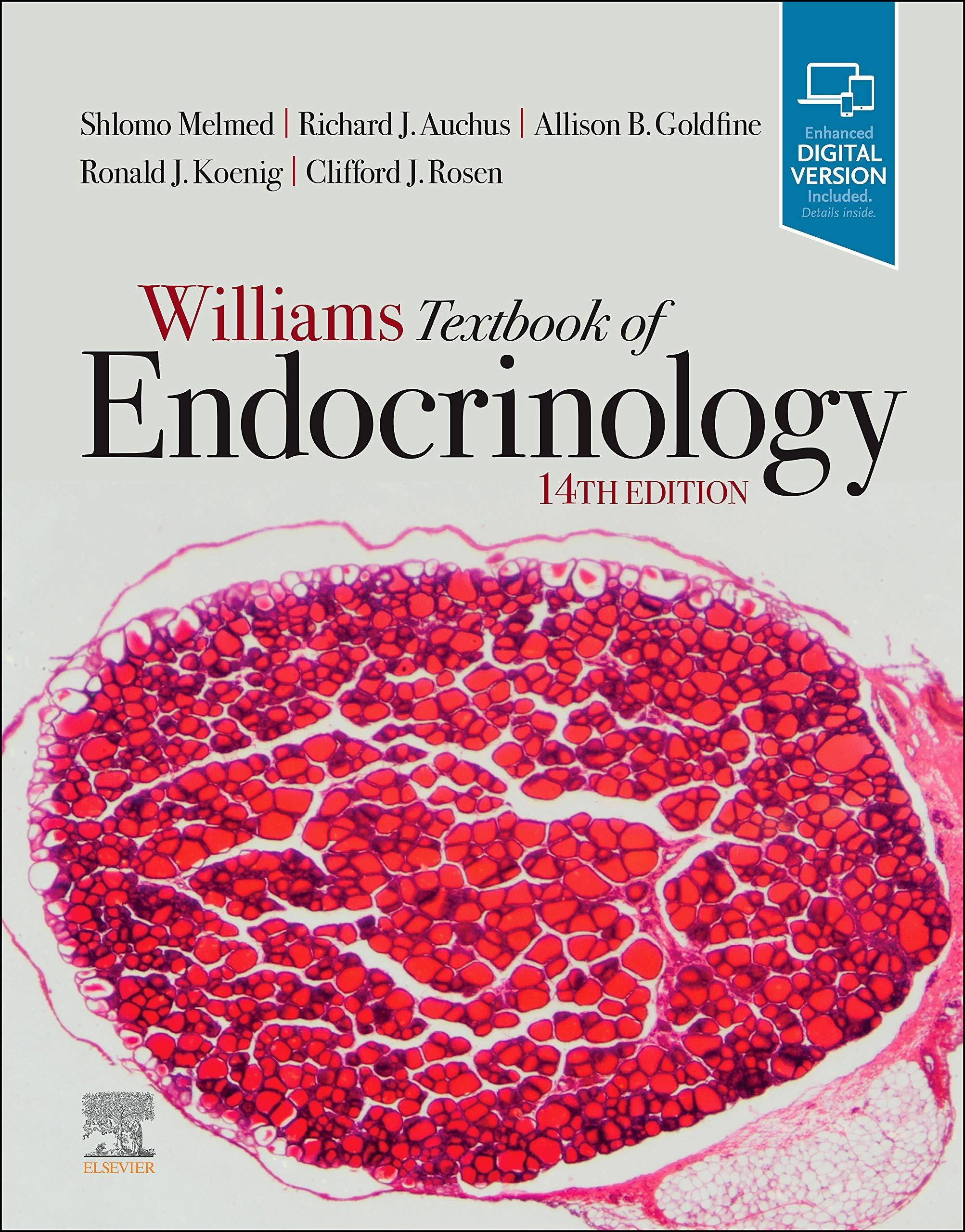
Williams Textbook of Endocrinology
14TH EDITION
Shlomo Melmed, MB ChB, MACP
Executive Vice President and Dean of the Medical Faculty
Cedars-Sinai Medical Center
Los Angeles, California
Richard J. Auchus, MD, PhD
Professor
Departments of Pharmacology and Internal Medicine
Division of Metabolism, Endocrinology, and Diabetes University of Michigan
Endocrinology Service Chief
Ann Arbor VA Healthcare System
Ann Arbor, Michigan
Allison B. Goldfine, MD
Associate Physician, Division of Endocrinology, Diabetes, and Hypertension
Brigham and Women’s Hospital
Lecturer, Part-Time
Harvard Medical School
Boston, Massachusetts
Director, Translational Medicine Cardiometabolic Disease
Novartis Institute of Biomedical Research
Cambridge, Massachusetts
Ronald J. Koenig, MD, PhD
Professor
Department of Internal Medicine
Division of Metabolism, Endocrinology, and Diabetes University of Michigan
Ann Arbor, Michigan
Clifford J. Rosen, MD
Professor of Medicine
Tufts University School of Medicine Boston, Massachusetts
Elsevier
1600 John F. Kennedy Blvd.
Ste 1800 Philadelphia, PA 19103-2899
WILLIAMS TEXTBOOK OF ENDOCRINOLOGY, FOURTEENTH EDITION
Copyright © 2020 by Elsevier, Inc. All rights reserved.
ISBN: 978-0-323-55596-8
No part of this publication may be reproduced or transmitted in any form or by any means, electronic or mechanical, including photocopying, recording, or any information storage and retrieval system, without permission in writing from the publisher. Details on how to seek permission, further information about the Publisher’s permissions policies and our arrangements with organizations such as the Copyright Clearance Center and the Copyright Licensing Agency, can be found at our website: www.elsevier.com/permissions.
This book and the individual contributions contained in it are protected under copyright by the Publisher (other than as may be noted herein).
Notice
Practitioners and researchers must always rely on their own experience and knowledge in evaluating and using any information, methods, compounds or experiments described herein. Because of rapid advances in the medical sciences, in particular, independent verification of diagnoses and drug dosages should be made. To the fullest extent of the law, no responsibility is assumed by Elsevier, authors, editors or contributors for any injury and/or damage to persons or property as a matter of products liability, negligence or otherwise, or from any use or operation of any methods, products, instructions, or ideas contained in the material herein.
Previous editions copyrighted 2016 by Elsevier Inc. and 2011, 2008, 2003, 1998, 1992, 1985, 1981, 1974, 1968, 1962, 1955, 1950 by Saunders, an affiliate of Elsevier Inc.
Copyright renewed 1990 by A.B. Williams, R.I. Williams
Copyright renewed 1983 by William H. Daughaday
Copyright renewed 1978 by Robert H. Williams
Library of Congress Control Number: 2019952358
Senior Content Strategist: Nancy Duffy
Senior Content Development Specialist: Rae Robertson
Publishing Services Manager: Catherine Jackson
Senior Project Manager: Claire Kramer
Design Direction: Amy Buxton
Printed in Canada
Last digit is the print number: 9 8 7 6 5 4 3 2 1
Contributors
John C. Achermann, MB, MD, PhD
Wellcome Trust Senior Research Fellow in Clinical Science and Professor of Pediatric
Endocrinology
Department of Genetics and Genomic Medicine
UCL GOS Insititute of Child Health
University College London London, Great Britain
Andrew J. Ahmann, MD, MS
Professor of Medicine
Division of Endocrinology, Diabetes, and Clinical Nutrition
Director, Harold Schnitzer Diabetes Health Center
Oregon Health & Science University Portland, Oregon
Lloyd P. Aiello, MD, PhD
Professor of Ophthalmology
Harvard Medical School
Director, Beetham Eye Institute
Joslin Diabetes Center Boston, Massachusetts
Erik K. Alexander, MD
Chief, Thyroid Section
Brigham and Women’s Hospital
Professor of Medicine
Harvard Medical School
Boston, Massachusetts
Rebecca H. Allen, MD, MPH
Associate Professor
Department of Obstetrics and Gynecology
Warren Alpert Medical School of Brown University
Providence, Rhode Island
Bradley D. Anawalt, MD
Professor and Vice Chair
Department of Medicine
University of Washington
Seattle, Washington
Mark S. Anderson, MD, PhD
Professor
Diabetes Center
University of California, San Francisco
San Francisco, California
Ana María Arbeláez, MD, MSCI
Associate Professor
Department of Pediatrics
Washington University School of Medicine
Pediatrician, St. Louis Children’s Hospital
St. Louis, Missouri
Mark A. Atkinson, PhD
American Diabetes Association Eminent Scholar for Diabetes Research
Departments of Pathology and Pediatrics
Director, University of Florida Diabetes Institute
Gainesville, Florida
Richard J. Auchus, MD, PhD
Professor
Departments of Pharmacology and Internal Medicine
Division of Metabolism, Endocrinology, and Diabetes
University of Michigan
Endocrinology Section Chief
Ann Arbor VA Healthcare System
Ann Arbor, Michigan
Jennifer M. Barker, MD
Associate Professor
Department of Pediatrics
University of Colorado
Aurora, Colorado
Rosemary Basson, MD, FRCP(UK)
Clinical Professor
Department of Psychiatry
University of British Columbia
Director, University of British Columbia
Sexual Medicine Program
British Columbia Centre for Sexual Medicine
Vancouver, British Columbia, Canada
Sarah L. Berga, MD
Professor and Director
Division of Reproductive Endocrinology and Infertility
University of Utah School of Medicine
Salt Lake City, Utah
Sanjay K. Bhadada, MBBS, MD, DM
Professor Department of Endocrinology
Nehru Hospital
Post Graduate Institute of Medical Education and Research
Chandigarh, India
Arti Bhan, MD
Division Head, Endocrinology, Diabetes, Bone and Mineral Disorders
Henry Ford Health System
Detroit, Michigan
Shalender Bhasin, MB, BS
Professor of Medicine
Harvard Medical School
Director, Research Program in Men’s Health: Aging and Metabolism
Director, Boston Claude D. Pepper Older Americans Independence Center
Brigham and Women’s Hospital
Boston, Massachusetts
Dennis M. Black, PhD
Department of Epidemiology and Biostatistics
University of California, San Francisco
San Francisco, California
Annabelle Brennan, MBBS, LLB (Hons)
Department of Obstetrics and Gynaecology
Royal Women’s Hospital
Melbourne, Australia
Andrew J.M. Boulton, MD, FACP, FRCP
Professor
Centre for Endocrinology and Diabetes
University of Manchester
Manchester, Great Britain
Visiting Professor
Division of Endocrinology, Metabolism, and Diabetes
University of Miami
Miami, Florida
Glenn D. Braunstein, MD
Professor of Medicine
Cedars-Sinai Medical Center
Professor of Medicine Emeritus
The David Geffen School of Medicine at University of California, Los Angeles
Los Angeles, California
Gregory A. Brent, MD
Professor of Medicine and Physiology
Chief, Division of Endocrinology, Diabetes, and Metabolism
The David Geffen School of Medicine at University of California, Los Angeles Los Angeles, California
F. Richard Bringhurst, MD
Physician and Associate Professor of Medicine
Endocrine Unit
Massachusetts General Hospital
Harvard Medical School
Boston, Massachusetts
Juan P. Brito, MD, MS
Associate Professor
Department of Medicine
Division of Endocrinology
Knowledge and Evaluation Research Unit in Endocrinology
Mayo Clinic Rochester, Minnesota
Todd T. Brown, MD, PhD
Professor of Medicine and Epidemiology
Division of Endocrinology, Diabetes, and Metabolism
Johns Hopkins University Baltimore, Maryland
Michael Brownlee, MD
Anita and Jack Saltz Chair in Diabetes Research Emeritus
Professor Emeritus, Medicine and Pathology
Associate Director for Biomedical Sciences
Emeritus
Einstein Diabetes Research Center
Albert Einstein College of Medicine
Bronx, New York
Serdar E. Bulun, MD
JJ Sciarra Professor of Obstetrics and Gynecology and Chair
Department of Obstetrics and Gynecology
Northwestern University Feinberg School of Medicine
Chicago, Illinois
David A. Bushinsky, MD
Professor of Medicine and of Pharmacology and Physiology
Department of Medicine
University of Rochester School of Medicine
Rochester, New York
Christin Carter-Su, ScB, PhD
The Anita H. Payne Distinguished University Professor of Physiology
The Henry Sewall Collegiate Professor of Physiology
Professor of Molecular and Integrative Physiology
Professor of Internal Medicine
University of Michigan Medical School
Associate Director
Michigan Diabetes Research Center
Ann Arbor, Michigan
Yee-Ming Chan, MD, PhD
Associate Physician
Department of Pediatrics
Division of Endocrinology
Boston Children’s Hospital
Assistant Professor of Pediatrics
Harvard Medical School
Boston, Massachusetts
Ronald Cohen, MD
Associate Professor
Department of Medicine
University of Chicago
Chicago, Illinois
David W. Cooke, MD
Associate Professor
Department of Pediatrics
Johns Hopkins University School of Medicine
Baltimore, Maryland
Philip E. Cryer, MD
Professor of Medicine Emeritus
Department of Medicine
Washington University School of Medicine
Physician
Barnes-Jewish Hospital
St. Louis, Missouri
Eyal Dassau, PhD
Director, Biomedical Systems Engineering Research Group
Senior Research Fellow in Biomedical Engineering in the Harvard John A. Paulson School of Engineering and Applied Sciences
Harvard University
Cambridge, Massachusetts
Mehul T. Dattani, MD, MBBS, DCH, FRCPCH, FRCP
Professor
Department of Paediatric Endocrinology
Great Ormond Street Hospital for Children
NHS Foundation Trust
Genetics and Genomic Medicine Programme
University College London Institute of Child Health
London, Great Britain
Mark E. Cooper, AO, MB BS, PhD, FRACP
Professor and Head
Department of Diabetes
Central Clinical School
Monash University
Melbourne, Australia
Ewerton Cousin, MSc
Postgraduate Program in Epidemiology
Universidade Federal do Rio Grande do Sul
Porto Alegre, Brazil
Francisco J.A. de Paula, MD, PhD
Associate Professor of Endocrinology and Metabolism
Department of Internal Medicine
Ribeirão Preto Medical School
University of São Paulo
Ribeirao Preto, Brazil
Marie B. Demay, MD
Physician and Professor of Medicine
Endocrine Unit
Massachusetts General Hospital
Harvard Medical School
Boston, Massachusetts
Sara A. DiVall, MD
Associate Professor
Departments of Pediatrics
Division of Endocrinology
University of Washington
Seattle, Washington
Bruce B. Duncan, MD, MPH, PhD
Department of Social Medicine and Postgraduate Program in Epidemiology
School of Medicine
Universidade Federal do Rio Grande do Sul
Porto Alegre, Brazil
Eva L. Feldman, MD, PhD
Russell N. DeJong Professor of Neurology
Director, Program for Neurology Research and Discovery
University of Michigan Medical School
Ann Arbor, Michigan
Ele Ferrannini, MD
Professor of Medicine
Institute of Clinical Physiology
National Research Council Pisa, Italy
Heather A. Ferris, MD, PhD
Assistant Professor of Medicine
University of Virginia Charlottesville, Virginia
Sebastiano Filetti, MD
Full Professor of Internal Medicine
Department of Translational and Precision Medicine
Sapienza University of Rome Rome, Italy
Laercio J. Franco, MD, MPH, PhD
Professor of Social Medicine
Ribeirão Preto Medical School–University of São Paulo
Ribeirão Preto, Brazil
Ezio Ghigo, MD
Professor of Endocrinology
Division of Endocrinology, Diabetology, and Metabolism
University of Turin
Turin, Italy
Ira J. Goldberg, MD
Clarissa and Edgar Bronfman Jr. Professor
New York University School of Medicine
Director, Division of Endocrinology, Diabetes, and Metabolism
New York University Langone Health
New York, New York
Allison B. Goldfine, MD
Associate Physician, Division of Endocrinology, Diabetes, and Hypertension
Brigham and Women’s Hospital
Lecturer, Part-Time
Harvard Medical School
Boston, Massachusetts
Director, Translational Medicine
Cardiometabolic Disease
Novartis Institute of Biomedical Research
Cambridge, Massachusetts
Evelien F. Gevers, MD, PhD
Department of Pediatric Endocrinology
Royal London Children’s Hospital
Barts Health NHS Trust Centre for Endocrinology
William Harvey Research Institute
Queen Mary University of London London, Great Britian
Peter A. Gottlieb, MD
Professor Department of Pediatrics University of Colorado
Aurora, Colorado
Steven K. Grinspoon, MD
Professor of Medicine
Harvard Medical School
Chief, Metabolism Unit
Massachusetts General Hospital Boston, Massachusetts
Sabine E. Hannema, MD, PhD
Paediatric Endocrinologist
Department of Paediatrics
Leiden University Medical Centre
Leiden, The Netherlands
Department of Paediatric Endocrinology
Erasmus Univeristy Medical Centre
Rotterdam, The Netherlands
Frances J. Hayes, MB BCh, BAO
Associate Professor of Medicine
Harvard Medical School
Clinical Director, Endocrine Division
Massachusetts General Hospital
Boston, Massachusetts
Martha Hickey, MD, BA(Hons), MSc, MBChB, FRCOG, FRANZCOG
Professor
Department of Obstetrics and Gynaecology
University of Melbourne
Melbourne, Australia
Joel. N. Hirschhorn, MD, PhD
Chief, Division of Endocrinology
Concordia Professor of Pediatrics and Professor of Genetics
Harvard Medical School Boston, Massachusetts
Ken Ho, MD, FRACP, FRCP (UK), FAHMS
Emeritus Professor
St. Vincent’s Hospital
Garvan Institute of Medical Research
University of New South Wales
Sydney, Australia
Anthony Hollenberg, MD
Sanford I. Weill Chair
Joan and Sanford I. Weill Department of Medicine
Professor of Medicine
Physician-in-Chief
New York Presbyterian/Weill Cornell Medical Center
New York, New York
Ieuan A. Hughes, MD, MA
Emeritus Professor of Paediatrics
Department of Paediatrics
University of Cambridge Cambridge, Great Britain
Harshini Katugampola, PhD, BSc, MBBS, MRCPCH, MSc
Department of Paediatric Endocrinology
Great Ormond Street Hospital for Children
NHS Foundation Trust
Genetics and Genomic Medicine Programme
University College London Institute of Child Health
London, Great Britain
Andrew M. Kaunitz, MD, FACOG
Professor and Associate Chairman
Department of Obstetrics and Gynecology
University of Florida College of Medicine–Jacksonville Jacksonville, Florida
Ronald J. Koenig, MD, PhD
Professor
Department of Internal Medicine
Division of Metabolism, Endocrinology, and Diabetes
University of Michigan
Ann Arbor, Michigan
Peter A. Kopp, MD
Professor of Medicine
Division of Endocrinology, Metabolism, and Molecular Medicine
Center for Genetic Medicine
Northwestern University
Feinberg School of Medicine
Chicago, Illinois
C. Ronald Kahn, MD
Chief Academic Officer
Joslin Diabetes Center
Mary K. Iacocca Professor of Medicine
Department of Medicine
Harvard Medical School Boston, Massachusetts
Ursula Kaiser, MD, FACP
Chief, Division of Endocrinology, Diabetes, and Hypertension
George W. Thorn, MD, Distinguished Professor in Endocrinology
Department of Medicine
Brigham and Women’s Hospital
Professor of Medicine
Harvard Medical School
Boston, Massachusetts
Henry M. Kronenberg, MD
Physician and Professor of Medicine
Endocrine Unit
Massachusetts General Hospital
Boston, Massachusetts
Lori Laffel, MD, MPH
Chief, Pediatric, Adolescent and Young Adult
Section
Senior Investigator and Head, Section on Clinical, Behavioral and Outomes Research
Joslin Diabetes Center
Professor of Pediatrics
Harvard Medical School
Boston, Massachusetts
Steven W.J. Lamberts, MD, PhD
Professor of Internal Medicine
Erasmus Medical Center
Rotterdam, The Netherlands
Fabio Lanfranco, MD, PhD
Division of Endocrinology, Diabetology, and Metabolism
Department of Medical Sciences
University of Turin
Turin, Italy
P. Reed Larsen, MD, FRCP
Professor of Medicine
Harvard Medical School
Senior Physician
Division of Endocrinology, Diabetes, and Metabolism
Brigham and Women’s Hospital
Boston, Massachusetts
Sophie Leboulleux, MD, PhD
Department of Nuclear Medicine and Endocrine Oncology
Gustave Roussy
Villejuif, France
Ronald M. Lechan, MD, PhD
Professor of Medicine
Department of Medicine
Division of Endocrinology
Tufts Medical Center
Boston, Massachusetts
Amit R. Majithia, MD
Assistant Professor
Departments of Medicine and Pediatrics
University of California San Diego School of Medicine
La Jolla, California
Spyridoula Maraka, MD, MSc
Assistant Professor of Medicine
Division of Endocrinology and Metabolism
University of Arkansas for Medical Sciences
Department of Medicine
Central Arkansas Veterans Healthcare System
Little Rock, Arkansas
Knowledge and Evaluation Research Unit in Endocrinology
Mayo Clinic
Rochester, Minnesota
Eleftheria Maratos-Flier, MD
Professor Emerita
Department of Medicine
Beth Israel Deaconess Medical Center
Harvard Medical School
Boston, Massachusetts
Director, Translation Medicine
Cardiovascular-Metabolic Disease
Novartis Institutes of Biomedical Research
Cambridge, Massachusetts
Andrea Mari, PhD
Institute of Neuroscience
National Research Council
Padua, Italy
Alvin M. Matsumoto, MD
Professor of Medicine
University of Washington School of Medicine
Seattle, Washington
Dayna E. McGill, MD
Research Associate, Section on Clinical, Behavioral, and Outcomes Research
Pediatric Endocrinologist, Pediatric, Adolescent, and Young Adult Section
Joslin Diabetes Center
Instructor of Pediatrics
Harvard Medical School
Boston, Massachusetts
Shlomo Melmed, MB ChB, MACP
Executive Vice President and Dean of the Medical Faculty
Cedars-Sinai Medical Center
Los Angeles, California
Victor Montori, MD, MS
Professor of Medicine
Division of Endocrinology, Diabetes, and Nutrition
Knowledge and Evaluation Research Unit in Endocrinology
Mayo Clinic
Rochester, Minnesota
Martin G. Myers, Jr., MD, PhD
Professor
Department of Internal Medicine
University of Michigan
Ann Arbor, Michigan
John D.C. Newell-Price, MA, PhD, FRCP
Professor of Endocrinology, Oncology, and Metabolism
The Medical School
University of Sheffield Sheffield, England
Paul J. Newey, MBChB (Hons), BSc (Hons), DPhil, FRCP
Senior Lecturer in Endocrinology
Division of Molecular and Clinical Medicine
Jacqui Wood Cancer Centre
Ninewells Hospital and Medical School
University of Dundee Dundee, Scotland
Joshua F. Nitsche, MD, PhD
Associate Professor
Division of Maternal-Fetal Medicine
Department of Obstetrics and Gynecology
Wake Forest School of Medicine
Winston Salem, North Carolina
Kjell Öberg, MD, PhD
Professor
Department of Endocrine Oncology
University Hospital
Uppsala, Sweden
David P. Olson, MD, PhD
Associate Professor
Department of Pediatrics
University of Michigan
Ann Arbor, Michigan
Brian T. O’Neill, MD, PhD
Assistant Professor
Fraternal Order of Eagles Diabetes Research Center
Department of Internal Medicine
Division of Endocrinology
University of Iowa
Iowa City, Iowa
Naykky Singh Ospina, MD, MS
Assistant Professor
Department of Medicine
Division of Endocrinology
Department of Medicine
University of Florida
Gainesville, Florida
Jorge Plutzky, MD
Director, Preventive Cardiology
Director, The Vascular Disease Prevention Program
Division of Cardiovascular Medicine
Brigham and Women’s Hospital
Harvard Medical School Boston, Massachusetts
Kenneth S. Polonsky, MD
Richard T. Crane Distinguished Service
Professor
Dean of the Division of the Biological Sciences and the Pritzker School of Medicine
Executive Vice President for Medical Affairs
University of Chicago Chicago, Illinois
Sally Radovick, MD
Professor of Pediatrics
Rutgers Robert Wood Johnson Medical School
New Brunswick, New Jersey
Ajay D. Rao, MD, MMSc
Associate Professor of Medicine
Section of Endocrinology, Diabetes, and Metabolism
Lewis Katz School of Medicine at Temple University Philadelphia, Pennsylvania
Sudhaker D. Rao, MBBS
Section Head, Bone and Mineral Disorders
Division of Endocrinology, Diabetes, and Bone and Mineral Disorders
Director, Bone and Mineral Research Laboratory
Henry Ford Health System
Detroit, Michigan
Visit https://ebookmass.com today to explore a vast collection of ebooks across various genres, available in popular formats like PDF, EPUB, and MOBI, fully compatible with all devices. Enjoy a seamless reading experience and effortlessly download highquality materials in just a few simple steps. Plus, don’t miss out on exciting offers that let you access a wealth of knowledge at the best prices!
Matthew C. Riddle, MD
Professor of Medicine
Division of Endocrinology, Diabetes, and Clinical Nutrition
Oregon Health & Science University
Portland, Oregon
Rene Rodriguez-Gutierrez, MD, MS
Professor of Medicine
Plataforma INVEST Medicina UANL-KER
Unit (KER Unit Mexico)
Facultad de Medicina
Endocrinology Division
Hospital Universitario “Dr. José E. Gonzalez”
Universidad Autónoma de Nuevo León
Monterrey, México
Knowledge and Evaluation Research Unit in Endocrinology
Mayo Clinic
Rochester, Minnesota
Clifford J. Rosen, MD
Professor of Medicine
Tufts University School of Medicine
Boston, Massachusetts
Evan D. Rosen, MD, PhD
Chief, Division of Endocrinology, Diabetes, and Metabolism
Beth Israel Deaconess Medical Center
Professor of Medicine
Harvard Medical School
Boston, Massachusetts
Institute Member
Broad Institute of Harvard and MIT
Cambridge, Massachusetts
Stephen M. Rosenthal, MD
Professor of Pediatrics
Division of Endocrinology and Diabetes
Medical Director, Child and Adolescent Gender Center
Benioff Children’s Hospital
University of California, San Francisco
San Francisco, California
Mahmoud Salama, MD, PhD
Adjunct Assistant Professor
Department of Obstetrics and Gynecology
Northwestern University
Chicago, Illinois
Domenico Salvatore, MD, PhD
Professor of Endocrinology
Department of Public Health
University of Naples “Federico II”
Naples, Italy
Victoria Sandler, MD
Clinical Assistant Professor
Division of Endocrinology
NorthShore University Health System
Evanston, Illinois
Maria Inês Schmidt, MD, PhD
Professor
Department of Social Medicine and Postgraduate Program in Epidemiology
School of Medicine
Universidade Federal do Rio Grande do Sul Porto Alegre, Brazil
Clay F. Semenkovich, MD
Irene E. and Michael M. Karl Professor
Washington University School of Medicine
Chief, Division of Endocrinology, Metabolism, and Lipid Research
Washington University
St. Louis, Missouri
Patrick M. Sluss, PhD
Associate Director, Clinical Pathology Core Pathology Service
Massachusetts General Hospital
Associate Professor of Pathology
Harvard Medical School
Boston, Massachusetts
Christian J. Strasburger, MD
Professor of Medicine
Chief, Division of Clinical Endocrinology
Department of Endocrinology, Diabetes, and Nutritional Medicine
Charité Universitaetsmedizin Berlin, Germany
Dennis M. Styne, MD
Yocha Dehe Chair of Pediatric Endocrinology
Professor of Pediatrics
University of California
Davis, California
Jennifer K. Sun, MD, MPH
Associate Professor of Ophthalmology
Harvard Medical School
Chief, Center for Clinical Eye Research and Trials
Beetham Eye Institute
Investigator, Research Division
Joslin Diabetes Center Boston, Massachusetts
Vin Tangpricha, MD, PhD
Professor of Medicine
Division of Endocrinology, Metabolism, and Lipids
Emory University School of Medicine
Atlanta, Georgia
Staff Physician
Atlanta VA Medical Center
Decatur, Georgia
Rajesh V. Thakker, MD, ScD, FRCP, FRCPath, FRS, FMedSci
Academic Endocrine Unit
Radcliffe Department of Medicine
University of Oxford
Oxford Centre for Diabetes, Endocrinology and Metabolism (OCDEM)
Churchill Hospital
Oxford, United Kingdom
Robert L. Thomas, MD, PhD
Resident Physician
Department of Internal Medicine
University of California
San Diego, California
Christopher J. Thompson, MD, MBChB, FRCP, FRCPI
Professor of Endocrinology
Academic Department of Endocrinology
Beaumont Hospital/Royal College of Surgeons in Ireland Medical School
Dublin, Ireland
R. Michael Tuttle, MD
Clinical Director
Endocrinology Service
Department of Medicine
Memorial Sloan Kettering Cancer Center
New York, New York
Annewieke W. van den Beld, MD, PhD
Department of Internal Medicine
Groene Hart Hospital
Gouda, The Netherlands
Department of Endocrinology
Erasmus Medical Center
Rotterdam, The Netherlands
Joseph G. Verbalis, MD
Professor of Medicine
Georgetown University
Chief, Endocrinology and Metabolism
Georgetown University Hospital
Washington, District of Columbia
Anthony P. Weetman, MD, DSc
Emeritus Professor of Medicine
Department of Human Metabolism
University of Sheffield
Sheffield, Great Britain
Wilmar M. Wiersinga, MD, PhD
Professor of Endocrinology
Department of Endocrinology and Metabolism
Academic Medical Center
Amsterdam, The Netherlands
Teresa K. Woodruff, PhD
Thomas J. Watkins Professor of Obstetrics and Gynecology
Northwestern University Chicago, Illinois
William F. Young, Jr., MD, MSc
Professor of Medicine, Tyson Family
Endocrinology Clinical Professor
Division of Endocrinology, Diabetes, Metabolism, and Nutrition
Mayo Clinic
Rochester, Minnesota
Preface
The Editors are delighted to welcome you to the 69th anniversary 14th edition of Williams Textbook of Endocrinology. In this new edition, we have strived to maintain Robert Williams’ original 1950 mandate to publish “a condensed and authoritative discussion of the management of clinical endocrinopathies based upon the application of fundamental information obtained from chemical and physiological investigation.” With the passing of the decades, our scholarly goal has been enriched by the addition of genetic, molecular, cellular, and population sciences, together forming the basis of multiple new insights into both the pathogenesis and management of endocrine disorders. Editors of this textbook aim to provide a cogent navigation through the wealth of information emanating from novel medical discoveries that advance the field and bring new therapeutic approaches to patients with endocrine diseases. Our challenge remains to be both concise and didactic, while comprehensively covering relevant translational and clinical endocrine science in an accessible fashion.
With these goals in mind, we have once again assembled a team of outstanding authorities who each contribute their unique expertise to synthesize current knowledge in their respective topic area. For this edition, we have added new chapters on the global
burden of endocrine disease and the navigation of the prolific expert endocrine guidelines, as well as chapters devoted to transgender endocrinology, and osteomalacia. The section on diabetes mellitus has been expanded with dedicated chapters on the physiology of insulin secretion, as well as a comprehensive update on therapeutics of type 2 diabetes mellitus. These new contributions reflect the changing emphasis of endocrine practice today and the availability of a wealth of new knowledge and therapeutic options that together affect clinical care. Each section has undergone significant revision and updating to bring the most current information to our readers.
We are deeply appreciative of the valued co-workers in our respective offices, including Shira Berman, and Grace Labrado for their dedicated efforts. We also thank our colleagues at Elsevier— Rae Robertson and Nancy Duffy—for shepherding the production process so professionally. The final product of this exemplary text is due to their skilled navigation of the medical publishing world. We are confident that our combined efforts have succeeded in achieving the high standards set by previous editions that have made Williams the classic “go to” book for all those interested in endocrinology.
CHAPTER OUTLINE
The Evolutionary Perspective, 3
Endocrine Glands, 4
Transport of Hormones in Blood, 5
Target Cells as Active Participants, 6
Control of Hormone Secretion, 7
KEY POINTS
Endocrinology is a scientific and medical discipline with a unique focus on hormones that features a multidisciplinary approach to understanding normal and pathologic hormone production and action, as well as diseases related to abnormal hormone signaling.
Endocrine and paracrine systems differ in important respects that illustrate the evolutionary pressures on these distiact cellsignaling strategies.
Differentiated hormone-secreting cells are design ed to efficiently synthesize hormones and secrete them in a regu ated way.
Hormones in the bloodstream often are associ ~ with binding proteins to enhance their solubility, 12rotect them from degradation and renal excretion, and regula e thei r-stability in the extracellular space.
About a hundred years ago, Starling coined the term hormone to describe secretin, a substance secreted by the small intestine into the bloodstream to stimulate pancreatic secretion. In his Croonian Lectures, Starling considered the endocrine and nervous systems as two distinct mechanisms for coordination and control of organ function. Thus, endocrinology found its first home in the discipline of mammalian physiology. Over the next several decades, biochemists, physiologists, and clinical investigators characterized peptide and steroid hormones secreted into the bloodstream from discrete endocrine glands
Hormone Measurement, 10
Endocrine Diseases, 10
Future Perspectives, 12
HCrmones either act on receptors on the plasma membranes of target cells or move into cells to bind to intracellular receptors; in either case, the target cell is not a passive recipient of signals but rather has key roles in regulating hormonal responses.
Control of hormone secretion involves integrated inputs from multiple distant targets, nervous system inputs, and local paracrine and autocrine factors, all leading to complex patterns of circadian secretion, pulsatile secretion, secretion driven by homeostatic stimuli, or stimuli that lead to secular changes over the lifespan.
Endocrine diseases fall into broad categories of hormone overproduction or underproduction, altered tissue response to hormones, or tumors arising from endocrine tissue.
Hormones and synthetic molecules designed to interact with hormone receptors are administered to diagnose and treat diseases.
or other organs. Diseases such as hypothyroidism and diabetes could be treated successfully for the first time by replacing specific hormones. These initial triumphs of discovery formed the foundation of the clinical specialty of endocrinology.
Advances in cell biology, molecular biology, and genetics over the ensuing years began to reveal mechanisms underlying endocrine disease pathogenesis, hormone secretion, and action. Even though these advances have embedded endocrinology in the framework of molecular cell biology, they have not changed the essential subject of endocrinology-the signaling that coordinates
and controls functions of multiple organs and processes. Herein we survey general themes and principles that underpin diverse approaches used by clinicians, physiologists, biochemists, cell biologists, and geneticists to understand the endocrine system.
The Evolutionary Perspective
Hormones are broadly defined as chemical signals secreted into the bloodstream that act on distant tissues, usually in a regulatory fashion. Hormonal signaling represents a special case of the more general process of signaling between cells. Even unicellular organisms such as baker’s yeast, Saccharomyces cerevisiae, secrete short peptide mating factors that act on receptors of other yeast cells to trigger mating between the two cells. These receptors resemble the ubiquitous family of seven membrane-spanning mammalian receptors that respond to diverse ligands such as photons and glycoprotein hormones. Because these yeast receptors trigger activation of heterotrimeric G proteins just as mammalian receptors do, this conserved signaling pathway was likely to have been present in the common ancestor of yeast and humans.
Signals from one cell to adjacent cells, termed paracrine signals, often use the same molecular pathways used by hormonal signals. For example, the sevenless receptor controls the differentiation of retinal cells in the Drosophila eye by responding to a membrane-anchored signal from an adjacent cell. Sevenless is a membrane-spanning receptor with an intracellular tyrosine kinase domain that closely resembles signaling by hormone receptors such as the insulin receptor tyrosine kinase. As paracrine factors and hormones can share signaling machinery, it is not surprising that hormones can, in some settings, act as paracrine factors. Testosterone, for example, is secreted into the bloodstream but also acts locally in the testes to control spermatogenesis. Insulinlike growth factor 1 (IGF1) is a polypeptide hormone secreted into the bloodstream from the liver and other tissues but is also a paracrine factor produced locally in most tissues to control cell proliferation. Furthermore, one receptor can mediate actions of a hormone, such as parathyroid hormone (PTH), and of a paracrine factor, such as parathyroid hormone–related protein. In some cases, the paracrine actions of “hormones” exhibit functions quite unrelated to the hormonal functions. For example, macrophages synthesize the active form of vitamin D, 1,25-dihydroxyvitamin D3 (1,25[OH]2D3), which can then bind to vitamin D receptors in the same cells and stimulate production of antimicrobial peptides.1 This example illustrates that the vitamin D 1α-hydroxylase (P450 27B1) responsible for activating 25-hydroxyvitamin D is synthesized in tissues in which its function is unrelated to the calcium homeostatic actions of the 1,25(OH)2D3 hormone.
Target cells respond similarly to signals that reach them from the bloodstream (hormones) or from adjacent cells (paracrine factors); the cellular response machinery does not distinguish between sites of origin of hormone signals. The shared final common pathways used by hormonal and paracrine signals should not, however, obscure important differences between hormonal and paracrine signaling systems (Fig. 1.1). Hormone synthesis occurs in specialized cells designed specifically for their production, and the hormone then travels in the bloodstream and diffuses in effective concentrations into tissues. Therefore, hormones must be produced in much larger amounts to act as hormones relative to the amounts needed to act as paracrine factors, which act at specific local locations. Hormones must be able to travel to and be protected from degradation in transit from the site of
production to the distant site of action. Therefore, for example, lipophilic hormones bind to soluble proteins that allow them to travel in the aqueous media of blood at relatively high concentrations. The ability of hormones to diffuse through the extracellular space implies local hormone concentrations at target sites will rapidly decrease when glandular secretion of the hormone ceases. As hormones quickly diffuse throughout extracellular fluid, hormonal metabolism can occur in specialized organs, including liver and kidney, in a manner that determines the effective hormone concentration in other tissues.
Paracrine factors have rather different constraints. Paracrine signals do not travel very far; consequently, the specific site of origin of a paracrine factor determines where it will act and provides specificity to that action. When the paracrine factor bone morphogenetic protein 4 (BMP4) is secreted by cells in the developing kidney, BMP4 regulates differentiation of renal cells; when the same factor is secreted by cells in bone, it regulates bone formation. Thus the site of origin of BMP4 determines its physiologic role. In contrast, because hormones are secreted into the bloodstream, their sites of origin are often divorced from their functions. Like BMP4, thyroid hormone, for example, acts in many tissues but the site of origin of thyroid hormone in a gland in the neck is not functionally relevant to the sites of action of the hormone.
Because specificity of paracrine factor action is so dependent on its precise site of origin, elaborate mechanisms have evolved to regulate and constrain the diffusion of paracrine factors. Paracrine factors of the hedgehog family, for example, are covalently bound to cholesterol to constrain diffusion of these molecules in the extracellular milieu. Most paracrine factors interact with binding proteins that block their action and control their diffusion. For example, chordin, noggin, and many other distinct proteins bind to various members of the BMP family to regulate their action. Proteases such as tolloid then destroy the binding proteins at specific sites to liberate BMPs so that they can act on appropriate target cells.
Thus, hormones and paracrine factors have several distinct strategies regulating biosynthesis, sites of action, transport, and metabolism. These differing strategies may partly explain why a hormone such as IGF1, unlike its close relative insulin, has multiple binding proteins to control its action in tissues. IGF1 exhibits a double life as both a hormone and a paracrine factor. Presumably the IGF1 actions mandate an elaborate binding protein apparatus to enable appropriate hormone signaling.
All the major hormonal signaling programs—G protein–coupled receptors, tyrosine kinase receptors, serine/threonine kinase receptors, ion channels, cytokine receptors, nuclear receptors—are also used by paracrine factors. In contrast, several paracrine signaling programs are used only by paracrine factors and not by hormones. For example, Notch receptors respond to membrane-based ligands to control cell fate, but no known blood-borne ligands use Notch-type signaling. Perhaps the complex intracellular strategy used by Notch, which involves cleavage of the receptor and subsequent nuclear actions of the receptor’s cytoplasmic portion, is too inflexible to serve the purposes of hormones.
Analyses of the complete genomes of multiple bacterial species, the yeast Saccharomyces cerevisiae, the fruit fly Drosophila melanogaster, the worm Caenorhabditis elegans, the plant Arabidopsis thaliana, humans, and many other species have allowed a comprehensive view of the signaling machinery used by various forms of life. S. cerevisiae uses G protein–coupled receptors; this organism,
Regulation of signaling: endocrine
Source: gland Distribution: bloodstream
• No contribution to specificity of target
• Synthesis/secretion
• Universal—almost
• Importance of dilution
Target cell
• Receptor: source of specificity
• Responsiveness:
Number of receptors
Downstream pathways
Other ligands
Metabolism of ligand/receptor
All often regulated by ligand
Nontarget organ
• Metabolism
• Liver
Regulation of signaling: paracrine
Source: adjacent cell
• Major determinant of target
• Synthesis/secretion
Target cell
• Receptor:
Specificity and sensitivity
Diffusion barrier
Determinant of gradient
• Induced inhibitory pathways, ligands, and binding proteins
Distribution: matrix
• Diffusion distance
• Binding proteins: BMP, IGF
• Proteases
• Matrix components
• Fig. 1.1 Comparison of determinants of endocrine and paracrine signaling. BMP, bone morphogenetic protein; IGF, insulin-like growth factor.
however, lacks tyrosine kinases, used in the insulin signaling pathway, and nuclear receptors that resemble the estrogen/thyroid receptor family. In contrast, the worm and fly share with humans the use of each of these signaling pathways, although with substantial variation in numbers of genes committed to each pathway. For example, the Drosophila genome encodes 21 nuclear receptors, the C. elegans genome encodes about 284, and the human genome encodes 48 such receptors. These patterns suggest ancient multicellular animals must already have established the signaling systems that are the foundation of the endocrine system as we know it in mammals.
Our understanding of endocrine systems and novel physiologic biology continues to expand. Even before the sequencing of the human genome, sequence analyses had made clear that many receptor genes are found in mammalian genomes for which no clear ligand or function was known. Analyses of these “orphan” receptors broadened current understanding of hormonal signaling. For example, the liver X receptor (LXR) was one such orphan receptor found when searching for unknown putative nuclear receptors. Subsequent experiments found oxygenated derivatives of cholesterol are the ligands for LXR, which regulates genes involved in cholesterol and fatty acid metabolism.2 The examples of LXR and many others raise the question of what constitutes a hormone. The classic view of hormones is that they are synthesized in discrete glands and have no function other
than activating receptors on cell membranes or in the nucleus. Cholesterol, which is converted in cells to oxygenated derivatives that activate the LXR receptor, in contrast, uses a hormonal strategy to regulate its own metabolism. Other orphan nuclear receptors similarly respond to ligands such as bile acids and fatty acids. These “hormones” have important metabolic roles quite separate from their signaling properties, although hormonelike signaling permits regulation of the metabolic function. The calcium-sensing receptor is an example from the G protein–coupled receptor family that responds to a nonclassic ligand, ionic calcium. Calcium is released into the bloodstream from bone, kidney, and intestine and acts on the calcium-sensing receptor on parathyroid cells, renal tubular cells, and other cells to coordinate cellular responses to calcium. Thus, many important metabolic factors have hormonal properties as part of a regulatory strategy within complex organisms. Broadened understanding of these new metabolic factors is leading to new therapeutic approaches to treat or prevent human diseases.
Endocrine Glands
Hormone formation may occur either in the endocrine glands, which are localized collections of specific cells, or in cells that have additional roles. Many protein hormones, such as growth hormone (GH), PTH, prolactin (PRL), insulin, and glucagon, are
produced in dedicated cells by standard protein synthetic mechanisms common to all cells. These secretory cells contain specialized secretory granules designed to store large amounts of hormone and to release the hormones in response to specific signals. Hormones made in these glands and specialized cells are considered to be classic endocrine systems. Formation of small hormone molecules initiates with commonly found precursors, usually in specific glands such as the adrenals, gonads, or thyroid. In the case of the steroid hormones, the precursor is cholesterol, which is modified by various cytochrome P450–based hydroxylations and carboncarbon bond cleavages and by specific oxidoreductases to form the glucocorticoids, androgens, estrogens, and their biologically active derivatives.
However, not all hormones are formed in dedicated and specialized endocrine glands; the adipose, enteroendocrine, and other systems are now also recognized to be complex endocrine systems. Thus with the discovery of novel peptides and amino acid or steroid-based molecules and their regulatory functions, the field of endocrinology and metabolism has recently been greatly expanded. For example, the protein hormone leptin, which regulates appetite and energy expenditure, is formed in adipocytes, providing a specific signal reflecting the nutritional state of the organism to the central nervous system. The enteroendocrine system comprises a unique hormonal system in which peptide hormones that regulate metabolic and other responses to oral nutrients are produced and secreted by specialized endocrine cells scattered throughout the intestinal epithelium. The cholesterol derivative, 7-dehydrocholesterol, the precursor of vitamin D3, is converted in skin keratinocytes to previtamin D3 by a photochemical reaction.
Thyroid hormone synthesis occurs via a unique pathway. The thyroid cell synthesizes a 660,000-kDa homodimer, thyroglobulin, which is then iodinated at specific tyrosines Some iodotyrosines combine enzymatically to form the iodothyronine molecule within thyroglobulin, which is then stored in the lumen of the thyroid follicle. For tyrosine iodination to occur, the thyroid cell must concentrate trace quantities of iodide from the blood and oxidize it via a specific peroxidase. Release of thyroxine (T4) from thyroglobulin requires phagocytosis and cathepsin-catalyzed digestion by the same cells.
Hormones are synthesized in response to biochemical signals generated by various modulating systems. Many of these systems are specific to the effects of the hormone product; for example, PTH synthesis is regulated by the concentration of ionized calcium, and insulin synthesis is regulated by the concentration of glucose. For others, such as gonadal, adrenal, and thyroid hormones, control of hormone synthesis is achieved by the homeostatic function of the hypothalamic-pituitary axis. Cells in the hypothalamus and pituitary monitor circulating hormone concentrations and secrete trophic hormones, which activate specific pathways for hormone synthesis and release. Typical examples are GH, luteinizing hormone (LH), follicle-stimulating hormone (FSH), thyroid-stimulating hormone (TSH), and adrenocorticotropic hormone (ACTH).
These trophic hormones increase rates of hormone synthesis and secretion, and they may induce target cell division, thus causing enlargement of the various target glands. For example, in hypothyroid individuals living in iodine-deficient areas of the world, TSH secretion causes a marked hyperplasia of thyroid cells. In such regions, the thyroid gland may be 20 to 50 times its normal size. Adrenal hyperplasia occurs in patients with genetic deficiencies in cortisol formation. Hypertrophy and hyperplasia
of parathyroid cells, initiated by an intrinsic response to the stress of hypocalcemia, occur in patients with renal insufficiency or calcium malabsorption.
Hormones may be fully active when released into the bloodstream (e.g., GH or insulin), or they may require activation in specific cells to produce biologic effects. These activation steps are often highly regulated. For example, T4 released from the thyroid cell is a prohormone that must undergo specific deiodination to form the active 3,5,3'-triiodothyronine (T3). This deiodination reaction can occur in target tissues, such as in the central nervous system; in thyrotrophs, where T3 provides feedback regulation of TSH production; or in hepatic and renal cells, from which it is released into the circulation for uptake by all tissues. A similar postsecretory activation step catalyzed by a 5α-reductase causes tissue-specific activation of testosterone to dihydrotestosterone in target tissues, including the male urogenital tract, prostate, genital skin, and liver. Vitamin D undergoes hydroxylation at the 25 position in the liver and in the 1 position in the kidney. Both hydroxylations must occur to produce the active hormone, 1,25-dihydroxyvitamin D. Activity of 1α-hydroxylase, but not 25-hydroxylase, is stimulated by PTH and hypophosphatemia but is inhibited by calcium, 1,25-dihydroxyvitamin D, and fibroblast growth factor 23 (FGF23).
Most hormones are synthesized as required on a daily, hourly, or minute-to-minute basis with minimal storage, but there are significant exceptions. One is the thyroid gland, which contains enough stored hormone to last for about 2 months. This storage permits a constant supply of thyroid hormone despite significant variations in the availability of iodine. However, if iodine deficiency is prolonged, normal T4 reservoirs can be depleted.
Feedback signaling systems exemplified earlier enable the hormonal homeostasis characteristic of virtually all endocrine systems. Regulation may include the central nervous system or local signal recogniton mechanisms in the glandular cells, such as the calcium-sensing receptor of the parathyroid cell. Disruption of hormonal homeostasis due to glandular or central regulatory system dysfunction has both clinical and laboratory consequences. Recognition and correction of disorders of these systems are the essence of clinical endocrinology.
Transport of Hormones in Blood
Protein hormones and some small molecules, such as catecholamines, are water soluble and readily transported via the circulatory system. Others are nearly insoluble in water (e.g., steroid and thyroid hormones), and their distribution presents special problems. Such molecules are tightly bound to 50-kDa to 60-kDa carrier plasma glycoproteins such as thyroxine-binding globulin (TBG), sex hormone–binding globulin (SHBG), and corticosteroid-binding globulin (CBG) or weakly bound to abundant albumin. Ligand-protein complexes serve as hormone reservoirs, ensure ubiquitous distribution of water-insoluble ligands, and protect small molecules from rapid inactivation or excretion in urine or bile. Protein-bound hormones exist in equilibria with the often-minute quantities of hormone in the aqueous plasma, with the “free” fraction of the circulating hormone taken up by the target cell. For example, if tracer thyroid hormone is injected into the portal vein in a protein-free solution, it binds to hepatocytes at the periphery of the hepatic sinusoid. When the same experiment is repeated with a protein-containing solution, uniform distribution of the tracer hormone occurs throughout the hepatic lobule.3 Despite the very high affinity of some binding proteins for their
respective ligands, one specific protein may not be essential for hormone distribution. For example, in humans with congenital TBG deficiency, other proteins—transthyretin (TTR) and albumin—subsume its role. As the affinity of these secondary thyroid hormone transport proteins is several orders of magnitude lower than that of TBG, it is possible for the hypothalamic-pituitary feedback system to maintain free thyroid hormone in the normal range at a much lower total hormone concentration. The fact that the free hormone concentration is normal in individuals with TBG deficiency indicates that the hypothalamic-pituitary axis defends the free, active hormone.4
Availability of gene-targeting techniques allows specific assessments of the physiologic roles of hormone-binding proteins. For example, mice with targeted inactivation of the vitamin D–binding protein (DBP) have been generated.5 Although the absence of DBP markedly reduces circulating concentrations of vitamin D, the mice are otherwise normal. However, they have enhanced susceptibility to a vitamin D–deficient diet due to the reduced reservoir of this sterol. In addition, the absence of DBP markedly reduces the half-life of 25-hydroxyvitamin D by accelerating its hepatic uptake, making the mice less susceptible to vitamin D intoxication.
Protein hormones and some small ligands (e.g., catecholamines) produce their effects by interacting with cell surface receptors. Others, such as steroid and thyroid hormones, must enter the cell to bind to cytosolic or nuclear receptors. In the past, it has been thought that much of the transmembrane transport of hormones was passive. Evidence now demonstrates specific transporters involved in cellular uptake of thyroid and some steroid hormones,7 providing yet another mechanism for regulating the distribution of a hormone to its site of action. Studies in mice devoid of megalin, a large, cell surface protein in the low-density lipoprotein (LDL) receptor family, suggest estrogen and testosterone bound to SHBG use megalin to enter peripheral tissues while still bound to SHBG.8 In this scenario, the hormone bound to SHBG, rather than “free” hormone, is the active moiety that enters cells. It is unclear how frequently this apparent exception to the “free hormone” hypothesis occurs.
MicroRNAs (miRNAs) have recently also been shown to elicit remote metabolic actions. For example, exosomal miRNA derived from adipose tissue regulates distant tissue gene expression, glucose tolerance, and circulating fibroblast growth factor 21 (FGF21) levels. MiRNAs may thus function as circulating adipokines.9 Other small lipid signaling molecules are being discovered, especially for their role in activating or suppressing what were previously designated as orphan receptors.
Target Cells as Active Participants
Hormones determine cellular target actions by binding with high specificity to receptor proteins. Whether a peripheral cell is hormonally responsive depends to a large extent on the presence and function of specific and selective hormone receptors and the downstream signaling pathway molecules. Thus receptor expression and intracellular effector pathways activated by the hormone signal are key determinants for which cells will respond, and how. Receptor proteins may be localized to the cell membrane, cytoplasm, or nucleus. Broadly, polypeptide hormone receptors are cell membrane associated, but steroid hormones selectively bind soluble intracellular proteins (Fig. 1.2).
Membrane-associated receptor proteins usually consist of extracellular sequences that recognize and bind ligand,
transmembrane-anchoring hydrophobic sequences, and intracellular sequences, which initiate intracellular signaling. Intracellular signaling is mediated by covalent modification and activation of intracellular signaling molecules (e.g., signal transducers and activators of transcription [STAT] proteins) or by generation of small molecule second messengers (e.g., cyclic adenosine monophosphate) through activation of heterotrimeric G proteins. Subunits of these G proteins (α-subunits, β-subunits, and γ-subunits) activate or suppress effector enzymes and ion channels that generate the second messengers. Some of these receptors (e.g., those for somatostatin) may in fact exhibit low constitutive activity and have been shown to signal in the absence of added ligand.
Several growth factors and hormone receptors (e.g., for insulin) behave as intrinsic tyrosine kinases or activate intracellular protein tyrosine kinases. Ligand activation may cause receptor dimerization (e.g., GH) or heterodimerization (e.g., interleukin 6), followed by activation of intracellular phosphorylation cascades. These activated proteins ultimately determine specific nuclear gene expression.
Both the number of receptors expressed per cell and their responses are regulated, thus providing a further level of control for hormone action. Several mechanisms account for altered receptor function. Receptor endocytosis causes internalization of cell surface receptors; the hormone-receptor complex is subsequently dissociated, resulting in abrogation of the hormone signal. Receptor trafficking may then result in recycling back to the cell surface (e.g., as for the insulin receptor) or the internalized receptor may undergo lysosomal degradation. Both these mechanisms triggered by activation of receptors effectively lead to impaired hormone signaling downregulation of the receptors. The hormone signaling pathway may also be downregulated by receptor desensitization (e.g., as for epinephrine); ligand-mediated receptor phosphorylation leads to a reversible deactivation of the receptor. Desensitization mechanisms can be activated by a receptor’s ligand (homologous desensitization) or by another signal (heterologous desensitization), thereby attenuating receptor signaling in the continued presence of ligand. Receptor function may also be limited by action of specific phosphatases (e.g., Src homology phosphatase [SHP]) or by intracellular negative regulation of the signaling cascade (e.g., suppressor of cytokine signaling [SOCS] proteins inhibiting Janus kinase/signal transducers and activators of transcription [JAK-STAT] signaling). Certain ligand-receptor complexes may also translocate to the nucleus.
Mutational changes in receptor structure can also determine hormone action. Constitutive receptor activation may be induced by activating mutations (e.g., TSH receptor) leading to endocrine organ hyperfunction, even in the absence of ligand. Conversely, inactivating receptor mutations may lead to endocrine hypofunction (e.g., testosterone or vasopressin receptors). These syndromes are well characterized (Table 1.1) and are well described in subsequent chapters.
The functional diversity of receptor signaling results in overlapping or redundant intracellular pathways. For example, GH, PRL, and cytokines each activate JAK-STAT signaling, whereas the distal effects of these stimuli clearly differ. Thus, despite common upstream signaling pathways, hormones can elicit highly specific cellular effects. Tissue-type or cell-type genetic programs or receptor-receptor interactions at the cell surface (e.g., hetero-oligomerization of dopamine D2 with somatostatin receptor, or insulin with IGF1 receptor) may also confer a specific cellular response to a hormone and provide an additive cellular effect.10 In addition, effector protein expression may differ in select cells to modulate
• Fig. 1.2 Hormonal signaling by cell surface and intracellular receptors. The receptors for the water-soluble polypeptide hormones, luteinizing hormone (LH), and insulin-like growth factor 1 (IGF1) are integral membrane proteins located at the cell surface. They bind the hormone-utilizing extracellular sequences and transduce a signal by the generation of second messengers: cyclic adenosine monophosphate (cAMP) for the LH receptor and tyrosine-phosphorylated substrates for the IGF1 receptor. Although effects on gene expression are indicated, direct effects on cellular proteins (e.g., ion channels) are also observed. In contrast, the receptor for the lipophilic steroid hormone progesterone resides in the cell nucleus. It binds the hormone and becomes activated and capable of directly modulating target gene transcription. AC, adenylyl cyclase; ATP, adenosine triphosphate; G, heterotrimeric G protein; mRNAs, messenger RNAs; PKA, protein kinase A; R, receptor molecule; TF, transcription factor; Tyr, tyrosine found in protein X; X, unknown protein substrate. (From Mayo K. Receptors: molecular mediators of hormone action. In: Conn PM, Melmed S, eds. Endocrinology: Basic and Clinical Principles. Totowa, NJ: Humana Press; 1997:11.)
hormonal response. For example, the glucose transporter-4 protein, which leads to insulin-mediated glucose uptake, is most abundantly expressed in muscle, hepatic and adipose tissues, causing these tissues to be the most sensitive tissues for insulinmediated glucose disposal.
A final mechanism of nuclear receptor modulation is prereceptor regulation via intracellular enzymes that convert the circulating molecules to more or less potent hormones. In addition to the activation of T4 and testosterone described earlier, selective hormone inactivation occurs in some cells. In the distal nephron, the enzyme 11β-hydroxysteroid dehydrogenase type 2 converts the mineralocorticoid-receptor ligand cortisol to inactive cortisone, thus preventing receptor activation. This mechanism allows aldosterone, which is not a substrate for the enzyme, to regulate mineralocorticoid activity in the kidney despite circulating aldosterone concentrations 1000 times lower than those of cortisol.
Control of Hormone Secretion
Anatomically distinct endocrine glands are composed of highly differentiated cells that synthesize, store, and secrete hormones. Circulating hormone concentrations are a function of glandular secretory patterns and hormone clearance rates. Hormone secretion is tightly regulated to attain circulating levels that are most conducive to elicit the appropriate target tissue response. For example, longitudinal bone growth is initiated and maintained by exquisitely regulated levels of circulating GH, yet mild GH hypersecretion results in gigantism, and GH deficiency causes growth retardation. Ambient circulating hormone concentrations are not uniform, and secretion patterns determine appropriate physiologic function. Thus, insulin secretion occurs in short pulses elicited by nutrient and other signals; gonadotropin secretion is episodic, determined by a hypothalamic pulse generator; and PRL
Diseases Caused by Mutations in G Protein–Coupled Receptors.
Retinitis pigmentosa
Nephrogenic diabetes insipidus
Familial glucocorticoid deficiency
Rhodopsin AD/AR
Vasopressin V2 X-linked
ACTH AR Loss
Color blindness Red/green opsins X-linked
Familial precocious puberty LH AD (male) Gain
Familial hypercalcemia
Neonatal severe parathyroidism
Dominant form hypocalcemia
Ca2+ sensing AD
Ca2+ sensing AR
Ca2+ sensing
Congenital hyperthyroidism TSH
Hyperfunctioning thyroid adenoma TSH
Metaphyseal chondrodysplasia
Hirschsprung disease
PTH-PTHrP
Somatic Gain
Endothelin-B Multigenic Loss
Coat color alteration (E locus, mice) MSH AD/AR Loss and gain
Dwarfism (little locus, mice)
GHRH AR Loss
aAll are human conditions with the exception of the final two entries, which refer to the mouse.
bLoss of function refers to inactivating mutations of the receptor, and gain of function to activating mutations.
ACTH, Adrenocorticotropic hormone; AD, autosomal dominant inheritance; AR, autosomal recessive inheritance; FSH, follicle-stimulating hormone; GHRH, growth hormone–releasing hormone; LH, luteinizing hormone; MSH, melanocyte-stimulating hormone; PTH-PTHrP, parathyroid hormone and parathyroid hormone–related peptide; TSH, thyroid-stimulating hormone.
From Mayo K. Receptors: molecular mediators of hormone action. In: Conn PM, Melmed S, eds. Endocrinology: Basic and Clinical Principles. Totowa, NJ: Humana Press; 1997:27.
secretion appears to be relatively continuous, with secretory peaks elicited during suckling.
Hormone secretion also adheres to rhythmic patterns. Circadian rhythms serve as adaptive responses to environmental signals and are controlled by a circadian timing mechanism.11 Light is the major environmental cue adjusting the endogenous clock. The retinohypothalamic tract entrains circadian pulse generators situated within hypothalamic suprachiasmatic nuclei. These signals subserve timing mechanisms for the sleep-wake cycle and determine patterns of hormone secretion and action. Disturbed circadian timing results in hormonal dysfunction and may also be reflective of entrainment or pulse generator lesions. For example, adult GH deficiency due to a damaged hypothalamus or pituitary is associated with elevations in integrated 24-hour leptin concentrations and decreased leptin pulsatility, yet preserved circadian rhythm of leptin. GH replacement restores leptin pulsatility, promoting loss of body fat mass.12 Sleep is an important cue regulating hormone pulsatility. About 70% of overall GH secretion occurs during slow-wave sleep, and increasing age is associated with declining slow-wave sleep and concomitant decline in GH and elevation of cortisol secretion.13 Most pituitary hormones are secreted in a circadian (day-night) rhythm, best exemplified by ACTH peaks before 9 am, whereas ovarian steroids follow a 28-day menstrual rhythm. Disrupted episodic rhythms are often a hallmark of endocrine dysfunction. For example, loss of circadian ACTH secretion with high midnight cortisol levels is a feature of Cushing disease. Hormone secretion is induced by multiple specific biochemical and neural signals. Integration of these stimuli results in the net temporal and quantitative secretion of the hormone (Fig. 1.3). Signals elicited by hypothalamic hormones (growth
hormone–releasing hormone [GHRH], somatostatin), peripheral hormones (IGF1, sex steroids, thyroid hormone), nutrients, adrenergic pathways, stress, and other neuropeptides all converge on the somatotroph cell, resulting in the ultimate pattern and quantity of GH secretion. Networks of reciprocal interactions allow for dynamic adaptation and shifts in environmental signals. These regulatory systems involve the hypothalamic, pituitary, and target endocrine glands, as well as the adipocytes and lymphocytes. Peripheral inflammation and stress elicit cytokine signals that interface with the neuroendocrine system, resulting in hypothalamic-pituitary axis activation. Parathyroid and pancreatic secreting cells are less tightly controlled by the hypothalamus, but their functions are tightly regulated by the distal effects they elicit. For example, PTH secretion is induced when serum calcium levels fall and the signal for sustained PTH secretion is abrogated by rising calcium levels, whereas insulin secretion is induced when blood glucose rises but suppressed when glucose concentrations fall.
Several tiers of control subserve the ultimate net glandular secretion. First, central nervous system signals, including afferent stimuli, neuropeptides, and stress, signal the synthesis and secretion of hypothalamic hormones and neuropeptides (Fig. 1.4). Four hypothalamic-releasing hormones (GHRH, corticotropin-releasing hormone [CRH], thyrotropin-releasing hormone [TRH], and gonadotropin-releasing hormone [GnRH]) traverse the hypothalamic portal vessels and impinge upon their respective transmembrane trophic hormone-secreting cell receptors. These distinct cells express GH, ACTH, TSH, and gonadotropins, respectively. In contrast, hypothalamic somatostatin and dopamine suppress GH or PRL and TSH secretion, respectively. Trophic hormones
Visit https://ebookmass.com today to explore a vast collection of ebooks across various genres, available in popular formats like PDF, EPUB, and MOBI, fully compatible with all devices. Enjoy a seamless reading experience and effortlessly download highquality materials in just a few simple steps. Plus, don’t miss out on exciting offers that let you access a wealth of knowledge at the best prices!

