Hoonpongsimanont et al.
Effectiveness of Opthalmologic Examination Teaching Session
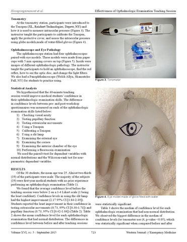
Tonometry At the tonometry station, participants were introduced to the Tonopen (XL, Reichert Technologies, Depew, NY) and how it is used to measure intraocular pressure (Figure 3). The instructor taught the participants to calibrate the Tonopen, apply the protective cover, and assess the intraocular pressure using globe models,made of water-filled gloves (Figure 4). Ophthalmoscope and Eye Pathology The ophthalmoscope station had four ophthalmoscopes paired with eye models. These models were made from paper cups with 5 mm opening covers on top (Figure 5). Inside were images of different ophthalmologic pathology. The instructor taught the participants to hold an ophthalmoscope, find the red reflex, how to see the optic disc, and change the light filters. We also had a Panophthalmoscope (Welch Allyn, Skaneateles Fall, NY) for students to practice using.
Figure 3. Tonometer.
Statistical Analysis We hypothesized that the 40-minute teaching session would improve medical students’ confidence in their ophthalmologic examination skills. The difference in confidence levels between pre- and post-workshop questionnaires was measured on each of the ophthalmologic examination skills listed below: 1) Checking visual acuity 2) Testing pupillary function 3) Testing extraocular movements 4) Using a Tonopen 5) Calibrating a Tonopen 6) Using a slit lamp 7) Examining the external eye 8) Examining the cornea 9) Examining the anterior chamber of the eye 10) Performing a fluorescein examination We used the paired t-test for dependent variables with normal distributions and the Wilcoxon-rank test for nonparametric dependent variables. RESULTS Of the 30 students, the mean age was 25. Almost two-thirds (19) of the participants were male. The majority of the subjects (19) were first-year medical students with no prior experience performing an ophthalmologic examination (Table 1). We found that the average confidence level before the teaching session were below 2 on a 1-4 Likert scale (1 being the least confident). Confidence levels in using the slit lamp had the highest improvement (2.17 95% CI [1.84-2.49]). Students reported the least improvement in their confidence in testing extraocular movements (0.73, 95% CI [0.30-1.71]) and pupillary function (0.73, 95% CI [0.42-1.04]) (Table 2). Table 2 shows the mean confidence level for each ophthalmologic examination that had normal distribution. The differences in confidence level between before and after teaching sessions Volume XVI, no. 5 : September 2015
Figure 4. Eye model made of glove filled with water.
were statistically significant. Table 3 shows the median of confidence level for each ophthalmologic examination that had non-normal distribution. We observed the biggest difference in the median of confidence levels for tonometer use (4, p-value <0.05), which was statistically significant when compared before and after
723
Western Journal of Emergency Medicine