6 minute read
In focus: the Visual Fixation System


The Visual Fixation System uses a mobile phone to display personalised targets for patients who find eye tests and focusing uncomfortable or challenging. Optometrist Simon Berry, who invented the system, explains how it works...

The Visual Fixation System (VFS) allows the clinician to complete clinical measurements more effectively with patients whose engagement may be variable – such as in patients with a disability, young children or patients with dementia.
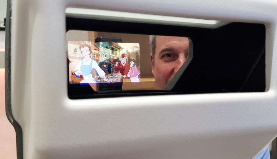
The system uses a mobile phone to display a specific target that will engage and/or relax the patient. The target is placed in direct line of sight of the clinician, allowing a number of clinical observations to be made.
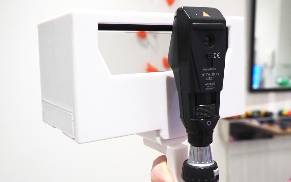
The VFS consists of a main unit and various different handles. The handles are interchangeable and should be selected depending on the clinical test required. There are currently two different handles for use with the device, with more under development.
Using A Phone As A Target
The VFS is compatible with most mobile phones. The flexibility of using a mobile phone means that an unlimited number of targets can be displayed and tailored to the particular needs of the patients. The sound from the phone can also be used to engage the patient.
There are a number of situations where this is useful, for example:
■ For a young child, their favourite TV show can be played through the phone, or photographs of family members can be shown. The parent’s phone can be borrowed and used for this purpose if needed
■ For a person with a learning disability, a target can be chosen that has the best chance of engaging the patient. This can be tailored to the patient’s interests
■ For an older person suffering with dementia, a clip of their favourite film or musical can be played. Music and photos can be displayed together
It should be noted that due to the optics of the device, the target shown to the patient will be a mirror image of that shown on the phone. This should not matter when displaying photos or videos, however, if the clinician wishes to display text then it will need to be adapted to be in the correct orientation for the patient to view.
To fit a mobile phone into the VFS, there is a sliding drawer that the phone fits into. It is advised to increase the brightness of the phone, disable any screensaver and change the orientation of the phone to landscape. The phone can be kept loose within the cartridge. However, this does mean that care needs to be taken when setting the VFS down on a desk.
Alternatively, the cartridge that holds the mobile phone features a velcro pad to keep the phone locked in place. A matching velcro pad can be stuck onto the back of the mobile phone. This will keep the phone more secure if required. Once the mobile phone is secure and displaying a target that will engage the patient, the VFS can be used for various different tests.
Measuring Accommodative Lag
Dynamic retinoscopy is a technique that allows the clinician to measure the patient’s accommodative response to a target. This is a useful measurement for many paediatric patients. It can help prescribing decisions for low to moderate hypermetropic prescriptions, and is a useful measurement when considering myopia control and the risk of a patient becoming increasingly myopic.
It is also known that certain patients are more prone to having problems with their accommodative response. For example, patients with Down syndrome or cerebral palsy are more likely to have an accommodative lag and these patients should be screened for the condition.
The VFS is the only device on the market that allows the clinician to examine directly along the visual axis. This allows accurate measurement of the accommodative response.
There are two quantitative ways of measuring accommodative lag. These are referred to as the monocular estimation method (MEM) and the Nott method. There are other methods sometimes described but they are modifications from these two basic techniques. The VFS allows measurements by both techniques. The clinician simply has to change the handle depending on their preferred method of measurement.
Other Clinical Observations Measurements
The VFS is useful as a means to grab a patient’s attention. It becomes invaluable in patients where co-operation is difficult, and allows the clinician a method of observation when other methods fail.
It can be used for a variety of different clinical observations and some of those observations are described below. These are not meant to be an exclusive list of possible tests but just descriptions of some of the techniques already used.
It should be noted that whilst some of the techniques described below might initially appear to be a crude measurement, in situations where patients cannot engage with the clinician any other way these methods are better than not being able to take any measurement at all.
Gross Examination
The VFS allows a gross examination of the ocular structures. It can be used, for example, to assess the fit of a spectacle frame, to take facial measurements or bifocal lens measurements.
It can be used with a 20D lens to increase the magnification and allow more accurate assessment of the external eye structures. This is useful for evaluating contact lens fits, or corneal assessments. It can be also used as a gross assessment of binocularity by observing the corneal reflections from the mobile target.
Checking Prescriptions
Once a prescription has been dispensed, if the result has been slightly rough due to the patient’s co-operation, then the VFS can be used to check the result after it has been prescribed. Anisometropia can be checked because with the prescription in place the reflex should be symmetrical between the right and left eye. Cylinder power and axis can be also be checked because with the correction in place a spherical reflex will be seen.
NEAR COVER TEST, PD AND OTHER MEASUREMENTS
The VFS can be used to take a variety of measurements whilst the patient is engaged in watching a target, for example, the near cover test, pupil reactions or 20D prism test. These measurements are crude, but better than not being able to complete the test. They can be made more accurate by keeping the target on the phone small. There is a balance to strike between cooperation of the patient, the size of the target and accuracy of the obtained result.
Fundus Examination
A fundus examination is possible using the VFS and a modified indirect ophthalmoscopy technique. An ophthalmoscope is placed in the instrument handle, set on 8D – depending on the preference of the clinician, the working range, and the field of view preferred when viewing the fundus. Using a 20D lens in front of the VFS and close to the patient, it is possible to obtain a view of the fundus using indirect ophthalmoscopy.
Cycloplegic Refraction
The VFS can be used for
Cycloplegic
refraction. Whilst under cycloplegia, the patient would not be able to focus properly on a target, but they are still attracted to the target even if they can’t see it properly. The VFS and retinoscope is held in one hand, and the lens rack held in the other.
Use With A Test Chart App
The VFS can also be used for more co-operative patients. A test chart can be displayed on the mobile phone and measurements of the accommodative response can be made. This is particularly useful in myopia control when a more detailed accommodative target is required.
advantage of the VFS is that the clinician is able to take measurements directly along the visual axis.
Tips On Using The Vfs
◼ Keep the room lights low. The more light behind the clinician, the more distraction there is for the patient viewing the target. When it is not possible to lower the room lights, there is an alternative mask provided for the viewing window. Using the VFS with this in place will make the target easier for the patient to view.
◼ Allow the patient a few moments to watch the target and adjust their viewing before starting any clinical examination. When a retinoscope beam is shone through the beam splitter, it is less distracting to the patient if they are already focused on the target.
◼ Be aware that many patients will reach for the target. They are being shown something that interests them.
◼ Keep the beam splitter clean to avoid stray reflections. This is particularly important when performing indirect ophthalmoscopy.
Simon Berry has been an optometrist for more than 27 years. As well as running a specialist paediatric clinic within a hospital, he owns an independent practice. He spent six years developing the Visual Fixation System, originally with support from Durham University, and launched it to the profession at 100% Optical last year. The system is patented in the UK and USA, with patents pending in other markets.
For further details and purchasing enquiries, email simon@simonberry.co.uk or telephone 0191 375 75 44. ■


