2 minute read
Revo FC OCT: pushing the boundaries

By Tim Baker, group CEO, BIB
As a Revo user, you can enjoy all the latest updates with many new and exciting features and the clinical benefits they bring. This ensures that your investment stays at the forefront of technology for the life of your device.
WHAT'S NEW AND IMPROVED?
Posterior and Anterior Full Range
The new Full Range feature pushes the retinal scan (Posterior B Scan) capability from a 12mm line scan to a 15mm line scan. This provides a 25 per cent increase in retinal visualisation from one single scan, facilitating greater clinical analysis and saving time.
Anterior (Anterior B Scan) Full Range 16mm and Anterior Chamber Radial Full Range examination now provides complete visualisation of the anterior chamber – and is a world first for which was essentially a posterior OCT. This ground-breaking new capability provides a complete cataract, cornea and glaucoma analysis with measurement and recording facility. Scans are achieved without the use of any adaptor or additional forehead bar (Revo FC only).
DeNoise Algorithm
Improved visualisation of all tomograms comes with the new Artificial Intelligence (AI) DeNoise algorithm, which filters out noise from the tomogram for the highest and smoothest image quality. The AI DeNoise algorithm enhances the visibility of morphological structures by processing the original image, and provides greater detail within the retinal layers thereby improving clinical diagnosis.
IOL Formulas
Measuring axial length utilising the optional biometry measurement software has until now only been for used for visualisation and measurement, ideal for those involved in myopia management. For clinics that want intraocular lens (IOL) calculations and formulas, Optopol has now made
OPTIONAL SOFTWARE MODULES
The following optional software modules are available: this possible with the add-on software IOL Formulas, which includes Hoffer Q, Holladay I, Haigis, Theoretical T and Regression II formulas. In addition, the WTW&P feature is available. White-towhite measures the distance from limbus to limbus measured horizontally through the centre of the pupil. Pupil diameter can also be measured horizontally through the centre of the pupil.
◼ Angiography OCT: This module allows visualisation of the retinal microvasculature. Angiography SOCT is a non-invasive, dye-free technique providing 3D image of retinal blood circulation.
◼ Biometry OCT: Myopia Management: B-OCT is an innovative method of using the posterior OCT device to measure ocular structure along eye axis.
◼ Topography OCT: The Topography OCT module provides the analysis of both surfaces based on corneal curvature, dioptric power, elevation and real power analysis based on both surfaces and local cornea thickness (ray tracing).
Post-tacking feature
The new post-tracking feature offers improved correlation using the fovea or optic disc centre for difficult cases.
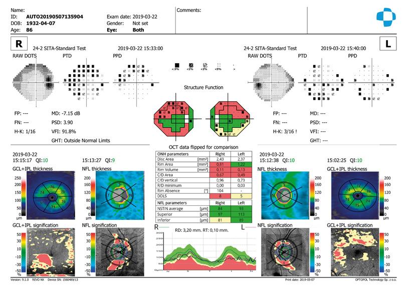
Structure and Function
Updated and improved Structure and Function Analysis allows the user to compare 10/2, 24/2 and 30/2 visual field results. It is designed to be used with Optopol's PTS visual field screeners.
Updated Software And Enhancements
The latest Revo FC OCT comes with a real-time hardware eye-tracking featuring, redesigned OCT optics and fundus optical systems to provide a better quality of tomograms and fundus photographs. The modified hardware, together with the latest version of software 11.5.0, provides an array of new functionalities and improvements.
For further information on your Revo OCT products from Optopol, please contact BIB on 01438 740823. ■
