An examination of the human remains within the Alfred Denny Collection as part of the More Than Human project.
A symbol of having a person there who understands your situation when you need it most. The author would like to thank Dr Emma Hughes, The University of Sheffield Department of Biosciences and Arts Council England for making this possible
Contents
Introduction
The Fellowship
-
The Full Skeleton
- The Articulated Hand
- The Articulated Foot
- The Infant Skull
- The Disarticulated Skeleton
- The Opened Skull
- The Bisected Skull
Fraternity
More Than Human
The Alfred Denny Museum is a stunning collection of zoological specimens collected by Alfred Denny in the late 19th century, and added to in later years. It’s particularly notable for its exhibits which combine taxidermy (the art of posing animal skins in a lifelike way) with osteology (which exposes the articulated skeletons of the animal) giving students the opportunity to see both the familiar form and inner structure of a creature in a single object.
The museum itself was hit by the bombs of World War Two, and many specimens (together with the museum’s original site) were lost, with it now occupying a space in the University of Sheffield Alfred Denny building. I was drawn to the collection via an old photograph on the Department of Biosciences website, showing a human skeleton standing next to a great ape. I contacted the University to enquire as to whether the skeleton still existed within the collection, and Dr Emma Hughes confirmed the individual was still there and that there were other human remains within the museum.
On my first visit I saw four individuals – the full skeleton, an articulated hand, an articulated foot, and the skull of an infant prepared in the Beauchêne technique. Dr Hughes very kindly allowed me to photograph these for reference, and was happy for me to research their potential origins – the skeleton and the extremities still bore the identification of the companies who prepared them, and so I was hoping to be able to potentially identify where these people may have come from, and the sources these osteologists used for their trade.
When I returned to make photographs for this book Dr Hughes told me since my last visit she had located the remains of three further people within the archive of the museum – two adult skulls, one bisected vertically, the other horizontally, and a complete disarticulated infant skeleton in a frame prepared by the same company as the adult skeleton.
In this context the skeletons in this particular closet are essential as examples of comparative anatomy – observing the differences between ourselves and our biological cousins greatly benefits our understanding of the world around us and our place within it. However in order to ensure the best use of these objects in the future the skulls and the infant remains have been transferred to the Human Anatomy department, where they will continue to save countless lives by training future medical practitioners and scientists.
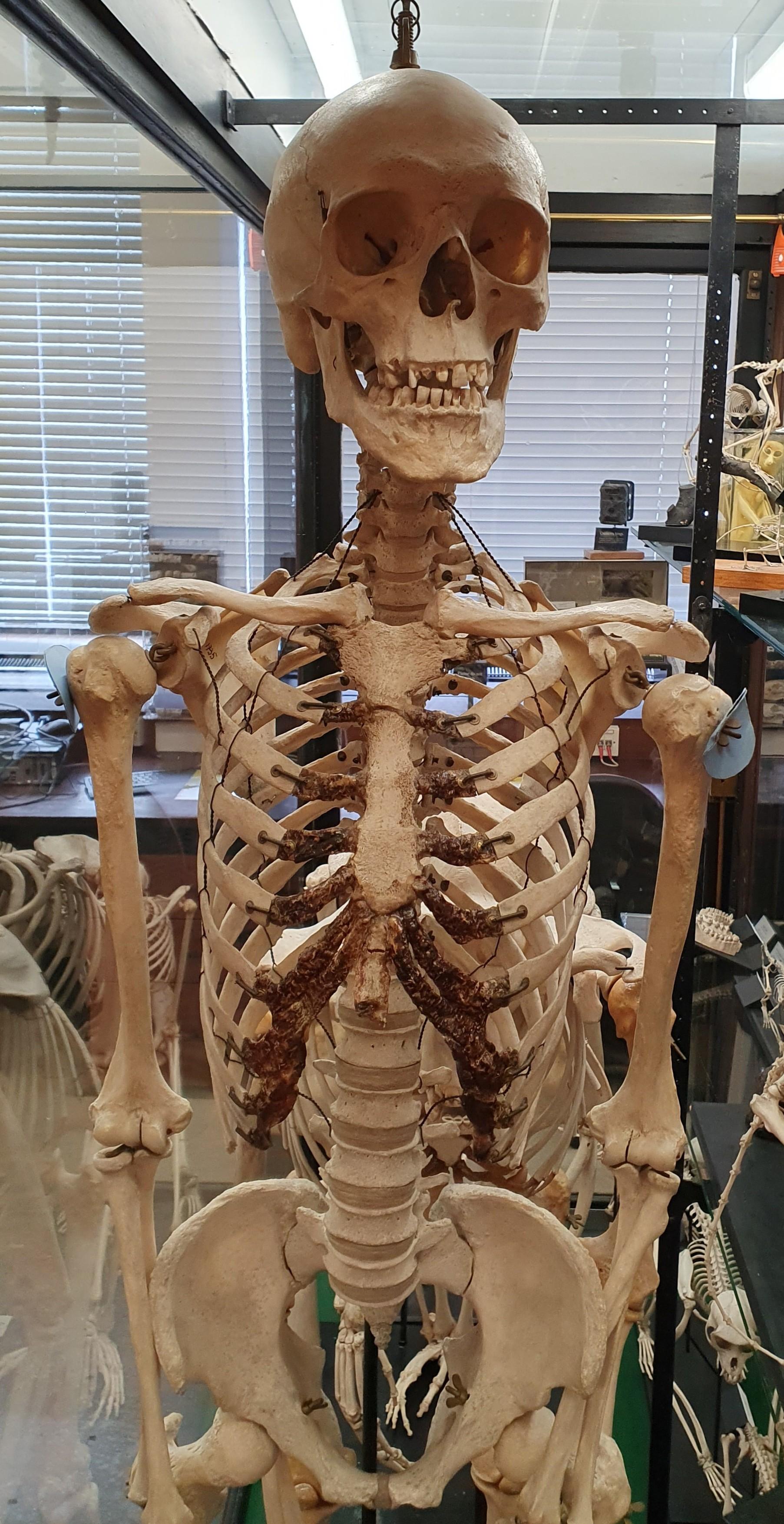
This is a fully articulated human skeleton. Most likely that of a man, and an executed criminal –broken vertebrae at the back of the neck suggests this individual was hanged, but the death was instantaneous.
A partially removed label on the left side of the skull indicates that this individual was prepared in France by Maison Travond, 9 Rue De L’Ecole De Medecine, Paris. The site is now occupied by an Italian Restaurant who’s Google reviews claim it is either perfection or horrendous.
After a hundred years and a bombing it has a few signs of wear and tear, but is still in excellent teaching condition.
This individual continues to reside in the Alfred Denny Museum.
T
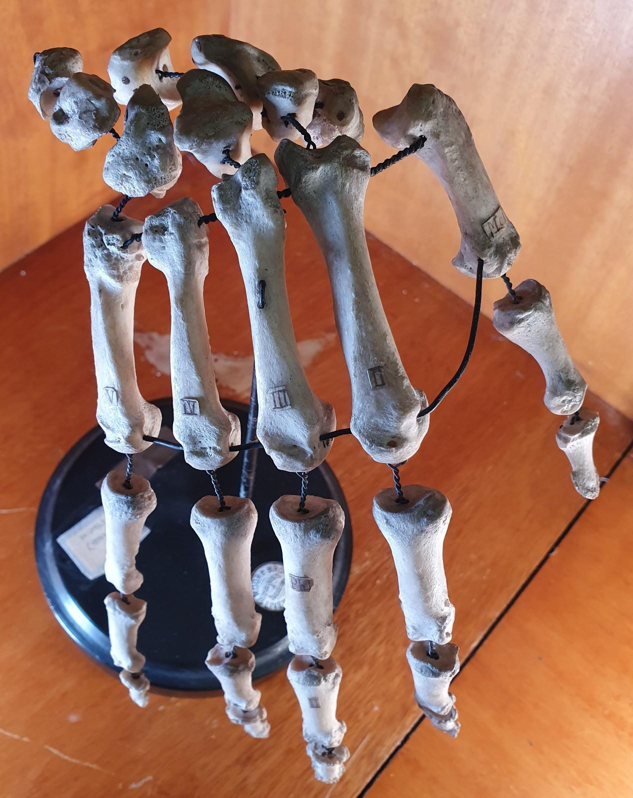
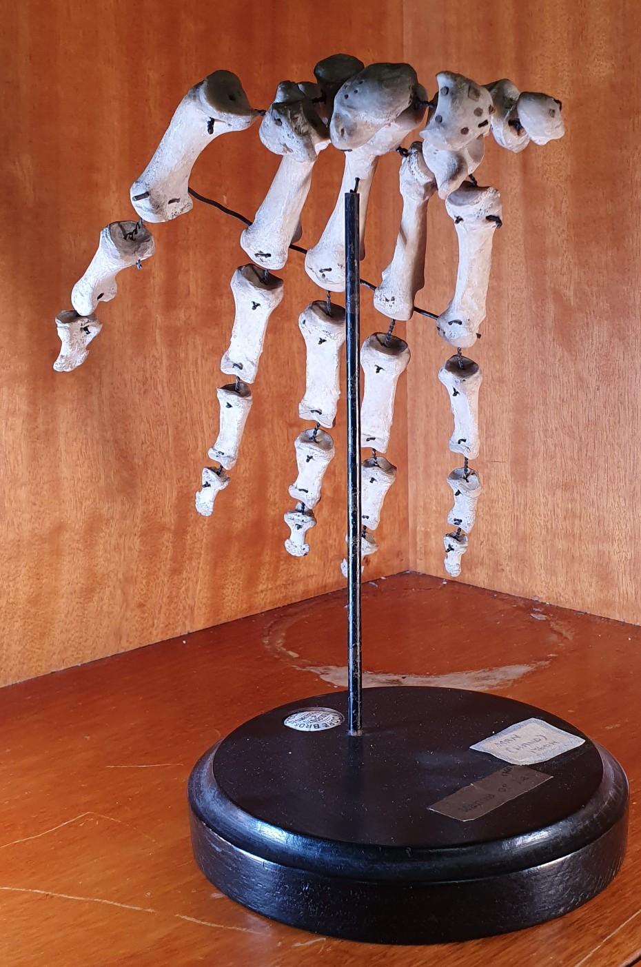
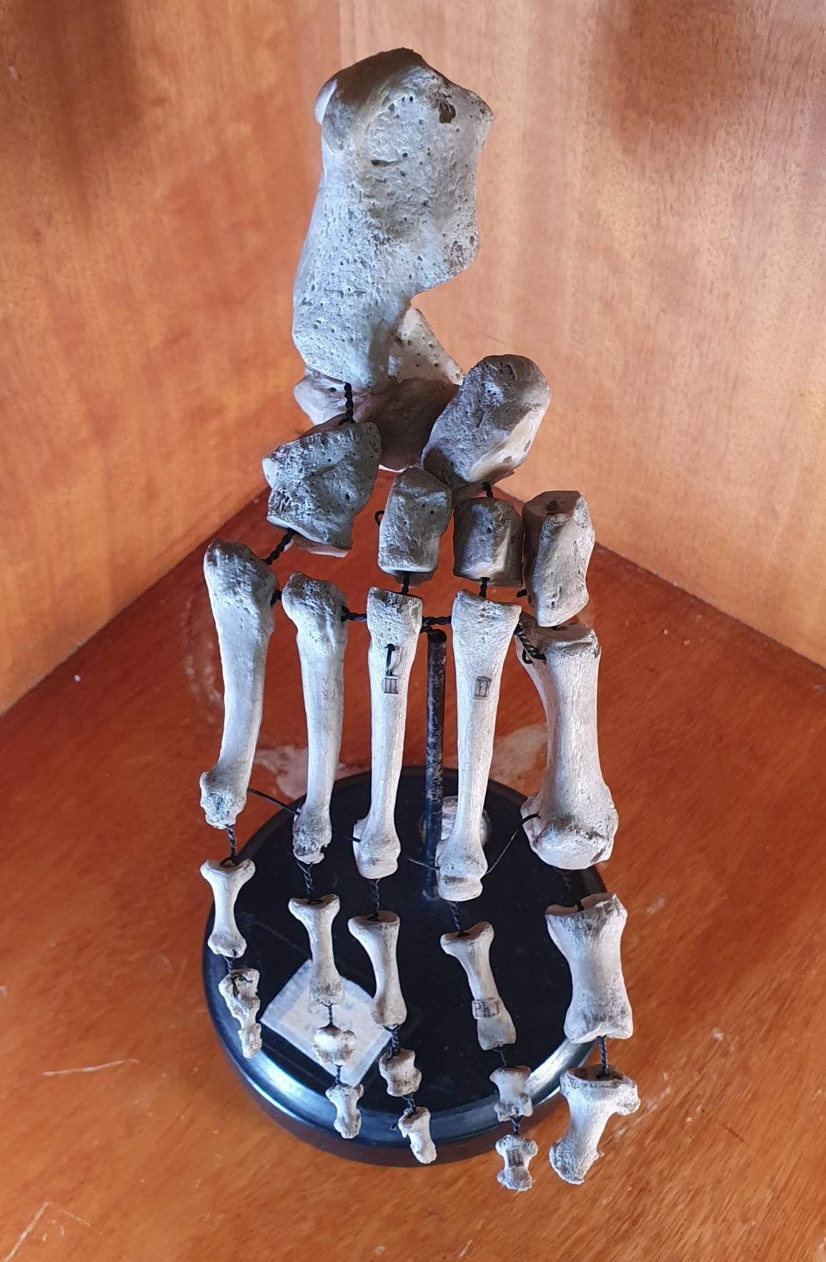
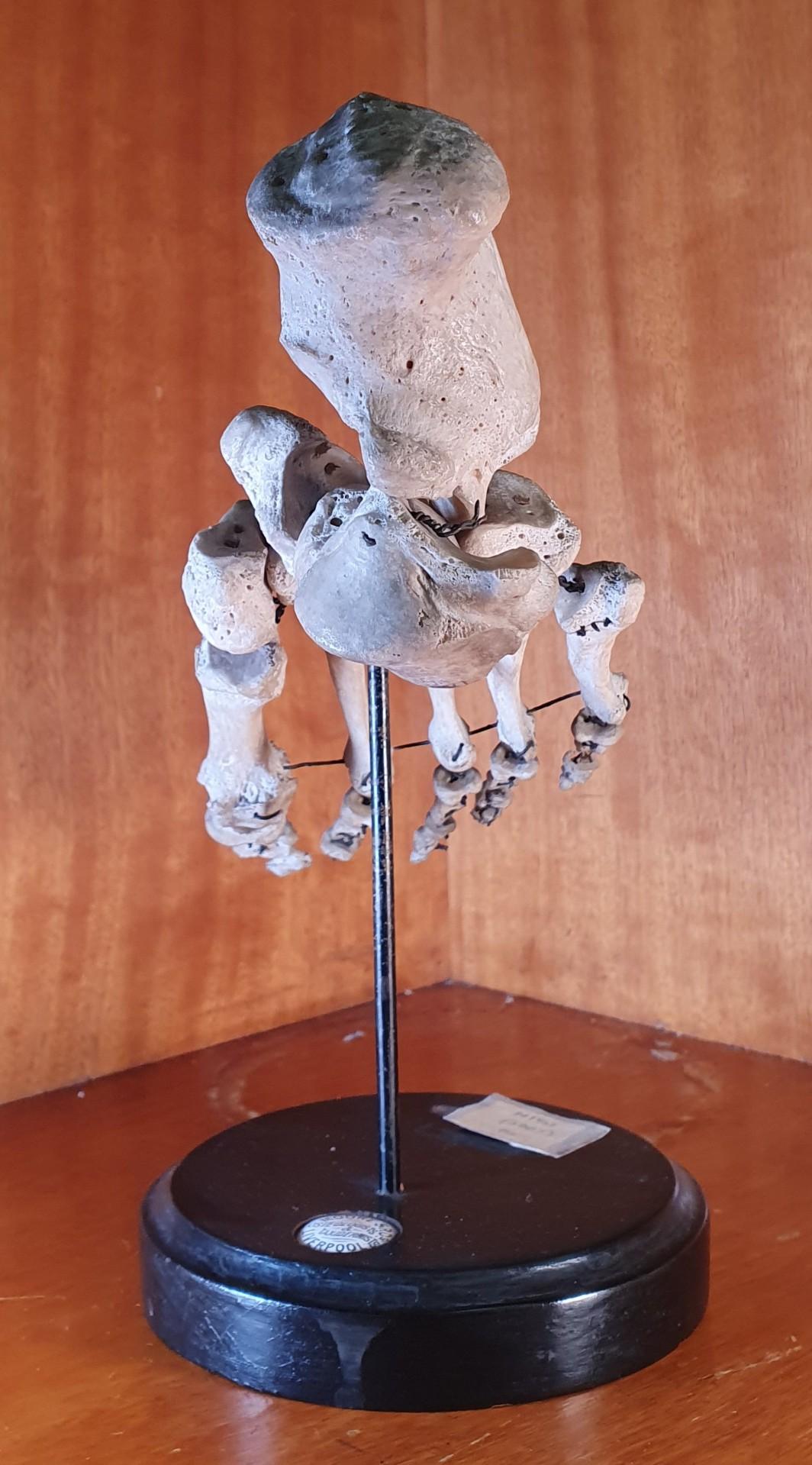
HE ARTICULATED HAND
A right hand, articulated but slightly spaced. All the metacarpal and some of the phalange bones have been labelled using Roman numerals.
The base indicates that this individual was prepared by Moore Brothers, Osteologists and Taxidermists, Liverpool.
This individual continues to reside in the Alfred Denny Museum.

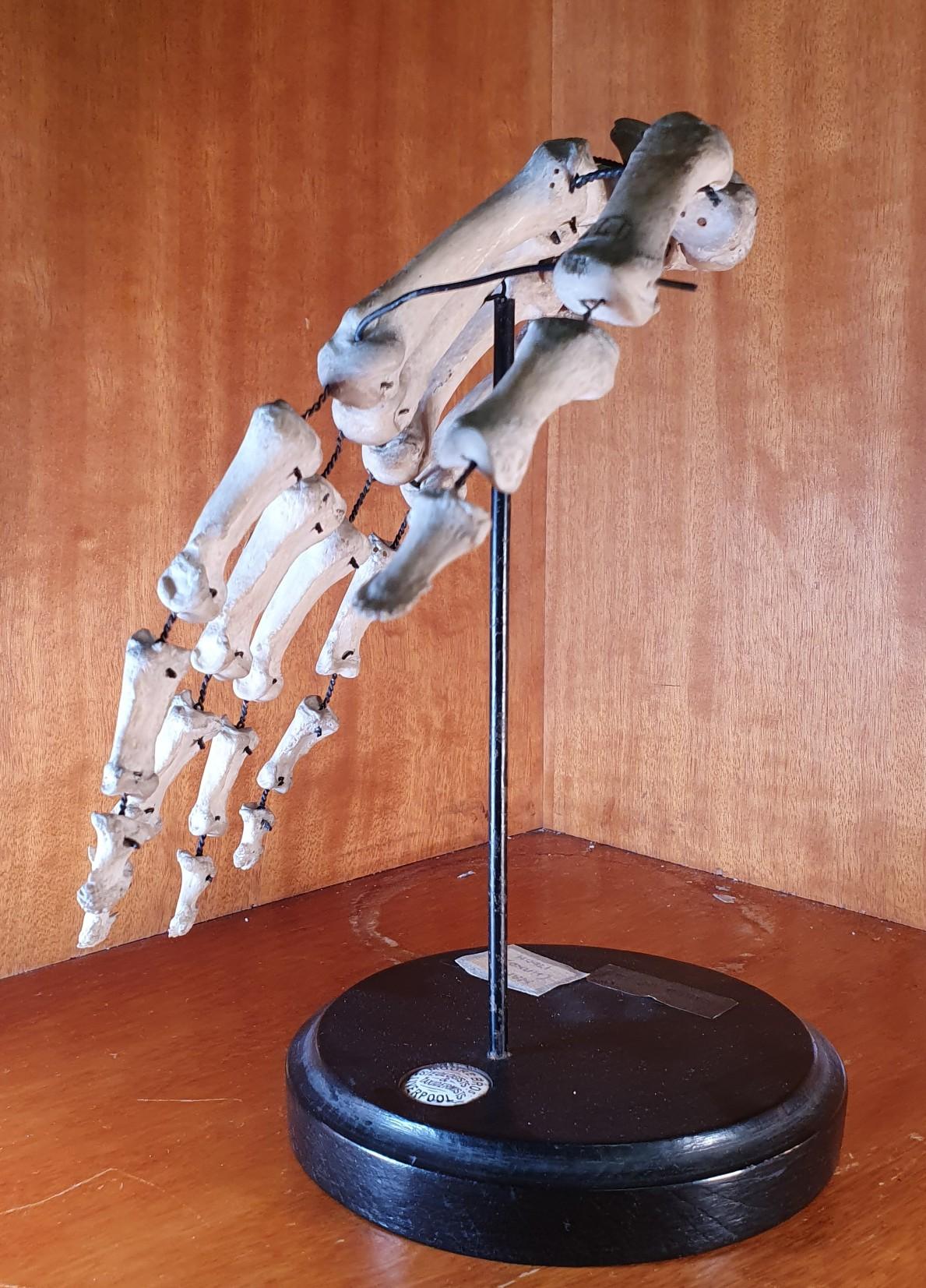
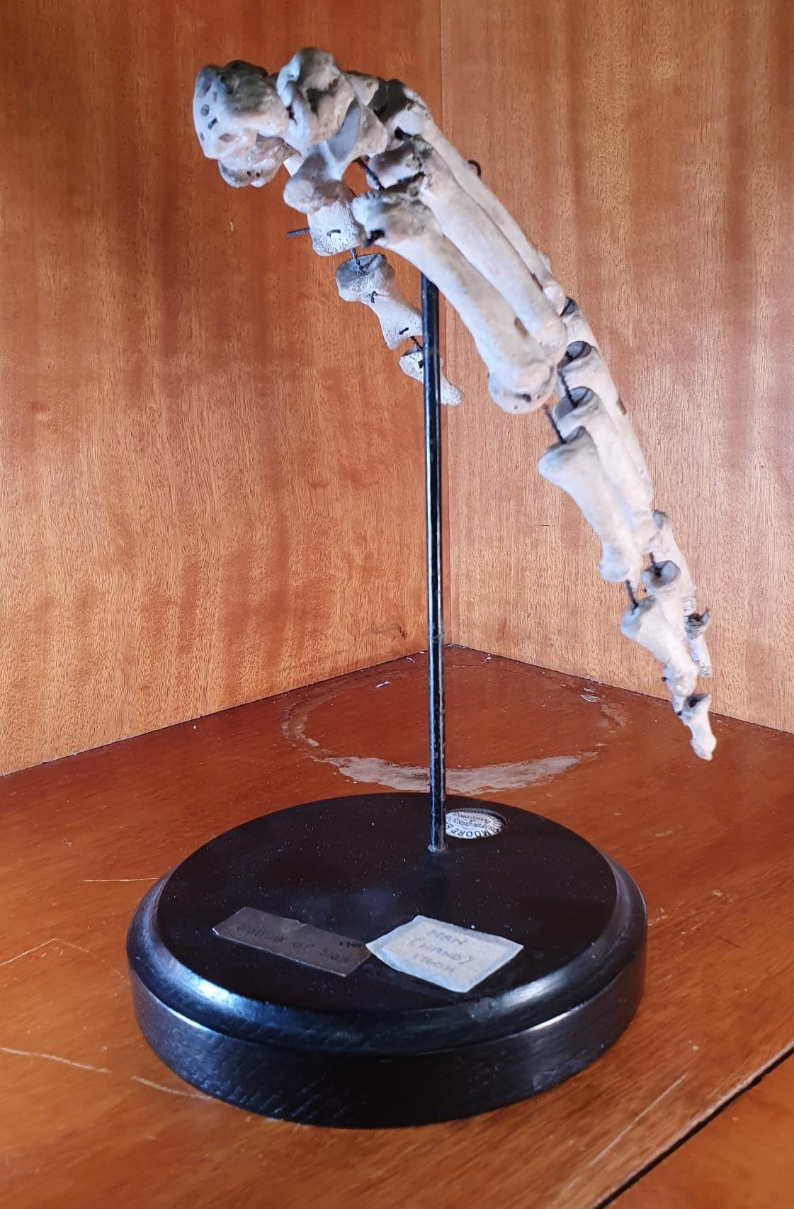
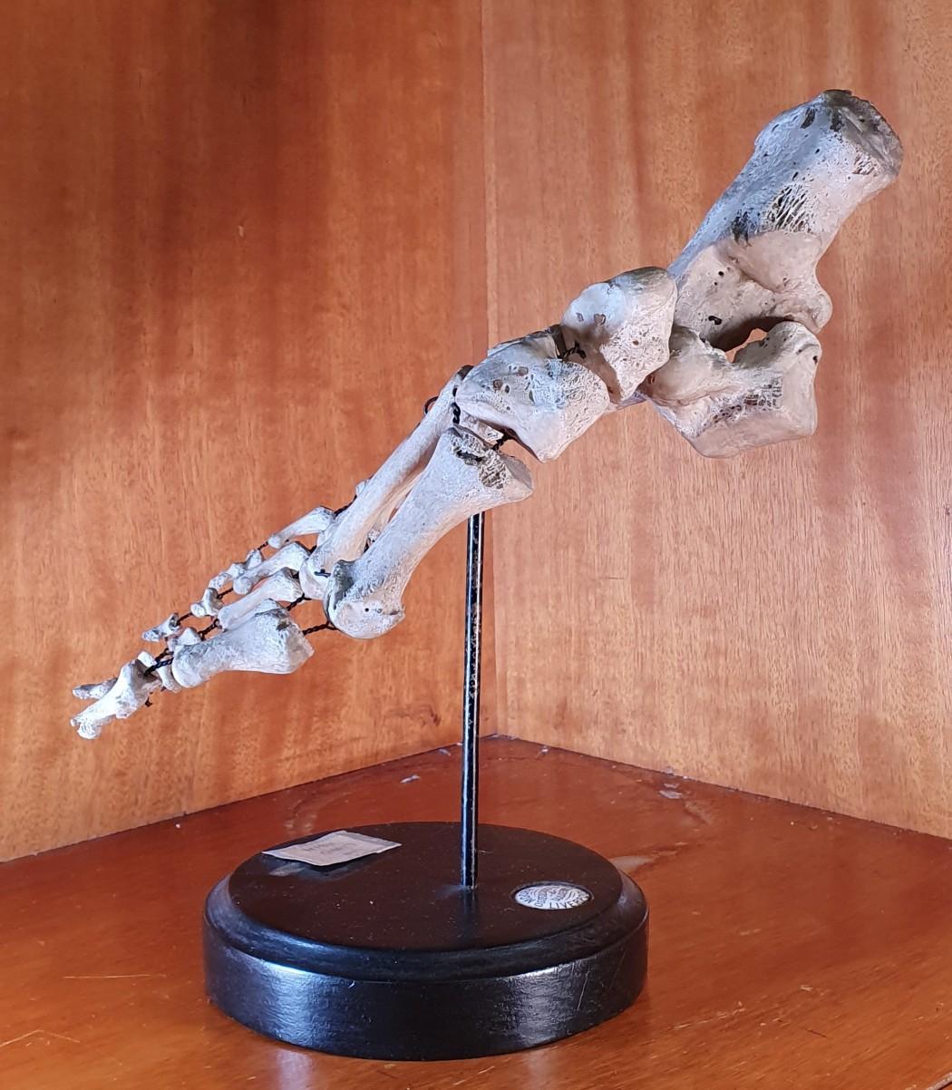
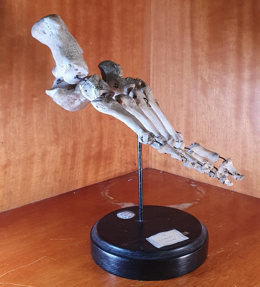
THE ARTICULATED FOOT
A right foot, articulated but slightly spaced. All the metatarsal bones have been labelled using Roman numerals. The base indicates that this individual was prepared by Moore Brothers, Osteologists and Taxidermists, Liverpool.
This individual continues to reside in the Alfred Denny Museum.

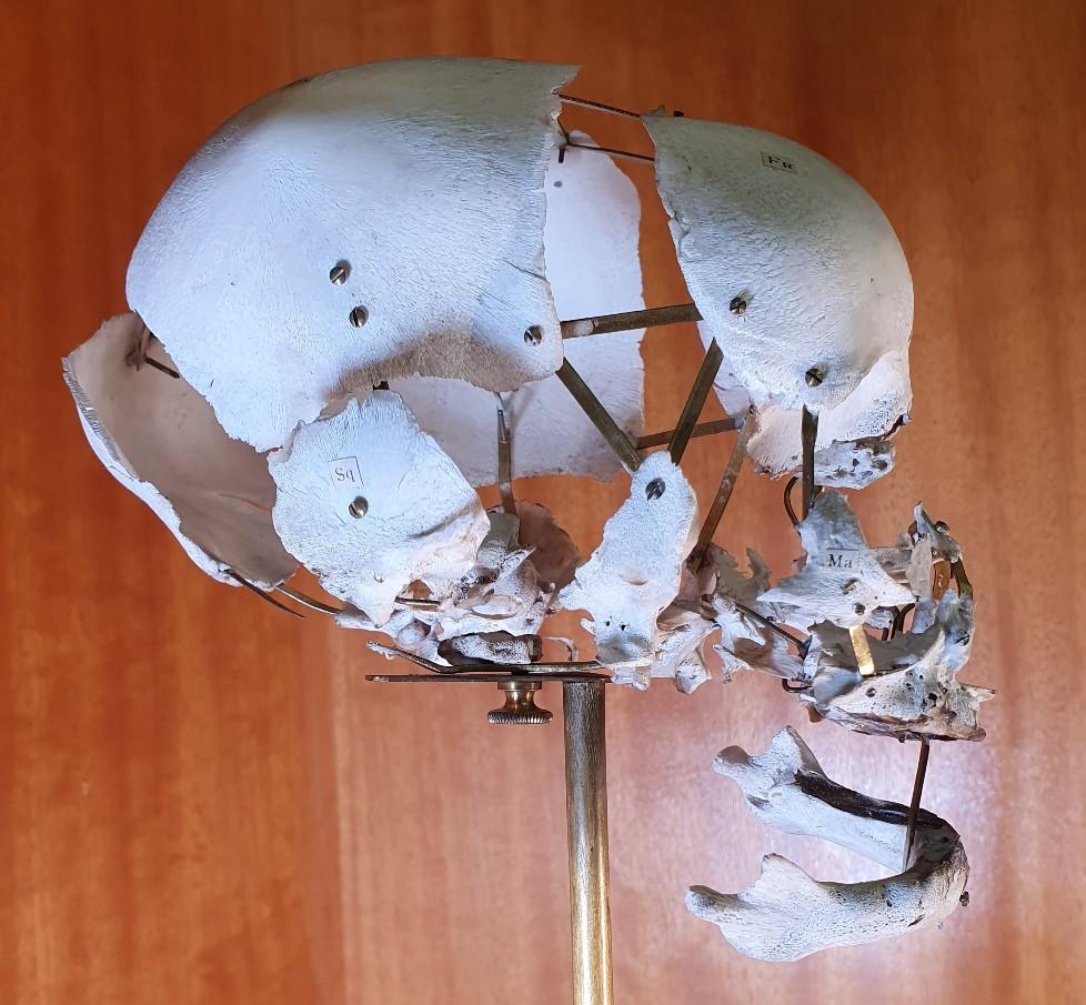
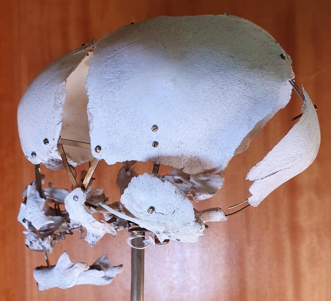
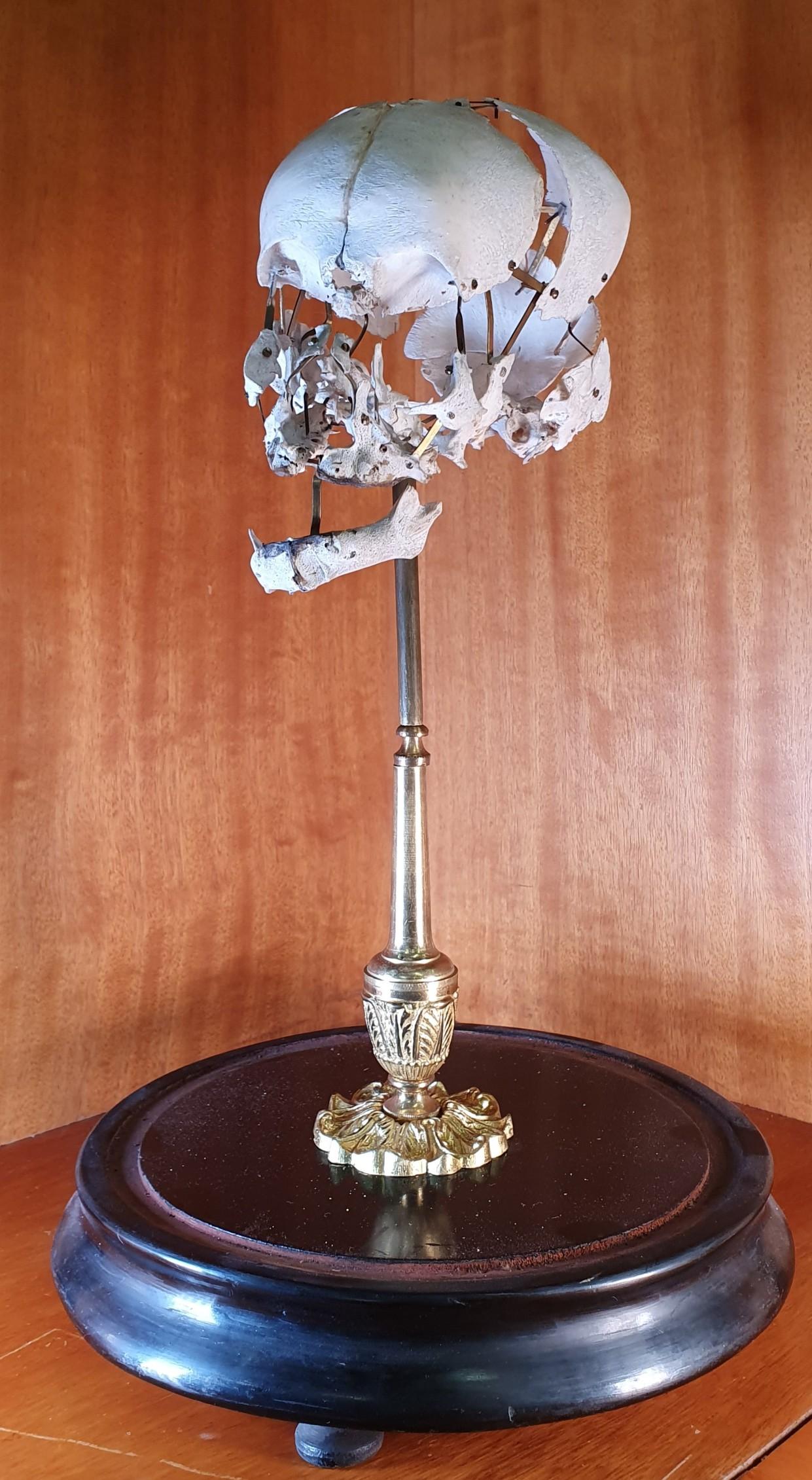
A disarticulated skull of an infant prepared in the Beauchêne technique. The stand is ornate brass and it is surmounted with a glass dome, suggesting this individual was prepared with concerns as much aesthetic as educational.
This individual was useful in teaching the individual plates of the skull, together with the delicacy of infant bones. The Beauchêne technique involves soaking the skull until the plates begin to separate and the osteologist can separate them – a process far easier if carried out before the skull plates fuse at a few months old.
There are no markings or other means of identifying the makers. As Beauchêne is a French technique, and other individuals in the collection, (including another infant with a disarticulated skull) originated in France it would be likely that this individual is also French.
This individual now resides in the department of Human Anatomy, and will help future scientists and medical professionals understand the human body into the future.

T
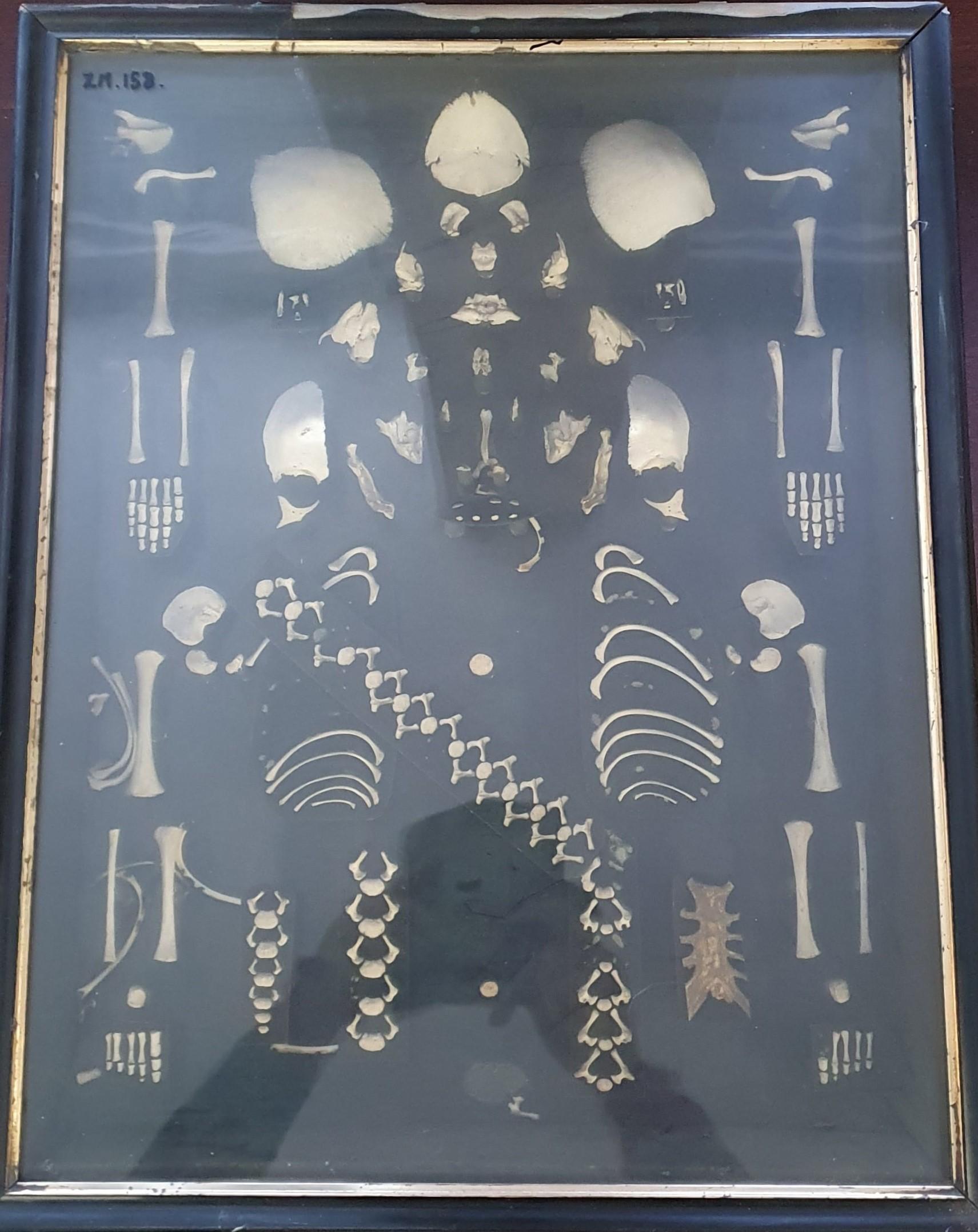
HE DISARTICULATED SKELETON
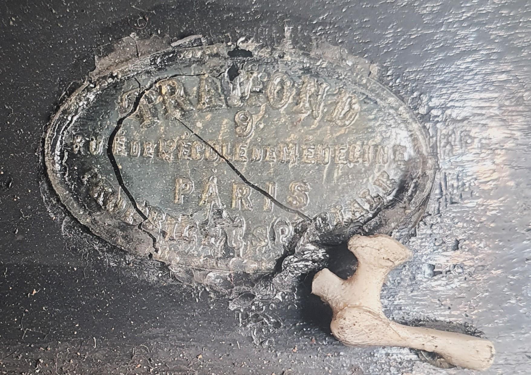
A complete infant skeleton, fully disarticulated and presented in a frame. A brass label at the base of the frame indicates that this individual was prepared in France by Maison Travond, 9 Rue De L’Ecole De Medecine, Paris.
The disarticulation is complete to the extent of separation of skull plates (suggesting a variation of the Beauchêne technique), individual bones of the ear and vertebrae. The composition of the bones is particularly interesting – while the presentation keeps the bones in the correct placement for human anatomy it’s somewhat stylised, again seemingly being presented with more concern for appearance then function.
This individual now resides in the department of Human Anatomy, and will help future scientists and medical professionals understand the human body into the future.
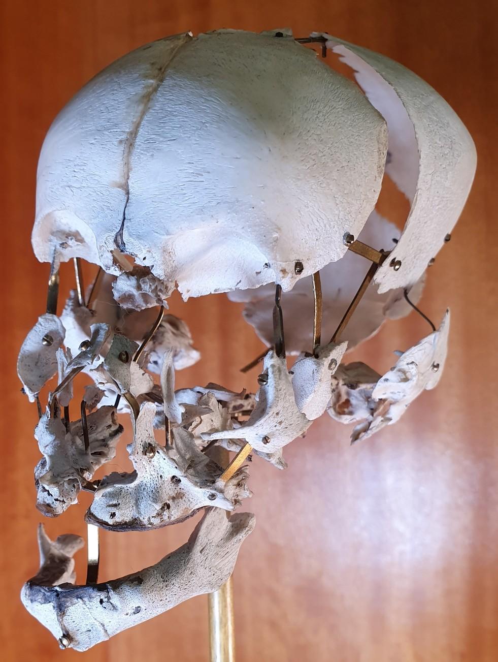
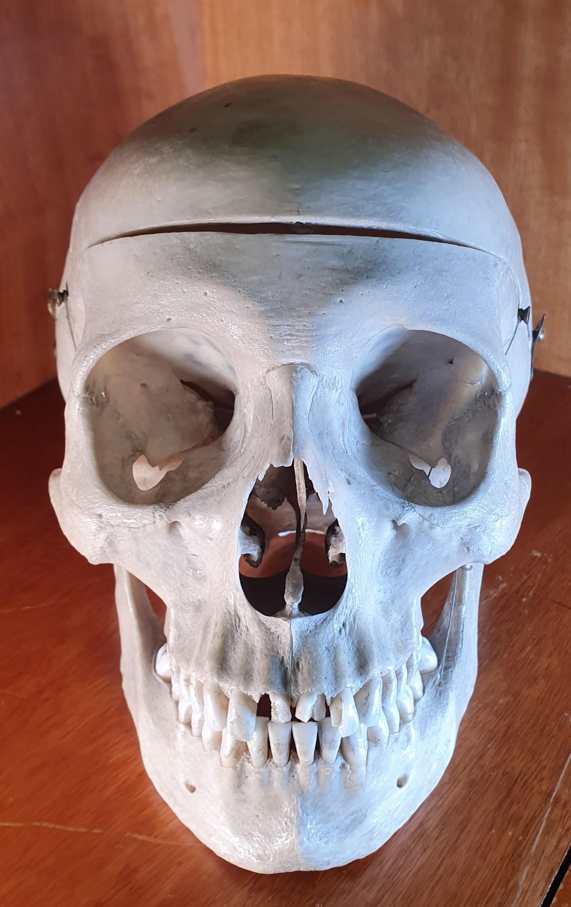
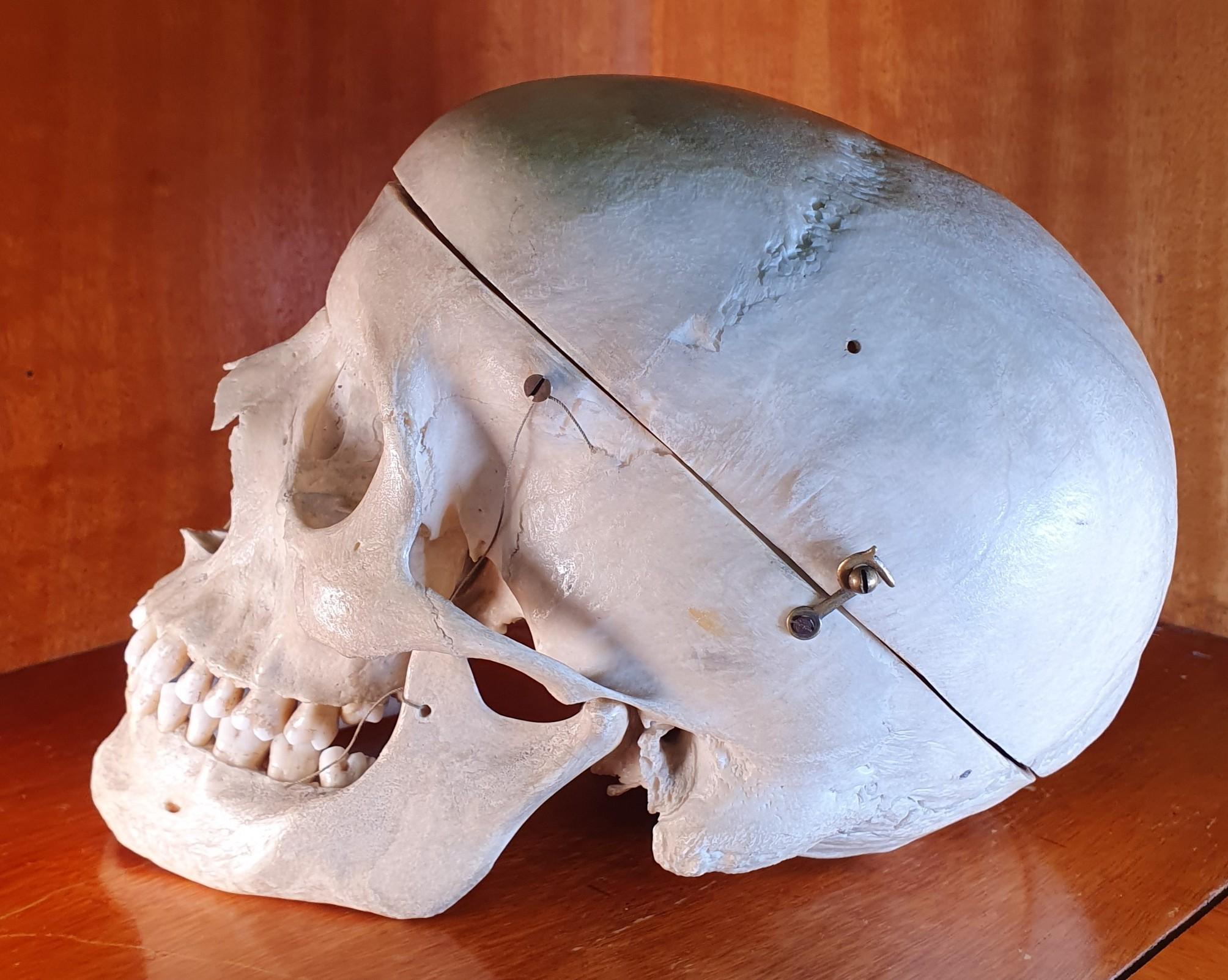
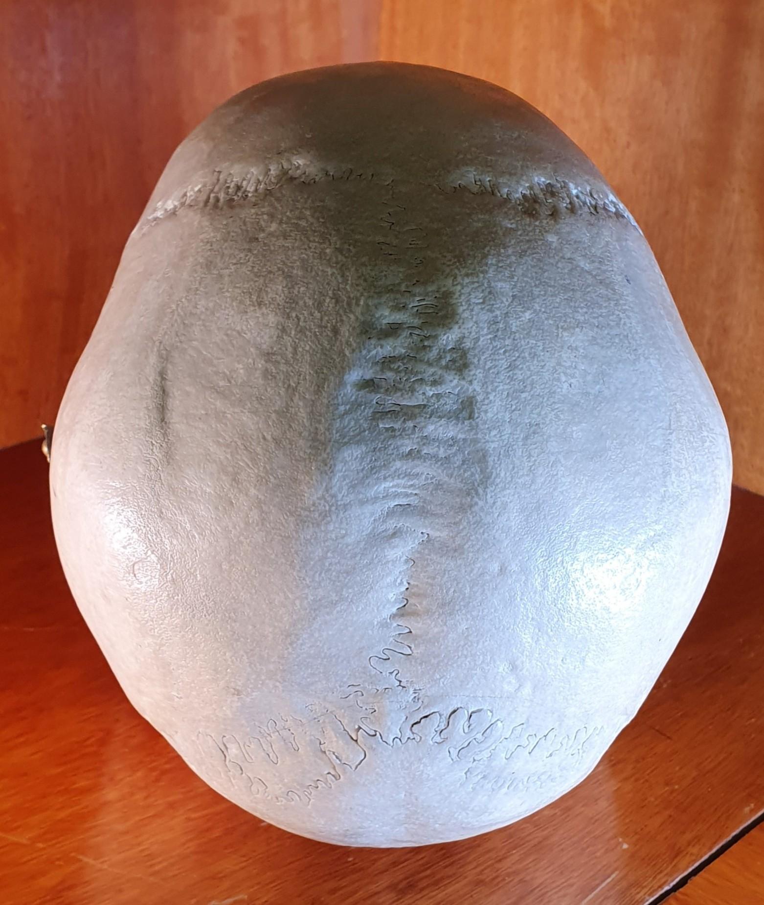
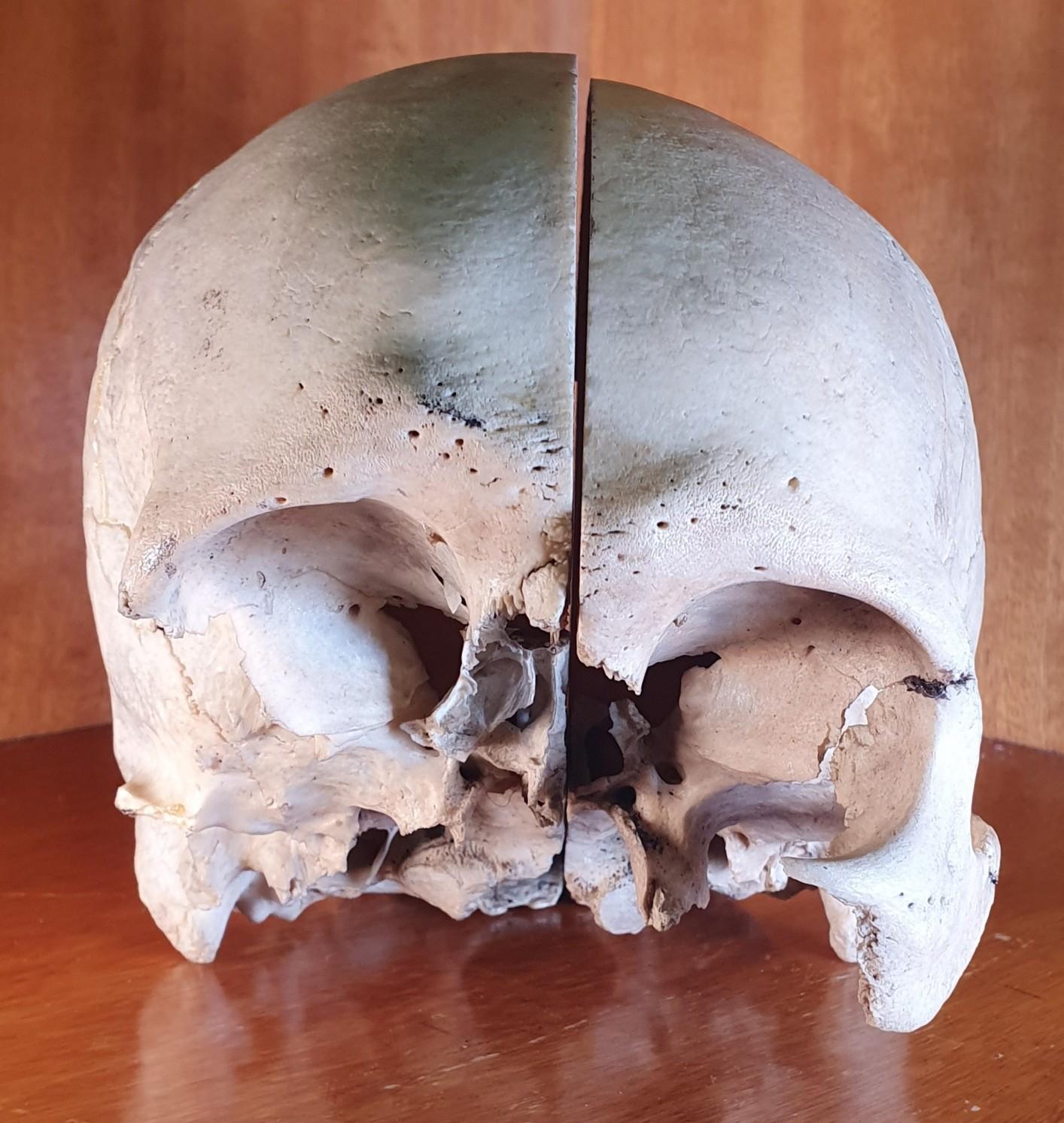
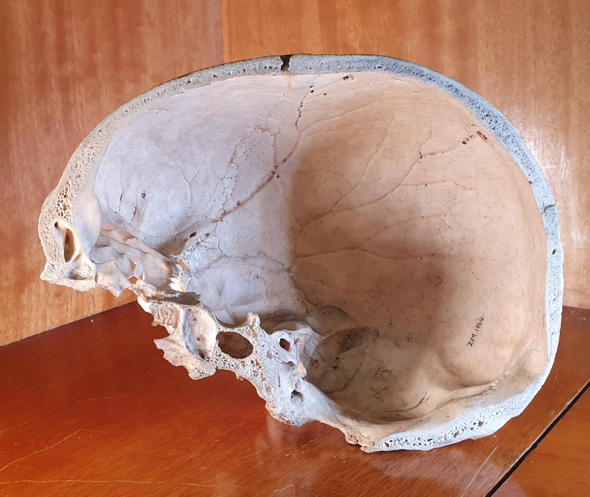
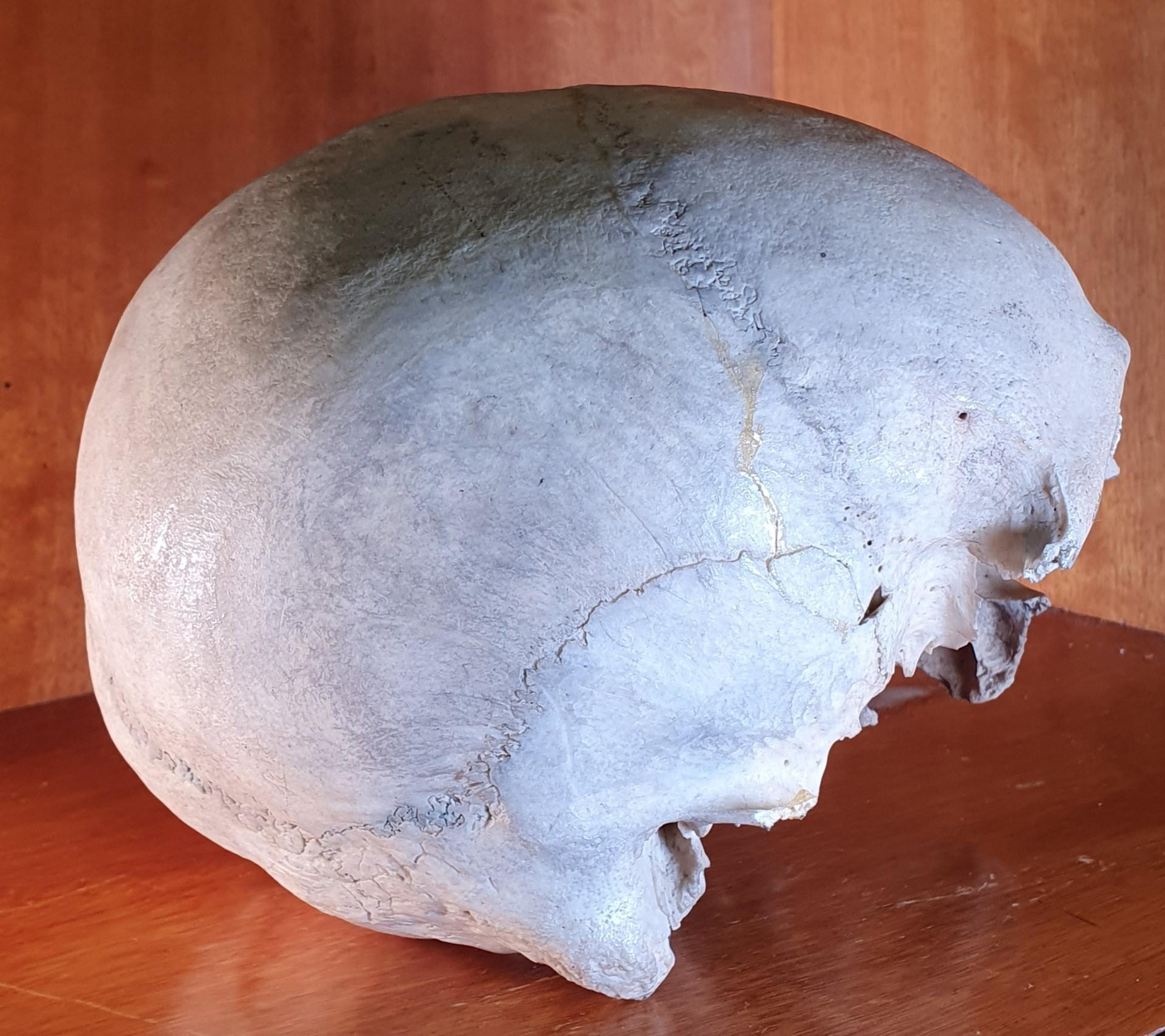
A human skull, not including jawbone, cut horizontally just above the browline to make the top of the skull removable, exposing the internal structure of the skull from the top.
There are no markings or other means of identifying the makers.
This individual now resides in the department of Human Anatomy, and will help future scientists and medical professionals understand the human body into the future.
 © Vicky Roden 2024