
Souvenir 2022ASSOCIATION OF ORAL AND MAXILLOFACIAL SURGEONS OF INDIA Cranio maxillofacial Surgery reworking the future..... 18 14 - 16 October 2022 th ANNUAL OF AOMSI KERALA CHAPTER Hotel Tripenta, KOZHIKODE, Kerala STATE CONFERENCE


Souvenir 2022ASSOCIATION OF ORAL AND MAXILLOFACIAL SURGEONS OF INDIA Cranio maxillofacial Surgery reworking the future..... 1814 - 16 October 2022 th ANNUAL OF AOMSI KERALA CHAPTER Hotel Tripenta, KOZHIKODE Kerala STATE CONFERENCE CONTENTS 18 14 - 16 October 2022 th ANNUAL OF AOMSI KERALA CHAPTER Hotel Tripenta, KOZHIKODE, Kerala STATE CONFERENCE 4 Organizing Committee 9 Messages 20 Editorial 21 Office Bearers of AMOSI Kerala chapter 23 Activity Report 21-22 26 Scientfic Schedule 30 Chief Guest 31 Oration 32 A Brief History of OMFS In India Prof. Dr. Varghese Mani 34 The Kerala Aomsi & Me Prof. Dr. Krishnamurthy Bonanthaya 35 Oral Oncology - Oral & Maxillofacial Surgeon The Pivot Point Prof. Dr. Senthil Murugan 38 Delegate Papers Prize Category 40 Delegate Papers Free Category 41 Postgraduate Papers Prize Category 52 Postgraduate Papers Free Category 3AOMSI Kerala
AOMSI KERALA STATE CONFERENCE 2022 ORGANIZING COMMITTEE MEMBERS


ORGANISING COMMITTEE
Dr. THOMAS JOSEPH Dr. GEORGE VARGHESE Dr. AJU OOMEN JACOB
PATRON
Dr. VARGHESE MANI
ADVISORY COMMITTEE SECRETARY Dr. MANOJ BHASKAR
ORGANIZING SECRETARY
Dr.
MANOJ KUMAR K.P
ORGANIZING CHAIRMAN Dr. UMMAR MANGALATH
TREASURER Dr. MANOJ JOSEPH MICHAEL
ORGANIZING CO-CHAIRMAN
Dr.
RAVINDRAN NAIR K.S
SCIENTIFIC COMMITTEE CHAIRMAN Dr. SOUMITHRAN C. S 4 AOMSI Kerala


ORGANISING COMMITTEE
ORGANIZING JOINT SECRETARY
Dr.
MOHAMMED YAHIYA
Dr. IKRAM BIN ISMAIL
CO-
CHAIRMAN Dr. SAJU N.S
Dr. NAMITHA S. PREM
Dr. SHALINI KRISHNAN
Dr. FAZMIYA. M
RECEPTION COMMITTEE CHAIRMAN Dr. BENNY JOSEPH
CO-CHAIRMAN
Dr. SUDHEESH
MANOHARAN
Dr.
PRAVISH VISHNUDAS
PRE-CONFERENCE COMMITTEE CHAIRMAN Dr. PRASANTH PANIKER SCIENTIFIC COMMITTEE 5AOMSI Kerala


ORGANISING COMMITTEE
Dr. NIKHIL O GOVINDAN Dr. DEEPTI KRISHNAN KUTTY
SOUVENIR CO-CHAIRMAN
Dr. ANROOP ANIRUDHAN Dr. ASWATHI VINOD Dr. SUNU V.S
RECEPTION COMMITTEE
Dr. T.A SHIYAS
CHAIRMAN Dr.
RAVINDRAN V.
HOSPITALITY 6 AOMSI Kerala


ORGANISING COMMITTEE
Dr. MANU MATHEW
Dr. SANJU LAKSHMANAN Dr. BINSHAD BASHEER
Dr. ABCISH KRISHNAN
GIFTS Dr.
SUBIN VARUGHESE
MEMENTOS
Dr. VIJAYAKUMAR DEPESH
BANQUET TRANSPORT & ACCOMODATION Dr. MITHILESH K.V Dr.
CHANCHALESH M.C
BROCHURE & ADVERTISEMENT
Dr.
VIPIN DAS
A.P
CHAIRMAN Dr. RAJESH MANUEL TRADE FAIR 7AOMSI Kerala
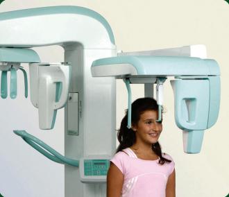
the digital dental imaging at Janatha


Now Experience
NABL & NABH Accredited Main road Chemmad 0494 246 0494 / 246 5509 / 246 3020 www.janathadiagnostics.com CBCT OPG TMJ IOPA 8 AOMSI Kerala
I am happy to know that the Association of Oral and Maxillofacial Surgeons of India [AOMSI] Kerala, intends to publish a Souvenir in connection with its 18th Annual State Conference to be held from 14th to 16th October 2022 at Kozhikode. I compliment the people behind this venture and wish the Conference and the Souvenir all success.


[Arif Mohammed Khan]

MESSAGE 07 September 2022 MESSAGE
9AOMSI Kerala


MESSAGE 10 AOMSI Kerala


MESSAGE 11AOMSI Kerala
Dr. NARASIMHUGARI TL REDDY IAS

District Collector & District Magistrate Kozhikode

I am happy to learn that Association of Maxillofacial Surgery of India ( AOMSI ) Kerala chapter is Publishing a souvenir is connection with their state conference being held at Calicut on 14,15,16.2022.
I hope that the conference would serve as a platform to experience upcoming trends and also help in evolving innovative ideas in the field of Maxillofacial surgery.
MESSAGE
12 AOMSI Kerala
Dr. SANJIV NAIR Vice President IAOMS

Dear colleagues,
The annual conference of the Kerala State branch of the AOMSI is scheduled from October 14th to 16th 2022 in Kozhikode. As one of the pioneer state chapters, Kerala state unit is known for its scientific extravaganza and social activities. The selection of experienced speakers will be of immense value to the delegates and trainees. It also gives a great opportunity for the surgeons to show case their valuable work to their peers.
Kozhikode also known as the pearl of the Arabian Sea is a well chosen venue for the meeting . A foodies paradise it is of historical importance and a must place to visit for most tourists who travel to the south. I wish the organising committee headed by Ummer Mangalath and Soumithran will for sure have a great scientific meeting organised for the delegates. Best wishes to the President Joe Edwards and the executive for the year long progressive activities to promote awareness of the speciality amongst the people of Kerala.
 Dr. SANJIV NAIR
MESSAGE 13AOMSI Kerala
Dr. MANJUNATH RAI National President AOMSI

Dear colleagues,
Greetings from AOMSI head office. I am delighted to pen down my message for the souvenir to be released during the 18th Annual Kerala State Chapter conference of AOMSI, from 14th to 16th of october, 2022 at Kozhikode, Kerala.
On this occasion, I would like to congratulate the Kerala state office bearers for actively participating in all the AOMSI head office activities. Other than that, there were quite a few online webinar series from the state and unique program like starting of "AOMSIKares" - "Kerala Active Response in Emergency Situation'' in different cities of Kerala State to deal with hospital violence.
I would like to congratulate the Org. Chairman Dr. Ummer Mangalath, org. Sec. Dr. Manoj Kumar K.P. and their entire team for their efforts to make this conference a grand success. I hope that the conference will wittnes enthusiastic participation of trainees, delegates and traders.
Best wishes for the successful organisation of the
MESSAGE 14 AOMSI Kerala
Dr. S GIRISH RAO General Secretary AOMSI

At the outset, I would like to congratulate the organizing committee comprising of organizing chairman Dr. Ummar M, organizing secretary Dr. Manoj Kumar K P, and the office bearers of Kerala State Chapter- President Dr. Joseph Edward, Secretary Dr. Akhilesh Pratap, Treasurer Dr. Latha P Rao and all the executive committee members for hosting a scientifically enriching state conference on 14th, 15th and 16th October 2022 at Calicut.
Kerala was the 1st and the pioneer state chapter to host AOMSI state conference. Kerala has been a leader in promoting maxillofacial surgery across the country. Maxillofacial surgeons of Kerala have taken many initiatives to contribute to the growth of our speciality. We hope that these endeavours continue to inspire further generations of maxillofacial surgeons to serve the society.

Wishing the conference a great success. Let us all join hands and come together and make Maxillofacial Surgery a household name.
Jai Hind!
MESSAGE 15AOMSI Kerala
Dr. JOSEPH EDWARD President AOMSI Kerala State

Dear Delegates,
It gives me immense pleasure to address & welcome each one of you to the 18th Annual State Conference of AOMSI –Kerala State Chapter held at KPM Tripenta Hotel, Kozhikode from 14th to 16th October 2022.
The field of Oral & Maxillofacial Surgery in particular is going through lots of challenges at present. It is essential for us to create a niche for ourselves especially in the fields where there is considerable overlap with other specialities. To put together an agenda organizing conferences, bringing everyone under one umbrella to gain knowledge and network with relevant people is always salient.
Numerous colourful enlightening CDE’s with various activities through my presidential year have been conducted and I am glad that the term is ending with a mega conference .This conference is tailored to provide postgraduates and delegates an exclusive opportunity to create a general rapport, share, improve their knowledge and get updated through eminent renowned Surgeons under one roof .Trade fairs including national and international traders to showcase and demonstrate their new products and services are noteworthy.
I take this opportunity to wholeheartedly congratulate of Kozhikode members for the excellent organization. Top event organizers should specially be appreciated Dr. .Ummer M; Organizing Chairman, Dr. Manoj Kumar K.P; Organizing Secretary and team members in particular Dr. Soumithran C.S, Scientific committee Dr. Manoj Joseph Michael Treasurer and the full organization team for the utmost efforts to make this conference well-organized, well-balanced with a very topical agenda. In this occasion, I would also like to extend my gratitude and thanks to all the members for their constant support and inputs to sustain the growth of our association and also for their active participation with their generous support and abiding friendship making my term fruitful and this conference a memorable one .
I congratulate my colleagues in choosing picturesque venue and outstanding faculty.
Looking forward to meet you all in Kozhikode
Sincere Regards
Dr. JOSEPH EDWARD
MESSAGE
16 AOMSI Kerala
Dr. AKHILESH PRATHAP Hon. Secretary AOMSI Kerala State

Dear Members,
I take this opportunity to thank you all for attending the eighteenth state conference of Aomsi Kerala. AOMSI Kerala state branch has played an important role in being the voice of oral and maxillofacial surgeons across the state independently and also along with the national association. We have grown as an association both in numbers and in activities. There are new challenges cropping up in our profession and these need to be addressed together as a single entity.
In the near future we should be able to conduct more programs targeted at specific sub communities to increase the relevance of our existence.it is also important that we conduct more programs to create awareness about our specialty amongst general public. I congratulate and sincerely thank members of the organizing committee for a memorable conference. A Special word of appreciation to Dr. Nikhil Govindan, Editor of this souvenir.
Alone we can go fast, Together we can go Far
Jai AOMSI

MESSAGE 17AOMSI Kerala
Dr. UMMAR MANGALATH Organising Chairman 18th Annual State Conference
Kerala
Dear Friends,
At the outset, let me thank the members of AOMSI Kerala for selecting me as the organizing chairman of this 18th state conference 2022.Iam humbled by the confidence shown in handing over this responsibility to me.
I have great pleasure to welcome you all to the 18th annual conference of Kerala Chapter of AOMSI being held at Kozhikode. With its unique culture and friendly ambiance, Kozhikode is a wonderful destination for all.Organising committee has spent several months of hard work with sincerity and commitment to organize this conference and make it colourful and memorable one.
The most important aspect of any conference is its academic session.We have given more importance to panel discussions in the scientific programme .The scientific session has eminent speakers from in and out of Kerala covering different specialties of maxillofacial surgery. I express my deepest gratitude and thanks to all faculty members and post graduates for presenting their topics .My sincere thanks is due to, Dr.Soumithran C.S, the scientific chairman, who has taken tremendous efforts to make the scientific program great.
I take this opportunity to congratulate our beloved editor Dr.Nikhil, who is instrumental in bringing out this souvenir .His selfless effort and dedication deserves a very special appreciation from all of us.
I take this opportunity to appreciate the work done by the subcommittee-chairmans and COC members, to make this conference a remarkable one. But of course, the biggest credit should be given to our vibrant organizing secretary Dr.Manoj Kumar K.P for all the great work he has done.Special thanks to our treasurer Dr.Manoj Joseph Michael.
I hope and pray that all you great people will enjoy the hospitality and friendship of Kozhikode and take back fond memories of this conference.

With regards
DR. UMMAR MANGALATH

MESSAGE
AOMSI
18 AOMSI Kerala
Dr. MANOJ KUMAR K.P. Organising Secretary 18th Annual State Conference

Kerala
Dear delegates
Warm greetings!!!
On behalf of AOMSI Kerala State & organizing committee, I would like to cordially welcome you to the state conference on REWORKING THE FUTURE , 2022 .
Kozhikode is one of the best cities in Kerala. Endowed with lush green country sides, serene beaches, historic sites, rivers, and hills, this city enchants both its inhabitants and its guests. With its unique culture and friendly ambiance, Kozhikode is a wondrous destination for all and the centre of Kozhikode is chosen as our venue.

Associations Provide Opportunities to meet and engage with Peers and Colleagues, which is the most important benefit associations can provide. Associations are made up of people who share similar challenges and opportunities. Our association should grow bigger and stronger each day. Since our speciality is between dental and medical, our speciality is not able to fledge to its fullest. Many of the other specialities have encroached into our speciality and started their practices. We should mutually help and help ourselves to become the best amongst us.
Learning never exhausts the mind. For the things we have to learn before we can do them, we learn by doing them. The beautiful thing about learning is that nobody can take it away from you. The scientific committee has made an extraordinary exceptional feast to enrich our delegates and our beloved budding surgeons. One of the easiest ways to help others is to simply share your knowledge. Every day there is an opportunity to educate someone about your area of expertise. The key is to keep educating yourself so you can stay ahead of the curve. .So my humble request use this opportunity wisely and lets sharpen our skills.
We are trying our best to ensure that your time and stay in the city of Kozhikode during the conference be one of the most memorable one and you go back with rich information and as a proud stakeholder. I welcome you, your family and friends again to this wonderful gathering and make the maximum out of it. I thank each and every one of you who are contributing to the success of the conference and looking forward to seeing you all soon
Warm regards, Dr. MANOJ KUMAR KP
AOMSI
MESSAGE 19AOMSI Kerala
Dr. NIKHIL O GOVINDAN

Editor, Souvenir

Annual State Conference
Kerala
Dear delegates, Warm greetings!
On behalf of the association of Oral & Maxillofacial surgeons of India, Kerala Chapter and the Organizing Committee , I would like to cordially welcome you to the 18th annual conference of AOMSI. The response from your side has been overwhelming . Being able to host this prestigious event in Calicut is honourable as well as exciting for the Organizing Committee. Calicut presently rechristened as Kozhikode, ruled by the Zamorins for centuries and with the Arabian sea nestling its shores, the city is an electic blend of different traditions ,religion and customs where the past and present merge seamlessly making it a melting pot in God’s own State..
You all know the word ‘Change’ is the only thing that will not change with time.
Modern science is ever evolving. It has developed on the foundation of evidence that we have gathered through our work. This scientific meeting is an opportunity to showcase your evidences and skills to maxillofacial society. It helps to improve,
EDITORIAL
18th
AOMSI
20 AOMSI Kerala
OFFICE BEARERS AOMSI KERALA 21-22
President Dr. JOSEPH EDWARD
Nuface Dental HealthCare SA1 Sankar Nagar Residency Road, Kollam-691001 Phone: 9446572285 email:drjosephedward@gmail.com
Imm. Past President
Dr. MANOJ BHASKAR
Vice President Dr. RAM MOHAN. A
Hon. Joint Secretary Dr. JAYAKRISHNAN.V
Rep. to National AOMSI Dr. SUJITH HARSHAN
Hon. General Secretary
Dr. AKHILESH PRATHAP
Chandrakantham Mathilbhagam Thiruvalla 689101 Phone:9946661016 email:secretaryaomsiks@ gmail.com

President Elect
Dr. ARUN BABU
Hon. Treasurer
Dr. LATHA P RAO Editor
Dr. MURALEEKRISHNAN M
Exe. Comm. Members
Dr. Ajith Samson
Dr. Arjun Krishnadas
Dr. Binu Augustine
Dr. Eldo Markose
Dr. Justin Mathew
Dr. Mohammed Yahiya
Dr. Ravi Veeraraghavan
Dr. SreejithV. P
Dr. Sudheesh Manoharan
Dr. Varun Menon
Dr. Anand Shekhar
Dr. Bobby John
Dr. Eapen Thomas
Dr. Joseph Lijo
Dr. Manoj Kumar K. P
Dr. Prasanth Panicker
Dr. Saju N.S
Dr. Sreekumar
Dr. Ummar M
Dr. Yeshaswini T
OFFICE BEARERS
21AOMSI Kerala


AOMSI
ACTIVITY REPORT
The office bearers and exec utive team of AOMSI Kerala State Branch under the leadership of Dr. Joseph Edward was installed during 17th state conference held at Alappuzha on 6th November 2022.
The state branch conducted the first webinar on Social Security Scheme on 11.12.2021 for creating awareness amongst members and to clarify their doubts. Dr. Ramdas Balakrishna was the faculty for the program. A promotional video was created by Dr. Varun Nambiar and circulated to promote Social Security Scheme. By the end of the initial offer 116 members from Kerala Aomsi had joined the scheme.
AOMSI Kerala conducted their second webinar on Medical Negligence and Informed Consent on 21.01. 2022. The session was moderated by Dr. Ravi Veeraraghavan. Dr. George Paul and Dr. George Skariah were the faculty. During the discussion, a proposal for the formation of a quick response team for members’ support during emergencies was put forward by the National AOMSI President Dr. Manjunath Rai and Dr. George Paul. This later paved the way for the formation of AOMSI KARES (Kerala Active Response in Emergency Situations).
On World Cancer Day (February 4th) AOMSI Kerala conducted a webinar on the topic, ‘Role of a Maxillofacial Surgeon in Oncosurgery’. The faculty for the program was Dr. Moni Abraham Kuriakose. The session was moderated by Dr. Sudheesh Manoharan.

February thirteenth, the International OMFS day was celebrated across the state with sixteen dental colleges, numerous private & government hospitals and dental clinics participating in the program. Many local branches of Indian Dental Association, Kerala state branch also participated in the program.
The official function of the state was conducted at Azeeziya Dental college,
Kollam, under the leadership of Dr. Joseph Edward, President, AOMSI Kerala. An E poster highlighting the significance of OMFS day and activities of AOMSI and an awareness video on tongue tie directed by Dr. Chanchalesh were released.
A road safety awareness video was also released on Feb 13th in association with IDA Valluvanadu. AOMSI Kerala was represented by Dr. Arun V in the event (SAFETY FIRST).
Senior Maxillofacial Surgeon Dr. Ummar Mangalath was honored on OMFS day in a function held at Korambayil Hospital,Manjeri. The program was organized by AOMSI Kerala members Dr. Jabir Kottammal, Dr. Sabu Rahman and Dr. Naveen O P. A Road Safety awareness program for the public also was conducted (ROTRAAP).
Dr. Saju N S, executive committee member, AOMSI Kerala, was honored for his contributions to society in a function conducted by IDA Vatakara Branch. An awareness talk on oral and maxillofacial surgery and planting of saplings were conducted in government dental college, Kasaragod, by Dr. Shalini P Krishnan, AOMSI member.
In the talent show conducted by National AOMSI, Dr. Kishore M A, Dr. Shalini Krishnan and Dr. Sruthi Nandakumar participated from AOMSI Kerala.
An OMFS awareness talk was given by Dr. Joseph Edward
ACTIVITY REPORT 2021-2022
Dr. AKHILESH PRATHAP Hon. Secretary AOMSI Kerala State
By the end of the initial offer 116 members from Kerala Aomsi had joined the social security scheme
KERALA STATE BRANCH
2021-2022 23AOMSI Kerala
on radio Benzigner which was live telecasted. Dr. Segin Chandran highlighted the scope and relevance of Oral and Maxillofacial Surgery in a live program on Amritha TV.
The first ever AOMSI KARES program was launched through a webinar conducted on Feb 13th.The Session was inaugurated by the AOMSI National president Dr. Manjunath Rai. The necessity and importance of such a program was conveyed to the members by the chief faculty for the day, Dr. George Paul.
IDA Thiruvalla branch conducted a webinar along with AOMSI Kerala on four decades of oral and maxillofacial surgery by Dr. Varghese Mani on 21st February.
A webinar was conducted for post graduate training on the topic condylar hyperplasia and treatment planning on February 18th 2022. The panelists were Dr. Sherry Peter, Dr. Manoj Kumar K P, Dr. Joseph Edward and Dr. Sooraj Santhakumar. Case presentations were done by Dr. Anju I S, Azeeziya Dental college, And Dr. Aparna Nair, KMCT Dental college. Dr. Sankar Vinod moderated the session.
With the Covid 19 situation under control, AOMSI Kerala ventured back into physical meetings and held a number of well attended CDE programs.
The first offline CDE program of AOMSI Kerala (TMJ SYMPOSIUM) was held at Thiruvananthapuram and was organized by Department of OMFS, NIMS Dental College on 2nd April 2022 at hotel Residency Tower. Dr. Balakrishnan Nair, senior Maxillofacial Surgeon was honored during the program. 111 delegates attended the program. Panel Discussion was moderated By Dr. Ravi Veera Raghavan.
The second Offline CDE Program of Aomsi Kerala (BASAL IMPLANTOLOGY) was organized by Department of OMFS, Anjaneya college of Dental Sciences on 8th and 9th of April,2022, in Calicut. Dr. Prashanth Pillai was the chief faculty and about 105 participants attended the program.
The third offline CDE program of AOMSI Kerala (OCULOS 2022) was organized in Thiruvananthapuram by Department of Oral and Maxillofacial Surgery and Department of Ophthalmology of Sree Gokulam Medical College Hospital on 24th April 2022 .97 participants attended the program. Dr. Oommen Aju Jacob and Dr. Mahadevan moderated the panel discussion. Dr. Milind Nayak was the chief faculty for the program.
The fourth offline CDE program of AOMSI Kerala (OBSTRUCTIVE SLEEP APNOEA) was held at Kochi and was directly organized by the state as a free program for the members on 11.09. 2022. Various aspects of the topic were presented by Dr. Sherry Peter , Dr. Sobha Subramaniam , Dr. Sujith Keshavan and Dr. Renuka Balu & Dr. Pramod Subash. There was an informative and active panel discussion at the end of
the program moderated by Dr. Pramod Subash.
The fifth offline CDE program of AOMSI Kerala State (RE SKILL 2022) was held at Edappal and was organized by the Dept of OMFS at Malabar Dental college and Hospital on 25th and 26th of September. Dr. Eapen Thomas was the chief faculty for the program.
The first offline meeting of AOMSI KARES was held on 7th August 2022 at Quilon Beach Hotel, Kollam. Dr. George Paul and Dr. Biju Nelson (IMA Act Force, Kollam) were the chief faculty for the program. The program was attended by about 35 members and was well appreciated. The panel discussion was moderated by Dr. Oommen Aju Jacob. Ida state President Dr. Shibu Rajagopal and IDA Hope Secretary Dr. Premjith graced the occasion by their presence and participation.
AOMSI Kerala state released the first issue of the official newsletter STAPLES for the year 2022 on 7th August 2022. The newsletter was compiled by the Editor Dr. Muraleekrishnan and included all the activities of the state till then.
AOMSI Kerala conducted its first ever onam celebrations on 11.09.2022 after the CDE program on OSA. Attendees were served traditional Onasadya as part of the celebrations.
Three offline meetings and six online meetings were conducted during present office term.
DR. AKHILESH PRATHAP
ACTIVITY REPORT 2021-2022
secretaryaomsiks@gmail.com
The first ever AOMSI KARES program was launched through a webinar conducted on Feb 13th.
24 AOMSI Kerala
21(472/12), A.V.S. Compound, 4th Block, Koramangala, Bangalore - 560 034 WILSON & W. WILSON Mfrs. of Surgical Instruments wilson.surgical@yahoo.com Mob:+91-98440 29750 Fax: +91-80-25525115 +91-80-25531939 +91-80-41306990 +91-80-25522873 Off: Resi : 25AOMSI Kerala
SCIENITFIC SCHEDULE
18th ANNUAL STATE CONFERENCE, AOMSI, KERALA
PRECONFERENCE DAY Friday, 14th October 2022
TIME PROGRAMME PROGRAMME DIRECTOR
9:00am - 4:00pm
1:00pm - 5:00pm
9:00am - 4:00pm
5:00pm - 7:00pm
Hands-on Programme in Basal Implantology using Models
Virtual Surgical Planning in Orthognathic Surgery – A Problem Based Approach
AO CMF Introductory Seminar on Management of Facial Trauma
Surgical Skill Assessment For Post Graduate Students
Dr. Prasanth Pillai
Dr. Pramod Subash
Dr. Yeshaswini Thelekkat
Dr. Arjun Shenoy
DAY 1 – Saturday, 15th October 2022
TIME PROGRAMME MODERATOR MEMBERS
Dr. Rajeev M P
Dr. Jayakumar K
8:30am - 9:15am Life Threatening
Maxillofacial Infections
Dr. George Philip
Dr. Suma R
Dr. Arun Babu Dr. Jayakrishnan
Dr. George Skariah
9:15am - 9:30am
Managing Concomitant Fractures of The Orbital Floor And Medial Wall
Dr. Ananthanarayanan Parameswaran
Chaired By Dr. Sujith Harshan Dr. S Mohan
9:30am - 10:15am
Trauma Session 1 - Orbital Injuries and Orbital Floor Reconstruction Dr. Prasanth Panicker
Dr. Shaju George Dr. Jayakumar N
Dr. Binu Augustine
Dr. Saju Simon
Dr. S Sreekumar
Dr. Bhagvandas Rai
10:15am - 11:00am
Trauma Session 2Challenges in Maxillofacial Trauma Management Dr. Eapen Thomas
Dr. Surej Kumar L K Dr. Mathew Jose Dr. Sooraj S
Dr. Yeshaswini T Dr. Manoj Bhaskar
11:00am - 11:15am
Virtual Planning in Maxillofacial Surgery Dr. John Nesan
11:15am - 12:00pm
Orthognathic SurgerySingle Jaw vs Bi-Jaw Dr. Mustafa Khader
Dr. Bindu R Dr. Satheesh Balan
Dr. Latha P Rao
Dr. Sony Jacob Mevada
Dr. Pramod Subash
Dr. Ushass Puthalath Dr. Shreyasi Tiwari
SCHEDULE
26 AOMSI Kerala
12:00pm12:45pm
Facial Assymmetry - Quo Vadis
Dr. Sankar Vinod
Dr. Varghese Mani
Dr. Ram Mohan
Dr. Shaji Thomas
Dr. Justin Mathew
Dr. Elbe Peter
Dr. Adarsh S Indra
12:45pm - 1:00pm
1:00pm - 1:15pm
Surgical Management of Obstructive Sleep Apnoea Dr. Sherry Peter
Dental Implant - When To Place? When To Load?
Dr. Segin Chandran
1:15pm - 2:00pm Lunch Break
Chaired By Dr. Varghese Mani
Dr. Arun George
Chaired By
Dr. Ajith Samson Dr. Lin Jacob
Dr. Ajith Kumar
Dr. Akhilesh Prathap
2:00pm - 2:45pm
Surgery for Internal Derangement of TMJ
Dr. A Thangavelu
Dr. Ananthanarayanan
Dr. P G Antony
Dr. Bobby John Dr. Sachin Aslam
2:45pm - 3:00pm
Management of Medially Displaced /Dislocated Condylar Fracture
Dr. Elavenil Panneer selvam
Dr. Thomas Joseph Dr. Satheesh Balan
Dr. Sherry Peter
3:00pm - 3:45pm
Alveolar Bone Grafting In Cleft Lip and Palate
3:45pm - 4:30pm
7:00pm10:00pm
Role of Implants in Maxillofacial Reconstruction
Dr. Krishnamurthy Bo nanthaya
Dr. Mathew P C Dr. Arun T J
Dr. Manoj Kumar V Dr. Varun Menon
Dr. K K Raja
Dr. Joji Thomas
Dr. Prasanth Pillai
6:00pm - 7:00pm Inauguration
Late. Prof. N
Dr. Joseph Edward
Dr. Segin Chandran Dr. Mahendra Perumal
SCHEDULE
S Rajeevan Oration by Dr. Philip Mathew & Banquet thereafter 18 14 - 16 October 2022 th ANNUAL OF AOMSI KERALA CHAPTER Hotel Tripenta, KOZHIKODE, Kerala STATE CONFERENCE 27AOMSI Kerala
DAY 2 – Sunday, 16th October 2022
TIME PROGRAMME MODERATOR MEMBERS
Dr. K K Raja
8:30am - 9:15am
Challenges in Maxillofacial Reconstruction
Dr. Pramod Subash
Dr. Abdul Majeed Kavarodi
Dr. Senthil Murugan Dr. Ajay Haridas Dr. Col Sudeep S
Dr. Varghese Mani
9:15am - 10:00am
Reworking The FutureWhat Next After Maxillofacial Surgery Interactive Session
Dr. George Paul Dr. Krishnamurthy Bonanthaya Dr. Girish Rao Dr. Manjunath Rai Dr. Eapen Thomas
10:00am10:45am
Management of Malignant Odontogenic Tumors
Dr. Ravi Veerara ghavan
Dr. George Paul Dr. Muralikrishnan Dr. Dr. Vinayakrishna Kolari
Dr. Deepthi Simon Dr. Mili James
10:45am - 11:30am
Morbidities in The Management of Oral Cancer
Dr. Krishnakumar Thankappan
Dr. Sajith Babu Dr. Krishnakumar K S Dr. Arun Lal Dr. Sudheesh Manoharan Dr. Manju V
11:30am - 11:40am
Recent Advances in Immediate Loading Implantology at Anterior Esthetic Zone
11:40am - 11:55pm
AOMSI KARES - AOMSI Kerala Active Response In Emergency Situations
Dr. Sherin A Khalam
Chaired By Dr. Jaeson Mohanan Painatt
Dr. Joseph Edward (President, AOMSI Kerala)
Dr. Akhilesh Prathap (Hon. Secretary, AOMSI Kerala)
11:55pm - 1:15pm Annual General Body Meeting – AOMSI Kerala State
1:15pm - 1:45pm Lunch Break
SCHEDULE
28 AOMSI Kerala
1:45pm - 2:30pm
Post-Operative Patient Management
Dr. Rajesh M C
Dr. Kishore K
Dr. Paul V J
Dr. Sunny Alex Dr. Abdul Gafoor Dr. Roshni A Dr. Sreejith V P
2:30pm - 2:45pm
2:45pm - 3:00pm
Diagnostic and Treatment Challenges of NonOdontogenic Facial Pain
Dr. Nishad P K
Recent Trends in Orofacial Reconstruction Dr. Krishnakumar K S
3:00pm - 3:15pm
Diagnostic Imaging of TMJ
Dr. Krishnakiran
Chaired By Dr. Geetha Ashok Dr. Usha Sashidaran
Chaired By Dr. Suresh Babu Dr. Muhammed Shiju
Chaired By Dr. Rajeev S Dr. Vikas Ellias Kuruvilla
3:15pm - 3:45pm Facial Esthetic Surgery
3:45pm - 4:30pm
Autogenous Bone Grafting - Is It A Thing of The Past?
1. Facial FillersDr. Abdul Majeed Kavarodi
2. Hair TransplantDr. Deepu Satish Babu
Dr. Vinayakrishna Kolari
Chaired By Dr. Nikhil K Kurien Dr. Jayalakshmi P S
Dr. Sachin Aslam
Dr. Bindu R Dr. Anju Gopinath Dr. Deepti Simon
18 14 - 16 October 2022 th ANNUAL OF AOMSI KERALA CHAPTER Hotel Tripenta, KOZHIKODE, Kerala STATE CONFERENCE SCHEDULE 29AOMSI Kerala
CHIEF GUEST FOR THE INAUGURATION Sri. RISHI RAJ SINGH IPS

CHIEF GUEST
30 AOMSI Kerala
ORATION 2022
AN ODYSSEY IN TO THE ARCHIVES OF MAXILLOFACIAL SURGERY
Dr. PHILIP MATHEW
Prof. & Head of Maxillofacial Department Jubilee Mission Medical College, Thrissur Consultant Smile Train Surgeon, Charles Pinto centre, Jubilee Director for orthognathic fellowship, AOMSI Former National President AOMSI.

ORATION
THE
31AOMSI Kerala
A BRIEF HISTORY of OMFS in India
Good day to you all my dear young colleagues.
Belonging to the older generation, I had the good fortune to train some, to the new millennium. As a person who has been in the profession for the last 50 years, may I take the liberty, to tell you about the progress maxillofacial surgery has made during the last half a century.
In early 1970’s the department of Oral Surgery, GDC Trivandrum, true to the word, was doing only Oral Surgery. (those days the department was known not as Maxillofacial Surgery but as Oral Surgery). The department was headed by Dr. P I John and other staff were Dr. MRP Menon, Dr. Balakrishnan, and Dr. Venugopal. Dr. Kuriakose at Govt Medical College, Kottayam and Dr. J I Chacko at Ernakulam were the other Surgeons of repute.

There was only one dental college in Kerala and one batch had only 30 students of which 15 were from outside Kerala and abroad. There was no PG course in Oral Surgery. No major procedures were done those days. Extraoral incisions were mostly for abscess drainage. (Those were plenty those days). Fractures were mainly managed by Intermaxillary fixations. No plates, no rigid fixations. Major procedures were done with the help of General or Plastic surgeons. Post graduate course was started in the year 1975 or so. When I joined Govt Medical college Calicut, as a tutor in 1979, I was the seventh maxillofacial surgeon in the state and the only one in the north of Cochin. By then things started changing. The name of the
speciality got changed to ‘Oral and Maxillofacial Surgery’.
Maxillofacial surgeons by then have started doing open reductions by themselves and even started taking up oral cancer. Our own colleagues from other dental specialities were not very confident about us doing major procedures. A senior colleague told me, “If I have a tumour of the jaw I will
go to a Surgeon and not to you or any other Oral Surgeon.” He also advised me to forget about major procedures and concentrate on general Dental practice. Finally, after a lot of coaxing and pressurising, ‘a theatre day once a week’ was allotted as there was lot of trauma cases. Things gradually changed and other surgeons started accepting OMFS as a surgical speciality.
ARTICLE 32 AOMSI Kerala
Dr. Unnikrishnan, professor of Anaesthesia was very supportive and so was other heads of the departments. I am indebted to them.
To briefly mention about the progress, we have made - up to mid-seventies we were only oral surgeons managing fractures, infections, impactions, cysts and other minor surgical procedures. Gradually we graduated to maxillofacial surgeons. By eighties and nineties, we started taking up tumours, oral cancer and performing neck dissections, orthognathic surgery and such major procedures. I am proud and happy to say that our “FACE Workshops’ were instrumental in propagating Orthognathic surgery in the country. Though it was started in Thrissur in 1994,
Dr. Kishore Nayak and Dr. Gunaseelan Rajan joined hands and we were able to conduct the workshops in all over the country and train a lot of young graduates. Face workshops were conducted often twice a year with hands-on training till the pandemic struck the country.
Maxillofacial surgeons started taking up cleft lip and palate surgeries. We are probably better than plastic surgeons since we have a better understanding about occlusion and the architectural foundation of the face. At this point I have to mention Dr. Adenwalla, world renowned cleft surgeon, who was a great support and mentor to Maxillofacial Surgeons. His demise was a great loss for Maxillofacial surgeons of India and to me personally, as I had the good fortune to introduce maxillofacial surgery to him and have him as a mentor and guide. I do mourn the loss of that great soul, who devoted his life for cleft children. He has trained many maxillofacial surgeons in cleft lip and palate. Now in the country many smile-train centres are headed by maxillofacial surgeons like Dr. Krishnamoorthy. He is Vice president of International board
for certification of specialists in OMFS and a member of Smile Train Innovation and Research Council.
Our profession has reached great heights and gained international recognition. Dr. Kishore Nayak was the first Indian to become President of International Association of Oral and Maxillofacial Surgeons, and Dr. Sanjiv Nair is the present president elect. Dr. Gunaseelan Rajan is the president of Asian Association.
Dr. Gopalakrishnan is the chairman of AO foundation Asia Pacific. These all evince the international recognition India achieved in our speciality. Our
surgeons, facial plastic surgeons and Craniofacial surgeons. Our Association has started fellowship programs in most of these sub specialities. We have formed ‘Indian Board of OMFS’ with the intend of advancing our profession.
However certain lacunae exist in our speciality. The question that is to be posed here is ‘Is our training adequate?’ Most of the European countries follow a double degree program to become a consultant maxillofacial surgeon. This may be a little superfluous, but learning whole body Anatomy, Physiology and basic subjects and a formal learning of General Surgery and General Medicine (in par with the medical graduates), is imperative to become a complete Maxillofacial surgeon, even if it amounts to increasing the course duration. Advances in technology, association with stalwarts from inside and outside the country powered the progress of maxillofacial surgery in our country. With knowledge, skill and passion success is ensured. Our conferences and workshops impart inspiration and knowledge to the elders and youngsters alike.
We have greater heights to reach and tread. youngsters are our hope and they are the future of our speciality.
journal is highly recognized all over the world. It is a personal pride that I am the first person to publish OMFS journal regularly. Subsequent editors followed suit and improved it. It is now indexed and a major journal of international repute.
Turn of this century has seen the rapid progress in our speciality and we have taken up Craniofacial syndromes and many maxillofacial surgeons have become reputed head and neck surgeons, microvascular reconstructive
We have greater heights to reach and tread. youngsters are our hope and they are the future of our speciality.
ARTICLE 33AOMSI Kerala
The KERALA AOMSI & Me
Eapen reminded me a few weeks ago,was his first organisational venture, nicely done.
I clearly remember demonstrating a LEFORT 1 distraction with an RED at the surgical workshop there (with the help of my friend Anant from Chennai) and then organising

a few, the wonderful scientific feast at a Palghat conference many years ago and the concept of “back to basics “panel discussions that Aju Oommen engineered at the first Trivandrum meeting, of which I was the moderator.
It is indeed an honour to write down these stray, nostalgic thoughts regarding the Kerala AOMSI. I am thankful to Nikhil Govindan for requesting me to write, and for coaxing me to send it despite not doing so after a few reminders!.
I have been a regular fixture at most of the previous state conferences and consider myself an honorary member of the Kerala chapter by now. There are a whole lot of sweet memories that flood my thought process as I write this.
I distinctly remember being part of the first meeting at a crowded Renaissance hotel ,where Rammohan organised the meeting and the last one I attended in 2018, again at Kochi ,just escaping the wrath of the floods around Independence Day!!.The meeting at Kottayam in the early days, which my friend
a homemade puttu and Kadala breakfast for him courtesy our good friend Sajish’s mother!. (Anant by the way is a vegetarian who landed in Kottayam!).Also that was the last extra oral RED device I used on a patient!.
I also fondly remember the scientific highlights from many of the meetings and to identify
I also had the privilege of listening to Mr Madhavan Menon ,one of the pioneer educationists of this country, lay down the vision for educational reforms in our country as a chief guest, at the same Palghat meeting one evening. Alas a lot of those plans have not seen the light of the day as we blunder along on the educational front.
The fellowship at these meetings has been exceptional and have drawn me and my ‘Keralite ‘colleague Sanjiv back together to the Kerala meetings year after year. I know I am at that stage of my career now where I have started sounding repetitive, and uninspiring to the youngsters and I hope I shall not overstay the generous welcome from this fraternity.
Prof. Dr. KRISHNAMURTHY BONANTHAYA
Yours truly
Krishnamurthy Bonanthaya ARTICLE
I have been a regular fixture at most of the previous state conferences and consider myself an honorary member of the Kerala chapter by now
34 AOMSI Kerala
Oral Cancer surgery and reconstruction constantly pose many challenges due to the intricate anatomy of the orofacial structures and their complex functions. Maxillofacial surgeons are accepted as pertinent clinicians to execute the treatment for oral cancer. Identifying the disease at various levels (including the Pre cancerous state), sound knowledge in anatomy and functions are the major vital factors for a Maxillofacial surgeon to confidently approach the patients.
Hailing from dental educational background, Dental & Maxillofacial clinics are more commonly visited out setup by the patients. The spike in incidence of oral cancer in india demands more of Maxillofacial oncologists to balance the patient inflow and proper management. Though the Maxillofacial surgeon’s role is limited to surgery of cancer resection and reconstruction, meticulous execution of the procedure is certainly attracting more of other fraternity specialists as well as the
POINT
patients to refer or get treated.
CHALLENGES FOR A MAXILLOFACIAL SURGEON IN ORAL ONCOLOGY :
Numerically, more of cancer patients are reporting to dental surgeons than the other specialities . However,the patients seek the other specialities for their treatment due to lack in comprehensive hospital environment (most of the maxillofacial oncologists are independent practitioners).
Despite so many resources 36 AOMSI Kerala available for an OMFS, the adequacy lacks in basic supports like recognition in a multi speciality hospitals (better in cosmopolitans). On the other hand the team practice among Omfs on oral oncology has to develop like other faculties, where the cohesive team approach enhances their strength.
with undue expectations on the results. By knowing the fact that any ablative procedures require basic closure of the defect which may not be cosmetically appealing.
ARE CANCER SURGERIES
ECONOMICALLY LUCRATIVE?
The recurrent nature of the disease is certainly not a strange for any clinician however, meeting the patient expectations on the outcome is really a challenge. Sometimes the entanglement exists whether to opt Surgery first or next to protect the reputation.
COSMESIS VS CURE:
Being a dental and maxillofacial surgeon, the restoration of form and function of the defective face is the prime concern of the operator. Such intentions of the surgeon leads the patients
Among all the medical and allied specialities, omfs practitioners with dental degree has the advantage of having independent practice without much of support from the super speciality. But when oncology is in practice, the dependency increases by the way of the allied super specialities in order to combine radiation and chemotherapy. Thus the overheads increase in bringing altogether. In Indian scenario, economy on health sectors have only two set of groups. In which, high standards of people will have periodic screening and substandards will have their treatment when they become symptomatic. Early reporting patients choose to get treated in a hospitals with corporate environment and affordability is practical. Meanwhile the treatment is also easy on stage basis as the disease is identified much earlier. In case of people with financial constraints, the treatment intensity and hospital stay increase (due to the advanced stage of the disease) which has direct impact on the economy. This eventually results in overall hospital expenditure but more on the clinicians. While concluding, oral cancer and reconstruction are the two integrated fascinating fields for a Maxillofacial surgeon next to their conventional practice. When the team practice is encouraged with proper post operative multi speciality care, the maxillofacial Onco surgery practice will be a movement in the forthcoming era.

The spike inincidence of oral cancer in india demands more of Maxillofacial Oncologists
Prof. Dr.
SENTHIL MURUGAN ARTICLE THE PIVOT
ORAL ONCOLOGY - ORAL & MAXILLOFACIAL SURGEON 35AOMSI Kerala


36 AOMSI Kerala

DELEGATE PRIZE PAPERS DELEGATE FREE PAPERS POSTGRADUATES PRIZE PAPERS POSTGRADUATES FREE PAPERS ABSTRACTS
DELEGATE PAPERS PRIZE CATEGORY
1. A COMPARATIVE STUDY USING ELECTROSURGERY AND 810nm DIODE LASER IN VESTIBULOPLASTY
Dr. Anagha S Nath, Assistant Professor, Sri Sankara Dental College, Varkala
Introduction: Many operators still prefer the use of scalpel as golden standard technique for soft tissue surgeries, as it is easily available in any simple operatory. However excessive unnecessary cuts or incisions/ exposure, blood at the site of surgery and impaired operatory site visibility are its disadvantages.Hence it is wise for an operator to use the modern bloodless field surgical techniques especially if the equipments are readily available in the operatory.Our present study is indented to evaluate and compare Electrosurgery with 810nm diode laser vestibuloplasty in terms of soft tissue healing and gain in vestibular depth.
Materials and methods:20 patients were selected as per selection criteria. 10 patients were randomly grouped into each group, Group 1 Electrosurgery group and group 2 Laser group.
Results and discussion:Data were analyzed using computer software,Statistically significant results were obtained from 7 th and 14 th post operative day. By comparing the soft tissue healing in Electrosurgery and in laser group both showed significant wound healing statistically. There was no statistically significant difference noted in obtained vestibular depth.
Conclusion:By this study we were able to conclude that Electrosurgery can be used
as analternative technique to laser and conventional scalpel technique if used with proper knowledge. As Electrosurgical unit cost only a small fraction of the price of laser unit to perform many soft tissues surgical procedures and the less operative time and patient comfort.
2. HUMAN PSYCHOLOGY & PHILOSOPHY IN FACIAL SURGERY- THE STOIC SURGEONS PRESPECTIVE
Dr. Arjun Shenoy, MDS, FIBOMS, FIBCSOMS, FCMFT, FAOCMF
Face being the primary identity of individuality also functions as an mirror of the mind through its display of varied emotions and plays a profound role for the individual in a society. Any disfigurement often results in significant psychological adversity and invites negative social response . however examination of the mental state of these patients is hardly recorded and for each individual the journey is as different as their individual response to the event.
The stoic philosophy for the surgeon attempts to renders keen objective observational analysis of such events allowing for breakdown of complicated human emotional responses across timelines.
This paper attempts to comprehensively deconstruct the impact of facial trauma on inducing PTSD and generalized anxiety disorders, patients with underlying body dysmorphic disorders seeking aesthetic surgeries, role of age and gender and socio-economic factors in emotional responses
to surgeries, severity of injuries and the post-operative behavior of in-patients, the various assessment tool to assess mental state of patients.
The paper attempts to formulate strategies to the maxillofacial surgeon in enhancing patient care for these patients by early identification of underlying mental state disorders with broader understanding of the psychological spectrum while maintaining a reassuring empathetic attitude with clear communications of realistic outcomes of surgery.
3. EVALUATION OF EFFICIENCY OF ANTI OXIDANTS IN THE MANAGEMENT OF PATIENTS OF TEMPOROMANDIBULAR DYSFUNCTION – A RANDOMIZED CONTROL TRIAL
Dr. Arun V, Consultant Maxillofacial Surgeon, Valluvanad Hospital Ottapalam
B
ackground: Temporomandibular joint (TMJ) disorders is a common disease affecting 5 to 12% of the population. Multiple reasons are attributed towards the etiology of this condition. Physiological and biochemical analysis has shown that there is an increase in the oxidative stress in the affected joints. Hence we conducted this study to evaluate effectiveness of antioxidants in TMD.
Aims and objectives: To evaluate the effect of antioxidants in the management of symptoms in TMD patients as compared to the conventional regimen.
Materials and methods: 50 patients were randomly divided
DELEGATE PAPERS PRIZE
38 AOMSI Kerala
into two groups- control group where in standard regimen of analgesics and exercises were advised and a control group where in additional antioxidants were prescribed. Patients were evaluated at the end of 28 days and 90 days and the results were subjected to statistical analysis
Results:There was a significant improvement in pain and inter incisal opening in the study group where in anti oxidants were prescribed at the end of 28 days
Conclusion:Addition of dietary or therapeutic doses of anti oxidants could yield a symptomatic relief in patients suffering the menace of temporomandibular joint disorder.
4. DILEMMA IN THE ANTERIOR MANDIBLE - A CASE REPORT
Dr. Tasneem Shah, Assistant Professor, Sri Sankara Dental College, Varkala
Multilocular lesions always create a dilemma in diagnosis and management. Central odontogenic fibroma (CODF) is a rare, benign, slow-growing intra-osseous odontogenic tumor of the jaw bone with an indolent growth leading to cortical expansion and accounts for about 6.1% of all central odontogenic tumours. It can be often confused with various other entities, such as dentigerous cysts, keratocysts, ameloblastomas, and odontogenic myxomas. Complete enucleation, curettage and a thorough peripheral ostectomy are the treatments of choice to ensure the lowest possible chances of recurrence. Presenting a case report of this rare entity where in a 13 year old boy had reported with a swelling in the lower front jaw region that developed since one month with no associated pain with non-contributory medical, social, cultural records and no history of any trauma.
This case report highlights the unique site of Central Odontogenic Fibroma in the anterior mandible, its slowgrowing behaviour coupled with the deciduous teeth. The great variability in the radiological appearance of the Central Odontogenic Fibroma directs us to consider this entity in the differential diagnosis of all jaw radiolucencies including anterior mandible. The case presented here reveals how difficult it can be to reach a preliminary diagnosis considering the age factor and the site involved.
5. ANALYSIS WITH THE OUTCOME OF CLOCKWISE AND COUNTER CLOCKWISE ROTATIONS OF MAXILLOMANDIBULAR COMPLEX.
Dr. Preeti Sharma, Fellow (Cleft & Craniofacial Surgery) Institute of Craniofacial Anomalies Mangalore
I
NTRODUCTION:
Rotation is differential vertical movements of maxillomandibular complex in a sagittal plane that facilitates linear changes further away from occlusal plane. This concept of rotation can be used in occlusal limiting cases.
AIM AND OBJECTIVES:
To analyse the outcome of clockwise CWR, and counterclockwise rotation CCWR, vis-à-vis, the quantum of movement QOM, relapse, effect on pharyngeal airway including the aesthetic outcome.
MATERIAL AND METHOD: This retrospective study includes 20 patients, 10 patients each of CWR and CCWR respectively. Pre and post cephalogram taken at:
1. T0, Pre-operative 2. T1, Immediate post op: 4th day 3. T2, Late post op: 8 -12 months
1. QOM, by super-imposition of T1-T0. Change in SNA for CWR
and SNB, SNPog for CCWR.
2. Relapse T2-T1, using same landmarks.
3. Airway assessment using McNamaras analysis and corelating with point A and POG.
4. Esthetic assessment, analysing various parameters with pre and post facial photographs.
RESULT AND CONCLUSIONS:
1. The average QOM assessed by SNA change in CWR and SNPog in CCWR is 7.63 and 7.2 respectively which reflected as aesthetic improvement in all parameters, p-value<0.001
2. Relapse in vertical plane 1.3 was higher than in sagittal plane 0.95 in both groups. The relapse of palatal plane 0.3o for CWR and Mandibular plane 0.45o for CCWR was minimal. However, relapse was greater in CCWR as compared to CWR, p value 0.0292.
3. There was significant improvement in lower airway with sagittal advancement at pog in both groups, p <0.05 with no change in upper airway.
DELEGATE PAPERS PRIZE
39AOMSI Kerala
DELEGATE PAPERS FREE CATEGORY
1. APPROACH TO SUB - CONDYLAR FRACTURES - AN OVERVIEW
Dr. Midhun Sai E, Fellow
Craniofacial Trauma, AOMSI, Rajagiri Hospital, Aluva , Kerala
The management of condylar fractures in adults remains controversial. Even with a consensus developing on the preference for open reduction and internal fixation of these fractures, the clinician is still faced with the dilemma about an optimal approach to the ramus-condylar unit.
The treatment approaches for condylar fractures of the mandible include functional, closed reduction and open reduction–internal fixation. Recently endoscopic management of condylar fractures has been emphasized in the literature. This paper intends to give an overview of the various approaches to condylar fractures with special emphasis on the – transmasseteric anterior parotid approach.
2. COMPARISON OF INTRAORAL WOUND HEALING USING FIBRIN SEALANT AND SILK SUTURES IN MANDIBULAR THIRD MOLAR SURGERY- A SPLIT MOUTH TRIAL
Dr. Ramya A Government Dental College, Kottayam
I
ntroduction :The most common method of wound closure in impacted
mandibular third molar surgery is suturing. Novel materials including staples, tapes, and tissue adhesives have been utilized as potential substitutes for sutures. Amongst tissue adhesives, fibrin sealants, have gathered much attention.
Aims and objectives: To compare time taken, wound healing and postoperative swelling, pain, trismus in mandibular third molar surgery using fibrin sealant and silk sutures.
Materials and methods:50 patients with almost bilaterally symmetrical mandibular impaction were recruited for the study. A split-mouth study design was used. The patients were randomly allocated into two groups. In group A (study group) the fibrin sealant was used for wound closure and in group B (control group) conventional simple interrupted suturing was done using 3-0 black braided silk.
Results: The study group demonstrated a statistically significant reduction in time to taken for wound closure and wound healing in comparison with the control group. There was significant mean decrease in maximum interincisal opening on 3rd day in study group and statistically significant increase in magnitude of swelling on 3rd and 5th day in the study group.
Discussion: With improving technologies, patient comfort and surgeon’s easiness can be increased by using fibrin sealants for wound closure. However, further studies with larger sample size would delineate the definitive use of sealant.
DELEGATE PAPERS FREE
40 AOMSI Kerala
POST GRADUATE PAPERS PRIZE CATEGORY
1. POSTERIOR PHARYNGEAL AIRWAY SPACE DIMENSIONS FOLLOWING MANDIBULAR SETBACK SURGERYA CEPHALOMETRIC ASSESSMENT
Dr. Anjali. S
Postgraduate
PMS College of Dental Science and Research, Trivandrum
Introduction: Orthognathic surgery is performed for patients who exhibit malocclusion and jaw skeletal discrepancies for which orthodontic treatment alone cannot produce a harmonious occlusion and normal facial proportions. An important aspect of Orthognathic surgery is the effect that skeletal movements and changes in the position of the hyoid bone and tongue may have on the oropharyngeal airway.
Aims and objectives: The aim of the study was to assess the surface area and width of the pharyngeal airway space (PAS) after surgical setback of the mandible and to find a correlation between the amount of mandibular setback and subsequent relapse and the change in the PAS area and width and change in the positions of the tongue and hyoid bone.
Methodology: 14 patients with skeletal class III deformity who underwent mandibular setback surgery the immediate and 6 month postoperative changes to the pharyngeal airway space dimensions were analyzed using lateral cephalographs.
Results: The preoperative PAS area was 944.64 sq.mm. Immediate post operative it changed to 880.5 sq.mm. 6 month post operative 950.6
sq.mm.
Discussion: In our study the pharyngeal airway space (PAS) area decreased in all the subjects during the immediate postoperative period, ranging from 4.5% to 10.9%, which might be due to the mandibular setback. This finding was consistent with that of previous studies.
2. DICLOFENAC TRANSDERMAL PATCH AS AN ALTERNATIVE FOR ORAL TABLETSPLITMOUTH STUDY
Dr. Ahana Mol UK Postgraduate
PMS College Of Dental sciences and Research, Trivandrum
were statistically analysed
Results: Statistical analysis proved Transdermal patch is more effective than oral tablet with less side effects.
Discussion: Even though there are different routes of administrations, oral route is considered as the most common route of administration. There were gastrointestinal side effects associated in patients receiving oral NSAIDs with first-pass metabolism. To avoid this transdermal patch can be used as an alternative and seems more effective with less complications and patients friendly.
I
ntroduction: Management of postoperative pain is an essential part of care given to patients. Commonly we use opioid analgesics, NSAIDS to control pain in many forms. Transdermal patch is one among, which offers the advantage of sustained drug delivery with reduced incidence of systemic adverse effects due to lower plasma concentrations.
Aim: To evaluate the effectiveness of diclofenac transdermal patch over oral medication after posterior tooth extraction.
Materials and methods: Four subjects with bilateral extraction of posterior tooth were selected as per the inclusion and exclusion criteria. And divided into 2 groups- Study group and Control group. After extraction, Control group was advised to take 50mg oral diclofenac one hour after and 12 th hourly for next3days. In study group 100mg transdermal diclofenac patch was given. Postoperative assessment was done using Verbal pain score chart. Results
3. BUR VERSUS ER: YAG LASER: A COMPARATIVE STUDY ON POST OPERATIVE SEQUELAE
Dr. Sneha Susan Zachariah Postgraduate PMS College Of Dental sciences And Research, Trivandrum
I
ntroduction: Surgical removal of impacted third molar involves the manipulation of hard tissues as well as soft tissues which leads to pain, trismus, and swelling in the post-operative period and these start off from an inflammatory process due to surgical trauma. Er: YAG lasers have shown successful ablation of hard tissues as well as soft tissues.
Aim: To compare the postoperative sequelae following surgical removal of partially impacted third molar with surgical bur and Er: YAG laser. Materials and methods:20 subjects including both genders were selected based on inclusion and exclusion criteria and alternatively arranged into laser group and surgical burgroup. Osteotomy was done
POSTGRADUATE PAPERS PRIZE 41AOMSI Kerala
POSTGRADUATE PAPERS PRIZE
with surgical bur and Er: YAG laser for each group.
Result: Statistical analysis shows a significant reduction in pain and swelling while using laser than the surgical bur technique. We were unable to notice any significant difference in trismus, but the laser group showed a comparative increase in mouth opening
Discussion: Erbium laser surgery provides excellent field visibility, hemostasis, precision, and enhanced infection control
Conclusion: Based on the observations, pain, and swelling associated with mandibular third molar impaction were significantly lesser when bone removal was done with Er: YAG laser than that with a surgical bur. The difference observed in trismus was statistically insignificant
4. SALIVARY CORTISOL - A STRESS MARKER?
Dr. Faizal S Postgraduate PMS College Of Dental Science And Research, Trivandrum
surgical removal of mandibular third molars and control group consisted of subjects where nosurgical procedures were performed. The unstimulated Saliva collected thrice from the study group and once from the control group. Each sample was labelled, frozen, stored andsend to lab. The collected sample estimated with cortisol ELISA kit and comparison of stress level was carefully analysed and recorded.
Result: There was statistically significant increase of cortisol seen in the study group compared to the control group and also within the study group from the pre-operative level to post-operative level.
Discussion: salivary cortisol is an indicator of stress and people who underwent surgical removal of molars produced high level of anxiety and stress.
pain score after TMJ arthroscopy in patients with chronic TMD refractive to conservative management.
Study design: Non randomised control trial - Patients within age group of 20 – 50 years with Chronic TMD. Sample size - 25
Data collection: Patients diagnosed with TMD were initially treated by conservative management if no significant improvement - undergo TMJ arthroscopy. The parameters of MO,lateral excursion, mandibular protrusion in mm and pain (VAS score) were recorded.
Discussion: The conservative management doesn’t give commendable improvement in the quality of life of the patient. In minimally invasive arthroscopic intervention - constant improvement of parameters which was statistically and clinically significant and has helped to lead a better life.
I
ntroduction: Surgical removal of third molar ignites high levels of anxiety and stress. Stress activates steroid production which prepare our body to withstand these situations. Many researchers suggested that evaluation of salivary cortisol level helps to assess the variation of stress level.
Aim and objectives: Evaluation of an increase in salivary cortisol as an indicator of stress inpatient undergoing minor surgical procedure.
Materials and Methods: 30 subjects were selected randomly including both females and males as per inclusion and exclusion criteria and divided into two equal groups- the studyand control group. The study group includes the subjects who had undergone
5. EFFECTIVENESS OF TMJ ARTHROSCOPY IN COMPARISION TO CONSERVATIVE MANAGEMENT IN TREATING CHRONIC TEMPOROMANDIBULAR DISORDER
Dr. Vilva Karthick M Postgraduate GDC Kottayam
6. EVALUATION OF POSTOPERATIVE COMPLICATIONS ASSOCIATED WITH LEFORT AND ANTERIOR MAXILLARY OSTEOTOMY: A PROSPECTIVE STUDY
I
ntroduction:The treatment of Temporomandibular disorder (TMD) can be non-invasive, minimally invasive & invasive & depends on stage and severity of disease. This study provides better insight into effectiveness of arthroscopy with TMD.
Aim:To evaluate effectiveness of TMJ arthroscopy in comparison to conservative management in treating patients with chronic TMD reporting to Government Dental College.
Objective: To access degree of improvement of mouth opening(MO), lateral excursion and protrusion of mandible &
Dr. Athira K S Postgraduate Mar Baselios Dental College
I
ntroduction: Lefort AMO is a widely used treatment modality in orthognathic surgery especially in Keralite population due to prevalence of bimaxillary protrusion, literature reports complications either of LeFort l orAMO. Compications range from minor problems like dental hypersensitivity upto the loss of entire osteotomy segment by avascular necrosis.
Aims and Objectives: Study is conducted to report types and frequencies of post-operative complications associated
42 AOMSI Kerala
with combined LeFort AMO procedures for correction of vertical and anteroposterior maxillary excess.
Methods: 25 Subjects who underwent LeFort I with AMO was included in the study and followed up for 6 months postoperatively. Complications such as: neurosensory deficit, unfavorable fractures, maxillary sinusitis, vascular complications, periodontal complications, aseptic necrosis, pulpal sensibility, ophthalmic complications, non-union and relapse were assessed.
Results: The overall complication rate in our study is 4%. 96% recovery noted in 6 months. Neurosensory alterations recovery occurred within 6 months to almost preoperative sensation. Tooth sensibility recovered in 6 months postoperatively, but less than preoperative values. Maxillary sinusitis present preoperatively must be addressed prior.
Discussion: Kramer et al found that overall complication rate to be 6.4 % in a prospective study on intra and perioperative complications of LeFort I osteotomy in a series of patients. Panula et al reviewed 655 patients retrospectively and found that the most common complication is neurosensory deficit and most serious complication is intraoperative bleeding.
7. COMPARATIVE EVALUATION OF RETROMANDIBULAR APPROACH VERSUS TRANSMASSETERIC APPROACH FOR OPEN REDUCTION AND INTERNAL FIXATION OF CONDYLAR FRACTURES FOR ITS MANAGEMENT
Dr. Geethu Philo Varghese Postgraduate Pushpagiri College Of Dental Sciences
Thiruvalla
Fractures of the mandibular condyle are the commonest type of fractures of the mandible with a frequency that ranges from 26% to 57%. It leads to the displacement of the condyle and loss of the height of the ramus. It has been noted that the condylar fractures are the most controversial fractures regarding their management. The decision for open reduction and fixation relies on the age of the patient, unilateral or bilateral fracture, level and displacement of the fracture, and presence of teeth. A retro mandibular approach is the most commonly used for open reduction and internal fixation. In our study we aimed to compare the complications associated with a retro andibular approach with a transmasseteric anterior parotid approach for their management. A comparative evaluation of postoperative occlusion, maximum mouth opening, range of movement, facial nerve function, visibility, convenience of plating, and time taken for exposure, fixation, and closure were studied. Incidence of complications such as wound dehiscence, wound infection, hematoma, sialocele formation, Frey’s syndrome, and hypertrophic scars were also evaluated. As there are only few studies about comparison of these approaches our study becomes relevant as it is necessary to identify the best approach with the least complications.
Keywords: Condylar fracture, transmasseteric, retromandibular
8.COMPARATIVE EVALUATION OF MIDLID AND SUBCILIARY APPROACH FOR ORBITAL FLOOR AND INFRAORBITAL RIM FRACTURE
Dr. Aiswarya RB Postgraduate Pushpagiri College Of Dental Sciences Thiruvalla
Fractures involving orbit make a considerable percentage of facial trauma (30%–40%). The floor of the orbit is made of zygomatic, maxillary, and palatine bones. Infraorbital groove, on the orbital surface of maxilla contains infraorbital nerves and vessels, is prone to get fracturedeasily in floor fractures or during surgery in this region.Approaches to infraorbital rim can be broadly classified in to transcutaneous and transconjuctival approaches consists of infraorbital,midlid or subtarsal and subciliary approaches. Each of these approaches has its advantages and disadvantages that make it more or less appealing to use depending on the patient’s age and severity of fracture. Also depends on other factors such as visibility, esthetics, postoperative scarring, complications, and operative time. Hence, there is always a dilemma to the treating surgeon, which approach to be carried out.
In this study we are compared the midlid and subciliary incisions for orbital floor and infraorbital rim fractures.
Keywords: Orbital rim fractures, subciliary and midlid incisions.
9. SQUAMOUS CELL CARCINOMA OF MANDIBLE WITH POST OPERATIVE METASTASIS TO CONTRALATERAL NECK- CASE REPORT
Dr. Anjali S Nair
Postgraduate
Sree Anjaneya Institute Of Dental Sciences
I
ntroduction:Squamous cell carcinoma is the second most common cause of skin cancer. It can also affect soft tissue, bone. Squamous cell carcinoma can spread to other parts of body like lymph nodes and lungs.
Aims: The aim is to completely remove the cancerous lesion
POSTGRADUATE PAPERS PRIZE 43AOMSI Kerala
in a 53 year old male patient diagnosed with squamous cell carcinoma in the lower right alveolus.
Objectives: To assess the level of lymph node involvement.
Methods: Ipsilateral enlargement 0f level Ib and level II were noted. No significant nodes noted on contralateral side. CT scan reveals lesion infiltrating into the mandible. Clinically staging of the lesion to be T4N2bM0. Segmental mandibulectomy with MND (type III) was done and reconstructed with PMMC flap. Postoperative period was eventful with patient developing MI which was medically managed.
Results: Patient presented with facial flap necrosis after 1 week and was managed with wound debridement and iv antibiotics. After 2.5 months patient presented with contralateral lypmh node metastasis on level Ib and level II, inspite of the preoperative negative findings in USG and MRI of neck.
10. STEM CELL THERAPY FOR OSMF
Dr. Rony Philip Postgraduate Azeezia College Of Dental Sciences And Research
Stem cells are master cells that have specialized capability for self renewal, potency and capability to differentiate to many cell types. At present, the adult mesenchymal stem cells are being used in the head and neck region for orofacial regeneration and their use in the treatment of oral mucosal lesions is still in budding stages. The paper presentation will focus on the current knowledge about the role of stem cell therapies in oral mucosal lesions focusing “oral submucosa fibrosis (OSMF)’’ and could facilitate new advancements in this area.
Oral submucous fibrosis is a chronic, insidious disease with an increased risk of malignancy.
Stem cells play a major role in improving signs and symptoms through neoangiogenesis, anti-inflammatory effect and fibrinolytic effect with no side effects and thereby can be a better alternative for steroid and surgical therapy.
Through further understanding of OSMF pathogenesis and related carcinogenesis, with the development of scientific research and medical level, especially in stem cells, exosomes, and other related emerging fields, it is possible to decrease the incidence, recurrence rate, and malignant transformation rate of OSMF.
11. CAT SCRATCH DISEASE WITH LYMPHADENOPATHY -A CASE REPORT
Dr. Jincy Nazar Postgraduate Azeezia College Of Dental Sciences And Research
Cat-scratch disease (CSD) is an infrequent benign regional lymphadenitis classically associated with being scratched, bitten, or licked by cats. The causative agent is Bartonella henselae, and it affects mostly children and young adults.
The lymphadenitis usually resolves spontaneously without antibiotics in otherwise healthy patients. Imaging and serological studies in correlation with a clinical history of cat contact may facilitate the diagnosis and avoid unnecessary invasive procedures. However, the imaging features of lymphadenopathy in CSD may be confused with those of neoplasms.
Clinically a wide spectrum of differential diagnosis such as tuberculosis, Ebstein Barr virus infection, malignancies like lymphomas and metastases may be considered and should be ruled out before the treatment is decided. We report this case, as
the patient presented with a very large sized lymph node in left submandibular region, giving a clinical suspicion of lymphoma.
We report a case of a 6 year boy who presented with a bosselated swelling in left side of neck measuring 8 X 7 cm in the left submandibular region and excision lymph node biopsy was done which confirmed cat scratch disease.
12. COMPARISON OF SOFT AND HARD SPLINTS IN THE SYMPTOMATIC MANAGEMENT OF TEMPOROMANDIBULAR JOINT DISORDERS.
Dr. Roshna P.K Postgraduate Government Dental College, Kottayam
I
ntroduction: Temporomandibular joint disorders (TMDs) embracing a variety of clinical signs and symptoms may be classified as arthrogenous, myogenous, or combined type. Splint therapy is a non-invasive management of TMDs. We hypothesized that hard splints provide better symptomatic results in the management of TMDs than soft splints based on observation.
Aim: To evaluate and compare the effectiveness of hard and soft splints in the symptomatic management of TMDs .
Methods: This study included 91 patients and randomly allocated into two groups. Group 1: hard splint group, n = 42, 10 males and 32 females; group 2: soft splint group, n = 49, 13 males and 36 females. The anamnestic and clinical dysfunction component of the modified Helkimo index was used to evaluate the outcomes at baseline (T0), first month (T1), and third month (T2).
Results and discussion: Symptoms such as clicking and pain of the joints, and muscle
POSTGRADUATE PAPERS PRIZE 44 AOMSI Kerala
pain showed improvement in both groups. Restriction and deviation of mouth opening didn’t show significant improvement in either group. Patients who underwent hard splint therapy showed an early improvement of symptoms at T1. However, there was no statistical difference between hard and soft splints at T2. Hence, both hard and soft splints were effective. However, hard splints provide an earlier reduction of symptoms.
13. ASSOCIATION BETWEEN DEGREE OF DISPLACEMENT OF ANGLE AND BODY FRACTURES OF MANDIBLE AND NEUROSENSORY CHANGES
Dr. Rathish T K Postgraduate Government Dental College, Kottayam
Introduction: Fractures of mandible can result in damage of inferior alveolar nerve. These injuries cause paraesthesia that may or may not recover with time. Current literature lacks definite information regarding the progression and regression of neurosensory deficit mainly due to inconsistent sensory testing, lack of censored data and follow up, variable duration and scoring and different treatment techniques.
Aim: Assess the neurosensory changes with mandibular angle and body fractures, its relation with displacement and pattern of fractures.
Methods: This study is a descriptive study of 160 patients who presented with unilateral isolated angle and body fractures. Neurosensory testing were carried out immediately after trauma and after 1 week, 1 month and at 6 months. CBCT was used to measure the displacement between the fractured segments.
Results and discussion: About 62.5% were not associated with
any paresthesia. At the end of 6 months only 1.9% of patients had mild neurosensory changes and 0.6% had severe changes. There is a statistically significant association between the displacement, pattern of fracture and the neurosensory change. In those fractures with displacement <4 mm the neurosensory changes recovered fully within 1 week. In those fractures with displacement > 5 mm, it took about 1 month to recover and in those cases with deficit persisting even after 1 month it did not recover after 6 months.
14. EFFECTIVENESS OF INTRA-ARTICULAR PLATELET RICH PLASMA INJECTION FOLLOWING ARTHROSCOPY FOR TREATMENT OF TEMPOROMANDIBULAR JOINT DISORDERS
Dr. Vineeth John Postgraduate Government Dental College, Kottayam
POSTGRADUATE PAPERS PRIZE
this study and divided into two equal groups. Intra-articular platelet rich plasma injection given following arthroscopy in study group and conventional arthroscopy was done for control group. Several variables were assessed between two groups which includes degree of pain, mouth opening, clicking and lateral excursion movement.
Results: The study group demonstrated a statistically significant reduction in pain and clicking, improvement in mouth opening than control group.
Discussion: In this study, group in which intra-articular PRP were used, exhibited superior efficiency compared to control group regarding majority of parameters assessed. This study provides an insight in the role of PRP in TMJ disorders and recommends it to be used as an effective and a common tool at disposal, yielding wonderful results.
I
ntroduction: Temporomandibular joint (TMJ) disorders are progressive painful conditions that cause joint dysfunction, joint sounds, malocclusion, and mouth opening restriction. Treatment options vary from conservative to invasive techniques. Minimally invasive surgical procedure include arthrocentesis, intraarticular drug administration and arthroscopy. TMJ arthroscopy may be finished with intraarticular injections. Platelet rich plasma (PRP) is known to be an added benefit following arthroscopy to provide better symptomatic results.
Aim: To compare the effectiveness of intra-articular PRP injection following arthroscopy and conventional arthroscopy in management of TMJ disorders.
Materials And Methods: 24 participants were enrolled in
15. COMPARISON BETWEEN TRANSCONJUCTIVAL AND SUBCILIARY APPROACH FOR ORBITAL FLOOR FRACTURE REPAIR
Dr. Rinsha Paul Postgraduate Government Dental College, Kottayam
I
ntroduction: Surgical management of blow-out fracture of orbit includes open reduction and reconstruction of orbital floor. In this study, we compare the transconjuctival and subciliary approaches in terms of access to the defect, the postoperative esthetic outcome and complications to find superior approach.
Aim and objective: To evaluate and compare the clinical outcome between preseptal transconjunctival and subciliary approaches in orbital floor fracture reconstrauction.
Methods : 20 patients with orbital floor fractures were
45AOMSI Kerala
POSTGRADUATE PAPERS PRIZE
selected and randomly divided into 2 equal groups. In 1 group transconjunctival incision was given and in other subciliary was performed and evaluated eye fissure index(EFI), lower iris coverage(LIC), canthal tilt, scleral show, ectropion, entropion.
Results: In subciliary group there is a statistically significant difference in postop 1 month EFI, lower iris coverage, and scleral show between control and operated eye. Ectropion is noticed in 50% 1month postoperatively, and it persisted in 20% 3 months postoperatively. In transconjunctival group 1 month postop entropion was observed in 3 cases (30%). It is resolved within 3 months.
Discussion: In our study, we found that both transconjunctival and subciliary technique give adequate surgical exposure intraoperatively, transconjunctival approach has absolutely no scars, and subciliary technique has a minimum scar which is hidden in skin crease. The subciliary approach has got highest risk of lower lid retraction and ectropion.
16. EFFICACY OF INTRALESIONAL DRUG DELIVERY WITH DEXAMETHASONE USING INDWELLING CATHETER IN PEDIATRIC PATIENTS WITH CHRONIC NON BACTERIAL OSTEOMYELITIS
Dr. Shanmugapriya Ganesan Junior Resident Government Dental College, Kottayam
Introduction: Chronic nonbacterial osteomyelitis (CNO) is a rare, autoinflammatory disorder affecting single or multiple bones in paediatric population.
Varied treatment modalities are present ranging from conservative drug management with non-steroidal anti-
inflammatory drugs (NSAIDs), corticosteroids (dexamethasone or methyl prednisolone), antirheumatoid drugs, anti-bone resorptive drugs (pamidronate) to surgical resection of the involved bone.
Aims and Objectives: This study aims to qualitatively analyse the demographics, clinical presentation and the efficacy of periodic local drug delivery of dexamethasone using an indwelling catheter for treatment of CNO of mandible in children.
Materials and Methods: Patients with recurrent osteomyelitis that does not respond to antibiotics were selected. The patients were planned for decortication or debridement under general anaesthesia. A nasogastric tube was inserted in the form of an indwelling catheter to provide intra-lesion access for irrigation and local drug delivery of dexamethasone twice daily for two weeks. Post-operative clinical, haematological and radio graphic assessments were done periodically at the end of two weeks, one month and three months.
Result: Intra-lesional irrigation with corticosteroid provides anti-inflammatory action reducing the swelling and pain postoperatively.
Discussion: The antiinflammatory action of the corticosteroids appears to be more effective when delivered directly into the site of interest. Localized drug delivery via an indwelling catheter is a cost effective and efficient solution to control the symptoms of CNO.
17. RECURRENCE OF OKC- THE CONUNDRUM SOLVED BY CHEMOCAUTERIZATION
Dr. Kavya Chandra Postgraduate Government Dental College, Kottayam
I
ntroduction: Odontogenic keratocyst is a locally aggressive odontogenic cyst that deserves special attention. This pathologic entity has high recurrence rate and mandates meticulous treatment.
Aims and objectives: The study is intended to assess effectiveness of chemical cauterization (using 5-FU and carnoy’s solution) method in management of OKC. Various options are listed in the literature regarding management. It ranges from enucleation combined with adjuvant modalities to extensive resection. Among the adjuvants, chemical cauterizing agents yield excellent results in which 5 Fluorouracil and Carnoy’s solution are used mainly.
Material and methods: 10 patients were evaluated clinically and histologically. Pre and post-operative radiographs were analysed. Among 10 cases of enucleation, 5 cases where chemical cauterization was done using 5-FU and rest by carnoy’s solution were observed. Patients were on follow up and were watched for recurrence and post operative complications.
Results: No recurrence was observed during 3 year follow up. Among both, application of 5-FU had minimal nerve damage and other complications
Discussion: In most of the cases OKC was seen in posterior aspect of mandible. Most appropriate management for lesion remains controversial. The aggressive nature of the para keratinized lining was attributed to matrix metalloproteins, interleukin-1 alpha Bcl-2 and cytokeratin bringing about the mural expansion. Management by chemo cauterization treats OKC with less postoperative morbidity. As observational time period is short, elaborate time period may be required to assess emergence of recurrence.
18. TWIN FORK SHAPED MINIPLATE FOR FIXATION OF-
46 AOMSI Kerala
POSTGRADUATE PAPERS
FRACTURES AT TRANSITION ZONE OF PARASYMPHYSIS BODY REGION OF MANDIBLE
Dr. Angela Alex Postgraduate Government Dental College, Kottayam
Background and objective: Fractures through the mandible at the level of the parasymphysis extending obliquely and traversing through the transitional zone to body region are relatively common. The presence of mental neurovascular bundle poses complications during reduction and fixation, and frequently results in mental nerve paraesthesia if manipulated too much.
Aim: To compare and evaluate the clinical and radiological outcomes of twinfork shaped miniplate and conventional miniplates in fracture fixation at transition zone of parasymphysis body region.
Materials and methods:40 patients with mandibular fracture at transition zone of parasymphysis body region were selected and divided in to Group A and Group B. Group A was treated with conventional miniplates and Group B was treated with twin fork shaped miniplate. Operative time and postoperative reduction of fracture in OPG were evaluated. Occlusion and neurosensory disturbance were evaluated preoperatively and postoperatively.
Results and discussion: It was noted that there is a statistically significant difference between the two groups, twinfork shaped and the conventional miniplate group in terms of operative time, occlusion and postoperative reduction of fracture segments. Twin fork shaped miniplate group had less operative time, more stable occlusion and more number of excellent fracture reduction cases and more positive neurosensory response
compared to the conventional miniplate group. The above results favoured twin fork shaped miniplate in fracture fixation at transition zone of parasymphysis body region.
19. VOLUMETRIC ANALYSIS OF JAW CYST FOLLOWING DECOMPRESSION
Dr. K. Surya Postgraduate Amrita School Of Dentistry
INTRODUCTION: Cystic decompression has been introduced as a conservative treatment of odontogenic cyst, volumetric studies on bone regeneration in cystic cavities were conducted by measuring three maximum diameters and calculating a geometrical approximated volume.
AIM: To assess the change in cyst volume following decompression of jaw cyst.
MATERIALS AND METHODS:
All patients who underwent decompression therapy of jaw cyst from January 2012 to December 2020. The sample size is 40, total sites were 166. Pre and Post treatment CBCT were uploaded in In Vesalius software. Change in volume, Decompression rate of cyst, Rate of bone formation, Remodelling of nerve tissue, Proximity with anatomical structure, Improvement at various anatomical site and histology type in each jaw cyst were assessed.
RESULTS: In our study shrinkage rate was 38.7% at the end of 3rd month and was 76.9% at the end of 6th month. Shrinkage rate is more in large cystic cavity compared to small cysts. Best results of decompression at 7-8 months. Decompression was faster in younger age group. Out of all odontogenic cyst OKC reduced rapidly followed by DC, RC, CEOT and UA. Lesion involving maxilla resolves faster than mandible.
DISCUSSION: Three-dimensional CT evaluation with dedicated software are useful to assess decompression effect, which is very simple to perform and is generally well-accepted by surgeons. It help us to understand the timing and modality of bone restoration after decompression.
20. EVALUATION OF INJECTABLE PLATELET RICH FIBRIN IN TRAUMATIC FACIAL SCARS -A LONGITUDINAL STUDY
Linta Sara George
Postgraduate
Mar Baselios Dental College
I
ntroduction: Facial scar is an inevitable complication following traumatic facial wounds. Our study focus on the aesthetic method of treating traumatic facial wound that would otherwise lead to ghastly scar.
Aims And Objectives: The study aims to assess facial skin healing after traumatic injury and to evaluate the effect of platelet rich fibrin in trauma scar healing.
Methods:25 patients with traumatic scars in maxillofacial region within a time period of 3-month post trauma were randomly selected for the study based on the inclusion and exclusion criteria. I-PRF obtained from centrifugation of autologous blood at 700rpm for 3 minutes was injected to the facial wound after assessing the initial scar score. Vascularity, pigmentation, pliability and height of the wound was assessed after a single i-PRF injection every 15 days for 3 months using digital photography and clinical assessment.
Results: Statistically significant change was noted in the vascularity, pigmentation, pliability of scar and height of the scar score from initial measurement to final
PRIZE 47AOMSI Kerala
POSTGRADUATE PAPERS PRIZE
measurement after 3 months after the normal healing time of a facial scar
Discussion: We found that Single injection I-PRF can result in excellent changes of facial scar with promotion of angiogenesis compared to micro-needling, dermabrasion. Desai C B et al in 2013 and Hafsa G.M et al in 2018 conducted similar study and concluded that PRF treated site showed hastened healing with early wound contracture.
21. EFFICACY OF HIGH CONDYLECTOMY AS A LONE TREATMENT MODALITY IN THE MANAGEMENT OF ACTIVE CONDYLAR HYPERPLASIA
Dr. Subiramaniyan
Postgraduate Amrita School of Dentistry.
Introduction: Mandibular condylar hyperplasia is characterized by the increased growth of condyle consequently affecting facial asymmetry and dental occlusion and may be associated with pain and dysfunction. This condition is conventionally managed by mandibular osteotomies alone or along with high condylectomy.
Aim: To assess the efficacy of high condylectomy as a lone treatment modality in the management of bone scintigraphy proven active unilateral condylar hyperplasia based on clinical and cephalometric analysis.
Materials and method: This was prospective study of 10 patients with unilateral active condylar hyperplasia and underwent high condylectomy alone between 2018-2021 at Amrita institute of medical sciences. Growth centre activity determined based on bone scintigraphy. The patients were assessed on basis of clinical measurements, photographs, PA and lateral
ceph and orthopantomogram. Investigation was carried out immediate, at 6 and 12 months.
Results: In all the 10 patients, the mandibular midline moved significantly towards the facial midline. In 8 of them, it coincided with the midline. At 12 month follow-up, in almost all patients, almost similar Ramal length was achieved on both operated and unoperated side.
Discussion: Evaluation of clinical parameters revealed moderate to good improvement in facial asymmetry with deviated chin returning more or less to the midline, along with a correction in the occlusal plane tilt, thereby reducing the severity and making it amenable to smaller procedures to obtain an optimal outcome.
22. GLANDULAR ODOTOGENIC CYSTAN UNUSUAL ENTITY
Dr. Deepu Chandran Postgraduate
PMS College Of Dental Science and Research, Trivandrum
Introduction: Glandular odontogenic cyst is a rare type of cyst of the jaws, which shares histological features with that of lateral periodontal cyst, botryoid cyst, radicular cyst with mucous metaplasia, or low-grade mucoepidermoid carcinoma, making it difficult to come to a definitive diagnosis.
Case Report: 32 year old female reported with swelling in lower front teeth region Since 6 months. OPG revealed radiolucent lesion with ragged borders and <2cm lower Cortex border. Incisional biopsy and aspiration done confirming glandular odontogenic cyst. Curetting and osteotomy followed by application of Carnoy’s solution was done under GA.
Discussion: average age is 50 and Commonest site anterior region in mandibular lesion. Radiographic finding is very
important in diagnosis. High rate of recurrence. Lesion should be sent for biopsy to establish diagnosis before treatment decision.
Conclusion: GOC are often misdiagnosed and treated as benign lesion. Always histopathological analysis should be done to confirm the diagnosis and preparation of a prompt treatment plan at the earliest. Adequate and early treatment of GOC is required because of its aggressive biologic behaviour and propensity for recurrence.
23. UNICYSTIC AMELOBLASTOMA: A CASE REPORT
Dr. Gopika Sasidharan
Postgraduate
PMS College of Dental Science and Research, Trivandrum
I
ntroduction: The unicystic ameloblastoma was first described by Robinson and Martinez in 1977.It is the second and far less frequent benign variant in intraosseous ameloblastoma. They comprise about 6% of the ameloblastomas.
Case Report: 10-year male patient complains of pain and swelling in lower posterior region for past 2 months. History of dull intermittent pain and increased size. On inspection, diffuse swelling extending from lower border of mandible to 3 cm lateral to corner of mouth. On palpation, it has a hard consistency. Radiographically, a large unicystic radiolucency in the left posterior aspect of mandible was noted. Histologically, it was noted to be a plexiform variant. Hence cyst enucleation and chemical cauterisation was done with Carnoy’s solution.
Discussion: Unicystic ameloblastoma is a slow growing painless intra osseous tumor. It occurs in younger population usually in the second and third decade of life and usually seen in mandibular third molar
48 AOMSI Kerala
region. Other treatments include radical and conservative surgical excision, curettage, chemical and electric cauterization, radiation therapy or a combination of above and a long term follow up is necessary.
Conclusion: Early identification and treatment of the unicystic ameloblastoma plays a major role as it has a better prognosis and early intervention can reduce the recurrence of the tumor.
24. SINUS MUCOCELE: A POST EXTRACTION SEQUELAE
Dr. Ajimsha SB Postgraduate
PMS College of Dental Science and Research, Trivandrum
Introduction: Surgical ciliated cyst (sinus mucocele) is a rare clinicopathological lesion that appears in patients who undergo maxillofacial surgery. It is a type of true epithelial lined antral cyst, may become expansile and may expand or erode the walls of the sinus.
Case Report: 37 year old male reported to the Department of OMFS with the chief complaint of swelling in the right palatal region for 2 months. History of extraction of 17 done 7 years back. Mild swelling was evident extra orally. Well-circumscribed radiolucency with a fairly radio opaque structure in the antrum, separated from the unaffected part of the sinus was noted radiographically. Preoperative computed tomographic (CT) scan showed an extensive radiolucent unilocular cystic lesion within the right maxillary sinus. Aspiration biopsy was done. Later the surgical enucleation of lesion was the cyst was done and histopathological study revealed it to be surgical ciliated cyst.
Discussion: Entrapment of the sinus membrane during the closure of wounds after surgical procedures in the maxillary sinus region would result in
an inflammatory process that would stimulate the epithelial proliferation and the expansion of the cyst.
Conclusion: Surgical ciliated cyst is one of the rare sinus pathologies that have got multiple etiological factors and treatment modality is enucleation. The rationale behind this report is to emphasis even a traumatic extraction can damage the floor of the sinus and lead to the cyst formation.
25. THE TWO –THIRD’S TUMOUR: A CASE REPORT
Dr. Anitha Kurup N Postgraduate PMS College of Dental Science and Research, Trivandrum
Introduction: Adenomatoid odontogenic tumour (AOT) is an uncommon benign odontogenic lesion that affects young patients associated with an impacted tooth, usually canine. AOT represents 3–7% of all odontogenic tumours.
Case report: A 14 year old male reported for management of missing tooth and swelling in the left maxillary region since 4 months. No associated pain or history of trauma was reported. On inspection a prominent swelling of 2 x 1.5 cm size in relation to 13 and 12 region. On palpation, a firm & bony hard swelling was present on the anterior maxilla. Radiographic investigations revealed a well localized unilocular mixed lesion of size 33.4mm x 29.7mm with buccal and lingual plate thinning and impacted 13 and 12. Surgical removal of cyst with impacted canine was done and curettage of lesion was performed.
Discussion: AOT is a wellrecognised slow growing benign tumour occurring within the tooth bearing areas of jaws. It is more frequent in females than in males with a sex ratio of 2:1, and is mainly observed during the second decade of life. The lesion is usually unilocular
POSTGRADUATE PAPERS
and radiolucent with fine calcifications (snowflake), well demarcated with smooth cortical border and often found in association with impacted teeth.
Conclusion: Even though enucleation and curettage for AOT is the most common treatment modality, accurate histological diagnosis is advisable to avoid unnecessary radical surgery.
26. INFLAMMATORY
PARADENTAL CYST –A CASE REPORT
Dr. Fathima Shifa P P
Postgraduate
PMS College of Dental Science and Research, Trivandrum
Introduction: Craig is credited for describing the paradental cyst in 1976.The inflammatory paradental cyst typically occur distal or buccal of partially erupted mandibular third molar with a history of pericoronitis. It is a rare cyst, which is often mimics other non-benign lesions of odontogenic and nonodontogenic origin.
Case report: An 18 year old female patient complaints of pain and swelling in lower right back region since 2 months with associated pus discharge. Radiographically a large radiolucent lesion was seen extending from distal surface of right lower third molar to subcondylar region. Following which an incisional biopsy with aspiration was done and histopathological examination revealed it to be Inflammatory Paradental cyst. Cyst enucleation was done along with the extraction of right lower third molar preserving the inferior alveolar nerve.
Discussion: Paradental cyst is a rare entity, with an incidence of 1% to 5% in all odontogenic cysts. It is often misdiagnosed as mural ameloblastoma, odontogenic keratocyst, and dentigerous cyst. Association with mandibular molars seems to be a characteristic clinical feature
PRIZE 49AOMSI Kerala
of the Paradental cyst.
Conclusion: Correlating the clinical, radiographic, and histologic findings is required to obtain a final diagnosis of the Paradental cyst. Surgical removal of the tooth and the Paradental cyst has been considered the treatment of choice when the involved tooth is a third molar. Prognosis is excellent after enucleation of cyst.
27. HEAMODYNAMIC CHANGES DURING LOWER THIRD MOLAR SURGICAL REMOVAL: COMPARISION BETWEEN ARTICAINE AND LIGNOCAINE
Dr. Gopika I Manoj
Postgraduate
PMS College of Dental Science and Research, Trivandrum
Introduction: Local anaesthesia forms the backbone of the pain control techniques in dentistry. Anxiety and pain during the procedure causes variation in vitals. Its effect on heamodynamic changes is still unclear. This study is to compare the heamodynamic changes caused by 2%lignocaine and 4% articaine.
Aim: To compare the hemodynamic changes during the surgical removal of lower third molar using 2% lignocaine and 4% articaine.
Materials and methods: 15 subjects were allocated into two groups for double blind study. Parameters like blood pressure, heart rate and SpO2 were monitored using pulse-oximeter and sphygmomanometer.
Measurements were taken 30 mins before local anaesthesia, 2 mins after local anaesthesia, immediately after surgical procedure and one hour after the procedure.
Results: Statistical analyses shows no significant difference in haemodynamic changes
between two local anesthetics except in systolic blood pressure.
Discussion: Complications of articaine were minimal. No significant difference was seen in SpO2 between articaine and lignocaine perioperative period.
Conclusion: Pain scale articaine group proved to have better local anesthetic effect than lignocaine group. Since there was no significant haemodynamic difference between the two groups, and as articaine have better local anesthetic potency, it can be used as an alternative to lignocaine.
28. GIANT PLEOMORPHIC ADENOMA OF PAROTID GLAND; AN ENIGMA TO THE SURGEON
Dr. Reshma Gafoor Postgraduate KMCT Dental College
Salivary gland tumors comprise of less than 3% of all tumors of the head and neck region. Parotid tumours are uncommon, with the majority presenting as discrete lumps arising within the superficial portion of the gland. Pleomorphic adenoma represents 45-74% of all salivary gland tumors and 65% of them occur in the parotid gland. Conventional teaching prescribes removal of these tumours by superficial parotidectomy (SP), which encompasses facial nerve identification and en bloc removal of the superficial portion of the gland.
Owing to the close proximity of this tumor to the facial nerve, there have been various techniques for surgical management of this tumor in the literature Extracapsular dissection (ECD) is an alternative approach to the removal of such lumps involving meticulous dissection immediately outside the tumour capsule while still
preserving the facial nerve and is distinct from enucleation.
Extra capsular dissection of pleomorphic adenoma is a conservative treatment modality which spares the handling of the facial nerve. This paper reports a case of extracapsular dissection of pleomorphic adenoma in the superficial lobe of parotid gland in a 50 year old male with a 1 year follow up.
29. VIRTUAL SURGICAL PLANNING AND FREE FIBULA FLAP IN MANDIBULAR RECONSTRUCTION
Dr. Anahita Ann Koruth Postgraduate KMCT Dental College
Ameloblastoma is a tumor of the jaw that typically occurs in the third and fourth decade of life. Most ameloblastomas are multicystic, showing more aggressive behavior and a greater tendency to recur than unicystic ameloblastoma. The resection and reconstruction of ameloblastoma poses a challenge to the surgeon.
Mandibular reconstruction is one of the workhorse in head and neck reconstructive surgery, with the free fibula flap currently considered the gold standard technique.
The last two decades have seen considerable developments and a surge in popularity of virtual surgical planning (VSP). Starting with virtual performing of the surgery, the possibilities that this technology offers are enormous. Despite the high costs of this technology, it is helpful to surgeons in ablative and reconstructive surgery.
This paper reports a case of ameloblastoma of the lower jaw which was reconstructed with free fibula flap using virtual surgical planning.
POSTGRADUATE PAPERS PRIZE 50 AOMSI Kerala
30. VIRTUAL SURGICAL PLANNING IN ORTHOGNATHIC SURGERY
Dr. Shweta Sabu Postgraduate KMCT Dental College
Orthognathic surgery is a complex type of facial surgery that can have a profound impact on a patient's occlusal function and facial aesthetics. The best outcome of orthognathic surgery depends on surgery technique and accuracy of treatment plan.
Complex maxillofacial deformities continue to pose challenges in analysis and correction beyond modern technology. Traditional surgical planning (TSP) such as manual model surgery, the use of photographs, and twodimensional radiographs have been conventionally used at the preoperative stage of orthognathic surgery.
Virtual surgical planning (VSP) has gained popularity for preoperative orthognathic surgery planning over the last decade. VSP provides preoperative insight into the surgical intervention and the fabrication of cutting guides and surgical splints can help decrease intraoperative surgical inaccuracies.
The computer-aided surgical simulations used in VSP have offered surgeons a threedimensional (3D) facial skeleton, soft tissue, and dentition representation to facilitate virtual diagnosis and surgery. Virtual surgical planning aids in accurate midline correction, yaw correction, cant correction, differential impaction and thus enhance precision and intra operative efficiency. This paper delineates our experience in the use of virtual surgical planning in orthognathic surgery through case series of three cases and elucidates the advantages of virtual surgical planning over traditional surgical planning.
POSTGRADUATE PAPERS PRIZE 51AOMSI Kerala
POSTGRADUATE PAPERS FREE CATEGORY
1. COMPLEMENTARY MEDICINE AS AN ALTERNATIVE FOR ALLEVIATING ANXIETY IN DENTOALVEOLAR PROCEDURES UNDER REGIONAL ANAESTHESIA
Dr. Raymond Joseph Periera
Postgraduate Indira Gandhi Institute Of Dental Sciences, Pondicherry.
Introduction: Dentoalveolar procedures like surgical extraction of an impacted third molar, arch bar wiring as a prerequisite for treating maxillofacial fractures can cause severe anxiety in patients. Allopathy in alleviating anxiety is being used but it may lead to dependence which will be detrimental for the patient. Yoga and music therapy a part of complementary medicine, help to reduce anxiety by increasing the parasympathetic or vagal activity and reduce sympathetic adrenergic response and thus modify the threshold of pain. We tried the same as dentoalveolar procedures also induce anxiety at a higher scale.
Aim: To evaluate the effects of complementary medicine in alleviating anxiety
i) Effect of yoga therapy in alleviating anxiety during surgical extraction of third molar
ii) Effect of music therapy in alleviating anxiety during arch bar wiring
Methodology: 2 randomised control trial were conducted from January 2021 to June 2022. Anxiety was measured using a 10 point Likert scale and the cardiovascular parameters (Blood Pressure, Heart rate, Pulse
rate and Respiratory rate) were derived and compared in both the studies.
Results: A significant reduction in anxiety was noted in yoga and music therapy groups when compared to the control group in both the studies.
Conclusion: Complementary medicine can be used as an effective alternative for pharmacological intervention in alleviating anxiety.
2. USE OF A NOVEL “LOW & SHORT” MEDIAL CUT IN SAGITTAL SPLIT OSTEOTOMY: A CASE REPORT
Dr. Manju S Postgraduate MES Dental College, Perinthalmanna
Bilateral sagittal split osteotomy is one of the most frequently performed techniques in orthognathic surgery. The traditional ‘high and short’ medial cut of the sagittal ramus osteotomy (Hunsuck modification) is a frequent cause of lingual plate interferences inpatients undergoing mandibular yaw or cant corrections.
Objective: To describe how the new ‘low and short’ medial cut of the sagittal ramus osteotomy performed in our case was helpful in avoiding “bad splits” and improving passive alignment of the osteotomy segments.
Conclusion: The novel “low and short” medial cut modification of the sagittal ramus osteotomy used in our case was helpful in avoiding an unfavourable split and was not associated with any neurosensory disturbance
of the inferior alveolar nerve. This technique maybe also useful in improving the passive approximation of the sagittal ramus osteotomy segments as part of the orthognathic reconstruction without undue risk of unfavorable osteotomy site healing.
3. A STUDY OF EARLY DETECTION OF CYSTIC CHANGES IN IMPACTED LOWER THIRD MOLARS.
Dr. Akshay. M. V Postgraduate MES Dental College, Perinthalmanna
Background: The follicular tissue around impacted third molars has a potential to develop pathosis. Radiographic pathology is usually defined as a pericoronal radiolucency measuring about 2.5 mm or larger in any dimension.
Aim: 1.Histopathological evaluation for a radiographically normal dental follicle associated with impacted mandibular third molar.
2. To assess the correlation of cystic changes with depth of impaction and angular position of the impacted tooth.
Methods: A cross-sectional study was done on 80 patients with radiographically normal (pericoronal radiolucency <2.5mm) impacted mandibular third molar. The follicle tissue surrounding the impacted tooth was given for histopathologic analysis after surgical removal of the tooth.
Result: Cystic changes was found in 18.7% of cases out of 80 follicular tissue sent for
POSTGRADUATE PAPERS FREE 52 AOMSI Kerala
histopathologic evaluation. Odontogenic keratocystic and dentigerous cystic changes were found in 6.6% cases.
Conclusion: Clinical and radiographic features alone may not be a reliable indicator of absence of pathology. For better treatment outcome and to prevent future morbidity, the clinician may treat all impacted third molars with caution.
4. EVALUATION OF MICROBIAL ADHERENCE ON ANTIBACTERIAL SUTURE MATERIALS DURING INTRA ORAL WOUND HEALING.
Dr. Ayisha
Postgraduate MES Dental College, Perinthalmanna
Introduction: Sutures can be a source of surgical wound contamination owing to bacterial adherence to them and the suture acts as a means of transmitting bacteria to oral tissues. There are commercially available antiseptic-coated suture materials (eg. Vicryl PLUS) ,routine use of these cannot be justified owing to high cost factor.
Aims and objectives:
1. To assess the adherence of micro-flora to the antiseptic –coated silk sutures and triclosan coated polyglactin 910 suture after oral surgeries.
2. To compare quantitatively the micro flora adhered to antiseptic coated silk suture and polyglactin suture with silk sutures.
Materials and method: A prospective comparative study was done on 36 patients who required multiple interrupted sutures on same day anywhere on the mandible. The suture materials were grouped into control and experimental and 2mm of tail end of suture was send for microbiological study on 3 rd and 7 th postoperative day.
Result: A statistically significant reduction of microbial adherence in the triclosan coated polyglactin 910 suture with no much difference when compared with antiseptic – coated silk suture.
Discussion: The findings of the study showed a decreased microbial adherence with impregnated sutures which Is of the same effect of commercially available antiseptic- coated sutures, which is of cost –effective
5. ORBITO-ZYGOMATIC OSTEOTOMY WITH ILIAC CREST GRAFTING AND RECONSTRUCTION WITH ANTEROLATERAL MICROVASCULAR THIGH FLAP.
Dr. Adarsh M Postgraduate Sree Anjaneya Institute of Dental Sciences
grafted to left orbit and were anastomosed to superficial temporal vessels.
Results: Significant increase in the orbital volume was noted.
6. ANAESTHETIC EFFICACY OF 4% ARTICAINE INFILTRATION VERSUS 2% LIDOCAINE NERVE BLOCK DURING THE SURGICAL REMOVAL OF MANDIBULAR THIRD MOLARS- A RANDOMISED CONTROLLED TRIAL.
Dr. Rohan Singh Chadda Postgraduate Indira Gandhi College Of Dental Sciences, Puducherry
I
ntroduction: Over the years reconstruction of craniofacial defect has been a challenging task. The main aim in reconstruction of a facial defect is to restore facial form, function and aesthetics.
Aims: The aim is to restore the orbital volume to accommodate the prosthetic eyeball in a 20year old female patient, who had undergone left orbital enucleation at the age of 2 years for the management of retinoblastoma
Objectives: Patient had a reduced orbital volume (CT reveals 50% reduction in orbital volume), In order to increase the orbital volume; orbito-zygomatic osteotomy with iliac crest grafting has to be done.
Methods: Left orbito zygomatic osteotomy was done to lateralize the segment followed by iliac crest grafting, plating was done in the frontozygomatic and zygomaticomalar region using miniplates. Right anterolateral thigh flap were taken and
Background and aims: Local anaesthetic agents are the mainstay of intraoperative pain control for most oral surgical procedures. Articaine is one of the most recent local anaesthetic drugs made available to dentists worldwide. In this study, we are comparing the anesthetic efficacy of 4% articaine given as infiltration versus 2% lidocaine given as nerve block for surgical removal of mandibular third molars.
Methodology: A total sample size of 60 patients have been randomly divided into 2 groups of 30 each. study group received 4% articaine as local infiltration and control group received 2% lidocaine as an Inferior alveolar nerve block before the surgical extracion of impacted mandibular third molars. The parameters assessed were pain- during injection & procedure, onset, duration and re-anaesthesia.
Results: The statistical analysis showed that the pain on injection & during the procedure was less in the articaine group than the lignocaine group. The need for re-anaesthesia was higher in the articaine group (43%) whereas in the lidocaine group 23%. Other
POSTGRADUATE PAPERS FREE
53AOMSI Kerala
parameters did not show any significant difference between them.

Conclusion: The study concludes that, as 4% articaine has similar efficacy of providing local anaesthesia as good as the commercially used lignocaine formulation, hence can be used safety in our clinical practice as a viable alternative for lignocaine.
7. FRONTO-0RBITAL RECONSTRUCTION USING CAD TITANIUM PLATES & CALVARIAL GRAFT- CASE REPORT.
Dr. Arun Kumar AM Postgraduate Sree Anjaneya Institute of Dental Sciences
graft from the parietal region was taken and placed over the Ti plate on the floor of the orbit. Post operatively at 2 weeks there is no diplopia in the downward gaze.
I
ntroduction: Over the years reconstruction of craniofacial defect has been a challenging task.
Aims: The aim is to restore the facial defect, form, function and aesthetics of a 40y old male patient after his RTA 1 year before.
Case: A 40 yr old male patient reported with a complaint of
POSTGRADUATE PAPERS FREE 54 AOMSI Kerala


